Article

2H Solid-State NMR Analysis of the Dynamics and Organization of Water in Hydrated Chitosan

1
Key Laboratory of Functional Polymer Materials of Ministry of Education and College of Chemistry; Nankai University, Tianjin 300071, China
2
State Key Laboratory of Medicinal Chemical Biology, Nankai University, Tianjin 300071, China
4
Biophysics and Department of Chemistry, University of Michigan, Ann Arbor, MI 48109-1055, USA
3
Collaborative Innovation Center of Chemical Science and Engineering (Tianjin), Nankai University, Tianjin 300071, China
*
Author to whom correspondence should be addressed.
Polymers 2016, 8(4), 149; https://doi.org/10.3390/polym8040149
Submission received: 25 February 2016 / Revised: 7 April 2016 / Accepted: 12 April 2016 / Published: 19 April 2016
(This article belongs to the Collection Polysaccharides)

Abstract

:
Understanding water–biopolymer interactions, which strongly affect the function and properties of biopolymer-based tissue engineering and drug delivery materials, remains a challenge. Chitosan, which is an important biopolymer for the construction of artificial tissue grafts and for drug delivery, has attracted extensive attention in recent decades, where neutralization with an alkali solution can substantially enhance the final properties of chitosan films cast from an acidic solution. In this work, to elucidate the effect of water on the properties of chitosan films, we investigated the dynamics and different states of water in non-neutralized (CTS-A) and neutralized (CTS-N) hydrated chitosan by mobility selective variable-temperature (VT) 2H solid-state NMR spectroscopy. Four distinct types of water exist in all of the samples with regards to dynamic behavior. First, non-freezable, rigid and strongly bound water was found in the crystalline domain at low temperatures. The second component consists of weakly bound water, which is highly mobile and exhibits isotropic motion, even below 260 K. Another type of water undergoes well-defined 180° flips around their bisector axis. Moreover, free water is also present in the films. For the CTS-A sample in particular, another special water species were bounded to acetic acid molecules via strong hydrogen bonding. In the case of CTS-N, the onset of motions of the weakly bound water molecules at 260 K was revealed by 2H-NMR spectroscopy. This water is not crystalline, even below 260 K, which is also the major contribution to the flexibility of chitosan chains and thus toughness of materials. By contrast, such motion was not observed in CTS-A. On the basis of the 2H solid-state NMR results, it is concluded that the unique toughness of CTS-N mainly originates from the weakly bound water as well as the interactions between water and the chitosan chains.

Graphical Abstract

1. Introduction

Water plays a key role in controlling the structure and function of biomacromolecules in nature, such as switching of cell channels, directing protein folding, stabilizing structures, etc. [1,2]. In particular, nature uses an intriguing strategy to make high-performance and degradable biomaterials via subtle control of the interactions between water and biomacromolecules. One example is spider silk, which exhibits unparalleled properties of stiffness, tensile strength, and toughness due to exposure to water during stretching as well as a unique supercontraction phenomenon upon absorption of water [3]. Biopolymers are the best choice for the preparation of structural materials, such as scaffolds and replica bone due to their outstanding biodegradability and biocompatibility [4,5]. However, the properties of polymer materials are often strongly affected by the polymer–water interactions, the understanding of which remains one of the great challenges in modern macromolecular science, especially with respect to the dynamics and organization of water in the polymer matrix.
Chitosan, which is an N-deacetylated derivative of chitin, has been widely used in the field of tissue engineering [4,6,7,8]. Chitosan and other polysaccharides tend to adsorb water at high atmospheric humidity, which can drastically modulate the dynamics and mechanical properties of these biopolymers [9]. Therefore, in general, the mechanical properties of polysaccharides or polysaccharide-containing materials strongly depend on their water content, even if it is very low. As reported in the literature, the water absorbed in biopolymer systems can exist in three states: non-freezable water, freezable bound water (weakly bound water), and free water [10]. In particular, variation of the location of bound water molecules will induce considerable changes to the physicochemical properties of polymeric materials. Therefore, the locations of bound water molecules as well as the interactions among water and polymers molecules need to be well determined and understood. Moreover, the mobility of the absorbed water in hydrogels is important for their use in sustained release, tissue engineering, and other biomedical and biotechnological applications [11,12,13]. Therefore, the mobility of water in polymer hydrogel materials must be studied.
Water molecules often act as a plasticizer in the polymers, rendering the biomaterials softer. However, elucidating the molecular mechanism responsible for the interactions between water and biopolymers still remains a challenge. In the past, dielectric spectroscopy, infrared analysis neutron reflectivity and thermal analysis have been widely used to investigate water-polymer interactions [14,15,16]. In particular, differential scanning calorimetry (DSC) is a useful technique that is very sensitive to thermal transitions in multiphase systems and has been used to investigate the different states of water in hydrogels [17]. However, DSC is not sensitive at all when there is only a very small amount of water in biopolymer materials.
Solid-state NMR (SSNMR) spectroscopy is a nondestructive and powerful technique for studying the structure and dynamics of biopolymers as well as the water–biopolymer interactions [18,19,20]. In a previous study, 17O NMR was used to gain structural insights into the bound water in crystalline amino acids [21]. However, the low natural abundance (0.037%), small gyromagnetic ratio, and quadrupolar nature of the 17O nuclei make the detection of the states and dynamics of trace water in biopolymers almost impossible if hyperpolarization method was not used. Fortunately, the 2H solid-state NMR technique is a powerful tool for exploring the molecular-level structures and dynamics of water in hydrated systems [22,23,24,25]. For example, 2H NMR has been employed to characterize the local mobility of two different types of water molecules in a synthetic water channel [23], to study the behavior of D2O in hydrated nylon-6 fibers [26], and to investigate the water-polymer interactions in hydrated biomacromolecule materials [19,27]. Herein, we used 2H SSNMR to investigate the dynamics and different states of water in non-neutralized (CTS-A) and neutralized (CTS-N) hydrated chitosan films as well as the relationship between the states of water and the mechanical properties of chitosan films. To distinguish the different states of water associated with different molecular mobility and their temperature dependence, mobility selective variable-temperature (VT) 2H NMR was carried out from 190 to 320 K. Moreover, the temperature dependence of weakly bound water was determined for both samples in this study, which plays a key role in determining the toughness of chitosan materials.

2. Experimental Section

2.1. Materials

Chitosan powder was purchased from Sigma-Aldrich (Shanghai, China) (high molecular weight, Mη = 700,000 (by viscometric method) [28]); its degree of deacetylation was determined to be 86.3% using NMR spectroscopy as reported before [29]. D2O, NaOH and CH3COOH were analytically pure.

2.2. Preparation of Chitosan Films

Chitosan powder was dissolved in a 2 w/v % acetic acid solution with subsequent stirring to promote dissolution. The films were cast from these solutions and dried at 318 K under vacuum (named CTS-A); these films still contain residual acid. The dried films were then neutralized by immersion into a 4 w/v % solution of NaOH followed by adequate washing with distilled water and drying at 318 K under vacuum (named CTS-N). These two samples were immersed in D2O (99.9%) for 2 h and then dried at 318 K under vacuum. The resulting water content was approximately 8 wt % in both samples (i.e., CTS-A and CTS-N) as determined by the thermogravimetric analysis (TGA) experiments (see Figure S2 for the thermogravimetric analysis in Supplementary Materials). All of the samples were sealed in glass tubes in order to keep the water content. Notably, the chemical exchange between D2O and the chitosan –OH and –NH groups also occurred at room temperature. This type of 2H signal should be taken into account for interpretation of the spectra presented in this work. CTS-N films for tensile measurement were prepared by first immersed the films into water for 1 h, then taking the films out and dried at room temperature for 0, 1, and 2 h with the water content of 71.4%, 45%, and 28.2% determined by TGA (not included here), respectively.

2.3. Tensile Measurements

Tensile stress-strain tests were performed at room temperature on a Testometric AX universal strength testing machine (Testometric Co. Ltd., Manchester, UK) using a 50 mm/min strain rate. The dumbbell-shaped specimens were stamped from the chitosan films using gauge dimensions of 12 × 2 mm2 with a thickness of 0.3–0.4 mm, which was measured using an SFJ digital thickness tester for each sample. Each data point was the average from measurements of at least five samples.

2.4. X-ray Diffraction (XRD)

The XRD experiments were performed in reflection mode on a D/max-2500 X-ray powder diffractometer (Rigaku Corporation, Tokyo, Japan) equipped with a Cu Kα (λ = 0.154 nm) radiation source operated at 40 kV and 100 mA.

2.5. Solid-State NMR Experiments

The NMR experiments were performed at room temperature (25 °C) on a Varian Infinitplus-400 wide-bore (89 mm) NMR spectrometer (Agilent Technologies Inc., Santa Clara, CA, USA) at a frequency of 61.21 MHz for 2H. A HX CP/MAS probe with a rotor diameter of 4 mm was used, and samples with a volume of 52 μL were placed in a zirconia PENCIL rotor (Agilent Technologies Inc., Santa Clara, CA, USA). The variable-temperature experiments were carried out in the temperature range from 190 to 320 K using a Varian model L950 temperature controller. The temperature was allowed to equilibrate for 30 min at each temperature before the NMR spectra were recorded. For all of the T1-selective 2H experiments, three different pulse sequences were used. A quadrupolar echo (90°-τ-90°-τ-acquire) sequence was used to obtain a undistorted spectrum, which corresponds to all of the 2H signal species in the hydrated chitosan. To selectively excite the fast-relaxing species and distinguish different types of water, a series of saturation pulses were incorporated into the quadrupolar echo sequence ((90°)n-times1-90°-τ2-90°-τ2-acquire, where a short τ1 selects only the fast-relaxing (i.e., mobile) component. For all the experiments applied in this study, n = 20 and τ1 = 10 ms was applied [15]. The 90° pulse length was approximately 2.9 μs, and the pulse delay of the solid echo sequence (τ and τ2) was set to 40 μs. Furthermore, a Torchia’s T1 [30] was incorporated into the quadrupolar echo sequence [30] (90°-τ2-90°-τ2-90°-τT1-90°-acquire, named Torchia’s T1-filter quadrupolar echo here) to selectively acquire the 2H signal of rigid/crystalline water at low temperatures. Actually, the spectrum of mobile components could also be obtained by the differential of two spectra obtained with τT1 set to 0 and 2.5 s, respectively.

3. Results and Discussion

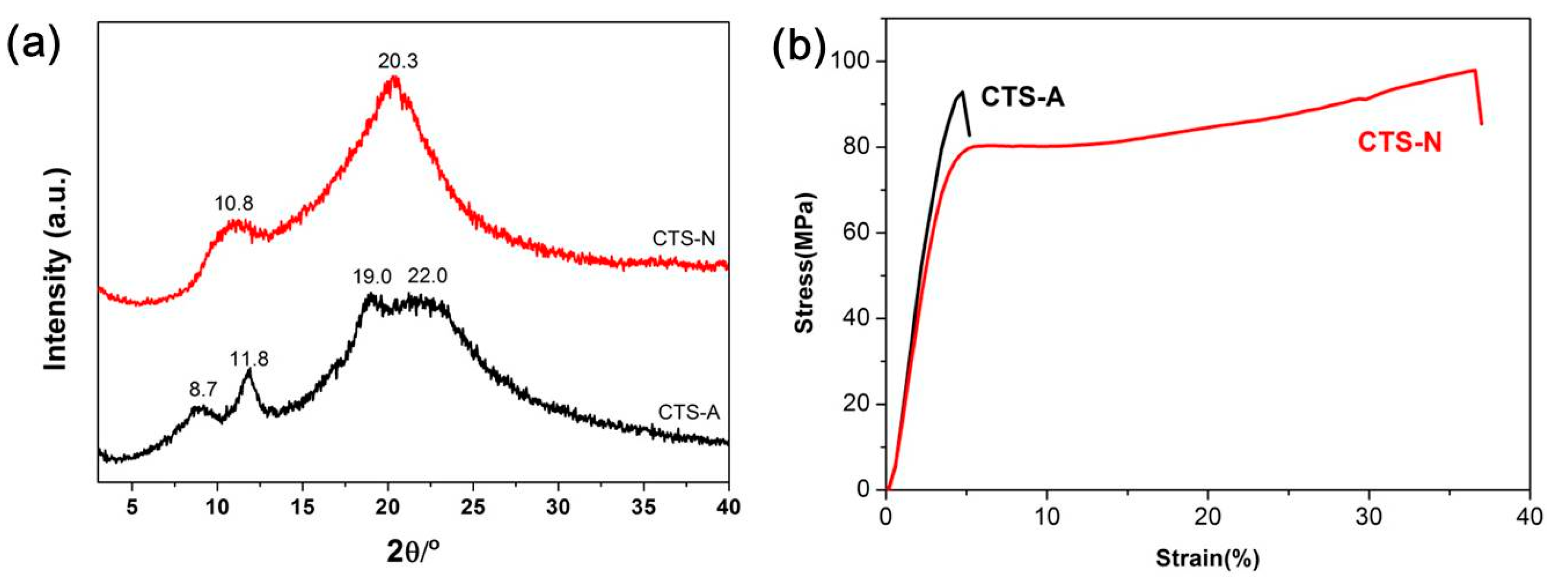
3.1. Crystallinity and Mechanical Properties of Hydrated Chitosan Films Based on XRD and Tensile Experiments

The crystallization behavior of the CTS-A and CTS-N films was characterized using XRD, as shown in Figure 1a. The X-ray powder pattern of chitosan in Figure 1 is consistent with the previously reported results [31]. The peaks at 2θ = 8.7°, 11.3° and 22.0° are the characteristic diffraction peaks of chitosan acetic salt, and the peaks at 2θ = 10.8° and 20.3° correspond to the equatorial (100) and (020) reflections, respectively, of neutralized chitosan. The degree of crystallinity was 14.8% for CTS-A and 25.8% for CTS-N on the basis of the peak-differentiation-imitating analysis of the two 1D profiles (see Figure S3 in the Supplementary Materials). The mechanical properties of the two CTS films were investigated using tensile measurements at room temperature, and the obtained stress–strain curves are shown in Figure 1b. The derived mechanical parameters, including the Young’s modulus, stress and elongation at break, are summarized in Table 1. Apparently, CTS-N exhibited a ductile fracture with a large elongation of 35.8%, while the elongation of CTS-A was much smaller than that of CTS-N. In fact, such crystalline and mechanical difference could be ascribed to the different dynamics and organization of water in the two hydrated chitosan films, as discussed in detail below. The effect of the water content on the mechanical properties of CTS films is shown in Figure S1 and listed in Table S1. For CTS-N films at water content greater than 8 wt %, an obvious decrease of the mechanical properties can be observed because of the plasticization effect of water.

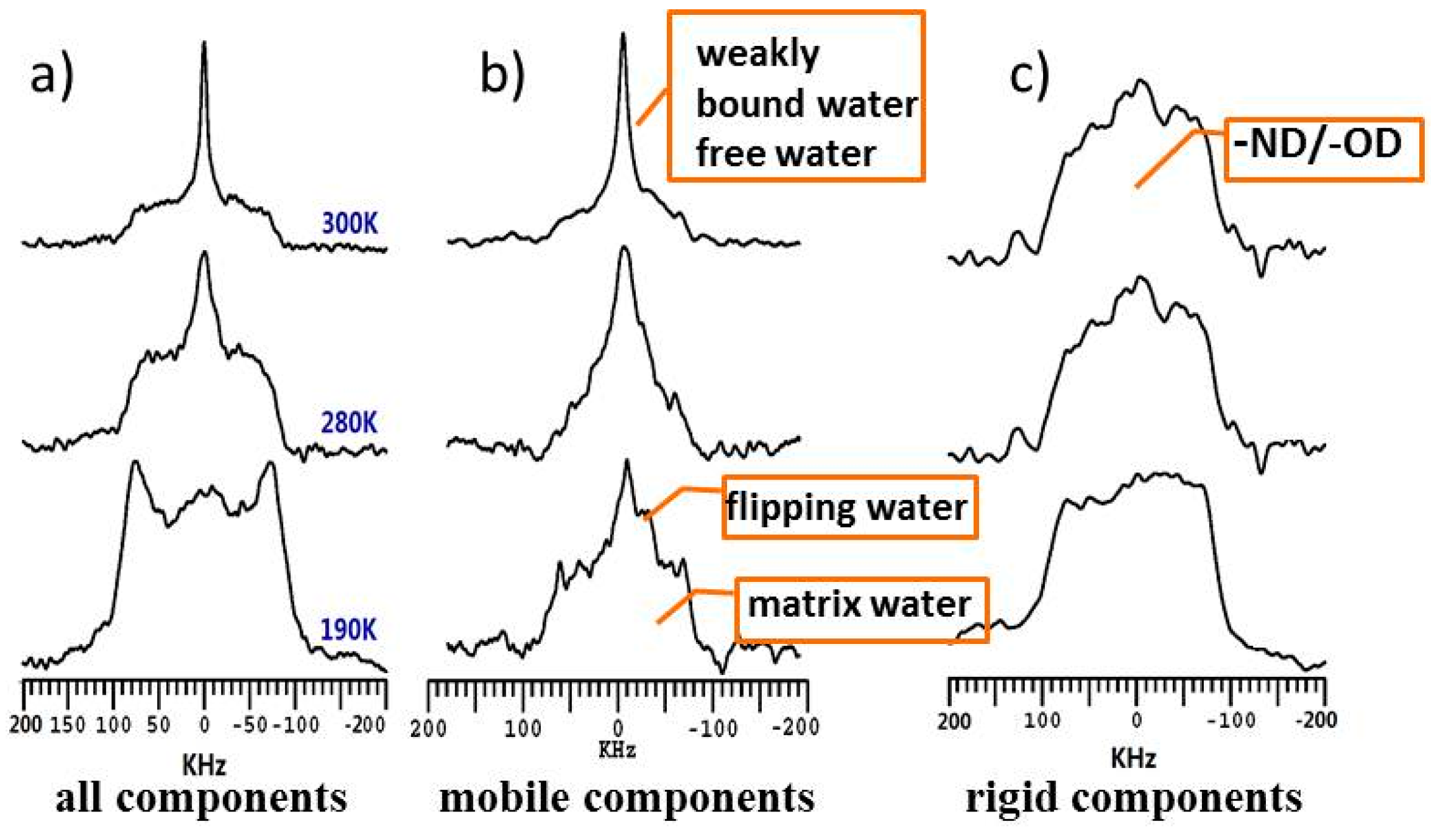
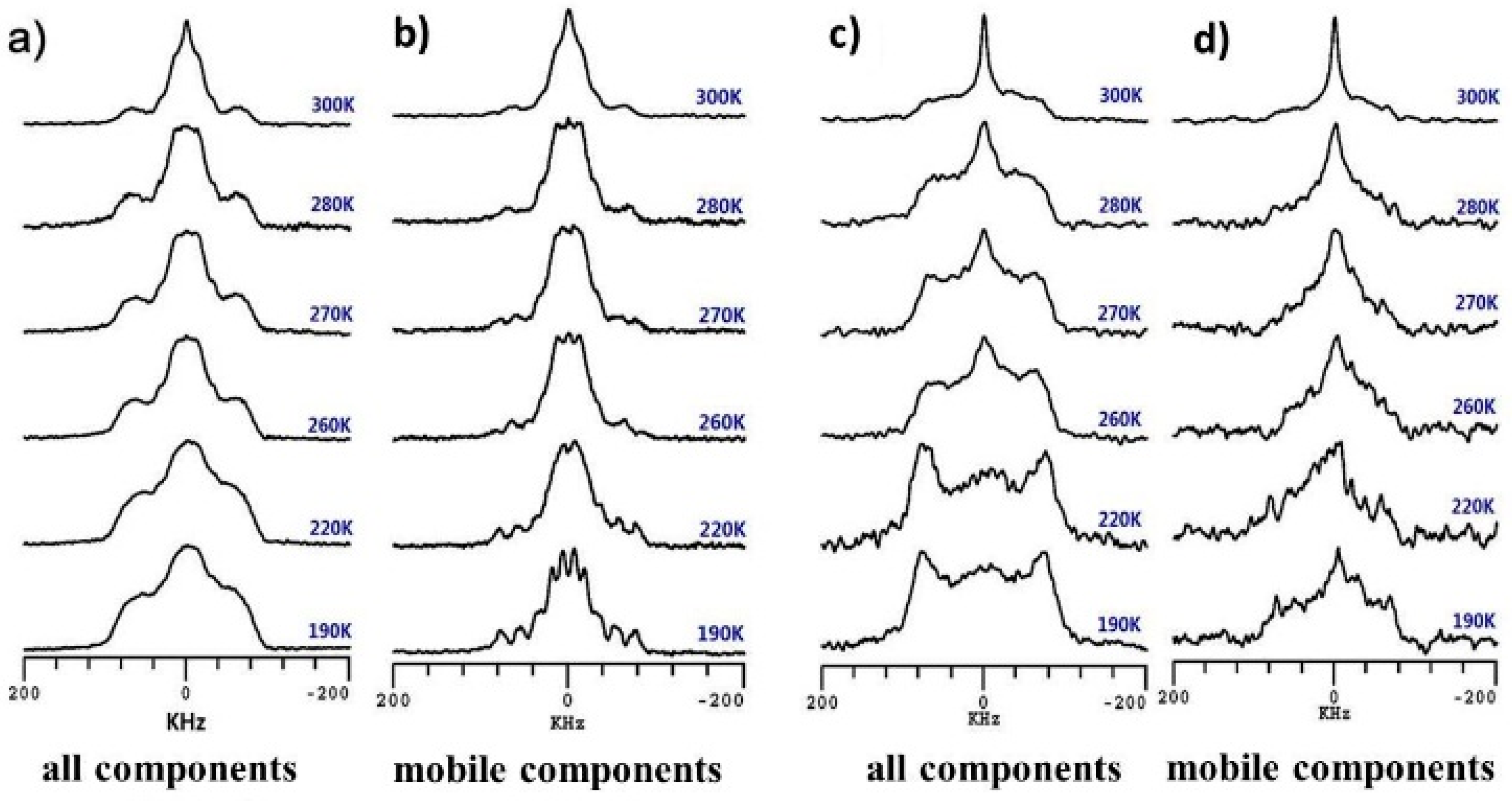
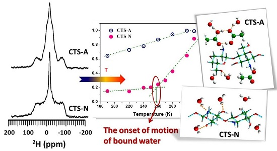
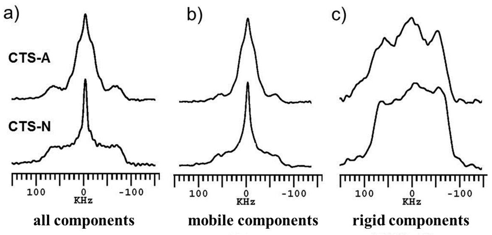
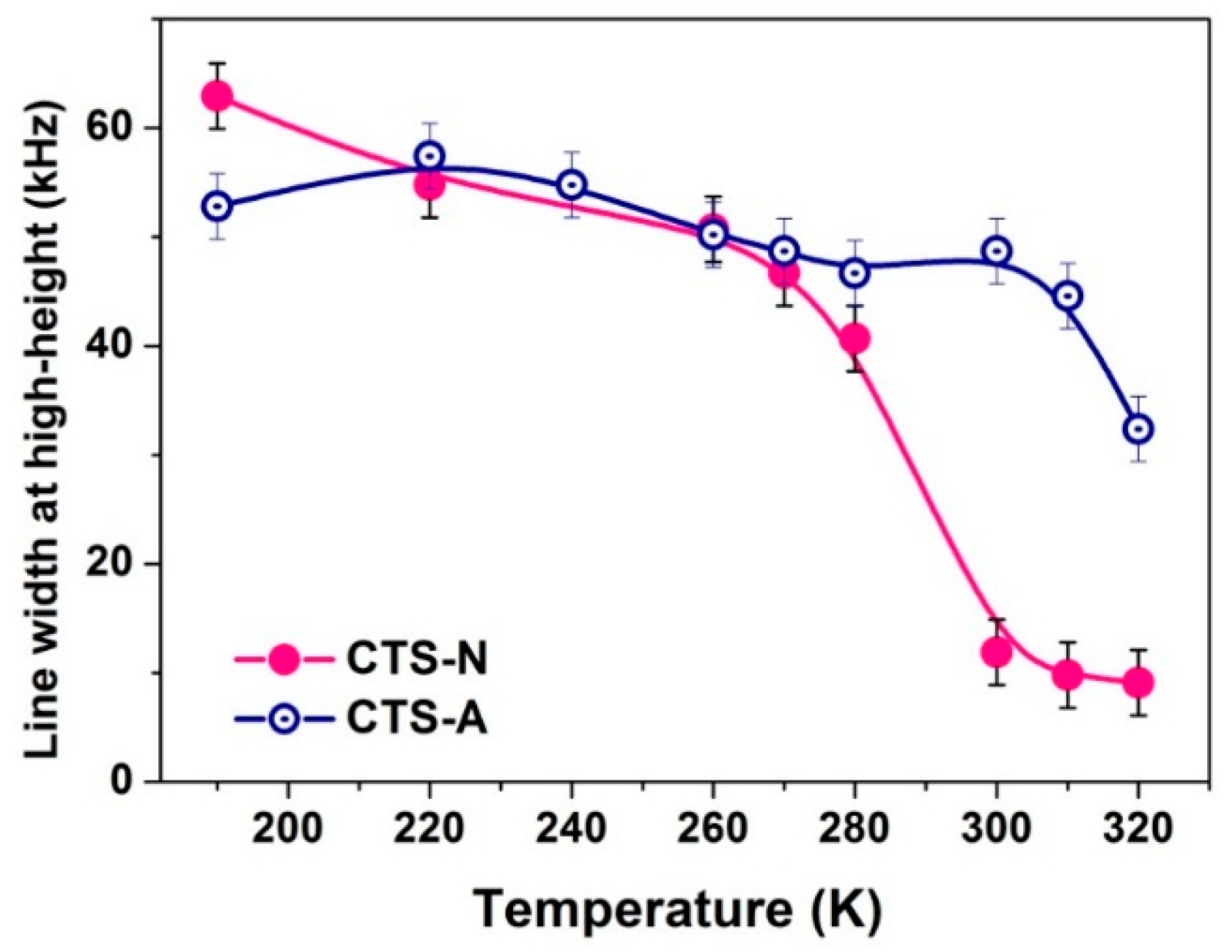
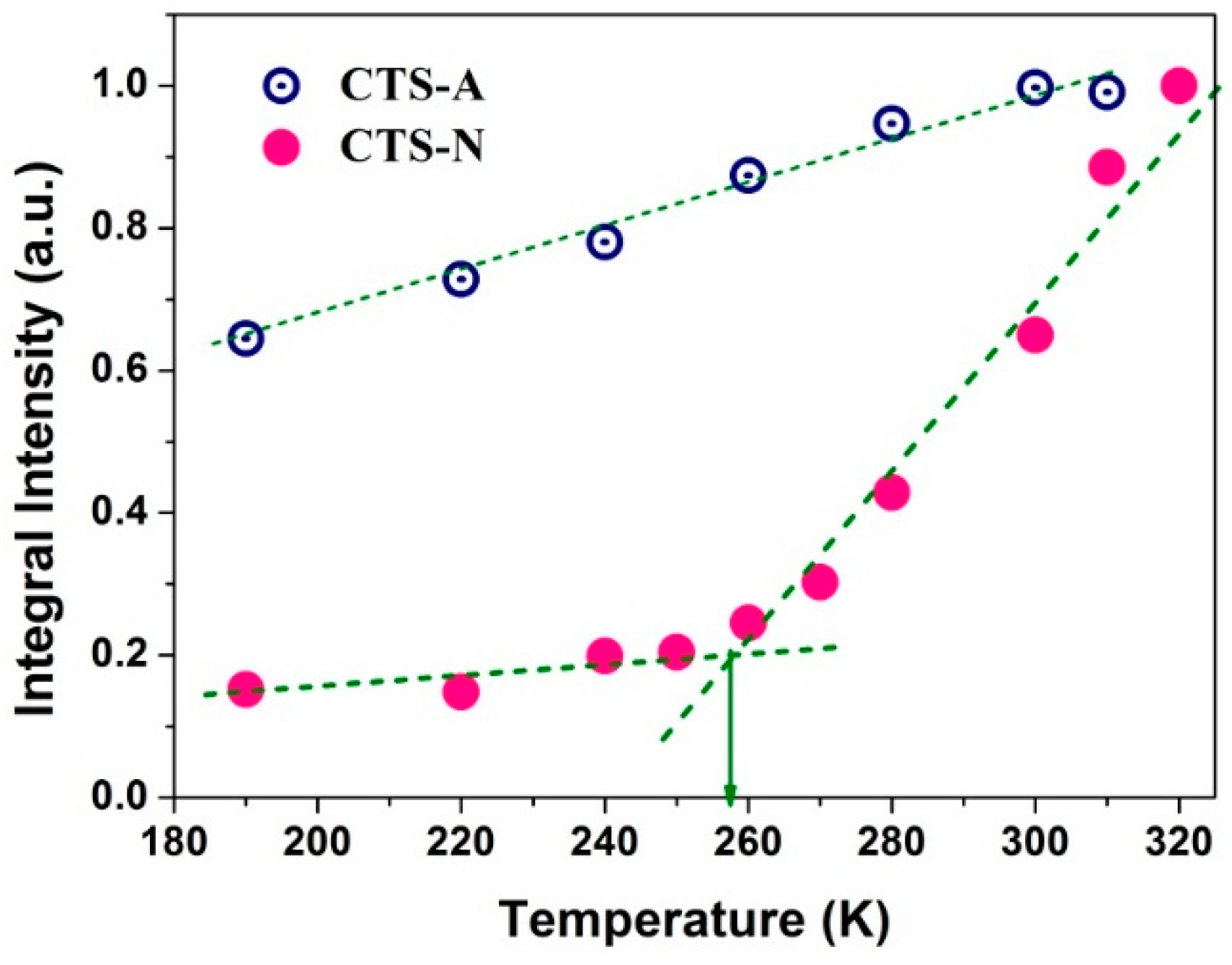
3.2. Dynamics and Different States of Water in Hydrated Chitosan before and after Neutralization Using Dynamic-Editing VT 2H NMR Experiments

2H NMR has been successfully used to reveal the orientational ordering and phase dynamics in a wide range of liquid crystals, polymers and biopolymers [32]. The 2H solid-state spectra are dominated by the quadrupole coupling of the deuteron spins. The splitting between the peaks in 2H spectra (ΔνQ) is determined by the quadrupolar interactions,
Δ ν Q = 3 4 ( e 2 q Q h ) ( 3 cos 2 θ ( t ) 1 )
where e2qQ/h is the quadruple coupling constant and θ is the angle of the 2H–N or 2H–O bond relative to the magnetic field in the chitosan samples. A distribution of angles, which is present in polycrystalline or amorphous samples, generates the so-called Pake pattern [33]. The molecular motions will induce the averaging of quadrupolar couplings, leading to the collapse of the Pake pattern into a narrow isotropic peak. According to the value of peak splitting, multiple components could be separated and determined based on the dynamic differences.
The dynamic-editing 2H NMR spectra of CTS-A and CTS-N at room temperature are shown in Figure 2, where different states of water are observed. First, the solid-echo sequence was used to obtain the undistorted spectra containing the signals of all 2H spins, as shown in Figure 2a, where the heterogeneous dynamics of different water species were observed from the peak splitting patterns. All of the spectra shown in Figure 2a contain overlapping peaks with a narrowed line at the center of the spectra and a broad Pake pattern at the bottom. This result indicates that the 2H nuclei in the CTS samples exhibit heterogeneous dynamic behavior. The mobile components with narrow lines (Figure 2a) can be separately detected via the saturation echo experiment with short τ1, as shown in Figure 2b. The results in Figure 2b indicate that the 2H spins with fast motion contain two species as follows: a broad peak corresponding to –ND/–OD groups resulted from the chemical exchange between chitosan and D2O and a narrow peak corresponding to water molecules. Notably, the spectral width of the central narrow peak of CTS-A was much larger than that of CTS-N, indicating a distinct difference in the mobile water dynamics and organization between the two samples. In addition, the mobile water molecules in CTS-A are strongly bound to the chitosan chains and led to remarkable line broadening, which will be further confirmed by VT 2H NMR below. The 2H spectra of the slow-relaxing (rigid) components were separately determined by Torchia’s T1 filter quadrupolar echo experiments, as shown in Figure 2c. For CTS-A and CTS-N, the very broad peak indicates the presence of strongly bound water in the chitosan films. According to a previous study on cellulose using 2H NMR [24], the broad peaks shown in Figure 2c may arise from room-temperature chemical exchange between D2O and the –OH and –NH groups of chitosan, which leads to isotopic labeling of chitosan. In addition, one type of water molecule exists in the chitosan crystalline domain as reported before [34]. Therefore, the strongly bound water in the crystalline phase should also contribute to the broad signal. We have also taken the deacetylation degree and molecular weight of chitosan samples. As shown in Figure S4, the spectra shape were almost the same, so this two aspects were not influence the conclusion below.
To further determine the temperature dependence of different water species, VT 2H NMR spectra were collected over the temperature range from 190 to 320 K. Figure 3 shows the dynamic-editing 2H spectra and peak assignment of CTS-N at 190, 280 and 300 K, and Figure 4 shows the 2H spectra for the total and mobile components at different temperatures. Briefly, the mobility of the different components is distinguished on the basis of their quadrupolar splitting in the 2H spectrum. The rigid components with confined mobility have strong quadruple interactions, which lead to a broad peak; the mobile components with high mobility exhibit a narrow peak due to the motion induced averaging of quadrupolar interactions. In general, as shown in Figure 3a, at room temperature (i.e., approximately 300 K), the broad peak of the rigid 2H component belong to –ND/–OD from chemical exchange between rigid chitosan chains and D2O, and the central narrow peak in the spectra of mobile components correspond to the weakly bound and free water. On the basis of the saturation recovery quadrupolar echo 2H spectra shown in Figure 3b, non-freezable water exhibited a high molecular mobility, even at 190 K. In addition, the rigid amorphous component at 190 K is classified as the water strongly bound to the polymer matrix in the crystalline region. Besides, the narrow peak of the highly mobile species in the center at 190 K implies the existence of flipping water undergoing 180° flips around their bisector axis, similar to that reported for some crystalline hydrates [24]. The broad peaks of the Torchia’s T1 filter quadrupolar echo spectrum at 190 K came from the signals of frozen D2O, and –OD and the –ND components in chitosan chains, both of which cannot be distinguished at this temperature. Overall, on the basis of the VT 2H results, four water species were observed: free water, weakly bound water, matrix water and flipping water that undergoes well-defined 180° flips in CTS-N and CTS-A.
On the basis of the VT 2H NMR spectra shown in Figure 4, the greatest difference in the water state between the CTS-A and CTS-N samples was mostly due to the weakly bound water. The line width at half height of the central narrow peak in the spectra of mobile components (Figure 4b,d) with increasing temperature is shown in Figure 5; these results indicate that the mobility of the bound water and free water increase with increasing temperature. As shown in Figure 5, the line width of the central narrow peak for CTS-N decreased substantially after 260 K, whereas that of CTS-A changed more slowly. The line width in the 2H spectrum for CTS-A was around 41 kHz at room temperature, which is much larger than that of CTS-N, ~10 kHz. This result indicates that the molecular motion of a portion of the water molecules is strongly affected by the mobility of the acetic acid. This experimental result should be attributed to fact that most of the water molecules (D2O) in the CTS-A sample were bound to CH3COOH because of the stronger hydrophilic force of carboxyl groups. This behavior will be further confirmed by curves showing relative intensity of the narrow components with increasing temperature.
To gain additional insight into the water dynamics, we also showed the integral intensity of the saturation recovery quadrupolar echo spectra (Figure 4b,d) with increasing the temperature as shown in Figure 6. For both CTS-A and CTS-N, the increase in the amount of mobile components with increasing temperature leads to an increase in the overall integral intensity of the saturation recovery quadrupolar echo spectra. In particular, an inflection point in the CTS-N curve was observed, which could be ascribed to the onset of the motion of the matrix water. As previously reported, the water molecules bound to the polymer matrix exhibit a glass-transition-like behavior at a temperature below 0 °C [35,36,37]. In contrast, in the case of CTS-A, the integral intensity of the mobile component increased linearly with increasing the temperature, because most of the water molecules were bound with the carboxyl groups in CH3COOH, which were non-freezable, even at 190 K. Few water molecules were bound to the polymer matrix, and thus no inflection point was observed.

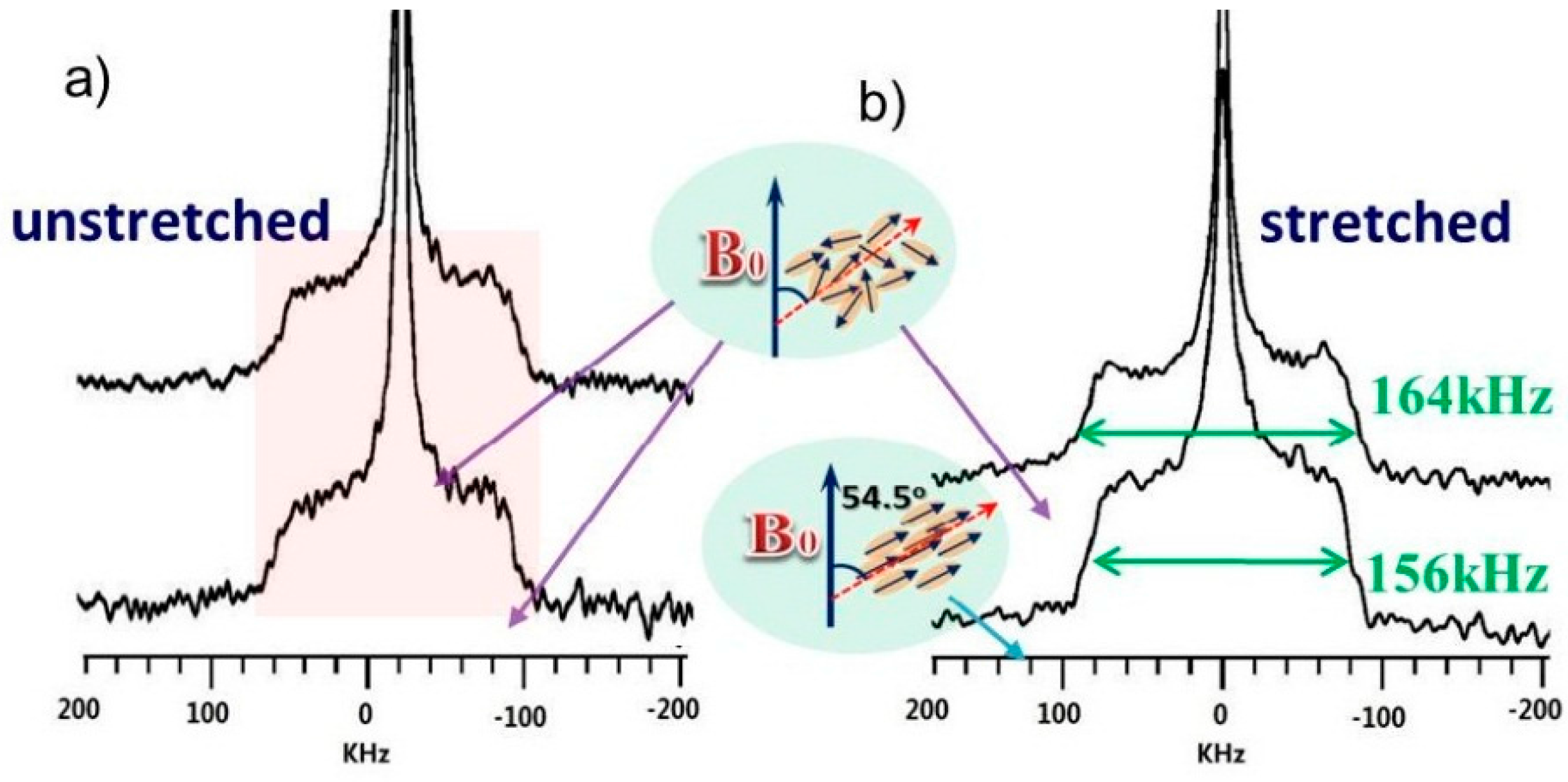
3.3. Effect of Stretching on the Hydrated and Neutralized Chitosan by 2H Experiments

Solid-state 2H NMR is an ideal probe for investigating preferential orientation distributions [32,38,39], as the quadrupolar splitting is sensitive to the orientation of O–D/N–D bond relative to the magnetic field, as shown in Equation (1). In general, the distribution of the spectral intensity across the 2H NMR line shape is directly correlated to the orientation distribution of the corresponding O–D/N–D bonds. For example, a randomly oriented powder sample gives rise to a characteristic powder line shape with a spherical distribution. However, if the O–D/N–D bonds were mostly along the magic angle (i.e., 54.7°), and the quadrupolar splitting will be largely reduced. The 2H NMR spectra of unstretched and stretched CTS-N obtained with random or oriented placement along the magic angle are shown in Figure 7. As is clearly shown in Figure 7b, the quadrupolar splitting of the 2H spectrum of the stretched CTS-N sample become smaller when the stretched sample was packed in the rotor along the magic angle, in comparison to that packed randomly, indicating that the polymer chains became oriented after stretching. As a control experiment, the 2H spectra of unstretched CTS-N were also acquired in two ways. Not surprisingly, the 2H spectra of unstretched CTS-N were basically the same whether the sample was packed randomly or along the magic angle. Therefore, a small amount of water could make some chitosan chains become more flexible, which would dissipate the stress in the materials and enhance the toughness. On the basis of these NMR results, the appropriate content of water bound with biopolymer chains is beneficial for enhancing the toughness of biomaterials.

4. Conclusions

The dynamics and different states of water in hydrated chitosan were investigated using dynamic-editing VT 2H solid-state NMR spectroscopy. Four distinct types of water in hydrated chitosan were detected in the chitosan samples: (1) non-freezable, rigid and strongly bound water in the crystalline domain; (2) highly mobile water that is weakly bound with polymer chains and exhibits isotropic motion, even below 260 K; (3) flipping water that undergoes well-defined 180° flips at 190 K; and (4) free water. In the case of neutralized and hydrated chitosan (CTS-N), the onset of motion of weakly bound water molecules at lower temperatures was revealed using 2H NMR spectroscopy, which is absent in the CTS-A sample due to the strong binding of water and carboxyl groups in CH3COOH molecules; these water molecules were not crystalline, even below 260 K. Furthermore, the 2H spectra of stretched and unstretched CTS-N samples suggested that the polymer chains became oriented after stretching. In summary, the water dynamics in chitosan films were investigated in detail by VT 2H spectra, which could play an important role in enhancing the mechanical properties of CTS-N sample.

Supplementary Materials

Supplementary materials can be found at www.mdpi.com/2073-4360/8/4/149/s1.

Acknowledgments

This work was supported by the National Science Fund for Distinguished Young Scholars (20825416), the National Natural Science Foundation of China (21534005 and 21374051), 973 program (2012CB821503), PCSIRT (IRT1257), and CERS-1-61.

Author Contributions

Fenfen Wang and Pingchuan Sun conceived and designed the experiments; Fenfen Wang performed the experiments, data analyses, and maunuscript writing; Rongchun Zhang corrected manuscript; Tiehong Chen and Pingchuan Sun carried out project development and leadership.

Conflicts of Interest

The authors declare no conflict of interest.

References

  1. Levy, Y.; Onuchic, J.N. Water and proteins: A love-hate relationship. Proc. Natl. Acad. Sci. USA 2004, 101, 3325–3326. [Google Scholar] [CrossRef] [PubMed]
  2. Li, T.; Hassanali, A.A.; Kao, Y.-T.; Zhong, D.; Singer, S.J. Hydration dynamics and time scales of coupled water−protein fluctuations. J. Am. Chem. Soc. 2007, 129, 3376–3382. [Google Scholar] [CrossRef] [PubMed]
  3. Liu, Y.; Shao, Z.; Vollrath, F. Relationships between supercontraction and mechanical properties of spider silk. Nat. Mater. 2005, 4, 901–905. [Google Scholar] [CrossRef] [PubMed]
  4. Li, Z.; Ramay, H.R.; Hauch, K.D.; Xiao, D.; Zhang, M. Chitosan–alginate hybrid scaffolds for bone tissue engineering. Biomaterials 2005, 26, 3919–3928. [Google Scholar] [CrossRef] [PubMed]
  5. Nazarov, R.; Jin, H.-J.; Kaplan, D.L. Porous 3-D scaffolds from regenerated silk fibroin. Biomacromolecules 2004, 5, 718–726. [Google Scholar] [CrossRef] [PubMed]
  6. Suh, J.K.F.; Matthew, H.W.T. Application of chitosan-based polysaccharide biomaterials in cartilage tissue engineering: A review. Biomaterials 2000, 21, 2589–2598. [Google Scholar] [PubMed]
  7. Wang, F.; Zhang, R.; Wu, Q.; Chen, T.; Sun, P.; Shi, A.-C. Probing the nanostructure, interfacial interaction, and dynamics of chitosan-based nanoparticles by multiscale solid-state NMR. ACS Appl. Mater. Interfaces 2014, 6, 21397–21407. [Google Scholar] [CrossRef] [PubMed]
  8. Geng, C.-Z.; Hu, X.; Yang, G.-H.; Zhang, Q.; Chen, F.; Fu, Q. Mechanically reinforced chitosan/cellulose nanocrystals composites with good transparency and biocompatibility. Chin. J. Polym. Sci. 2014, 33, 61–69. [Google Scholar] [CrossRef]
  9. Petridis, L.; O’Neill, H.M.; Johnsen, M.; Fan, B.; Schulz, R.; Mamontov, E.; Maranas, J.; Langan, P.; Smith, J.C. Hydration control of the mechanical and dynamical properties of cellulose. Biomacromolecules 2014, 15, 4152–4159. [Google Scholar] [CrossRef] [PubMed]
  10. Smyth, G.; Quinn, F.X.; McBrierty, V.J. Water in hydrogels. 2. A study of water in poly(hydroxyethyl methacrylate). Macromolecules 1988, 21, 3198–3204. [Google Scholar] [CrossRef]
  11. Pasqui, D.; de Cagna, M.; Barbucci, R. Polysaccharide-based hydrogels: The key role of water in affecting mechanical properties. Polymers 2012, 4, 1517–1534. [Google Scholar] [CrossRef]
  12. Ma, J.; Wang, H.; He, B.; Chen, J. A preliminary in vitro study on the fabrication and tissue engineering applications of a novel chitosan bilayer material as a scaffold of human neofetal dermal fibroblasts. Biomaterials 2001, 22, 331–336. [Google Scholar] [CrossRef]
  13. Rajabi-Siahboomi, A.R.; Bowtell, R.W.; Mansfield, P.; Davies, M.C.; Melia, C.D. Structure and behavior in hydrophilic matrix sustained release dosage forms: 4. Studies of water mobility and diffusion coefficients in the gel layer of HPMC tablets using NMR imaging. Pharm. Res. 1996, 13, 376–380. [Google Scholar] [CrossRef] [PubMed]
  14. Kamata, Y.; Parnell, A.J.; Gutfreund, P.; Skoda, M.W.A.; Dennison, A.J.C.; Barker, R.; Mai, S.; Howse, J.R.; Ryan, A.J.; Torikai, N.; et al. Hydration and ordering of lamellar block copolymer films under controlled water vapor. Macromolecules 2014, 47, 8682–8690. [Google Scholar] [CrossRef]
  15. González-Campos, J.B.; Prokhorov, E.; Luna-Bárcenas, G.; Fonseca-García, A.; Sanchez, I.C. Dielectric relaxations of chitosan: The effect of water on the α-relaxation and the glass transition temperature. J. Polym. Sci. Part B: Polym. Phys. 2009, 47, 2259–2271. [Google Scholar] [CrossRef]
  16. Huang, W.; Krishnaji, S.; Tokareva, O.R.; Kaplan, D.; Cebe, P. Influence of water on protein transitions: Thermal analysis. Macromolecules 2014, 47, 8098–8106. [Google Scholar] [CrossRef]
  17. Lin, C.; Gitsov, I. Synthesis and physical properties of reactive amphiphilic hydrogels based on poly(p-chloromethylstyrene) and poly(ethylene glycol): Effects of composition and molecular architecture. Macromolecules 2010, 43, 3256–3267. [Google Scholar] [CrossRef]
  18. Li, B.; Xu, L.; Wu, Q.; Chen, T.; Sun, P.; Jin, Q.; Ding, D.; Wang, X.; Xue, G.; Shi, A.C. Various types of hydrogen bonds, their temperature dependence and water-polymer interaction in hydrated poly(acrylic acid) as revealed by H-1 solid-state NMR spectroscopy. Macromolecules 2007, 40, 5776–5786. [Google Scholar] [CrossRef]
  19. Vogel, A.; Katzka, C.P.; Waldmann, H.; Arnold, K.; Brown, M.F.; Huster, D. Lipid modifications of a Ras peptide exhibit altered packing and mobility versus host membrane as detected by 2H solid-state NMR. J. Am. Chem. Soc. 2005, 127, 12263–12272. [Google Scholar] [CrossRef] [PubMed]
  20. Fu, R.Q.; Wang, X.S.; Li, C.G.; Santiago-Miranda, A.N.; Pielak, G.J.; Tian, F. In situ structural characterization of a recombinant protein in native escherichia coli membranes with solid-state magic-angle-spinning NMR. J. Am. Chem. Soc. 2011, 133, 12370–12373. [Google Scholar] [CrossRef] [PubMed]
  21. Michaelis, V.K.; Keeler, E.G.; Ong, T.-C.; Craigen, K.N.; Penzel, S.; Wren, J.E.C.; Kroeker, S.; Griffin, R.G. Structural insights into bound water in crystalline amino acids: Experimental and theoretical 17O NMR. J. Phys. Chem. B 2015, 119, 8024–8036. [Google Scholar] [CrossRef] [PubMed]
  22. Pope, J.C.; Sue, H.-J.; Bremner, T.; Blümel, J. High-temperature steam-treatment of PBI, PEEK, and PEKK polymers with H2O and D2O: A solid-state NMR study. Polymer 2014, 55, 4577–4585. [Google Scholar] [CrossRef]
  23. Fei, Z.; Zhao, D.; Geldbach, T.J.; Scopelliti, R.; Dyson, P.J.; Antonijevic, S.; Bodenhausen, G. A synthetic zwitterionic water channel: Characterization in the solid state by X-ray crystallography and NMR spectroscopy. Angew. Chemie 2005, 117, 5866–5871. [Google Scholar] [CrossRef]
  24. Radloff, D.; Boeffel, C.; Spiess, H.W. Cellulose and cellulose/poly(vinyl alcohol) blends. 2. Water organization revealed by solid-state NMR spectroscopy. Macromolecules 1996, 29, 1528–1534. [Google Scholar] [CrossRef]
  25. VanderSchee, C.R.; Ooms, K.J. Investigating water interactions with collagen using H-2 multiple quantum filtered NMR spectroscopy to provide insights into the source of double quantum filtered signal in tissue. J. Phys. Chem. B 2014, 118, 3491–3497. [Google Scholar] [CrossRef] [PubMed]
  26. Hutchison, J.L.; Murthy, N.S.; Samulski, E.T. Deuterium NMR studies of water in oriented nylon 6 fibers. Macromolecules 1996, 29, 5551–5557. [Google Scholar] [CrossRef]
  27. Schartel, B.; Wendling, J.; Wendorff, J.H. Cellulose/poly(vinyl alcohol) blends. 1. Influence of miscibility and water content on relaxations. Macromolecules 1996, 29, 1521–1527. [Google Scholar] [CrossRef]
  28. Maghami, G.G.; Roberts, G.A.F. Evaluation of the viscometric constants for chitosan. Die Makromol. Chemie 1988, 189, 195–200. [Google Scholar] [CrossRef]
  29. Hirai, A.; Odani, H.; Nakajima, A. Determination of degree of deacetylation of chitosan by 1H NMR spectroscopy. Poly. Bull. 1991, 26, 87–94. [Google Scholar] [CrossRef]
  30. Torchia, D.A. The measurement of proton-enhanced carbon-13 T1 values by a method which suppresses artifacts. J. Magn. Resonance (1969) 1978, 30, 613–616. [Google Scholar] [CrossRef]
  31. Saito, H.; Tabeta, R.; Ogawa, K. High-resolution solid-state carbon-13 NMR study of chitosan and its salts with acids: Conformational characterization of polymorphs and helical structures as viewed from the conformation-dependent carbon-13 chemical shifts. Macromolecules 1987, 20, 2424–2430. [Google Scholar] [CrossRef]
  32. Chowdhury, S.; Madsen, L.A.; Frazier, C.E. Probing alignment and phase behavior in intact wood cell walls using 2H NMR spectroscopy. Biomacromolecules 2012, 13, 1043–1050. [Google Scholar] [CrossRef] [PubMed]
  33. Pake, G.E. Nuclear resonance absorption in hydrated crystals: Fine structure of the proton line. J. Chem. Phys. 1948, 16, 327–336. [Google Scholar] [CrossRef]
  34. Okuyama, K.; Noguchi, K.; Miyazawa, T.; Yui, T.; Ogawa, K. Molecular and crystal structure of hydrated chitosan. Macromolecules 1997, 30, 5849–5855. [Google Scholar] [CrossRef]
  35. Quinn, F.X.; Kampff, E.; Smyth, G.; McBrierty, V.J. Water in hydrogels. 1. A study of water in poly(N-vinyl-2-pyrrolidone/methyl methacrylate) copolymer. Macromolecules 1988, 21, 3191–3198. [Google Scholar] [CrossRef]
  36. Cerveny, S.; Colmenero, J.; Alegría, A. Dielectric investigation of the low-temperature water dynamics in the poly(vinyl methyl ether)/H2O system. Macromolecules 2005, 38, 7056–7063. [Google Scholar] [CrossRef]
  37. Van den Dries, I.J.; van Dusschoten, D.; Hemminga, M.A. Mobility in maltose−water glasses studied with 1H NMR. J. Phys. Chem. B 1998, 102, 10483–10489. [Google Scholar] [CrossRef]
  38. Simmons, A.H.; Michal, C.A.; Jelinski, L.W. Molecular orientation and two-component nature of the crystalline fraction of spider dragline silk. Science 1996, 271, 84–87. [Google Scholar] [CrossRef] [PubMed]
  39. Spiess, H.W. Deuteron NMR—A new tool for studying chain mobility and orientation in polymers. In Characterization of Polymers in the Solid State I: Part A: NMR and Other Spectroscopic Methods Part B: Mechanical Methods; Kaush, H.H., Zachman, H.G., Eds.; Springer: Berlin, Germany; Heidelberg, Germany, 1985; pp. 23–58. [Google Scholar]
Figure 1. (a) XRD patterns of CTS-A and CTS-N at room temperature; and (b) stress–strain curves of CTS-A and CTS-N.
Polymers 08 00149 g001
Figure 2. Dynamic-editing 2H NMR spectra of CTS-A and CTS-N at room temperature: (a) solid-echo spectra recorded with a repetition time of 2 s; (b) saturation Recovery quadrupolar echo spectra with a saturation recovery delay τ1 = 10 ms to selectively observe signals of mobile components; and (c) Torchia’s T1-filter spectra to selectively observe signals of rigid components.
Polymers 08 00149 g002
Figure 3. Dynamic-editing VT 2H NMR spectra for CTS-N obtained from: (a) quadrupolar echo; (b) saturation recovery quadrupolar echo; and (c) Torchia’s T1 filter quadrupolar echo experiments, respectively.
Polymers 08 00149 g003
Figure 4. VT 2H spectra obtained from: (a,c) quadrupolar echo experiments to obtain signals of all components; and (b,d) saturation recovery quadrupolar echo experiments to obtain signals of mobile components for CTS-A (a,b) and CTS-N (c,d), respectively.
Polymers 08 00149 g004
Figure 5. Line width at half height of the narrow components of the solid-echo spectra for CTS-N (Figure 4).
Polymers 08 00149 g005
Figure 6. Integral signal intensity of the saturation recovery quadrupolar echo 2H spectra from Figure 4b,d. The onset of motion of the weakly bound matrix water is shown at 260 K.
Polymers 08 00149 g006
Figure 7. 2H NMR spectra of unstretched (a) and stretched (b) CTS-N sample. The top spectra correspond to the randomly oriented powder samples, while the bottom spectra correspond to sample placed along the magic angle.
Polymers 08 00149 g007
Table 1. Tensile properties of CTS-A and CTS-N samples.
SamplesYoung’s modulus (GPa)Yield stress (MPa)Break strength (MPa)Strain of break % (Elongation)
CTS-A2.86 ± 0.0584.3 ± 2.56.1 ± 0.8
CTS-N2.43 ± 0.0881.4 ± 1.698.7 ± 1.635.8 ± 5.3

