A Novel Self-Assembled Liposome-Based Polymeric Hydrogel for Cranio-Maxillofacial Applications: Preliminary Findings

Abstract
: Soft nanogels are submicron-sized hydrophilic structures engineered from biocompatible polymers possessing the characteristics of nanoparticles as well as hydrogels, with a wide array of potential applications in biotechnology and biomedicine, namely, drug and protein delivery. In this work, nanogels were obtained using the physical self-assembly technique or ‘layer-by-layer’ which is based on electrostatic interactions. Liposomal vesicles were coated with alternating layers of hyaluronic acid and chitosan yielding a more viscous hydrogel formulation that previously reported core-shell nanoparticulate suspension, via simply modifying the physico-chemical characteristics of the system. Structural features, size, surface charge, stability and swelling characteristics of the nanogel were studied using scanning electron microscopy and dynamic light scattering. With a specific cranio-maxillofacial application in mind, the hydrogel was loaded with recombinant human (rh) bone morphogenetic protein-7, also known as osteogenic protein-1 or rhOP-1 and release was monitored over an extended period of 60 days. This preliminary study reports promising results on the formulation of a novel core-shell polymeric nanogel.1. Background
Reconstruction of large bone defects continue to present a significant challenge for orthopedists, traumatologists and cranio-maxillofacial surgeons attending millions of patients around the globe [1]. Bone Tissue Engineering (BTE) attempts to induce de novo bone formation for bridging defects using void fillers or biomaterials have been applied to a great extent [2]. Yet, autologous bone grafts continue to be routinely employed clinically despite the well-documented shortcomings, such as limited graft accessibility and donor site morbidity, as well as increased costs. Hence, the drive for developing alternative methods for bone regeneration and repair continues [3]. Nanoparticulate systems are modern tools leading a revolution in the field of localized and release-controlled drug, gene and protein (or growth factor) delivery. Among the available nanosystems, self-assembled polymeric nanogels are particularly attractive, since they are easy to produce, affordable and may effectively incorporate a variety of therapeutic agents for diagnostic and treatment purposes [4]. Furthermore, they may be decorated with different kinds of molecules, improving the stability and target ability [5]. In a previous set of in vitro studies performed by the corresponding author during his doctoral studies, monodisperse, stable and cyto-compatible nanocapsules constituting a core of cationic liposomes (L) and a shell constructed through the layer-by-layer (L-b-L) self-assembly of alternating layers of anionic alginate (AL) and cationic chitosan (CH) were formulated, suitable for complex formation with anionic proteins [6], such as recombinant human (rh) bone morphogenetic protein-7, also known as osteogenic protein-1 or rhOP-1 [7]. A sustained tri-phasic release of the water-soluble growth factor was evident from the hybrid core-shell nanoparticulate system or L(AL-CH)3 for an extended period of 45 days with the bioactivity of the protein maintained via enhancing pre-osteoblast differentiation [7,8]. In a subsequent in vivo investigation, the effect of a single and early bolus injection of various low doses of rhOP-1 administered in combination with the L(AL-CH)3 delivery system on the rate, quantity and quality of new bone formation and consolidation during long bone lengthening in rabbits was performed, with favorable results [9]. The biocompatibility and rhOP-1 stimuli localization of the nanocapsules were then demonstrated in rats following intramuscular administration [10]. Yet, for other BTE applications, such as spinal fusion and craniofacial defect repair, a more appropriate system would be more malleable and viscous or gel-like rather than a free-flowing suspension. Hence, in this preliminary work, the physico-chemical characteristics of the original L(AL-CH)3 were modified. Sodium alginate was replaced with hyaluronic acid (HA), a natural polymer with a number of embryologic and wound healing properties attractive for BTE. Also, smaller liposomes were prepared and polymeric solution concentrations/ratios modified. Thus, we introduce herein the 2nd generation of the core-shell system; a liposomal-based hyaluronan-chitosan bioadhesive hydrogel or L(HA-CH) intended for cranio-maxillofacial applications. Samples with varying number of assembled layers were prepared, yet, only the characterization of L(HA-CH)3 gel and rhOP-1 loaded L(HA-CH)3 are reported, providing a comparison to our previous in vitro data, as a proof of concept where, to the best of knowledge, this is the first work on the self-assembly of HA and CH as shell on a liposome-based core.
2. Materials and Methods
The formulation and characterization of the hybrid core-shell nanoparticulate protein delivery system (1st generation) has been previously described [6,7]. For this study, a more viscous hydrogel (for defect malleability) was formulated via simply modifying the physico-chemical characteristics, polymeric material, concentrations and ratios of the liposomal core and multi-layered shell. Briefly, for the preparation of liposomes, 1,2-Dipalmitoyl-snglycero-3-phosphocholine (DPPC; phospholipid) was purchased from Genzyme Pharmaceuticals, Switzerland; cholesterol (CHOL; stabilizer) and dimethyldioctadecyl-ammonium bromide (DDAB; cationic surfactant) were obtained from Sigma-Aldrich Chemical (Dorset, UK). The extrusion apparatus was purchased from Avanti® Polar Lipids, and the 19-mm polycarbonate filters (150 nm pore size) were obtained from GE Osmonics. For the L-b-L coating, hyaluronan (HA: 90 kDa MW, 1.6 m3/Kg viscosity) and chitosan (CH: 90 kDa MW, 2.82 m3/Kg viscosity with 85% deacetylation) were obtained from Sigma- Aldrich Chemical. Liposomes (a mixture of DPPC:CHOL:DDAB in 7:2:1 molar ratio in chloroform:methanol) were formulated via the thin-film hydration technique followed by extrusion through double filters to yield smaller and more discrete/monodisperse liposomes. For the L-b-L build-up, fresh HA and CH solutions (0.01-2.0 mg/mL concentration range) were prepared in ultra-pure water (UPW). The cationic liposomal core was then coated with alternating layers of HA and CH until a shell composed of six polyelectrolyte layers was achieved; 3 bi-layers: L(HA-CH)3, herein. With the deposition of each polymeric layer, the solution was incubated at room temperature (RT) for 45 min and centrifuged at 15,000 rpm for 10 min for washing. Average hydrodynamic diameter and zeta (ζ) potential surface charge measurements followed using a dynamic low-angle laser light-scattering device (Malvern Instruments, UK). For protein incorporation into the hydrogel, a 0.5 μg concentration of rhOP-1 (15.7 kDa MW; purchased from ProSpec Technologies Inc, NJ, USA via Dong-in Biotech Co. Ltd., South Korea) solution was incorporated within the aqueous phase of nanogel emulsion via a simple direct addition method [11] to ensure entrapment within the core and the several compartments of the multi-layered shell. Aliquots of the L(HA-CH)3 and L(HA-CH)3 + rhOP-10.μg nanogel were then stored at 22 °C until further use. Similarly, size, dispersity and surface charge changes were recorded. Imaging using a cold field emission scanning electron microscope (S-4300, Hitachi High-Technologies Canada, Inc., ON, Canada) was also performed. Protein release was investigated at RT in UPW over 60 days. Suspensions were ultra-centifuged for 20 min at 180,000 g to separate the nanocapsules from the supernatant containing the released protein for quantitative analysis. Pellet was re-suspended in 1 mL of UPW and the procedure repeated over the planned time period, at pre-determined points. Absorbance was read at 450 nm using a microplate reader (details in [6]). Finally, swelling and degradation studies comparing volume phase transitions induced independently by UPW or PBS (pH: 7.4) were performed.
3. Results and Discussion
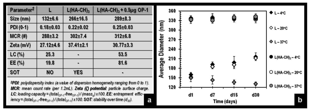
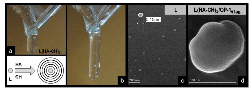
Polymeric hydrogels are widely investigated in the pharmaceutical and biomedical fields as carriers for drug delivery, tissue engineering and bionanotechnology because of their 3-D physical structure, mechanical properties, high-water content and biocompatibility [4,5]. A Nanogel can be defined as a colloidal system of aggregates in the sub-micrometer range prepared from hydrophilic polymers with gel-like characteristics, with high potential in gene and protein delivery. Several excellent articles are available on the topic for the interested reader [11,12]. Herein, self-assembled nanogels (<300 nm) composed of a liposomal cationic core coated with alternating layers of anionic hyaluronic acid and cationic chitosan were formulated. HA, a naturally-occurring polymer, is bioactive, cytocompatible, bio-/muco-adhesive and degradable enzymatically. It has been shown to provide intrinsic signals that can enhance tissue regeneration and repair [13]. More importantly, HA is one of the essential components of the extra-cellular matrix, playing a predominant role in tissue morphogenesis, cell migration, differentiation and adhesion [14]. Hence, it was chosen to replace alginate specifically for our planned in vivo studies in cranial defect repair where large or so-called critical-sized defects require a different system than that in bone lengthening. On the other hand, chitosan, another natural and bioadhesive polysaccharide, is known for its osteogenic properties [3,5,8,9,13], thus, was retained. As mentioned earlier, a smaller liposomal core was tailored using the multiple extrusion method. The L-b-L self-assembly technique based on electrostatic interactions (ionic gelation) yielded a viscous gel with compact, monodisperse and discrete nanocapsules, as displayed in Figure 1 with a cumulative hydrodynamic diameter of 266 ± 16.5 nm compared to 132 ± 6.6 nm core liposomes (∼130 nm shell). Besides a ∼40 nm reduction in liposomal core size, this was achieved via elevating the concentration of the polymeric solutions (reported herein: 2.0 mg/mL HA and CH hydrogel solutions prepared in UPW).
This difference increases slightly following the loading of rhOP-10.5ug. The in vitro characterization of the final L(HA-CH)3 hydrogel is presented in Figure 2(a) demonstrating the beneficence of the multiple compartments within the liposomal core and the shell that can be seen in the differences in loading capacity and encapsulation efficiency, 2–5 fold greater than bare liposomes. In addition, the stability of the system was noted with a surface charge of >30 mV, indicating strong repulsion forces among particles, thus preventing excessive aggregation [6,7]. Figure 2(b) further emphasizes stability findings where we evaluated the effect of temperature on the ‘aggregates’ within the nanogel. A significant difference can be seen between bare and coated liposomes (ρ < 0.05). The formulated L(HA-CH)3 hydrogel was not affected by thermal changes (4–37 °C) further confirming the reported surface charge results indicating a stable preparation, an important aspect in micro- and nano-gel formulations.
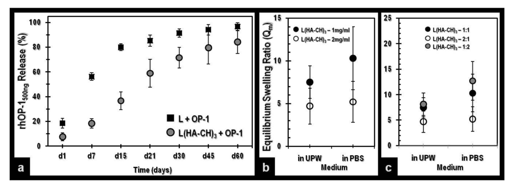
Conversely, bare liposomes accumulated with cooling and went through break down with the increase of temperature. The latter was visible to the naked eye, and expected. On the other hand, nano-structured bio-/muco-adhesive hybrid hydrogels with a biodegradable and porous network can be used as an artificial extra-cellular matrix for tissue regeneration and repair purposes as well as a therapeutic agent(s) delivery carrier for the localized and controlled release of single or multiple biomolecules via a diffusion mechanism. The release profiles of rhOP-1 from bare liposomes and L(HA-CH)3 nanogel were monitored over a prolonged period of 60 days and the cumulative views are displayed in Figure 3(a). A more controlled, linear and multi-phasic release was demonstrated by the L(HA-CH)3 nanogel when compared to the liposome-associated burst and faster release. This phenomenon can be explained by the ability of the multi-layers to control the diffusion of the growth factor from the core and through the different compartments. We have seen previously that with increasing number of layers, a more compact shell is formed due to the complexation of the polymers forming a denser network that can be further tailored for slower or faster agent release [6,7,9,10]. However, when comparing the present data with the earlier L(AL-CH)3, we noted that HA contributes to forming a softer material yet with a slightly slower release kinetic. This, we tend to believe, can be associated with the change in liposomal size in addition to the nature of the polymers themselves being of a more concentrated/thicker format than before, thus, degrading slower and therefore, magnifying the potential of this L(HA-CH)3 hydrogel for its intended application(s) without a present need for any functionalization or cross-linker.
As for most hydrogels, swelling and degradation properties are of key importance. The swelling ratio of some of the different L(HA-CH)3 samples analyzed (w/v and v/v) are reported in Figure 3(b). Degradability of the system is not shown. Triplicates were prepared either in UPW or PBS (pH: 7.4). The equilibrium mass swelling ratio (Qm) was calculated using the equation: Qm(%) = (ms)/(md × 100), reported by Benchrif et al. [15]; where ms is the swollen gel weight and md is that of the dried gel. For PBS samples, soluble salt fraction was subtracted, for a correct comparison with UPW. Our findings confirmed a significant difference (ρ < 0.05) between and not among groups. Collectively, increasing the core-shell nanogel content within the hydrogel matrix is directly proportional to an increase in the total solid content resulting in a greater impact on the swelling behavior and respective ratio of the preparation, i.e., less swelling was noted when HA and CH were prepared in a 1 mg/mL concentration and surrounded the liposomal core in a 2:1 ratio. Hence, tunable size, charge and water content (a modifiable surface area for a likely ligand-conjugation) are attainable.
4. Concluding Remarks
To the best of our knowledge, this is the first report on the formulation of a hybrid liposome-based hyaluronic acid-chitosan nanogel for the encapsulation and release of bone morphogenetic proteins. A viscous, sticky, stable, biodegradable and reproducible gel resulted. Linear/sustained protein release was evident over a prolonged duration of time. The self-assembly process or component organization into structurally well-defined aggregates, forming a hydrogel, can be characterized by numerous beneficial attributes: versatility, ease to manufacture/handle and potential cost-effectiveness. Our ongoing studies investigate the efficacy of this L(HA-CH)3 hydrogel system in a model of cranial BTE.


Acknowledgments
This work was supported by the South Korean Ministry of Knowledge and Education (MKE) and the Incheon Free Economic Zone (IFEZ) in the framework of several funding operating grants to the Utah-Inha Drug Delivery Systems (DDS) and Advanced Therapeutics Research Center, Repub. South Korea. The authors thank the INHA University Biomedical Polymers research team for their technical support.
References
- Weinstein, S.L. 2000–2010: The bone and joint decade. J. Bone Joint Surg. Am. 2000, 82, 1–3. [Google Scholar]
- Mussano, F.; Ciccone, G.; Ceccarelli, M.; Baldi, I.; Bassi, F. Bone morphogenetic proteins and bone defects: A systematic review. Spine 2007, 32, 824–830. [Google Scholar]
- Haidar, Z.S.; Hamdy, R.C.; Tabrizian, M. Delivery of recombinant bone morphogenetic proteins for bone regeneration and repair. Part B: Delivery systems for BMPs in orthopaedic and craniofacial tissue engineering. Biotechnol. Lett. 2009, 31, 1825–1835. [Google Scholar]
- Gonçalves, C.; Pereira, P.; Gama, M. Self-assembled hydrogel nanoparticles for drug deliveryapplications. Materials 2010, 3, 1420–1460. [Google Scholar]
- Haidar, Z.S. Bio-inspired/-functional colloidal core-shell polymeric-based nanosystems: Technology promise in tissue engineering, bioimaging and nanomedicine. Polymers 2010, 2, 323–352. [Google Scholar]
- Haidar, Z.S.; Hamdy, R.C.; Tabrizian, M. Protein release kinetics for core-shell hybrid nanoparticles based on the layer-by-layer assembly of alginate and chitosan on liposomes. Biomaterials 2008, 29, 1207–1215. [Google Scholar]
- Haidar, Z.S.; Azari, F.; Hamdy, R.C.; Tabrizian, M. Modulated release of OP-1 and enhanced pre-osteoblast differentiation using a core-shell nanoparticulate system. J. Biomed. Mater. Res. A 2009, 91, 919–928. [Google Scholar]
- Haidar, Z.S.; Hamdy, R.C.; Tabrizian, M. Delivery of recombinant bone morphogenetic proteins for bone regeneration and repair. Part A: Current challenges in BMP delivery. Biotechnol. Lett. 2009, 31, 1817–1824. [Google Scholar]
- Haidar, Z.S.; Tabrizian, M.; Hamdy, R.C. A hybrid OP-1 delivery system enhances new bone regeneration and consolidation in a rabbit model of distraction osteogenesis. Growth Factors 2010, 28, 44–55. [Google Scholar]
- Haidar, Z.S.; Hamdy, R.C.; Tabrizian, M. Biocompatibility and safety of a hybrid core-shell nanoparticulate OP-1 delivery system intramuscularly administered in rats. Biomaterials 2010, 31, 2746–2754. [Google Scholar]
- Shidhaye, S.; Lotlikar, V.; Malke, S.; Kadam, V. Nanogel engineered polymeric micelles for drug delivery. Curr. Drug Ther. 2008, 3, 209–217. [Google Scholar]
- Kabanov, A.V.; Vinogradov, S.V. Nanogels as pharmaceutical carriers: Finite networks of infinite capabilities. Angew. Chem. Int. Ed. Engl. 2009, 48, 5418–5429. [Google Scholar]
- Hubbell, J.A. Materials as morphogenetic guides in tissue engineering. Curr. Opin. Biotechnol. 2003, 5, 551–558. [Google Scholar]
- Patterson, J.; Siew, R.; Herring, S.W.; Lin, A.S.; Guldberg, R.; Stayton, P.S. Hyaluronic acid hydrogels with controlled degradation properties for oriented bone regeneration. Biomaterials 2010, 26, 6772–6781. [Google Scholar]
- Bencherif, S.A.; Siegwart, D.J.; Srinivasan, A.; Horkay, F.; Hollinger, J.O.; Washburn, N.R.; Matyjaszewski, K. Nanostructured hybrid hydrogels prepared by a combination of atom transfer radical polymerization and free radical polymerization. Biomaterials 2009, 29, 5270–5278. [Google Scholar]
© 2011 by the authors; licensee MDPI, Basel, Switzerland. This article is an open access article distributed under the terms and conditions of the Creative Commons Attribution license (http://creativecommons.org/licenses/by/3.0/).
Share and Cite
Joo, V.; Ramasamy, T.; Haidar, Z.S. A Novel Self-Assembled Liposome-Based Polymeric Hydrogel for Cranio-Maxillofacial Applications: Preliminary Findings. Polymers 2011, 3, 967-974. https://doi.org/10.3390/polym3020967
Joo V, Ramasamy T, Haidar ZS. A Novel Self-Assembled Liposome-Based Polymeric Hydrogel for Cranio-Maxillofacial Applications: Preliminary Findings. Polymers. 2011; 3(2):967-974. https://doi.org/10.3390/polym3020967
Chicago/Turabian StyleJoo, Victor, Thiruganesh Ramasamy, and Ziyad S. Haidar. 2011. "A Novel Self-Assembled Liposome-Based Polymeric Hydrogel for Cranio-Maxillofacial Applications: Preliminary Findings" Polymers 3, no. 2: 967-974. https://doi.org/10.3390/polym3020967
APA StyleJoo, V., Ramasamy, T., & Haidar, Z. S. (2011). A Novel Self-Assembled Liposome-Based Polymeric Hydrogel for Cranio-Maxillofacial Applications: Preliminary Findings. Polymers, 3(2), 967-974. https://doi.org/10.3390/polym3020967