<?xml version="1.0" encoding="UTF-8"?>
<!DOCTYPE article PUBLIC "-//NLM//DTD Journal Publishing DTD v2.3 20070202//EN" "journalpublishing.dtd">
<article xmlns:xlink="http://www.w3.org/1999/xlink" xml:lang="en" article-type="review-article">
<front>
<journal-meta>
<journal-id journal-id-type="publisher-id">Polymers</journal-id>
<journal-title>Polymers</journal-title>
<issn pub-type="epub">2073-4360</issn>
<publisher>
<publisher-name>Molecular Diversity Preservation International (MDPI)</publisher-name></publisher></journal-meta>
<article-meta>
<article-id pub-id-type="doi">10.3390/polym3010100</article-id>
<article-id pub-id-type="publisher-id">polymers-03-00100</article-id>
<article-categories>
<subj-group>
<subject>Review</subject></subj-group></article-categories>
<title-group>
<article-title>Chitosan-Based Hyaluronic Acid Hybrid Polymer Fibers as a Scaffold Biomaterial for Cartilage Tissue Engineering</article-title></title-group>
<contrib-group>
<contrib contrib-type="author">
<name><surname>Iwasaki</surname><given-names>Norimasa</given-names></name><xref ref-type="aff" rid="af1-polymers-03-00100"><sup>1</sup></xref><xref ref-type="corresp" rid="c1-polymers-03-00100"><sup>*</sup></xref></contrib>
<contrib contrib-type="author">
<name><surname>Kasahara</surname><given-names>Yasuhiko</given-names></name><xref ref-type="aff" rid="af1-polymers-03-00100"><sup>1</sup></xref></contrib>
<contrib contrib-type="author">
<name><surname>Yamane</surname><given-names>Shintarou</given-names></name><xref ref-type="aff" rid="af1-polymers-03-00100"><sup>1</sup></xref></contrib>
<contrib contrib-type="author">
<name><surname>Igarashi</surname><given-names>Tatsuya</given-names></name><xref ref-type="aff" rid="af1-polymers-03-00100"><sup>1</sup></xref></contrib>
<contrib contrib-type="author">
<name><surname>Minami</surname><given-names>Akio</given-names></name><xref ref-type="aff" rid="af1-polymers-03-00100"><sup>1</sup></xref></contrib>
<contrib contrib-type="author">
<name><surname>Nisimura</surname><given-names>Shin-ichiro</given-names></name><xref ref-type="aff" rid="af2-polymers-03-00100"><sup>2</sup></xref></contrib></contrib-group>
<aff id="af1-polymers-03-00100">
<label>1</label> Department of Orthopaedic Surgery, School of Medicine, Hokkaido University, Kita 15, Nishi 7, Sapporo 060-8638, Japan; E-Mails: <email>kasa14@rose.ocn.ne.jp</email> (Y.K.); <email>yamane629@yahoo.co.jp</email> (S.Y.); <email>iga@amber.plala.or.jp</email> (T.I.); <email>a-minami@med.hokudai.ac.jp</email> (A.M.)</aff>
<aff id="af2-polymers-03-00100">
<label>2</label> Graduate School of Advanced Life Science, Frontier Research Center for Post-Genomic Science and Technology, Hokkaido University, Kita 10, Nishi 8, Sapporo 060-0810, Japan; E-Mail: <email>shin@glyco.sci.hokudai.ac.jp</email> (S.N.)</aff>
<author-notes>
<corresp id="c1-polymers-03-00100">
<label>*</label> Author to whom correspondence should be addressed; E-Mail: <email>niwasaki@med.hokudai.ac.jp</email>; Tel.: +81-11-706-5937; Fax: +81-11-706-6054.</corresp></author-notes>
<pub-date pub-type="collection">
<year>2011</year></pub-date>
<pub-date pub-type="epub">
<day>27</day>
<month>12</month>
<year>2010</year></pub-date>
<volume>3</volume>
<issue>1</issue>
<fpage>100</fpage>
<lpage>113</lpage>
<history>
<date date-type="received">
<day>13</day>
<month>10</month>
<year>2010</year></date>
<date date-type="rev-recd">
<day>24</day>
<month>11</month>
<year>2010</year></date>
<date date-type="accepted">
<day>24</day>
<month>12</month>
<year>2010</year></date></history>
<permissions>
<copyright-statement>© 2010 by the authors; licensee MDPI, Basel, Switzerland.</copyright-statement>
<copyright-year>2011</copyright-year>
<license>
<p>This article is an open access article distributed under the terms and conditions of the Creative Commons Attribution license (http://creativecommons.org/licenses/by/3.0/).</p></license></permissions>
<abstract>
<p>An ideal scaffold material is one that closely mimics the natural environment in the tissue-specific extracellular matrix (ECM). Therefore, we have applied hyaluronic acid (HA), which is a main component of the cartilage ECM, to chitosan as a fundamental material for cartilage regeneration. To mimic the structural environment of cartilage ECM, the fundamental structure of a scaffold should be a three-dimensional (3D) system with adequate mechanical strength. We structurally developed novel polymer chitosan-based HA hybrid fibers as a biomaterial to easily fabricate 3D scaffolds. This review presents the potential of a 3D fabricated scaffold based on these novel hybrid polymer fibers for cartilage tissue engineering.</p></abstract>
<kwd-group>
<kwd>chitosan</kwd>
<kwd>hyaluronic acid</kwd>
<kwd>hybrid polymer fiber</kwd>
<kwd>cartilage tissue engineering</kwd>
<kwd>scaffold</kwd></kwd-group></article-meta></front>
<body>
<sec sec-type="intro">
<label>1.</label>
<title>Introduction</title>
<p>The substrate for most cells in living organisms is the extracellular matrix (ECM). The ECM adheres to cells via integrins, which are membrane-spanning heterodimeric receptors. Through the cell-matrix interactions, the ECM transduces physiological signals regulating cell differentiation, cell proliferation, cell apoptosis, matrix synthesis, and matrix remodeling to the cells [<xref ref-type="bibr" rid="b1-polymers-03-00100">1</xref>]. One of the notable characteristics of cartilage tissue is that a small number of chondrocytes, which are the sole cells in this tissue, are embedded in the rich ECM. Consequently, the ECM plays a crucial role in cartilage tissue development and regeneration.</p>
<p>The limited potential of articular cartilage for self-repair necessitates surgical procedures to treat injured cartilage [<xref ref-type="bibr" rid="b2-polymers-03-00100">2</xref>-<xref ref-type="bibr" rid="b5-polymers-03-00100">5</xref>]. However, no current procedures for cartilage repair have successfully regenerated long-lasting hyaline cartilage tissue to replace cartilaginous lesions. Tissue engineering techniques involving culturing isolated chondrocytes on biocompatible and biodegradable scaffold materials, including naturally occurring and synthetic materials, have been considered the ideal procedures for treating such lesions (<xref ref-type="table" rid="t1-polymers-03-00100">Table 1</xref>) [<xref ref-type="bibr" rid="b6-polymers-03-00100">6</xref>-<xref ref-type="bibr" rid="b13-polymers-03-00100">13</xref>]. These techniques require three important factors: scaffolds, cell sources, and signals, for successful tissue regeneration. A number of studies have also suggested the importance of selecting appropriate biomaterials as scaffolds for cell adhesion and proliferation [<xref ref-type="bibr" rid="b7-polymers-03-00100">7</xref>-<xref ref-type="bibr" rid="b15-polymers-03-00100">15</xref>]. For the reason mentioned above, in cartilage tissue engineering, scaffold materials should act as the tissue-specific ECM.</p>
<p>Scaffolds for cartilage tissue engineering require two different potentials to endure against biomechanically stressed conditions and to support chondrogenesis while maintaining the chondrocyte phenotype. Unfortunately, most scaffolds developed to date for cartilage regeneration conform to only one of these requirements. To meet these biomechanical and biological requirements, the authors have developed a novel three-dimensional (3D) scaffold fabricated from chitosan-based hyaluronic acid (HA) hybrid polymer fibers [<xref ref-type="bibr" rid="b16-polymers-03-00100">16</xref>-<xref ref-type="bibr" rid="b18-polymers-03-00100">18</xref>]. Previous studies have shown that cellular functions differ in 3D and two-dimensional culture systems [<xref ref-type="bibr" rid="b19-polymers-03-00100">19</xref>,<xref ref-type="bibr" rid="b20-polymers-03-00100">20</xref>]. In cartilage tissue engineering, a closer approximation to natural environments should be attained by culturing cells in 3D materials. To structurally mimic the environments of the cartilage tissue, a scaffold must be a 3D system with adequate mechanical strength.</p>
<p>Here, we mainly present the feasibility of our 3D scaffold fabricated from chitosan-based HA hybrid polymer fibers for cartilage tissue engineering. This review is based on the data derived from our previous <italic>in vivo</italic> and <italic>in vitro</italic> studies [<xref ref-type="bibr" rid="b16-polymers-03-00100">16</xref>-<xref ref-type="bibr" rid="b18-polymers-03-00100">18</xref>,<xref ref-type="bibr" rid="b21-polymers-03-00100">21</xref>]. In this paper, statistical comparisons were performed using one-way analysis of variance (ANOVA) and Fisher's PLSD tests. Differences were considered significant at p &lt; 0.05.</p></sec>
<sec>
<label>2.</label>
<title>Development of a 3D Scaffold Fabricated from Chitosan-Based HA Hybrid Polymer Fibers</title>
<sec>
<label>2.1.</label>
<title>Chitosan-Based HA Hybrid Polymer Fiber Preparation</title>
<p>Regarding cartilage regeneration, the ideal cell carrier substance is one that closely mimics the natural environment in the cartilage-specific ECM [<xref ref-type="bibr" rid="b12-polymers-03-00100">12</xref>]. Cartilage ECM mainly consists of type II collagen and glycosaminoglycans (GAGs). Given the importance of GAGs in enhancing chondrogenesis <italic>in vitro</italic>, the uses of GAGs or GAGs-like biomaterials as components of a scaffold material are likely to be a reasonable approach for enhancing chondrogenesis [<xref ref-type="bibr" rid="b11-polymers-03-00100">11</xref>,<xref ref-type="bibr" rid="b12-polymers-03-00100">12</xref>,<xref ref-type="bibr" rid="b22-polymers-03-00100">22</xref>].</p>
<p>Chitosan is a partially deacetylated derivative of chitin, the primary structural polymer in arthropod exoskeletons. This natural material is a linear polysaccharide consisting of β(1→4) linked D-glucosamine residues with a variable number of randomly located N-acetyl-glucosamine groups. The average molecular weight ranges from 50 to 1,000 kDa. The potential of chitosan as a biomaterial is based on its cationic nature and high charge density in solution. Madihally <italic>et al.</italic> [<xref ref-type="bibr" rid="b9-polymers-03-00100">9</xref>] reported that the cationic nature of chitosan allowed for electrostatic interactions with anionic GAGs, proteoglycans (PGAs), and other negatively charged species. These ionic interactions may serve as a mechanism for retaining and recruiting cells, growth factors, and cytokines within a tissue scaffold. Therefore, chitosan has been used as a good compatible material for tissue repair and wound healing [<xref ref-type="bibr" rid="b11-polymers-03-00100">11</xref>,<xref ref-type="bibr" rid="b23-polymers-03-00100">23</xref>-<xref ref-type="bibr" rid="b25-polymers-03-00100">25</xref>]. In cartilage tissue engineering, previous studies have shown that this material possesses promising potential as a carrier material for the transplant of chondrocytes [<xref ref-type="bibr" rid="b12-polymers-03-00100">12</xref>,<xref ref-type="bibr" rid="b26-polymers-03-00100">26</xref>-<xref ref-type="bibr" rid="b30-polymers-03-00100">30</xref>].</p>
<p>Since chitosan is considered to be a cationic polysaccharide showing excellent cell supporting properties, a hybrid material composed of chitosan combined with GAGs may be a novel class of polyion complex effective for cartilage specific scaffolds [<xref ref-type="bibr" rid="b11-polymers-03-00100">11</xref>]. Hyaluronan is a linear GAG composed, on average, of 1 × 10<sup>4</sup> disaccharide units of glucuronic acid and N-acetylglucosamine, with a molecular weight of 1,000–5,000 kDa. This GAG is a main component of the ECM of articular cartilage and plays a crucial role in regulating the behavior of chondrocytes. Concerning the biological roles of HA, Zimmermann <italic>et al.</italic> [<xref ref-type="bibr" rid="b31-polymers-03-00100">31</xref>] demonstrated that HA is an adhesion modulator molecule, which can mediate the early stage of cell-substrate interaction. CD 44 acts as a cell surface receptor for HA [<xref ref-type="bibr" rid="b32-polymers-03-00100">32</xref>,<xref ref-type="bibr" rid="b33-polymers-03-00100">33</xref>]. Murdoch <italic>et al.</italic> [<xref ref-type="bibr" rid="b34-polymers-03-00100">34</xref>] showed a dramatic increase in CD44 expression on chondrocytes isolated from the cartilage. These data indicate that scaffold materials introducing HA can provide excellent adhesivity for seeded chondrocytes and enhance the biological behavior of the chondrocytes on the scaffolds.</p>
<p>
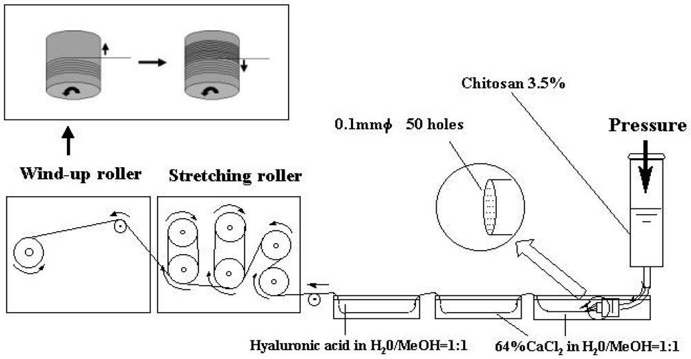
In articular cartilage tissue engineering, we must address that the articular cartilage is subject to excessive mechanical stress. Consequently, mechanical strength with high cellular adhesivity to maintain the number of seeded chondrocytes is a requirement for scaffold materials. To meet this requirement, we developed a novel chititosan-based HA introduced hybrid polymer fiber as a fundamental material for 3D fabricated scaffolds. Polymer fibers were developed by the wet spinning method as described by Tamura <italic>et al.</italic> [<xref ref-type="bibr" rid="b35-polymers-03-00100">35</xref>] with the following modification. Viscosity average molecular weight of HA was 2,400 kDa. The degree of deacetylation of the chitosan was 81%, and viscosity average molecular weight was 600 kDa. To prepare the polymer fibers, 7 g of chitosan powder was dissolved in 200 mL of 2% aqueous acetic acid solution to give 3.5% of polymer concentration. Dope of chitosan was spun into a calcium coagulant bath (64% CaCl<sub>2</sub> dissolved in 50% aqueous methanol solution) through a stainless steel spinnlet (0.1 mm diameter, 50 holes) at a winding speed of 4.4 m/min at room temperature. Then, 50% aqueous methanol solution was used as a second coagulation bath and hyaluronic acid dissolved in 50% aqueous methanol solution as a third coagulation bath. Using an original roller system (Okada Co., Inc., Sapporo, Japan), the resulting fibers were stretched and treated with 0.8% sodium hydroxide (NaOH) dissolved in 90% aqueous methanol solution to neutralize the acidity of the fibers (<xref ref-type="fig" rid="f1-polymers-03-00100">Figure 1</xref>). The fibers wound in the roller were washed with methanol and dried at room temperature. The diameter of each fiber was 30 μm (<xref ref-type="fig" rid="f2-polymers-03-00100">Figure 2</xref>). The developed hybrid fibers were sterilized in an autoclave at 135 °C for 20 minutes. The tensile strength of the hybrid polymer fiber was determined by the concentration of the introduced HA. The values of the HA hybrid fiber significantly increased compared with that of the chitosan fiber (87 N/mm<sup>2</sup> in the chitosan fibers, 168 N/mm<sup>2</sup> in the chitosan introduced with HA 0.04% fibers, and 144 N/mm<sup>2</sup> in the chitosan introduced with HA 0.07% fibers).</p>
<p>Regarding the diameter of fibers for fabricated scaffolds used in cartilage tissue engineering, Neurnberger <italic>et al.</italic> [<xref ref-type="bibr" rid="b36-polymers-03-00100">36</xref>] suggested that fiber sizes smaller than chondrocytes are beneficial in terms of cellular adhesion and maintainance of the chondrocyte phenotype. The diameter of chondrocytes is approximately 10 to 30 μm. As mentioned above, the diameter of our fibers in dry condition is 30 μm. Due to biodegradability, the diameter in a culture medium or living joints must be smaller than 30 μm. In view of this point, we determined the diameter of the developed fibers.</p></sec>
<sec>
<label>2.2.</label>
<title>Biological Effects of the Hybrid Polymer Fiber on Chondrocyte Behavior</title>
<p><xref ref-type="table" rid="t2-polymers-03-00100">Tables 2</xref> and <xref ref-type="table" rid="t3-polymers-03-00100">3</xref> summarize the data regarding the biological effects of the developed hybrid polymer fibers on cellular behaviors [<xref ref-type="bibr" rid="b16-polymers-03-00100">16</xref>]. The DNA content of the chondrocytes at seven days after cultivation indicates the degree of chondrocyte proliferation on the scaffold materials. The obtained data suggested that adhesion, proliferation, and ECM products of the chondrocytes significantly increased on the hybrid polymer fibers as compared to the non-hybrid chitosan fiber. Light microscopy and scanning electron microscopy (SEM) showed the maintenance of the characteristic round morphology of the cultured chondrocytes on the hybrid fibers (<xref ref-type="fig" rid="f3-polymers-03-00100">Figure 3</xref>). The SEM images also revealed dense fibrous tissue indicating type II collagen on the hybrid fibers (<xref ref-type="fig" rid="f3-polymers-03-00100">Figure 3</xref>). The obtained data suggest the superior biological effects of our novel hybrid fibers on chondrocyte activities.</p></sec>
<sec>
<label>2.3.</label>
<title>Determination of Adequate Pore Size for a Novel 3D Scaffold for Cartilage Tissue Regeneration</title>
<p>
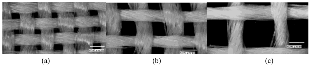
Fiber materials can provide adequate mechanical strength for a 3D scaffold of cartilage tissue engineering. Additionally, they easily control the macroscopic and microscopic scaffold structures. One of the most important factors for developing a 3D cartilage tissue engineering scaffold is the pore size of the scaffold material. To determine an adequate pore size, we performed an experiment to clarify the effects of pore size of the developed scaffolds on chondrocyte behavior. From the chitosan-based HA hybrid polymer fibers, which are introduced with 0.07% hyaluronic acid, the 3D scaffolds with three different pore sizes (100, 200, and 400 μm diameter) were woven by using the original apparatus (<xref ref-type="fig" rid="f4-polymers-03-00100">Figure 4</xref>). The effects on the ECM products of each scaffold material are summarized in <xref ref-type="table" rid="t4-polymers-03-00100">Table 4</xref> [<xref ref-type="bibr" rid="b17-polymers-03-00100">17</xref>]. The obtained data suggested that the current scaffold with 400 μm pore size significantly increased ECM synthesis by cultured chondrocytes.</p>
<p>Previous studies have shown that the scaffold pore size is highly interconnected with cell proliferation and ECM synthesis in chondrocyte culture [<xref ref-type="bibr" rid="b10-polymers-03-00100">10</xref>,<xref ref-type="bibr" rid="b37-polymers-03-00100">37</xref>,<xref ref-type="bibr" rid="b38-polymers-03-00100">38</xref>]. Nehrer <italic>et al.</italic> [<xref ref-type="bibr" rid="b10-polymers-03-00100">10</xref>] clarified the effects of scaffold pore sizes, from 20 to 86 μm, on chondrocyte behavior using collagen sponges. They suggested that the cultured cells on the material with small pore diameter lost the chondrocytic morphology over time. Using poly (ethylene-glycol)-terephthalate/poly (butylenes terephthalate) scaffolds with different pore sizes (182 μm and 525 μm), Malda <italic>et al.</italic> [<xref ref-type="bibr" rid="b38-polymers-03-00100">38</xref>] demonstrated that large pore size scaffolds significantly increased the GAG production of cultured chondrocytes. These previous and our results suggest great promise for the future of a fabricated scaffold with a relatively large pore size, such as 400 μm, for cartilage regeneration.</p></sec>
<sec>
<label>2.4.</label>
<title>Engineered Cartilage Development Using the Fabricated 3D Scaffold from the Chitosan-Based HA Hybrid Polymer Fibers</title>
<p>
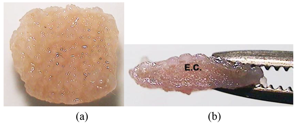
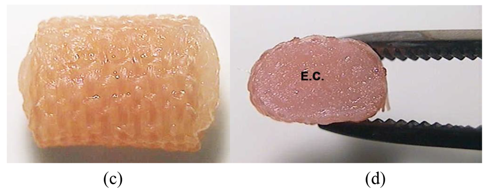
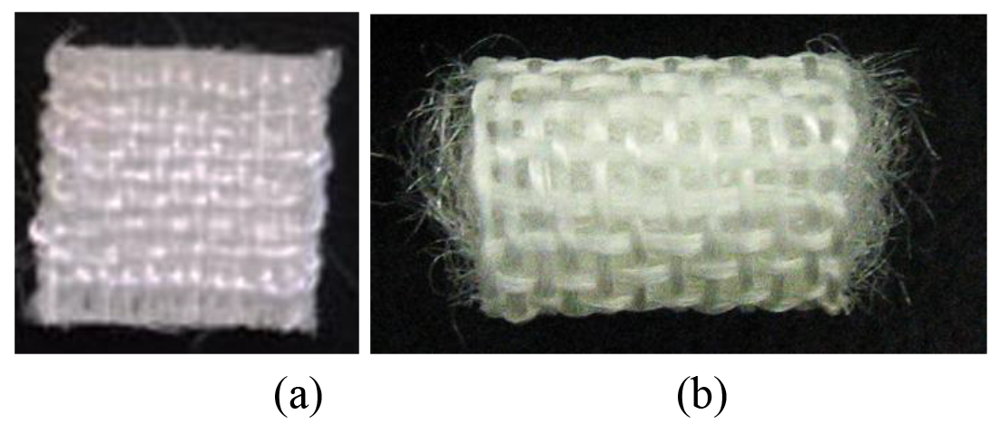
In clinical usage of the developed scaffold material, we must inhibit inflammatory reactions and an acute decrease in mechanical strength of the implanted tissue during the biodegradation process of the material. One of the ideal strategies for addressing this issue is to reduce the volume of scaffold material in engineered constructs with mechanically mature cartilage. For this strategy, we developed two types of 3D scaffolds fabricated with the chitosan-based HA hybrid polymer fibers [<xref ref-type="bibr" rid="b18-polymers-03-00100">18</xref>]. One was a cushion-type scaffold, consisting of two sheets, which were 8 × 8 mm wide and 1 mm thick. The other was a 10 mm high cylinder-type scaffold, which had a 6 mm diameter (<xref ref-type="fig" rid="f5-polymers-03-00100">Figure 5</xref>). Based on the obtained data, the pore size of both scaffolds was 400 μm. Although the volume of each scaffold material was minimized, both scaffolds retained the initial shape under a dynamic culture condition.</p>
<p>
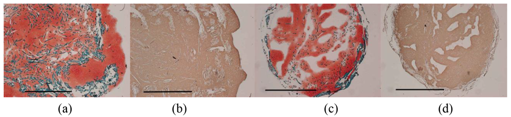
To prepare tissue-engineered constructs, chondrocytes were isolated from eight week-old Japanese white rabbits according to sequential enzyme digestion under sterile conditions as described previously [<xref ref-type="bibr" rid="b39-polymers-03-00100">39</xref>]. A chondrocyte suspension containing 3 × 10<sup>5</sup> and 7 × 10<sup>5</sup> cells was seeded onto the cushion-type (cushion group) and the cylinder-type (cylinder group) scaffold. The samples were placed on a 48-well plate in a 37 °C, humidified 5% CO<sub>2</sub> incubator for 1 hour and then overlaid with 1 mL of the culture medium. After one-week static culture, these samples were transferred into a disposable high aspect ratio vessel bioreactor (HARV, 50 mL; Synthecon, Inc., Houston, TX, U.S.) and dynamically cultured for a further four or seven weeks. <xref ref-type="table" rid="t5-polymers-03-00100">Table 5</xref> summarizes biochemical and biomechanical evaluations of engineered cartilage in both material groups at each time point [<xref ref-type="bibr" rid="b18-polymers-03-00100">18</xref>]. At eight weeks after cultivation, macroscopic (<xref ref-type="fig" rid="f6-polymers-03-00100">Figure 6</xref>) and histological (<xref ref-type="fig" rid="f7-polymers-03-00100">Figure 7</xref>) findings suggested successful hyaline-like cartilage regeneration with rich GAG and type II collagen products. Regarding the biomechanical properties of regenerated tissues, the cushion-shape scaffold significantly increased the Young's modulus of regenerated tissue from five to eight weeks after cultivation. On the other hand, the cylinder-shape scaffold did not alter the value during the culture period. Although the Young's modulus of the cylinder group was significantly inferior to that of the cushion group, the value was comparable to that of normal rabbit cartilage. Regarding the reason for the high stiffness of the engineered cartilage in the cushion-type scaffold, the stiffness of the scaffold material may affect the value of the engineered tissue. The obtained results indicate that our novel 3D scaffolds regenerate histologically and mechanically mature tissue.</p></sec></sec>
<sec>
<label>3.</label>
<title>Animal Model Experiments</title>
<p>For future clinical application of our scaffold materials, we assessed the reparative tissues treated with the implantation of mature engineered-cartilage constructs with the developed 3D scaffolds using a rabbit model. Mature female Japanese white rabbits weighing 2.6 to 3.1 kg were used for the current analysis. Under general anesthesia using intravenous pentobarbital (0.05 mg/kg) followed by isoflurane in oxygen gas anesthesia, through a medial parapatellar arthrotomy, a full-thickness osteochondral defect 5 mm in diameter and 2 mm deep was made by an electric-powered drill on the patellar groove of the bilateral distal femur. Previous studies have shown that such defects fail to repair spontaneously [<xref ref-type="bibr" rid="b40-polymers-03-00100">40</xref>,<xref ref-type="bibr" rid="b41-polymers-03-00100">41</xref>]. The tissue-engineered constructs using each scaffold—cushion-type or cylinder-type scaffold—were press-fit implanted into the defects after an eight week-cultivation in both treatment groups. Postoperatively, all animals were kept in a separate cage and allowed to move freely. For evaluation, animals were euthanized at 12 weeks postoperatively and the femoral condyles were harvested and fixed in 10% buffered formalin. Each experimental group consisted of seven rabbits.</p>
<p>
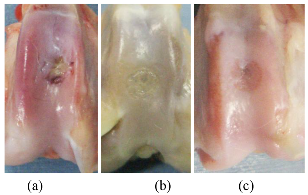
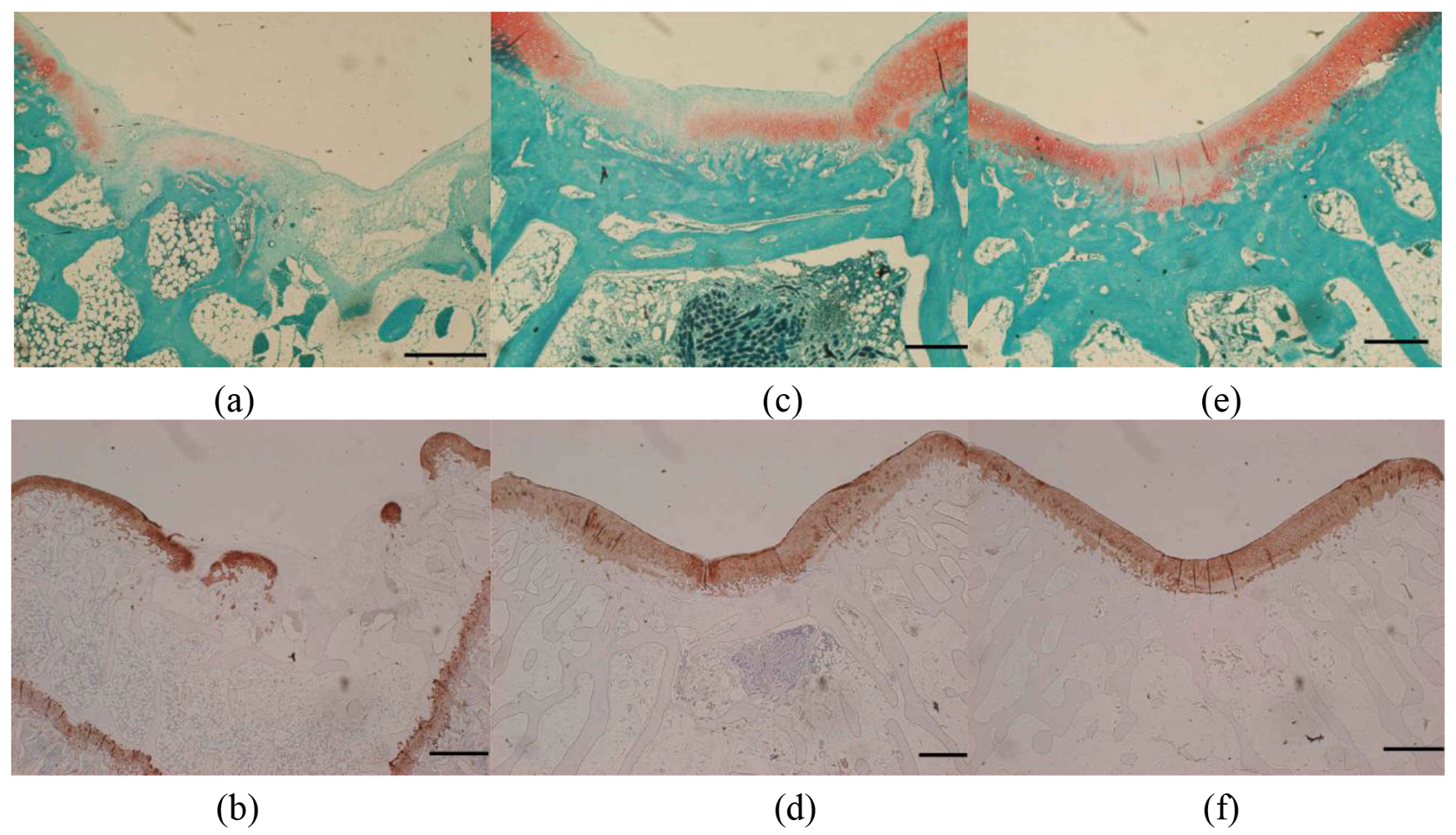
Apparent joint effusion and synovitis, which indicate inflammatory reaction, were not found in any animals at euthanasia. The gross appearance of osteochondral defects showed a repair with cartilage-like tissue in both treatment groups over the no treatment group (<xref ref-type="fig" rid="f8-polymers-03-00100">Figure 8</xref>) [<xref ref-type="bibr" rid="b18-polymers-03-00100">18</xref>]. Histological findings also demonstrated that the defects in both treatment groups were filled with hyaline-like cartilage or a combination of hyaline-like cartilage and fibrocartilage (<xref ref-type="fig" rid="f9-polymers-03-00100">Figure 9</xref>) [<xref ref-type="bibr" rid="b18-polymers-03-00100">18</xref>]. <xref ref-type="table" rid="t6-polymers-03-00100">Table 6</xref> summarizes the <italic>in vivo</italic> quantitative evaluation of reparative tissues [<xref ref-type="bibr" rid="b18-polymers-03-00100">18</xref>]. The histological score according to the criteria of Wayne [<xref ref-type="bibr" rid="b42-polymers-03-00100">42</xref>] and the Young's modulus of both treatment groups overcame significantly those of the no treatment group. The Young's modulus of the treatment groups showed no significant difference, as compared to that of normal cartilage. The obtained results suggest that the implantation of tissues regenerated with our novel scaffolds plays functional roles in repairing osteochondral defects in living joints.</p>
<p>We successfully developed two types of scaffolds while minimizing their volume. A cushion-shape scaffold can be used to regenerate a large tissue. This has potential for repairing large cartilaginous lesions including osteoarthritis (OA) or rheumatoid arthritis (RA). A tubular-shape scaffold can create a cylindrical regenerated tissue. The implantation of tissues regenerated with this scaffold is an alternative treatment to osteochondral plug grafts such as mosaicplasty. Both scaffolds created from chitosan-based HA hybrid polymer fibers with seeding chondrocytes were able to maintain their initial shape and support cartilage regeneration during the eight week cultivation under a dynamic condition. The current tissue engineering technique using our hybrid polymer fibers can be used to form engineered cartilage constructs for a variety of sized or shaped osteochondral defects.</p></sec>
<sec sec-type="conclusions">
<label>4.</label>
<title>Conclusions</title>
<p>Clinical experience with cartilage tissue engineering for patients with cartilaginous lesions currently exceeds 15 years. However, prospective, comparative, and clinical trials have shown no significant superiority of this technique over other surgical procedures for the treatment of cartilage defects. To address the issues of current tissue engineering techniques, we have developed an original 3D scaffold fabricated from chitosan-based HA hybrid polymer fibers and succeeded in regenerating hyaline-like cartilage with a combination of this scaffold and a bioreactor system. Our experiment using a rabbit model demonstrated the potential of this engineered cartilage construct to enhance cartilage repair in living joints. Because of its ease of handling and press-fitting to osteochondral defects without any coverage, the current technique using the developed scaffold will provide technical advantages to surgeons and better clinical outcomes for patients. Long-term assessments using a large animal model are required to adapt the current approach for use in humans.</p></sec></body>
<back>
<sec sec-type="display-objects">
<title>Figures and Tables</title>
<fig id="f1-polymers-03-00100" position="float">
<label>Figure 1.</label>
<caption>
<p>Our original roller system for creating the chitosan-based HA hybrid polymer fibers [<xref ref-type="bibr" rid="b16-polymers-03-00100">16</xref>].</p></caption>
<graphic xlink:href="polymers-03-00100f1.gif"/></fig>
<fig id="f2-polymers-03-00100" position="float">
<label>Figure 2.</label>
<caption>
<p>Scanning electron micrograph of the chitosan-based HA hybrid polymer fibers [<xref ref-type="bibr" rid="b16-polymers-03-00100">16</xref>].</p></caption>
<graphic xlink:href="polymers-03-00100f2.gif"/></fig>
<fig id="f3-polymers-03-00100" position="float">
<label>Figure 3.</label>
<caption>
<p>Light microscopy and SEM images of chondrocytes seeded on the hybrid polymer fibers at 14 days after cultivation. The light micrograph shows the proliferation of chondrocytes seeded on the hybrid polymer fibers (<bold>a</bold>) The SEM images demonstrate the characteristic round morphology of the chondrocytes (<bold>b</bold>) and the dense fibers of the type II collagen around the chondrocytes (<bold>c</bold>) White arrows indicate chondrocytes. F, hybrid polymer fibers [<xref ref-type="bibr" rid="b16-polymers-03-00100">16</xref>].</p></caption>
<graphic xlink:href="polymers-03-00100f3.gif"/></fig>
<fig id="f4-polymers-03-00100" position="float">
<label>Figure 4.</label>
<caption>
<p>Macroscopic appearance of 3D fabricated scaffolds from the chitosan-based HA hybrid polymer fibers: (<bold>a</bold>) 100 μm pore size, (<bold>b</bold>) 200 μm pore size, and (<bold>c</bold>) 400 μm pore size [<xref ref-type="bibr" rid="b17-polymers-03-00100">17</xref>].</p></caption>
<graphic xlink:href="polymers-03-00100f4.gif"/></fig>
<fig id="f5-polymers-03-00100" position="float">
<label>Figure 5.</label>
<caption>
<p>Three-dimensional scaffolds fabricated with the chitosan-based HA hybrid polymer fibers: (<bold>a</bold>) cushion-type scaffold, (<bold>b</bold>) cylinder-type scaffold.</p></caption>
<graphic xlink:href="polymers-03-00100f5.gif"/></fig>
<fig id="f6-polymers-03-00100" position="float">
<label>Figure 6.</label>
<caption>
<p>Macroscopic appearance of engineered cartilage in the cushion-type (<bold>a</bold>, <bold>b</bold>) and in the cylinder-type (<bold>c</bold>, <bold>d</bold>) scaffold at eight weeks after cultivation. The engineered cartilage can be easily handled with forceps due to its stable mechanical property. E.C., engineered cartilage in the scaffold [<xref ref-type="bibr" rid="b18-polymers-03-00100">18</xref>].</p></caption>
<graphic xlink:href="polymers-03-00100f6a.gif"/>
<graphic xlink:href="polymers-03-00100f6b.gif"/></fig>
<fig id="f7-polymers-03-00100" position="float">
<label>Figure 7.</label>
<caption>
<p>Histological and immunohistochemical appearance of engineered cartilage in the cushion-type (<bold>a</bold>, <bold>b</bold>) and in the cylinder-type (<bold>c</bold>, <bold>d</bold>) scaffold at eight weeks after cultivation. Safranin-O staining shows the formation of a proteoglycan-rich cartilaginous matrix (a, c). Immunohistochemical staining with antitype II collagen demonstrates the rich production of type II collagen in engineered cartilage (b, d). Scale bars indicate 5 mm [<xref ref-type="bibr" rid="b18-polymers-03-00100">18</xref>].</p></caption>
<graphic xlink:href="polymers-03-00100f7.gif"/></fig>
<fig id="f8-polymers-03-00100" position="float">
<label>Figure 8.</label>
<caption>
<p>The gross appearance of osteochondral defects at 12 weeks postoperatively. The gross findings show a repair with cartilage-like tissue in both the cushion-type (<bold>b</bold>) and the cylinder-type (<bold>c</bold>) treatment groups over the no treatment group (<bold>a</bold>) [<xref ref-type="bibr" rid="b18-polymers-03-00100">18</xref>].</p></caption>
<graphic xlink:href="polymers-03-00100f8.gif"/></fig>
<fig id="f9-polymers-03-00100" position="float">
<label>Figure 9.</label>
<caption>
<p>Histological and immunohistochemical appearance of the osteochondral defect in the patellar groove of rabbit knee joint at 12 weeks postoperatively. Safranin-O staining (<bold>a</bold>, <bold>c</bold>, <bold>e</bold>) and immunohistochemical staining with antitype II collagen (<bold>b</bold>, <bold>d</bold>, <bold>f</bold>) demonstrate the rich production of GAGs and type II collagen in the reparative tissue of both the cushion-type treatment group (c, d) and the cylinder-type treatment group (e, f), compared to the no treatment group (a, b). Scale bars indicate 1 mm [<xref ref-type="bibr" rid="b18-polymers-03-00100">18</xref>].</p></caption>
<graphic xlink:href="polymers-03-00100f9.gif"/></fig>
<table-wrap id="t1-polymers-03-00100" position="float">
<label>Table 1.</label>
<caption>
<p>Scaffold biomaterials available for clinical application of cartilage tissue engineering.</p></caption>
<table frame="hsides" rules="groups">
<thead>
<tr>
<th align="center" valign="top"><bold>Natural biomaterials</bold></th>
<th align="center" valign="top"><bold>Synthetic biomaterials</bold></th></tr></thead>
<tbody>
<tr>
<td align="center" valign="top">Collagen</td>
<td align="center" valign="top">Polyglycolic acid (PGA)</td></tr>
<tr>
<td align="center" valign="top">Hyaluronic acid</td>
<td align="center" valign="top">Polylactic acid (PLA)</td></tr>
<tr>
<td align="center" valign="top">Fibrin glue</td>
<td align="center" valign="top">PGA/PLA acd</td></tr>
<tr>
<td align="center" valign="top">Alginate</td>
<td align="center" valign="top">Polydioxanone</td></tr>
<tr>
<td align="center" valign="top">Chitosan</td>
<td align="center" valign="top"/></tr></tbody></table></table-wrap>
<table-wrap id="t2-polymers-03-00100" position="float">
<label>Table 2.</label>
<caption>
<p>Chondrocyte adhesion and DNA content on chitosan-HA hybrid polymer fibers [<xref ref-type="bibr" rid="b16-polymers-03-00100">16</xref>].</p></caption>
<table frame="hsides" rules="groups">
<thead>
<tr>
<th align="center" valign="top"><bold>Scaffold Materials (n = 5)</bold></th>
<th align="center" valign="top"><bold>Cell Adhesion</bold></th>
<th align="center" valign="top"><bold>DNA content at 7 days after cultivation</bold></th></tr></thead>
<tbody>
<tr>
<td align="left" valign="top">Non-hybrid chitosan fiber</td>
<td align="center" valign="top">79 ± 2 %</td>
<td align="center" valign="top">134 ± 4 μg/sample</td></tr>
<tr>
<td align="left" valign="top">Chitosan−rcnt;HA hybrid fiber</td>
<td align="center" valign="top">95 ± 1 %<xref ref-type="table-fn" rid="tfn2-polymers-03-00100">*</xref></td>
<td align="center" valign="top">142 ± 11 μg/sample</td></tr>
<tr>
<td align="left" valign="top">Chitosan−cnt;HA hybrid fiber</td>
<td align="center" valign="top">91 ± 3 %<xref ref-type="table-fn" rid="tfn2-polymers-03-00100">*</xref></td>
<td align="center" valign="top">240 ± 23 μg/sample<xref ref-type="table-fn" rid="tfn3-polymers-03-00100">**</xref></td></tr></tbody></table>
<table-wrap-foot><fn id="tfn1-polymers-03-00100">
<p>Mean ± standard deviation.</p></fn><fn id="tfn2-polymers-03-00100">
<label>*</label>
<p>p &lt; 0.05 <italic>vs.</italic> non-hybrid chitosan fiber.</p></fn><fn id="tfn3-polymers-03-00100">
<label>**</label>
<p>p &lt; 0.05 <italic>vs.</italic> non-hybrid chitosan fiber and chitosan−0.04% HA hybrid polymer fiber.</p></fn></table-wrap-foot></table-wrap>
<table-wrap id="t3-polymers-03-00100" position="float">
<label>Table 3.</label>
<caption>
<p>Chondrocyte gene expression of cartilage specific ECM on chitosan-HA hybrid polymer fibers at 14 days after cultivation [<xref ref-type="bibr" rid="b16-polymers-03-00100">16</xref>].</p></caption>
<table frame="hsides" rules="groups">
<thead>
<tr>
<th align="center" valign="top"><bold>Scaffold Materials (n = 5)</bold></th>
<th align="center" valign="top"><bold>Type II Collagen</bold></th>
<th align="center" valign="top"><bold>Aggrecan</bold></th>
<th align="center" valign="top"><bold>Type I Collagen</bold></th></tr></thead>
<tbody>
<tr>
<td align="left" valign="top">Non-hybrid chitosan fiber</td>
<td align="center" valign="top">1.51 ± 0.01</td>
<td align="center" valign="top">No expression</td>
<td align="center" valign="top">0.45 ± 0.04</td></tr>
<tr>
<td align="left" valign="top">Chitosan−0.04%HA hybrid fiber</td>
<td align="center" valign="top">1.59 ± 0.07</td>
<td align="center" valign="top">1.07 ± 0.17</td>
<td align="center" valign="top">0.60 ± 0.11</td></tr>
<tr>
<td align="left" valign="top">Chitisan−0.07%HA hybrid fiber</td>
<td align="center" valign="top">1.37 ± 0.12</td>
<td align="center" valign="top">1.59 ± 0.09<xref ref-type="table-fn" rid="tfn5-polymers-03-00100">*</xref></td>
<td align="center" valign="top">0.43 ± 0.11</td></tr></tbody></table>
<table-wrap-foot><fn id="tfn4-polymers-03-00100">
<p>Mean ± standard deviation. Values are mean normalized ratio (experimental integrated density/GAPDH integrated density) of mRNA.</p></fn><fn id="tfn5-polymers-03-00100">
<label>*</label>
<p>p &lt; 0.05 <italic>vs.</italic> chitosan−0.04% HA hybrid polymer fiber.</p></fn></table-wrap-foot></table-wrap>
<table-wrap id="t4-polymers-03-00100" position="float">
<label>Table 4.</label>
<caption>
<p>Effects of scaffold pore size on gene expression of cartilage specific ECM and glycosaminoglycans product of chondrocytes at 28 days after cultivation [<xref ref-type="bibr" rid="b17-polymers-03-00100">17</xref>].</p></caption>
<table frame="hsides" rules="groups">
<thead>
<tr>
<th align="center" valign="top"><bold>Pore Size</bold><break/><bold>(n = 5)</bold></th>
<th align="center" valign="top"><bold>Type II</bold><break/><bold>Collagen</bold></th>
<th align="center" valign="top"><bold>Aggrecan</bold></th>
<th align="center" valign="top"><bold>Type I</bold><break/><bold>Collagen</bold></th>
<th align="center" valign="top"><bold>Type II/I</bold><break/><bold>Collagen Ratio</bold></th>
<th align="center" valign="top"><bold>Glycosaminoglycans</bold><break/>(μg/sample)</th></tr></thead>
<tbody>
<tr>
<td align="center" valign="top">100 μm</td>
<td align="center" valign="top">0.66 ± 0.08</td>
<td align="center" valign="top">0.79 ± 0.05</td>
<td align="center" valign="top">0.61 ± 0.11</td>
<td align="center" valign="top">1.12 ± 0.35</td>
<td align="left" valign="top">35.9 ± 2.8</td></tr>
<tr>
<td align="center" valign="top">200 μm</td>
<td align="center" valign="top">0.67 ± 0.13</td>
<td align="center" valign="top">0.64 ± 0.14</td>
<td align="center" valign="top">0.44 ± 0.10</td>
<td align="center" valign="top">1.63 ± 0.48</td>
<td align="left" valign="top">45.2 ± 3.1<xref ref-type="table-fn" rid="tfn7-polymers-03-00100">*</xref></td></tr>
<tr>
<td align="center" valign="top">400 μm</td>
<td align="center" valign="top">0.79 ± 0.08</td>
<td align="center" valign="top">0.67 ± 0.16</td>
<td align="center" valign="top">0.46 ± 0.19</td>
<td align="center" valign="top">1.95 ± 0.78<xref ref-type="table-fn" rid="tfn7-polymers-03-00100">*</xref></td>
<td align="left" valign="top">56.2 ± 5.9<xref ref-type="table-fn" rid="tfn7-polymers-03-00100">*</xref><sup>,</sup><xref ref-type="table-fn" rid="tfn8-polymers-03-00100">**</xref></td></tr></tbody></table>
<table-wrap-foot><fn id="tfn6-polymers-03-00100">
<p>Mean ± standard deviation. Values in type II collagen, aggrecan, type I collagen are mean normalized ratio (experimental integrated density/GAPDH integrated density) of mRNA. Those in glycosaminoglycans mean the content of each sample.</p></fn><fn id="tfn7-polymers-03-00100">
<label>*</label>
<p>p &lt; 0.05 <italic>vs.</italic> 100 μm;</p></fn><fn id="tfn8-polymers-03-00100">
<label>**</label>
<p>p &lt; 0.05 <italic>vs.</italic> 200 μm.</p></fn></table-wrap-foot></table-wrap>
<table-wrap id="t5-polymers-03-00100" position="float">
<label>Table 5.</label>
<caption>
<p>Biochemical and biomechanical evaluations of engineered cartilage in both scaffold materials [<xref ref-type="bibr" rid="b18-polymers-03-00100">18</xref>].</p></caption>
<table frame="hsides" rules="groups">
<thead>
<tr>
<th align="left" valign="top" rowspan="3"/>
<th colspan="2" align="center" valign="top"><bold>Cushion-type Scaffold (n = 5)</bold></th>
<th colspan="2" align="center" valign="top"><bold>Cylinder-type Scaffold (n = 5)</bold></th></tr>
<tr>
<th valign="bottom" colspan="4">
<hr/></th></tr>
<tr>
<th align="center" valign="top"><bold>5 weeks</bold></th>
<th align="center" valign="top"><bold>8 weeks</bold></th>
<th align="center" valign="top"><bold>5 weeks</bold></th>
<th align="center" valign="top"><bold>8 weeks</bold></th></tr></thead>
<tbody>
<tr>
<td align="left" valign="top">Total amount of DNA (μg)</td>
<td align="center" valign="top">53.8 ± 1.4</td>
<td align="center" valign="top">95.5 ± 2.1<xref ref-type="table-fn" rid="tfn10-polymers-03-00100">*</xref></td>
<td align="center" valign="top">97.9 ± 3.2</td>
<td align="center" valign="top">132.3 ± 6.6<xref ref-type="table-fn" rid="tfn11-polymers-03-00100">†</xref></td></tr>
<tr>
<td align="left" valign="top">Total amount of protein (μg)</td>
<td align="center" valign="top">1108.4 ± 49.3</td>
<td align="center" valign="top">2,178.9 ± 114.5<xref ref-type="table-fn" rid="tfn10-polymers-03-00100">*</xref></td>
<td align="center" valign="top">1,655.9 ± 82.9</td>
<td align="center" valign="top">2,677.5 ± 356.0<xref ref-type="table-fn" rid="tfn11-polymers-03-00100">†</xref></td></tr>
<tr>
<td align="left" valign="top">Protein/DNA ratio</td>
<td align="center" valign="top">20.9 ± 1.7</td>
<td align="center" valign="top">22.9 ± 2.5</td>
<td align="center" valign="top">17.0 ± 1.0</td>
<td align="center" valign="top">20.1 ± 1.9</td></tr>
<tr>
<td align="left" valign="top">Young's modulus (MPa)</td>
<td align="center" valign="top">4.9 ± 1.1</td>
<td align="center" valign="top">12.2 ± 2.4<xref ref-type="table-fn" rid="tfn10-polymers-03-00100">*</xref><sup>,</sup><xref ref-type="table-fn" rid="tfn12-polymers-03-00100">**</xref></td>
<td align="center" valign="top">2.8 ± 0.5</td>
<td align="center" valign="top">3.2 ± 0.7</td></tr></tbody></table>
<table-wrap-foot><fn id="tfn9-polymers-03-00100">
<p>Mean ± standard error.</p></fn><fn id="tfn10-polymers-03-00100">
<label>*</label>
<p>p &lt; 0.01 <italic>vs.</italic> 5 weeks,</p></fn><fn id="tfn11-polymers-03-00100">
<label>†</label>
<p>p &lt; 0.05 <italic>vs.</italic> 5 weeks,</p></fn><fn id="tfn12-polymers-03-00100">
<label>**</label>
<p>p &lt; 0.001 <italic>vs.</italic> Cylinder type scaffold at 8 weeks.</p></fn></table-wrap-foot></table-wrap>
<table-wrap id="t6-polymers-03-00100" position="float">
<label>Table 6.</label>
<caption>
<p>Quantitative evaluations of reparative tissue at 12 weeks after operation [<xref ref-type="bibr" rid="b18-polymers-03-00100">18</xref>].</p></caption>
<table frame="hsides" rules="groups">
<thead>
<tr>
<th align="left" valign="top"/>
<th align="center" valign="top"><bold>No Treatment</bold><break/><bold>(n = 7)</bold></th>
<th align="center" valign="top"><bold>Cusion-type Scaffold</bold><break/><bold>(n = 7)</bold></th>
<th align="center" valign="top"><bold>Cylinder-type Scaffold</bold><break/><bold>(n = 7)</bold></th>
<th align="center" valign="top"><bold>Normal cartilage</bold><break/><bold>(n = 7)</bold></th></tr></thead>
<tbody>
<tr>
<td align="left" valign="top">Macroscopic score</td>
<td align="center" valign="top">8.6 ± 2.0</td>
<td align="center" valign="top">9.9 ± 0.9</td>
<td align="center" valign="top">9.1 ± 0.9</td>
<td align="center" valign="top">/</td></tr>
<tr>
<td align="left" valign="top">Histological score</td>
<td align="center" valign="top">5.3 ± 0.7</td>
<td align="center" valign="top">10.1 ± 1.4<xref ref-type="table-fn" rid="tfn14-polymers-03-00100">*</xref></td>
<td align="center" valign="top">9.3 ± 1.6<xref ref-type="table-fn" rid="tfn15-polymers-03-00100">**</xref></td>
<td align="center" valign="top">/</td></tr>
<tr>
<td align="left" valign="top">Young's modulus (MPa)</td>
<td align="center" valign="top">10.4 ± 3.8</td>
<td align="center" valign="top">1.9 ± 0.6</td>
<td align="center" valign="top">1.7 ± 0.6</td>
<td align="center" valign="top">3.2 ± 0.6</td></tr></tbody></table>
<table-wrap-foot><fn id="tfn13-polymers-03-00100">
<p>Mean ± standard error.</p></fn><fn id="tfn14-polymers-03-00100">
<label>*</label>
<p>p &lt; 0.001 <italic>vs.</italic> no treatment,</p></fn><fn id="tfn15-polymers-03-00100">
<label>**</label>
<p>p &lt; 0.005 <italic>vs.</italic> no treatment.</p></fn></table-wrap-foot></table-wrap></sec>
<ack>
<p>The authors thank Kazuo Harada and Sachiko Nonaka (Chemical Biology Institute, Sapporo, Japan) for their excellent material preparation and Mark Hamilton (Tokai University, Sapporo, Japan) for his enthusiastic assistance in manuscript preparation.</p></ack>
<ref-list>
<title>References</title>
<ref id="b1-polymers-03-00100"><label>1.</label><citation citation-type="journal"><person-group person-group-type="author"><name><surname>Hynes</surname><given-names>R.O.</given-names></name></person-group><article-title>Cell adhesion: Old and new questions</article-title><source>Trends Cell Biol.</source><year>1999</year><volume>9</volume><fpage>M33</fpage><lpage>M77</lpage><pub-id pub-id-type="doi">10.1016/S0962-8924(99)01667-0</pub-id><pub-id pub-id-type="pmid">10611678</pub-id></citation></ref>
<ref id="b2-polymers-03-00100"><label>2.</label><citation citation-type="journal"><person-group person-group-type="author"><name><surname>Pridie</surname><given-names>K.H.</given-names></name></person-group><article-title>A method of resurfacing osteoarthritic knee joints</article-title><source>J. Bone Joint Surg. Am.</source><year>1959</year><volume>41</volume><fpage>618</fpage><lpage>619</lpage></citation></ref>
<ref id="b3-polymers-03-00100"><label>3.</label><citation citation-type="journal"><person-group person-group-type="author"><name><surname>Steadman</surname><given-names>J.R.</given-names></name><name><surname>Rodkey</surname><given-names>W.G.</given-names></name><name><surname>Rodrigo</surname><given-names>J.J.</given-names></name></person-group><article-title>Microfracture: Surgical technique and rehabilitation to treat chondral defects</article-title><source>Clin. Orthop. Relat. Res.</source><year>2001</year><volume>391</volume><supplement>Suppl</supplement><fpage>S362</fpage><lpage>S369</lpage><pub-id pub-id-type="doi">10.1097/00003086-200110001-00033</pub-id><pub-id pub-id-type="pmid">11603719</pub-id></citation></ref>
<ref id="b4-polymers-03-00100"><label>4.</label><citation citation-type="journal"><person-group person-group-type="author"><name><surname>Johnson</surname><given-names>L.L.</given-names></name></person-group><article-title>Arthroscopic abrasion arthroplasty historical and pathologic perspective: Present status</article-title><source>Arthroscopy</source><year>1986</year><volume>2</volume><fpage>54</fpage><lpage>69</lpage><pub-id pub-id-type="doi">10.1016/S0749-8063(86)80012-3</pub-id><pub-id pub-id-type="pmid">3954840</pub-id></citation></ref>
<ref id="b5-polymers-03-00100"><label>5.</label><citation citation-type="journal"><person-group person-group-type="author"><name><surname>O'Driscoll</surname><given-names>S.W.</given-names></name><name><surname>Keeley</surname><given-names>F.W.</given-names></name><name><surname>Salter</surname><given-names>R.B.</given-names></name></person-group><article-title>Durability of regenerated articular cartilage produced by free autogenous periosteal grafts in major full-thickness defects in joint surfaces under the influence of continuous passive motion. A follow-up report at one year</article-title><source>J. Bone Joint Surg. Am.</source><year>1988</year><volume>70</volume><fpage>595</fpage><lpage>606</lpage><pub-id pub-id-type="pmid">3356727</pub-id></citation></ref>
<ref id="b6-polymers-03-00100"><label>6.</label><citation citation-type="journal"><person-group person-group-type="author"><name><surname>Brittberg</surname><given-names>M.</given-names></name><name><surname>Lindahl</surname><given-names>A.</given-names></name><name><surname>Nilsson</surname><given-names>A.</given-names></name><name><surname>Ohlsson</surname><given-names>C.</given-names></name><name><surname>Isaksson</surname><given-names>O.</given-names></name><name><surname>Peterson</surname><given-names>L.</given-names></name></person-group><article-title>Treatment of deep cartilage defects in the knee with autologous chondrocyte transplantation</article-title><source>N. Engl. J. Med.</source><year>1994</year><volume>331</volume><fpage>889</fpage><lpage>895</lpage><pub-id pub-id-type="doi">10.1056/NEJM199410063311401</pub-id><pub-id pub-id-type="pmid">8078550</pub-id></citation></ref>
<ref id="b7-polymers-03-00100"><label>7.</label><citation citation-type="journal"><person-group person-group-type="author"><name><surname>Aigner</surname><given-names>J.</given-names></name><name><surname>Tegeler</surname><given-names>J.</given-names></name><name><surname>Hutzler</surname><given-names>P.</given-names></name><name><surname>Campoccia</surname><given-names>D.</given-names></name><name><surname>Pavesio</surname><given-names>A.</given-names></name><name><surname>Hammer</surname><given-names>C.</given-names></name><name><surname>Kastenbauer</surname><given-names>E</given-names></name><name><surname>Naumann</surname><given-names>A.</given-names></name></person-group><article-title>Cartilage tissue engineering with novel nonwoven structured biomaterial based on hyaluronic acid benzyl ester</article-title><source>J. Biomed. Mater. Res.</source><year>1998</year><volume>42</volume><fpage>172</fpage><lpage>181</lpage><pub-id pub-id-type="doi">10.1002/(SICI)1097-4636(199811)42:2&lt;172::AID-JBM2&gt;3.0.CO;2-M</pub-id><pub-id pub-id-type="pmid">9773813</pub-id></citation></ref>
<ref id="b8-polymers-03-00100"><label>8.</label><citation citation-type="journal"><person-group person-group-type="author"><name><surname>Ishaug-Riley</surname><given-names>S.L.</given-names></name><name><surname>Okun</surname><given-names>L.E.</given-names></name><name><surname>Prado</surname><given-names>G.</given-names></name><name><surname>Applegate</surname><given-names>M.A.</given-names></name><name><surname>Ratcliffe</surname><given-names>A.</given-names></name></person-group><article-title>Human articular chondrocyte adhesion and proliferation on synthetic biodegradable polymer films</article-title><source>Biomaterials</source><year>1999</year><volume>20</volume><fpage>2245</fpage><lpage>2256</lpage><pub-id pub-id-type="doi">10.1016/S0142-9612(99)00155-6</pub-id><pub-id pub-id-type="pmid">10614931</pub-id></citation></ref>
<ref id="b9-polymers-03-00100"><label>9.</label><citation citation-type="journal"><person-group person-group-type="author"><name><surname>Madihally</surname><given-names>S.V.</given-names></name><name><surname>Matthew</surname><given-names>H.W.</given-names></name></person-group><article-title>Porous chitosan scaffolds for tissue engineering</article-title><source>Biomaterials</source><year>1999</year><volume>20</volume><fpage>1133</fpage><lpage>1142</lpage><pub-id pub-id-type="doi">10.1016/S0142-9612(99)00011-3</pub-id><pub-id pub-id-type="pmid">10382829</pub-id></citation></ref>
<ref id="b10-polymers-03-00100"><label>10.</label><citation citation-type="journal"><person-group person-group-type="author"><name><surname>Nehrer</surname><given-names>S.</given-names></name><name><surname>Breina</surname><given-names>H.A.</given-names></name><name><surname>Ramappa</surname><given-names>A.</given-names></name><name><surname>Shortkroff</surname><given-names>S.</given-names></name><name><surname>Young</surname><given-names>G.</given-names></name><name><surname>Minas</surname><given-names>T.</given-names></name><name><surname>Sledge</surname><given-names>C.B.</given-names></name><name><surname>Yannas</surname><given-names>I.V.</given-names></name><name><surname>Spector</surname><given-names>M.</given-names></name></person-group><article-title>Canine chondrocytes seeded in type I and type II collagen implants investigated <italic>in vitro</italic></article-title><source>J. Biomed. Mater. Res.</source><year>1997</year><volume>38</volume><fpage>95</fpage><lpage>104</lpage><pub-id pub-id-type="doi">10.1002/(SICI)1097-4636(199722)38:2&lt;95::AID-JBM3&gt;3.0.CO;2-B</pub-id><pub-id pub-id-type="pmid">9178736</pub-id></citation></ref>
<ref id="b11-polymers-03-00100"><label>11.</label><citation citation-type="journal"><person-group person-group-type="author"><name><surname>Sechriest</surname><given-names>V.F.</given-names></name><name><surname>Miao</surname><given-names>Y.J.</given-names></name><name><surname>Niyibizi</surname><given-names>C.</given-names></name><name><surname>Westerhausen-Larson</surname><given-names>A.</given-names></name><name><surname>Matthew</surname><given-names>H.W.</given-names></name><name><surname>Evans</surname><given-names>C.H.</given-names></name><name><surname>Fu</surname><given-names>F.H.</given-names></name><name><surname>Suh</surname><given-names>J.K.</given-names></name></person-group><article-title>GAG-augmented polysaccharide hydrogel: A novel biocompatible and biodegradable material to support chondrogenesis</article-title><source>J. Biomed. Mater. Res.</source><year>2000</year><volume>49</volume><fpage>534</fpage><lpage>541</lpage><pub-id pub-id-type="doi">10.1002/(SICI)1097-4636(20000315)49:4&lt;534::AID-JBM12&gt;3.0.CO;2-#</pub-id><pub-id pub-id-type="pmid">10602087</pub-id></citation></ref>
<ref id="b12-polymers-03-00100"><label>12.</label><citation citation-type="journal"><person-group person-group-type="author"><name><surname>Suh</surname><given-names>J.K.F.</given-names></name><name><surname>Matthew</surname><given-names>H.W.T.</given-names></name></person-group><article-title>Application of chitosan-based polysaccharide biomaterials in cartilage tissue engineering: A review</article-title><source>Biomaterials</source><year>2000</year><volume>21</volume><fpage>2589</fpage><lpage>2598</lpage><pub-id pub-id-type="doi">10.1016/S0142-9612(00)00126-5</pub-id><pub-id pub-id-type="pmid">11071608</pub-id></citation></ref>
<ref id="b13-polymers-03-00100"><label>13.</label><citation citation-type="journal"><person-group person-group-type="author"><name><surname>Vacanti</surname><given-names>C.A.</given-names></name><name><surname>Langer</surname><given-names>R.</given-names></name><name><surname>Vacanti</surname><given-names>J.P.</given-names></name></person-group><article-title>Synthetic polymers seeded with chondrocytes provide a template for new cartilage formation</article-title><source>Plast. Reconstr. Surg.</source><year>1991</year><volume>88</volume><fpage>753</fpage><lpage>759</lpage><pub-id pub-id-type="doi">10.1097/00006534-199111000-00001</pub-id><pub-id pub-id-type="pmid">1924560</pub-id></citation></ref>
<ref id="b14-polymers-03-00100"><label>14.</label><citation citation-type="journal"><person-group person-group-type="author"><name><surname>Hutmacher</surname><given-names>D.W.</given-names></name></person-group><article-title>Scaffolds in tissue engineering bone and cartilage</article-title><source>Biomaterials</source><year>2000</year><volume>21</volume><fpage>2529</fpage><lpage>2543</lpage><pub-id pub-id-type="doi">10.1016/S0142-9612(00)00121-6</pub-id><pub-id pub-id-type="pmid">11071603</pub-id></citation></ref>
<ref id="b15-polymers-03-00100"><label>15.</label><citation citation-type="journal"><person-group person-group-type="author"><name><surname>LeBaron</surname><given-names>R.G.</given-names></name><name><surname>Athanasiou</surname><given-names>K.A.</given-names></name></person-group><article-title><italic>Ex vivo</italic> synthesis of articular cartilage</article-title><source>Biomaterials</source><year>2000</year><volume>21</volume><fpage>2575</fpage><lpage>2587</lpage><pub-id pub-id-type="doi">10.1016/S0142-9612(00)00125-3</pub-id><pub-id pub-id-type="pmid">11071607</pub-id></citation></ref>
<ref id="b16-polymers-03-00100"><label>16.</label><citation citation-type="journal"><person-group person-group-type="author"><name><surname>Yamane</surname><given-names>S.</given-names></name><name><surname>Iwasaki</surname><given-names>N.</given-names></name><name><surname>Majima</surname><given-names>T.</given-names></name><name><surname>Funakoshi</surname><given-names>T.</given-names></name><name><surname>Masuko</surname><given-names>T.</given-names></name><name><surname>Harada</surname><given-names>K.</given-names></name><name><surname>Minami</surname><given-names>A.</given-names></name><name><surname>Monde</surname><given-names>K.</given-names></name><name><surname>Nishimura</surname><given-names>S.</given-names></name></person-group><article-title>Feasibility of chitosan-based hyaluronic acid hybrid biomaterial for a novel scaffold in cartilage tissue engineering</article-title><source>Biomaterials</source><year>2005</year><volume>26</volume><fpage>611</fpage><lpage>619</lpage><pub-id pub-id-type="doi">10.1016/j.biomaterials.2004.03.013</pub-id><pub-id pub-id-type="pmid">15282139</pub-id></citation></ref>
<ref id="b17-polymers-03-00100"><label>17.</label><citation citation-type="journal"><person-group person-group-type="author"><name><surname>Yamane</surname><given-names>S.</given-names></name><name><surname>Iwasaki</surname><given-names>N.</given-names></name><name><surname>Kasahara</surname><given-names>Y.</given-names></name><name><surname>Harada</surname><given-names>K.</given-names></name><name><surname>Majima</surname><given-names>T.</given-names></name><name><surname>Monde</surname><given-names>K.</given-names></name><name><surname>Nishimura</surname><given-names>S.</given-names></name><name><surname>Minami</surname><given-names>A.</given-names></name></person-group><article-title>Effect of pore size on <italic>in vitro</italic> cartilage formation using chitosan-based hyaluronic acid hybrid polymer fibers</article-title><source>J. Biomed. Mater. Res. A</source><year>2007</year><volume>81</volume><fpage>586</fpage><lpage>593</lpage><pub-id pub-id-type="pmid">17177288</pub-id></citation></ref>
<ref id="b18-polymers-03-00100"><label>18.</label><citation citation-type="journal"><person-group person-group-type="author"><name><surname>Kasahara</surname><given-names>Y.</given-names></name><name><surname>Iwasaki</surname><given-names>N.</given-names></name><name><surname>Yamane</surname><given-names>S.</given-names></name><name><surname>Igarashi</surname><given-names>T.</given-names></name><name><surname>Majima</surname><given-names>T.</given-names></name><name><surname>Nonaka</surname><given-names>S.</given-names></name><name><surname>Harada</surname><given-names>K.</given-names></name><name><surname>Nishimura</surname><given-names>S.</given-names></name><name><surname>Minami</surname><given-names>A.</given-names></name></person-group><article-title>Development of mature cartilage constructs using novel three-dimensional porous scaffolds for enhanced repair of osteochondral defects</article-title><source>J. Biomed. Mater. Res. A</source><year>2008</year><volume>86</volume><fpage>127</fpage><lpage>136</lpage><pub-id pub-id-type="pmid">17957716</pub-id></citation></ref>
<ref id="b19-polymers-03-00100"><label>19.</label><citation citation-type="journal"><person-group person-group-type="author"><name><surname>Hauselmann</surname><given-names>H.J.</given-names></name><name><surname>Fernandes</surname><given-names>R.J.</given-names></name><name><surname>Mok</surname><given-names>S.S.</given-names></name><name><surname>Schmid</surname><given-names>T.M.</given-names></name><name><surname>Block</surname><given-names>J.A.</given-names></name><name><surname>Aydelotte</surname><given-names>M.B.</given-names></name><name><surname>Kuettner</surname><given-names>K.E.</given-names></name><name><surname>Thonar</surname><given-names>E.J.</given-names></name></person-group><article-title>Phenotypic stability of bovine articular chondrocytes after long-term culture in alginate beads</article-title><source>J. Cell Sci.</source><year>1994</year><volume>107</volume><fpage>17</fpage><lpage>27</lpage><pub-id pub-id-type="pmid">8175906</pub-id></citation></ref>
<ref id="b20-polymers-03-00100"><label>20.</label><citation citation-type="journal"><person-group person-group-type="author"><name><surname>Kimura</surname><given-names>T.</given-names></name><name><surname>Yasui</surname><given-names>N.</given-names></name><name><surname>Ohsawa</surname><given-names>S.</given-names></name><name><surname>Ono</surname><given-names>K.</given-names></name></person-group><article-title>Chondrocytes embedded in collagen gels maintain cartilage phenotype during long-term cultures</article-title><source>Clin. Orthop. Rel. Res.</source><year>1984</year><volume>186</volume><fpage>231</fpage><lpage>239</lpage></citation></ref>
<ref id="b21-polymers-03-00100"><label>21.</label><citation citation-type="journal"><person-group person-group-type="author"><name><surname>Iwasaki</surname><given-names>N.</given-names></name><name><surname>Yamane</surname><given-names>S.</given-names></name><name><surname>Majima</surname><given-names>T.</given-names></name><name><surname>Minami</surname><given-names>A.</given-names></name><name><surname>Harada</surname><given-names>K.</given-names></name><name><surname>Nonaka</surname><given-names>S.</given-names></name><name><surname>Maekawa</surname><given-names>N.</given-names></name><name><surname>Tamura</surname><given-names>H.</given-names></name><name><surname>Tokura</surname><given-names>S.</given-names></name><name><surname>Monde</surname><given-names>K.</given-names></name><etal/></person-group><article-title>Feasibility of polysaccharide hybrid materials for scaffolds in cartilage tissue engineering: Evaluation of chondrocyte adhesion to polyion complex fibers prepared from alginate and chitosan</article-title><source>Biomacromolecules</source><year>2004</year><volume>5</volume><fpage>828</fpage><lpage>833</lpage><pub-id pub-id-type="doi">10.1021/bm0400067</pub-id><pub-id pub-id-type="pmid">15132668</pub-id></citation></ref>
<ref id="b22-polymers-03-00100"><label>22.</label><citation citation-type="journal"><person-group person-group-type="author"><name><surname>Guo</surname><given-names>J.F.</given-names></name><name><surname>Jourdian</surname><given-names>G.W.</given-names></name><name><surname>MacCallum</surname><given-names>D.K.</given-names></name></person-group><article-title>Culture and growth characteristics of chondrocytes encapsulated in alginate beads</article-title><source>Connect. Tissue Res.</source><year>1989</year><volume>19</volume><fpage>277</fpage><lpage>297</lpage><pub-id pub-id-type="doi">10.3109/03008208909043901</pub-id><pub-id pub-id-type="pmid">2805684</pub-id></citation></ref>
<ref id="b23-polymers-03-00100"><label>23.</label><citation citation-type="journal"><person-group person-group-type="author"><name><surname>Muzzarelli</surname><given-names>R.A.</given-names></name><name><surname>Biagini</surname><given-names>G.</given-names></name><name><surname>Bellardini</surname><given-names>M.</given-names></name><name><surname>Simonelli</surname><given-names>L.</given-names></name><name><surname>Castaldini</surname><given-names>C.</given-names></name><name><surname>Fratto</surname><given-names>G.</given-names></name></person-group><article-title>Osteoconduction exerted by methylpyrrolidinone chitosan used in dental surgery</article-title><source>Biomaterials</source><year>1993</year><volume>14</volume><fpage>39</fpage><lpage>43</lpage><pub-id pub-id-type="doi">10.1016/0142-9612(93)90073-B</pub-id><pub-id pub-id-type="pmid">8425023</pub-id></citation></ref>
<ref id="b24-polymers-03-00100"><label>24.</label><citation citation-type="book"><person-group person-group-type="author"><name><surname>Hirano</surname><given-names>S.</given-names></name><name><surname>Seino</surname><given-names>H.</given-names></name><name><surname>Akiyama</surname><given-names>Y.</given-names></name><name><surname>Nonaka</surname><given-names>I.</given-names></name></person-group><article-title>Chitosan: A biocompatible materials for oral and intravenous administrations</article-title><source>Progress in Biomedical Polymers</source><person-group person-group-type="editor"><name><surname>Gebelein</surname><given-names>C.G.</given-names></name><name><surname>Dunn</surname><given-names>R.L.</given-names></name></person-group><publisher-name>Plenum Publishing Co.</publisher-name><publisher-loc>New York, NY, USA</publisher-loc><year>1990</year><fpage>283</fpage><lpage>290</lpage></citation></ref>
<ref id="b25-polymers-03-00100"><label>25.</label><citation citation-type="journal"><person-group person-group-type="author"><name><surname>Mori</surname><given-names>T.</given-names></name><name><surname>Okumura</surname><given-names>M.</given-names></name><name><surname>Matsuura</surname><given-names>H.</given-names></name><name><surname>Ueno</surname><given-names>K.</given-names></name><name><surname>Tokura</surname><given-names>S.</given-names></name><name><surname>Okamoto</surname><given-names>Y.</given-names></name><name><surname>Minami</surname><given-names>S.</given-names></name><name><surname>Fujinaga</surname><given-names>T.</given-names></name></person-group><article-title>Effect of chitin and its derivatives on the proliferation and cytokine production of fibroblast <italic>in vitro</italic></article-title><source>Biomaterials</source><year>1997</year><volume>18</volume><fpage>947</fpage><lpage>951</lpage><pub-id pub-id-type="doi">10.1016/S0142-9612(97)00017-3</pub-id><pub-id pub-id-type="pmid">9199765</pub-id></citation></ref>
<ref id="b26-polymers-03-00100"><label>26.</label><citation citation-type="journal"><person-group person-group-type="author"><name><surname>Hoemann</surname><given-names>CD.</given-names></name><name><surname>Hurtig</surname><given-names>M.</given-names></name><name><surname>Rossomacha</surname><given-names>E.</given-names></name><name><surname>Sun</surname><given-names>J.</given-names></name><name><surname>Chevrier</surname><given-names>A.</given-names></name><name><surname>Shive</surname><given-names>M.S.</given-names></name><name><surname>Buschmann</surname><given-names>M.D.</given-names></name></person-group><article-title>Chitosan-glycerol phosphate/blood implants improve hyaline cartilage repair in ovine microfracture defects</article-title><source>J. Bone Joint Surg. Am.</source><year>2005</year><volume>87</volume><fpage>2671</fpage><lpage>1686</lpage><pub-id pub-id-type="doi">10.2106/JBJS.D.02536</pub-id><pub-id pub-id-type="pmid">16322617</pub-id></citation></ref>
<ref id="b27-polymers-03-00100"><label>27.</label><citation citation-type="journal"><person-group person-group-type="author"><name><surname>Kim</surname><given-names>S.E.</given-names></name><name><surname>Park</surname><given-names>J.H.</given-names></name><name><surname>Cho</surname><given-names>Y.W.</given-names></name><name><surname>Chung</surname><given-names>H.</given-names></name><name><surname>Jeong</surname><given-names>S.Y.</given-names></name><name><surname>Lee</surname><given-names>E.B.</given-names></name><name><surname>Kwon</surname><given-names>I.C.</given-names></name></person-group><article-title>Porous chitosan scaffold containing microspheres loaded with transforming factor-β 1: Implications for cartilage tissue engineering</article-title><source>J. Control. Release</source><year>2003</year><volume>91</volume><fpage>365</fpage><lpage>374</lpage><pub-id pub-id-type="doi">10.1016/S0168-3659(03)00274-8</pub-id><pub-id pub-id-type="pmid">12932714</pub-id></citation></ref>
<ref id="b28-polymers-03-00100"><label>28.</label><citation citation-type="journal"><person-group person-group-type="author"><name><surname>Lahiji</surname><given-names>A.</given-names></name><name><surname>Sohrabi</surname><given-names>A.</given-names></name><name><surname>Hungerford</surname><given-names>D.S.</given-names></name><name><surname>Frondoza</surname><given-names>C.G.</given-names></name></person-group><article-title>Chitosan supports the expression of extracellular matrix proteins in human osteoblasts and chondrocytes</article-title><source>J. Biomed. Mater. Res.</source><year>2000</year><volume>51</volume><fpage>586</fpage><lpage>595</lpage><pub-id pub-id-type="doi">10.1002/1097-4636(20000915)51:4&lt;586::AID-JBM6&gt;3.0.CO;2-S</pub-id><pub-id pub-id-type="pmid">10880106</pub-id></citation></ref>
<ref id="b29-polymers-03-00100"><label>29.</label><citation citation-type="journal"><person-group person-group-type="author"><name><surname>Nettles</surname><given-names>D.L.</given-names></name><name><surname>Elder</surname><given-names>S.H.</given-names></name><name><surname>Gilbert</surname><given-names>J.A.</given-names></name></person-group><article-title>Potential use of chitosan as a cell scaffold material for cartilage tissue engineering</article-title><source>Tissue Eng.</source><year>2002</year><volume>8</volume><fpage>1009</fpage><lpage>1016</lpage><pub-id pub-id-type="doi">10.1089/107632702320934100</pub-id><pub-id pub-id-type="pmid">12542946</pub-id></citation></ref>
<ref id="b30-polymers-03-00100"><label>30.</label><citation citation-type="journal"><person-group person-group-type="author"><name><surname>Park</surname><given-names>D.J.</given-names></name><name><surname>Choi</surname><given-names>B.H.</given-names></name><name><surname>Zhu</surname><given-names>S.J.</given-names></name><name><surname>Huh</surname><given-names>J.Y.</given-names></name><name><surname>Kim</surname><given-names>B.Y.</given-names></name><name><surname>Lee</surname><given-names>S.H.</given-names></name></person-group><article-title>Injectable bone using chitosan-alginate gel/mesenchymal stem cells/BMP-2 composites</article-title><source>J. Craniofac. Surg.</source><year>2005</year><volume>33</volume><fpage>50</fpage><lpage>54</lpage></citation></ref>
<ref id="b31-polymers-03-00100"><label>31.</label><citation citation-type="journal"><person-group person-group-type="author"><name><surname>Zimmerman</surname><given-names>E.</given-names></name><name><surname>Geiger</surname><given-names>B.</given-names></name><name><surname>Addadi</surname><given-names>L.</given-names></name></person-group><article-title>Initial stages of cell-matrix adhesion can be mediated and modulated by cell-surface hyaluronan</article-title><source>Biophys. J.</source><year>2002</year><volume>82</volume><fpage>1848</fpage><lpage>1857</lpage><pub-id pub-id-type="doi">10.1016/S0006-3495(02)75535-5</pub-id><pub-id pub-id-type="pmid">11916844</pub-id></citation></ref>
<ref id="b32-polymers-03-00100"><label>32.</label><citation citation-type="journal"><person-group person-group-type="author"><name><surname>Chow</surname><given-names>G.</given-names></name><name><surname>Knudson</surname><given-names>C.B.</given-names></name><name><surname>Homandberg</surname><given-names>G.</given-names></name><name><surname>Knudson</surname><given-names>W.</given-names></name></person-group><article-title>Increased expression of CD44 in bovine articular chondrocytes by catabolic cellular mediators</article-title><source>J. Biol. Chem.</source><year>1995</year><volume>270</volume><fpage>27734</fpage><lpage>27741</lpage><pub-id pub-id-type="doi">10.1074/jbc.270.46.27734</pub-id><pub-id pub-id-type="pmid">7499241</pub-id></citation></ref>
<ref id="b33-polymers-03-00100"><label>33.</label><citation citation-type="journal"><person-group person-group-type="author"><name><surname>Underhill</surname><given-names>C.</given-names></name></person-group><article-title>CD44: The hyaluronan receptor</article-title><source>J. Cell Sci.</source><year>1992</year><volume>103</volume><fpage>293</fpage><lpage>298</lpage><pub-id pub-id-type="pmid">1282514</pub-id></citation></ref>
<ref id="b34-polymers-03-00100"><label>34.</label><citation citation-type="confproc"><person-group person-group-type="author"><name><surname>Murdoch</surname><given-names>A.D.</given-names></name><name><surname>Oldershaw</surname><given-names>R.A.</given-names></name><name><surname>Hardingham</surname><given-names>T.E.</given-names></name></person-group><article-title>Differential regulation of cell-surface proteoglycans by chondrocytes during adaptation to cell culture</article-title><conf-name>Proceedings of the 49th Annual Meeting of the Orthopaedic Research Society</conf-name><conf-loc>New Orleans, LA, USA</conf-loc><conf-date>17–19 February 2003</conf-date><comment>No. 0220</comment></citation></ref>
<ref id="b35-polymers-03-00100"><label>35.</label><citation citation-type="journal"><person-group person-group-type="author"><name><surname>Tamura</surname><given-names>H.</given-names></name><name><surname>Tsuruta</surname><given-names>Y.</given-names></name><name><surname>Tokura</surname><given-names>S.</given-names></name></person-group><article-title>Preparation of chitosan-coated alginate filament</article-title><source>Mater. Sci. Eng. C.</source><year>2002</year><volume>20</volume><fpage>143</fpage><lpage>147</lpage><pub-id pub-id-type="doi">10.1016/S0928-4931(02)00024-3</pub-id></citation></ref>
<ref id="b36-polymers-03-00100"><label>36.</label><citation citation-type="journal"><person-group person-group-type="author"><name><surname>Nuernberger</surname><given-names>S.</given-names></name><name><surname>Cyran</surname><given-names>N.</given-names></name><name><surname>Albrecht</surname><given-names>C.</given-names></name><name><surname>Redl</surname><given-names>H.</given-names></name><name><surname>Vécsei</surname><given-names>V.</given-names></name><name><surname>Marlovits</surname><given-names>S.</given-names></name></person-group><article-title>The influence of scaffold architecture on chondrocyte distribution and behavior in matrix-associated chondrocyte transplantation grafts</article-title><source>Biomaterials</source><year>2010</year><volume>32</volume><fpage>1032</fpage><lpage>1040</lpage><pub-id pub-id-type="pmid">21074264</pub-id></citation></ref>
<ref id="b37-polymers-03-00100"><label>37.</label><citation citation-type="journal"><person-group person-group-type="author"><name><surname>Bhardwaj</surname><given-names>T.</given-names></name><name><surname>Pilliar</surname><given-names>R.M.</given-names></name><name><surname>Grynpas</surname><given-names>M.D.</given-names></name><name><surname>Kandel</surname><given-names>R.A.</given-names></name></person-group><article-title>Effect of material geometry on cartilaginous tissue formation <italic>in vitro</italic></article-title><source>J. Biomed. Mater. Res.</source><year>2001</year><volume>57</volume><fpage>190</fpage><lpage>199</lpage><pub-id pub-id-type="doi">10.1002/1097-4636(200111)57:2&lt;190::AID-JBM1158&gt;3.0.CO;2-J</pub-id><pub-id pub-id-type="pmid">11484181</pub-id></citation></ref>
<ref id="b38-polymers-03-00100"><label>38.</label><citation citation-type="journal"><person-group person-group-type="author"><name><surname>Malda</surname><given-names>J.</given-names></name><name><surname>Woodfield</surname><given-names>T.B.</given-names></name><name><surname>van der Vloodt</surname><given-names>F.</given-names></name><name><surname>Wilson</surname><given-names>C.</given-names></name><name><surname>Martens</surname><given-names>D.E.</given-names></name><name><surname>Tramper</surname><given-names>J.</given-names></name><name><surname>van Blitterswijk</surname><given-names>C.A.</given-names></name><name><surname>Riesle</surname><given-names>J.</given-names></name></person-group><article-title>The effect of PEGT/PBT scaffold architecture on the composition of tissue engineering cartilage</article-title><source>Biomaterials</source><year>2005</year><volume>26</volume><fpage>63</fpage><lpage>72</lpage><pub-id pub-id-type="doi">10.1016/j.biomaterials.2004.02.046</pub-id><pub-id pub-id-type="pmid">15193881</pub-id></citation></ref>
<ref id="b39-polymers-03-00100"><label>39.</label><citation citation-type="journal"><person-group person-group-type="author"><name><surname>Hsu</surname><given-names>S.H.</given-names></name><name><surname>Chang</surname><given-names>S.H.</given-names></name><name><surname>Yen</surname><given-names>H.J.</given-names></name><name><surname>Whu</surname><given-names>S.W.</given-names></name><name><surname>Tsai</surname><given-names>C.L.</given-names></name><name><surname>Chen</surname><given-names>D.C.</given-names></name></person-group><article-title>Evaluation of biodegradable polyesters modified by type II collagen and Arg-Gly-Asp as tissue engineering scaffolding materials for cartilage regeneration</article-title><source>Artif. Organs</source><year>2006</year><volume>30</volume><fpage>42</fpage><lpage>55</lpage><pub-id pub-id-type="doi">10.1111/j.1525-1594.2006.00179.x</pub-id><pub-id pub-id-type="pmid">16409397</pub-id></citation></ref>
<ref id="b40-polymers-03-00100"><label>40.</label><citation citation-type="journal"><person-group person-group-type="author"><name><surname>Sharpio</surname><given-names>F.</given-names></name><name><surname>Koibe</surname><given-names>S.</given-names></name><name><surname>Glimcher</surname><given-names>M.J.</given-names></name></person-group><article-title>Cell origin and differentiation in the repair of full-thickness defects of articular cartilage</article-title><source>J. Bone Joint Surg. Am.</source><year>1993</year><volume>75</volume><fpage>532</fpage><lpage>553</lpage><pub-id pub-id-type="pmid">8478382</pub-id></citation></ref>
<ref id="b41-polymers-03-00100"><label>41.</label><citation citation-type="journal"><person-group person-group-type="author"><name><surname>Otsuka</surname><given-names>Y.</given-names></name><name><surname>Mizuta</surname><given-names>H.</given-names></name><name><surname>Takagi</surname><given-names>K.</given-names></name><name><surname>Iyama</surname><given-names>K.</given-names></name><name><surname>Yoshitake</surname><given-names>Y.</given-names></name><name><surname>Nishikawa</surname><given-names>K.</given-names></name><name><surname>Suzuki</surname><given-names>F.</given-names></name><name><surname>Hiraki</surname><given-names>Y.</given-names></name></person-group><article-title>Requirement of fibroblast growth factor signaling for regeneration of epiphyseal morphology in rabbit full-thikness defects of articular cartilage</article-title><source>Dev. Growth Differ.</source><year>1997</year><volume>39</volume><fpage>143</fpage><lpage>156</lpage><pub-id pub-id-type="doi">10.1046/j.1440-169X.1997.t01-1-00003.x</pub-id><pub-id pub-id-type="pmid">9108328</pub-id></citation></ref>
<ref id="b42-polymers-03-00100"><label>42.</label><citation citation-type="journal"><person-group person-group-type="author"><name><surname>Wayne</surname><given-names>J.S.</given-names></name><name><surname>McDowell</surname><given-names>C.L.</given-names></name><name><surname>Shields</surname><given-names>K.J.</given-names></name><name><surname>Tuan</surname><given-names>R.S.</given-names></name></person-group><article-title><italic>In vivo</italic> response of polylactic acid-alginate scaffolds and bone marrow-derived cells for cartilage tissue engineering</article-title><source>Tissue Eng.</source><year>2005</year><volume>11</volume><fpage>953</fpage><lpage>963</lpage><pub-id pub-id-type="doi">10.1089/ten.2005.11.953</pub-id><pub-id pub-id-type="pmid">15998234</pub-id></citation></ref></ref-list></back></article>
