Article

Tetracarbonatodiruthenium Fragments and Lanthanide(III) Ions as Building Blocks to Construct 2D Coordination Polymers

by
Daniel Gutiérrez-Martín
1,
Miguel Cortijo
1,
Álvaro Martín-Humanes
1,
Rodrigo González-Prieto
1,
Patricia Delgado-Martínez
2,
Santiago Herrero
1,*,
José L. Priego
1,* and
Reyes Jiménez-Aparicio
1,*
1
Departamento de Química Inorgánica, Facultad de Ciencias Químicas, Universidad Complutense de Madrid, Ciudad Universitaria, E-28040 Madrid, Spain
2
Centro de Asistencia a la Investigación Difracción de Rayos X, Facultad de Ciencias Químicas, Universidad Complutense de Madrid, E-28040 Madrid, Spain
*
Authors to whom correspondence should be addressed.
Polymers 2019, 11(3), 426; https://doi.org/10.3390/polym11030426
Submission received: 31 January 2019 / Revised: 21 February 2019 / Accepted: 27 February 2019 / Published: 5 March 2019
(This article belongs to the Special Issue Magnetic Field in Polymer Research)

Abstract

:
Two-dimensional coordination polymers of [Pr(DMSO)2(OH2)3][Ru2(CO3)4(DMSO)(OH2)]·5H2O (Prα) and [Ln(OH2)5][Ru2(CO3)4(DMSO)]·xH2O (Ln = Sm (Smβ), Gd (Gdβ)) formulae have been obtained by reaction of the corresponding Ln(NO3)3·6H2O dissolved in dimethyl sulphoxide (DMSO) and K3[Ru2(CO3)4]·4H2O dissolved in water. Some DMSO molecules are coordinated to the metal atoms reducing the possibilities of connection between the [Ru2(CO3)4]3− and Ln3+ building blocks giving rise to the formation of two-dimensional networks. The size of the Ln3+ ion and the synthetic method seem to have an important influence in the type of two-dimensional structure obtained. Slow diffusion of the reagents gives rise to Prα that forms a 2D net that is built by Ln3+ ions as triconnected nodes and two types of Ru25+ units as bi- and tetraconnected nodes with (2-c)(3-c)2(4-c) stoichiometry (α structure). An analogous synthetic procedure gives Smβ and Gdβ that display a grid-like structure, (2-c)2(4-c)2, formed by biconnected Ln3+ ions and two types of tetraconnected Ru25+ fragments (β structure). The magnetic properties of these compounds are basically explained as the sum of the individual contributions of diruthenium and lanthanide species, although canted ferrimagnetism or weak ferromagnetism are observed at low temperature.

Graphical Abstract

1. Introduction

The first tetracarbonatodiruthenium compound, {Na3[Ru2(CO3)4]·6H2O}n, was described by Wilkinson et al. several years ago [1] although their crystal structure was reexamined by Cotton et al. [2] in order to elucidate the relationship between diruthenium(II,III) and diruthenium(III,III) carbonato complexes.
The very stable carbonate anion, [Ru2(CO3)4]3−, has two ruthenium atoms joined by four bridging carbonate ligands forming the typical paddlewheel structure with a Ru–Ru bond order of 2.5 (Figure 1). This anion is very similar to the diruthenium [Ru2(O2CR)4]+ cation and, in accordance with the theoretical calculations carried out by Norman et al. [3], a configuration σ2π4δ2π*2δ*1 is assumed. Due to the near degeneracy of the π* and δ* orbitals, the diruthenium(II,III) complexes with paddlewheel structure usually present three unpaired electrons (S = 3/2) [3,4,5,6]. A magnetic study shows that this complex presents a canted ferrimagnet behavior below 4.2 K [7].
As can be observed in Figure 1, in the [Ru2(μ-O2CO)4]3− anion each carbonate ligand has one free oxygen atom. This oxygen atom can be coordinated to other metal atoms giving heterometallic compounds. Thus, the combination of diruthenium dimers with S = 3/2 and metal complexes with different spins leads to solids with interesting magnetic properties [8,9,10,11,12,13,14,15,16,17,18]. Thus, the reaction of K3[Ru2(CO3)4]·4H2O and Ni(NO3)2·6H2O forms a 3D network with magnetic order at very low temperatures [8]. Three-dimensional networks of HxK1−xMII[Ru2(CO3)4](H2O)y(MeOH)z(M = Mn, Fe, Co, Ni, Mg) stoichiometry were obtained by the reaction of K3[Ru2(CO3)4]·4H2O with M2+ salts (M = Mn, Co, Ni, Cu, Fe, Mg). These compounds show magnetic order as canted ferrimagnets with very similar ordering temperatures but it has been proposed that the presence of M(II) cations does not significantly contribute to the magnetic coupling pathways [9].
The influence of chloride and bromide anions on the self-assembling of [Ru2(CO3)4]3− and Co2+ or Cu2+ ions in aqueous solution has been studied. This self-assembling leads to layer structures with different composition: [{Co(H2O)4}2Ru2(CO3)4(H2O)Cl]n·7.5nH2O, [{Co(H2O)4}2Ru2(CO3)4(H2O)2]n·[{Co(H2O)4}2Ru2(CO3)4Br2]n·10.5nH2O [10] and K2Li[Cu(H2O)2Ru2(CO3)4X2]·5H2O [X = Cl, Br) [11].
Other heterometallic complexes with metals as Co2+ [12], Cd2+ [13], Zn2+ [14] and Mn2+ [14,15,16,17] have been described displaying a great versatility to form different networks. Interestingly, the complex Mn4(H2O)16H[Ru2(CO3)4]2[Ru2(CO3)4(H2O)2]·11H2O [18] is a soft ferromagnet (Tc = 3 K) and K[Mg(H2O)4Ru2(CO3)4]·H2O shows magnetic ordering below 3.5 K and its coercivity improves when the particle size changes from the micrometer to the nanometer scale [19].
However, the number of complexes containing tetracarbonatodiruthenium and lanthanides species are very scarce. The first heteronuclear complexes of the type Ln[Ru2(CO3)4]·8H2O (Ln = Gd, Nd, Ho, Yb) were described by Miller et al. [20] although only microcrystalline solids were isolated. However, very recently the formation of single crystals of the complexes [Ln(OH2)4][Ru2(CO3)4(OH2)]·xH2O [Ln = Gd, Eu, Yb] and K3[Gd(H2O)4]2[Ru2(CO3)4]3·3.5H2O has been achieved. The resolution of the crystal structures of these compounds shows in all cases the formation of 3D coordination polymers [21].
In order to block some coordination positions and to obtain polymers with lower dimensionality than the previous 3D-compounds, we used dimethyl sulphoxide, which is a solvent with a strong donor character. Thus, using this solvent we prepared two-dimensional coordination polymers of the type [Pr(DMSO)2(OH2)3][Ru2(CO3)4(DMSO)(OH2)]·5H2O (Prα) and [Ln(OH2)5][Ru2(CO3)4(DMSO)]·xH2O (Ln = Sm (Smβ), Gd (Gdβ)). Moreover, we also prepared [Ln(OH2)4][Ru2(CO3)4(OH2)]·xH2O (Ln = Pr (Pr3D), Sm (Sm3D)) for comparative reasons. These complexes display a 3D polymeric structure and they are isostructural to the Gd, Eu and Yb derivatives that were previously reported by our research group [21]. The magnetic properties and crystal structures of the new complexes are described in this paper.

2. Materials and Methods

2.1. Materials and Physical Measurements

K3[Ru2(CO3)4]·4H2O was prepared following a published procedure [2]. The rest of the reagents were purchased from commercial sources and used as received without further purification. Elemental analyses were done by the Microanalytical Services of the Universidad Complutense de Madrid. FTIR spectra were measured using a Perkin–Elmer Spectrum 100 with a universal ATR accessory in the 4000–650 cm1 spectral range. Thermogravimetric measurements were perfomed using a PerkinElmer Pyris 1 TGA instrument under nitrogen atmosphere with a heating rate of 5 °C min–1. A Quantum Design MPMSXL Superconducting Quantum Interference Device (SQUID) magnetometer was used to obtain the variable temperature magnetic susceptibility data of finely ground crystals in the temperature range 2–300 K under 1 T. Magnetization measurements were collected at 2 K from −5 to 5 T. All data were corrected taking into account the signal of the sample holder and the diamagnetic contributions of the samples. The molar diamagnetic corrections were calculated on the basis of Pascal’s constants. Single crystal X-ray diffraction measurements were carried out with a Bruker Smart-CCD diffractometer at room temperature using a Mo Kα (λ = 0.71073 Å) radiation and a graphite monochromator. CCDC 1894711-1894714 contain the crystallographic data for the new compounds described in this work. These data can be obtained free of charge from the Cambridge Crystallographic Data Centre via www.ccdc.cam.ac.uk/data_request/cif. A summary of some crystal and refinement data are shown in Table 1. Powder X-ray diffraction (PXRD) measurements were carried out by the X-ray service of the UCM using a PANalytical X’Pert MPD diffractometer.

2.2. Synthesis

2.2.1. Synthesis of [Pr(DMSO)2(OH2)3][Ru2(CO3)4(DMSO)(OH2)]·5H2O (Prα)

Brownish yellow crystals were obtained after a few days by slow diffusion of a 15 mL water solution of 0.095 mmol (0.06 g) of K3[Ru2(CO3)4]·4H2O into a solution of 0.11 mmol of the corresponding Ln(NO3)3·6H2O in 15 mL of DMSO. The water and DMSO solutions were separated by 10 mL of water. The crystals were filtered, washed with acetone (3 × 15 mL) and dried under vacuum: Yield: 0.04 g (43%). Anal. Calcd (%) for PrRu2C10H26O19S3·5H2O (979.62 g·mol−1): C, 12.26; H, 3.70. Found (%): C, 12.01; H, 3.53. FT-IR (cm−1): 1672sh, 1639w, 1504s, 1415sh, 1326m, 1314m, 1251m, 1058m, 1010m, 991sh, 965m, 935m, 813m, 715m.

2.2.2. Synthesis of [Ln(OH2)5][Ru2(CO3)4(DMSO)]·3H2O (Ln = Sm (Smβ), Gd (Gdβ))

Orange crystals of these compounds were prepared following a similar procedure to that employed to prepare Prα using 0.11 mmol of the corresponding Ln(NO3)3·6H2O. (Smβ): Yield: 0.07 g (90%). Anal. Calcd (%) for SmRu2C6H16O18S·3H2O (814.80 g·mol−1): C, 8.84; H, 2.72; S, 3.94. Found (%): C, 8.45; H, 2.74; S, 3.78. FT-IR (cm−1): 1659w, 1629w, 1498sh, 1449s, 1346m, 1291m, 1261m, 1054m, 985s, 920m, 812m, 708m. (Gdβ) Yield: 0.04 g (51%). Anal. Calcd (%) for GdRu2C6H16O18S·3H2O (821.68 g·mol−1): C, 8.77; H, 2.70; S, 3.90. Found (%): C, 8.48; H, 2.74; S, 3.68 FT-IR (cm−1): 1654w, 1626w, 1500sh, 1449s, 1347m, 1291m, 1261m, 1055m, 985m, 921m, 813m, 708m.

2.2.3. Synthesis of [Ln(OH2)4][Ru2(CO3)4(OH2)]·xH2O (Ln = Pr (Pr3D), Sm (Sm3D))

Method a: A solution of 0.32 mmol (0.20 g) of K3[Ru2(CO3)4]·4H2O in 20 mL of water was added dropwise to a solution of 0.35 mmol of the corresponding Ln(NO3)3·xH2O in 20 mL of water. The mixture was stirred overnight and the brown solid obtained was filtered, washed with water, methanol and diethyl ether and dried under vacuum. (Pr3D): Yield: 0.14 g (60%). Anal. Calcd (%) for PrRu2C4H12O18·2H2O (727.207 g·mol−1): C, 6.61; H, 2.22. Found (%): C, 6.24; H, 2.13. (Sm3D) Yield: 0.20 g (85%). Anal. Calcd (%) for SmRu2C4H12O18·2H2O (736.65 g·mol−1): C, 6.52; H, 2.19. Found (%): C, 6.20; H, 2.15.
Method b: Pr3D and Sm3D were obtained after a few days by slow diffusion of a solution of 0.05 mmol (0.03 g) of K3[Ru2(CO3)4]·4H2O in 20 mL of water into a solution of 0.07 mmol of the corresponding Ln(NO3)3·xH2O in 15 mL of water. The two solutions were separated by 10 mL of water. The crystals were washed with water, methanol and diethyl ether. (Pr3D): Yield: 0.02 g (54%). Anal. Calcd (%) for PrRu2C4H12O18·3H2O (745.223 g·mol−1): C, 6.45; H, 2.43. Found (%): C, 6.14; H, 2.28. (Sm3D) Yield: 0.035 g (93%). Anal. Calcd (%) for SmRu2C4H12O18·3H2O (754.665 g·mol−1): C, 6.37; H, 2.40. Found (%): C, 6.15; H, 2.19.
Pr3D: FT-IR (cm−1): 1651w, 1597w, 1494s, 1461s, 1320w, 1252m, 1051m, 814w, 762w, 716w. Sm3D: FT-IR (cm−1): 1649w, 1598w, 1503s, 1463s, 1255m, 1051w, 814w, 716w.

3. Results and Discussion

3.1. Synthesis

As previously reported, the equimolecular reaction of K3[Ru2(CO3)4]·4H2O and Ln(NO3)3·xH2O in water under different conditions (direct mixture, layering synthesis or solvothermal synthesis with or without microwave radiation) leads to the formation of 3D structures with [Ln(OH2)4][Ru2(CO3)4(OH2)]·xH2O composition [21]. This approach has been also successfully employed to prepare Pr3D and Sm3D in this work. In order to reduce the dimensionality of that structure, the same reaction was assayed by dissolving the lanthanide salts in a strong donor solvent such as dimethyl sulphoxide (DMSO) with the aim of blocking some coordination positions of the metals. The layering method was selected to prepare the new compounds because it was successfully used to obtain single crystals of [Ln(OH2)4][Ru2(CO3)4(OH2)]·xH2O. Changing the solvent of the rare earth salt solutions was sufficient to form two other structures, one with LnRu2(CO3)4·3DMSO·9H2O composition for the lighter lanthanide (Pr) and LnRu2(CO3)4·DMSO·8H2O composition for the heavier ones (Sm, Gd and Dy). The decrease of the lanthanide radius could be the explanation of that change.
Before adding the water solution of K3[Ru2(CO3)4]·4H2O, 10 mL of neat water was added to avoid the precipitation of the compounds at the interface of both solutions. This procedure permits the slow diffusion of the reactants leading to the direct formation of single crystals, suitable for X-ray diffraction analysis, with acceptable yields. It should be also taken into account that the insolubility of the compounds prevents their recrystallization. Powder X-ray diffraction measurements show that a single phase is obtained; α structure in the case of Prα and β structure in the case of Smβ and Gdβ (Figure 2). The IR spectra of Prα, Smβ and Gdβ are shown in Figures S1 and S2.
The direct mixing of the reagents, K3[Ru2(CO3)4]·4H2O in water and Ln(NO3)3·xH2O in DMSO, instantaneously produces precipitation of a solid that presents the same single phase for Pr, Sm and Gd, as demonstrated by powder X-ray diffraction analysis (Figure 3). This phase is the same as the one obtained for praseodymium by the layering method (α structure). However, no completely satisfactory elemental analyses have been obtained for these samples. Nevertheless, these results point out that LnRu2(CO3)4·3DMSO·9H2O is the kinetic compound, whereas LnRu2(CO3)4·DMSO·8H2O is thermodynamically more stable, as least when Ln = Sm and Gd.

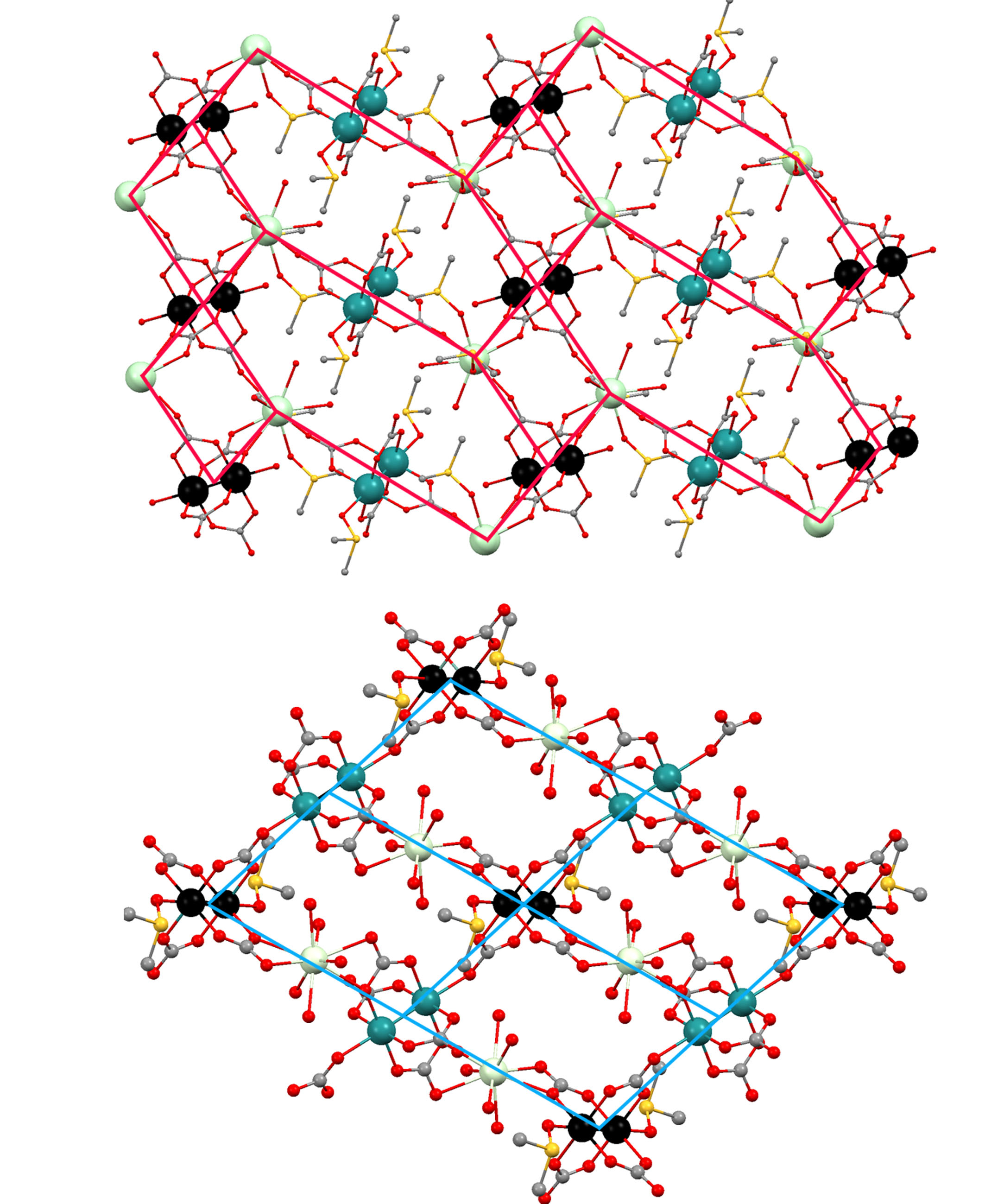
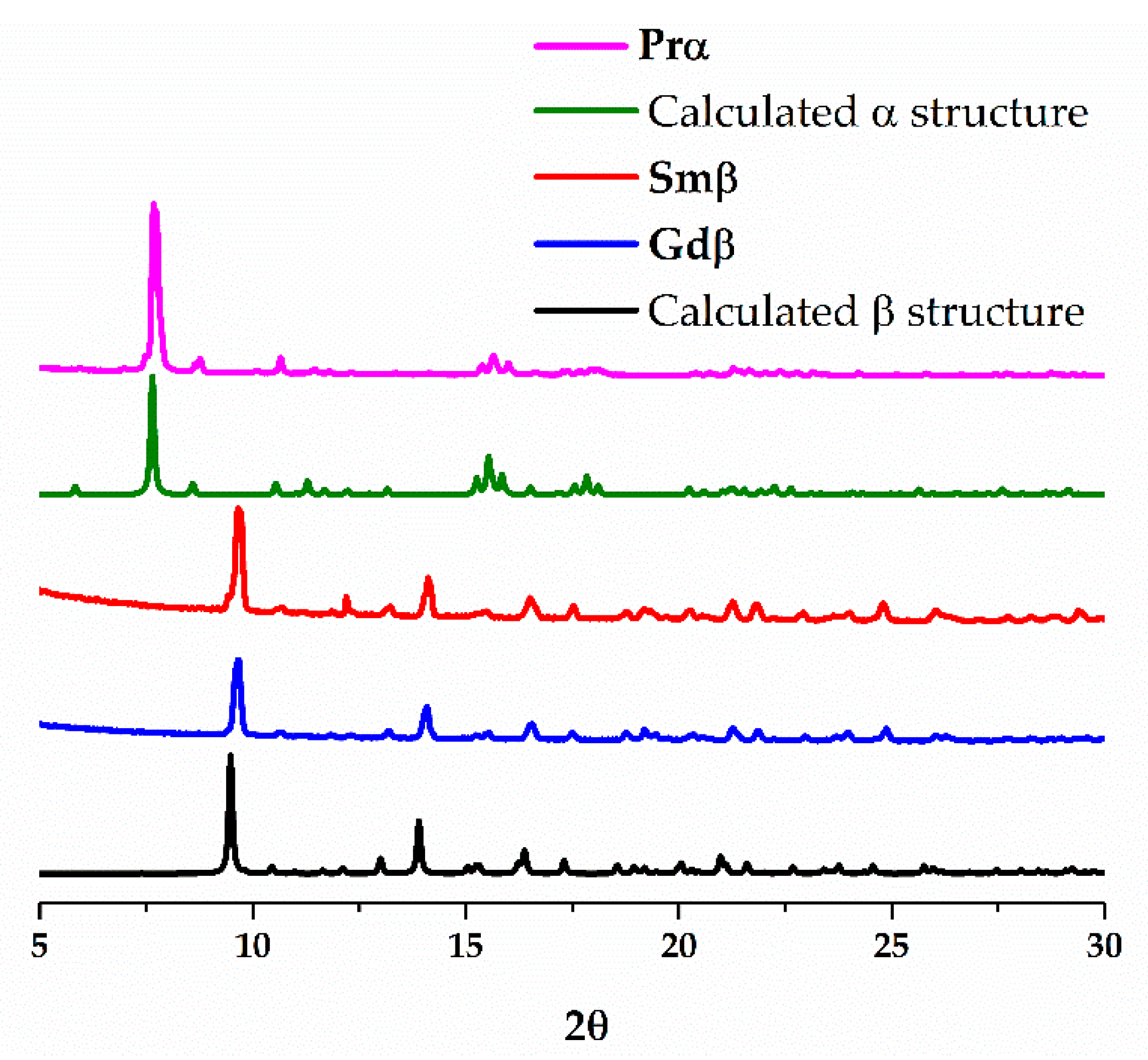
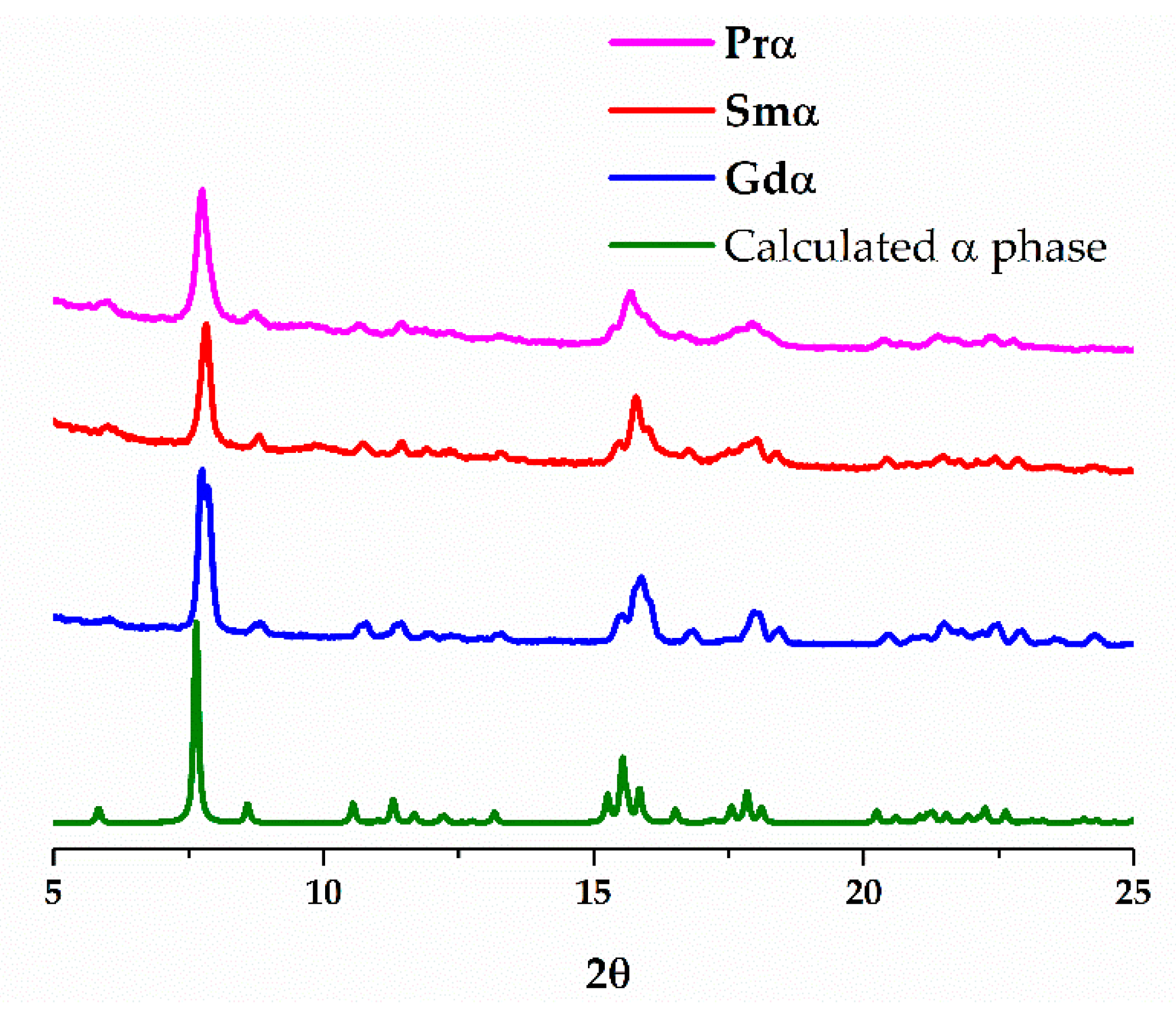
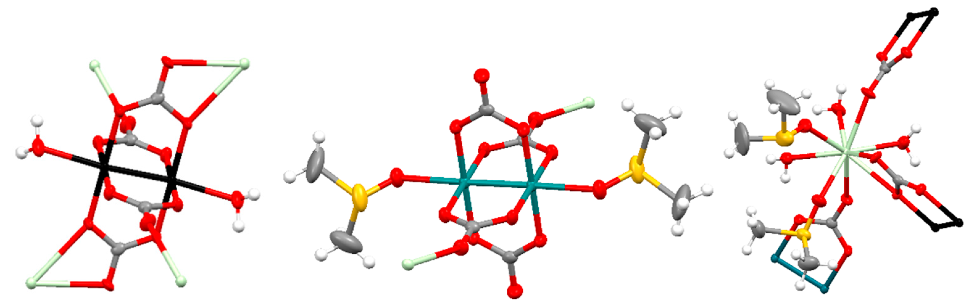
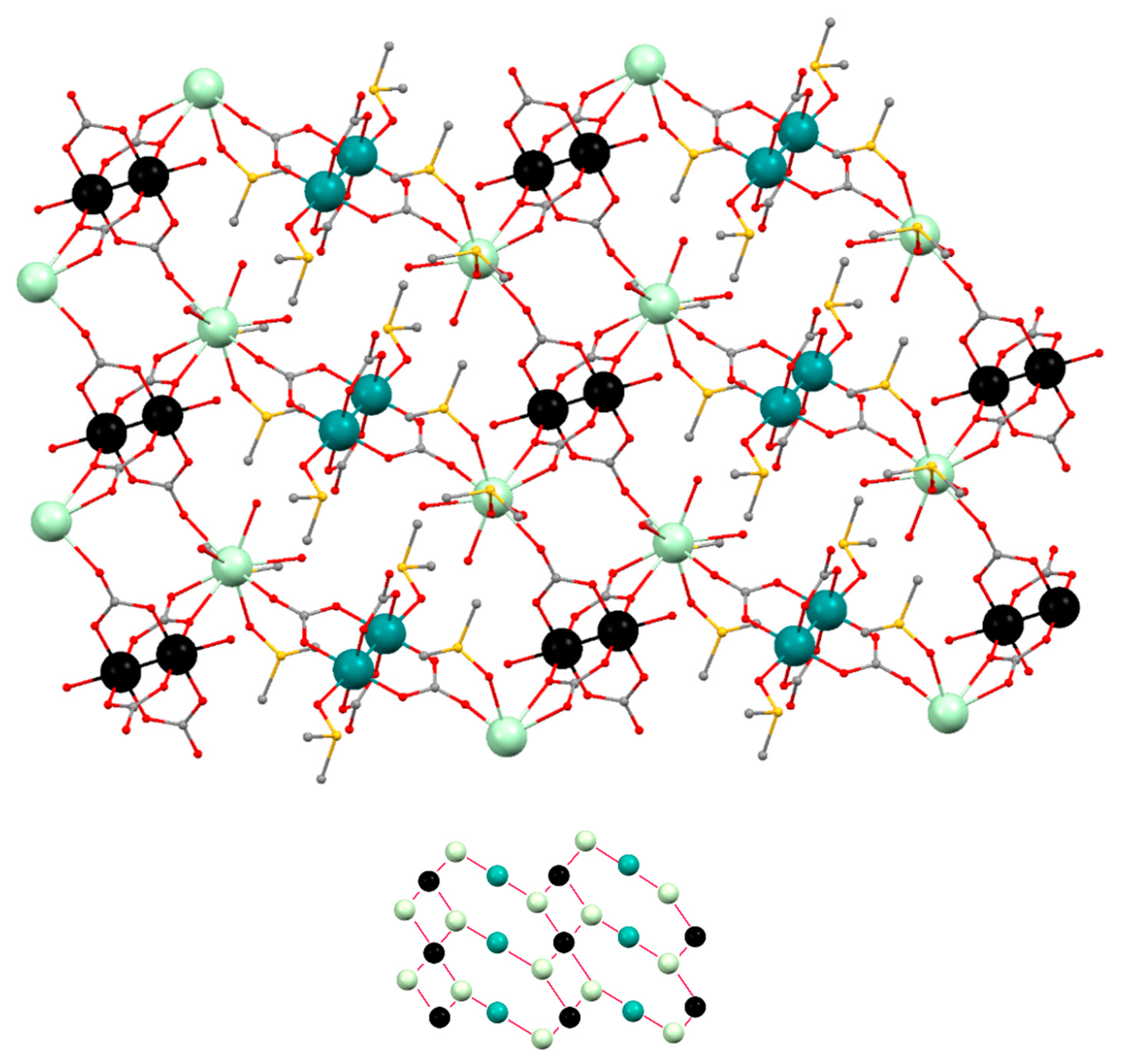
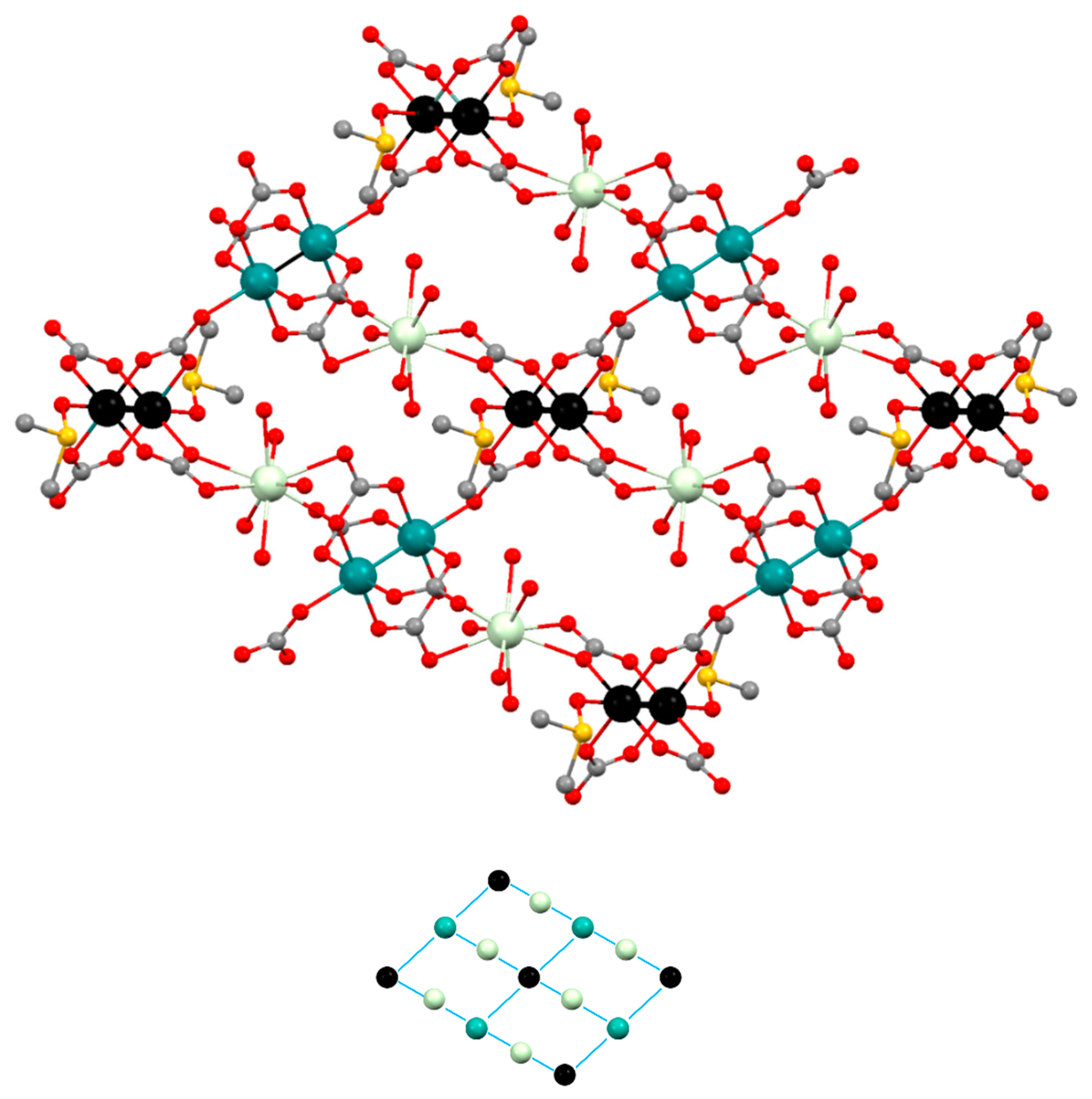
3.2. Structural Description

The crystal structure of Prα, Smβ and Gdβ was determined from single crystal X-ray diffraction. They crystallize in the P-1 space group but two types of structures were found: [Pr(DMSO)2(OH2)3][Ru2(CO3)4(DMSO)(OH2)]·5H2O (Prα) (α structure) and [Ln(OH2)5][Ru2(CO3)4(DMSO)]·xH2O (Ln = Sm (Smβ), Gd (Gdβ)) (β structure). The α structure has a significantly lower density (2.142 g·cm3) than the β structure (2.447, 2.426 g·cm3, Table 1). The structure of [Sm(OH2)4][Ru2(CO3)4(OH2)]·2H2O (Sm3D) was determined by single crystal X-ray diffraction and is isostructural to the previously reported Gd, Eu and Yb derivatives (See Figures S3–S5) [21]. The PXRD of [Pr(OH2)4][Ru2(CO3)4(OH2)]·2H2O (Pr3D) shows that it is isostructural with Sm3D (See Figure S6).
The structure of Prα (α structure) is formed by [Ru2(CO3)4(OH2)2]3−, [Ru2(CO3)4(DMSO)2]3− and [Pr(DMSO)2(OH2)3]3+ units in a 1:1:2 ratio giving a neutral 2D net (Figure 4). The two types of diruthenium units display a paddlewheel structure with two ruthenium atoms bridged by four carbonate ligands and two water or two DMSO molecules at the axial positions (Figure 4, left and center). Each carbonate ligand of the [Ru2(CO3)4(OH2)2]3− units is also coordinated to a Pr3+ ions in such a way that two of the carbonates, in trans disposition, display a µ3-1κO,2κO′,3κO″ coordination mode while the other two carbonates display a µ3-1κO,2:3κ2O′O″,3κO″ coordination mode (Figure 4, left). Two of the carbonate ligands, in trans disposition, of the [Ru2(CO3)4(DMSO)2]3− bridge a Pr3+ ion and two ruthenium atoms with a µ3-1κO,2κO′,3κO″ coordination mode (Figure 4, center). The Ru–Ru distances are 2.264 and 2.258 Å, which are in the range 2.238–2.272 Å found for other tetracarbonatodiruthenium compounds [2,10,13,16,18,20,21]. The Pr3+ ions have a coordination number of nine and are surrounded by the three oxygen atoms of water molecules, two oxygen atoms of DMSO molecules and 4 oxygen atoms of 3 carbonate ligands (Figure 4, right).
The combination of the building blocks that form the structure of Prα gives rise to a 2D polymeric structure (Figure 5). If each type of Ru25+ unit and the Ln3+ units are considered as nodes, the resulting net is built by triconected (Pr3+ ions, Figure 4 right), biconnected (Ru25+ ions, Figure 4 center), and tetraconnected (Ru25+ ions, Figure 4 left) nodes with a (2-c)(3-c)2(4-c) stoichiometry (Figure 5, bottom). Interestingly, the polymeric 2D structure of Prα is related to that of [Ln(OH2)4][Ru2(CO3)4(OH2)]·xH2O (Ln = Nd, Eu, Gd, Yb), Pr3D and Sm3D [21] that display a 3D net formed by mono and dimetallic nodes that are tri-, tetra- and hexaconnected (Figure S7).
Five water molecules per formula that do not belong to the 2D network have been found in the crystal structure of Prα. These water molecules establish multiple hydrogen bonds with neighbor water molecules and carbonate, DMSO and water ligands belonging to the 2D network. Interestingly, the thermogravimetric analysis of Prα (heating rate of 5 °C min–1, Figure S8) shows a weight loss in the 35–65 °C range that corresponds to ca. five water molecules per formula. Then, a plateau is observed until 90 °C, when a loss that corresponds to 3–4 water molecules is observed. The framework is stable until 190 °C when it begins to decompose.
The neutral layers that form the structure of Smβ and Gdβ are built with paddlewheel [Ru2(CO3)4(DMSO)2]3− and [Ru2(CO3)4]3− units and [Ln(OH2)5]3+ units combined in a 1:1:2 ratio. The ruthenium atoms in the two types of diruthenium fragments are bridged by four carbonate ligands and the axial positions are occupied by DMSO molecules or two carbonate ligands (Figure 6, left and center). Two trans carbonate ligands of the [Ru2(CO3)4(DMSO)2]3− units are also coordinated to a Ln3+ ion displaying a µ3-1κO,2:3κO′O″,3κO″ coordination mode while the other trans carbonate ligands are coordinated to a Ru atom of the [Ru2(CO3)4]3− units with a µ3-1κO,2κO′,3κO″ coordination mode (Figure 6, left). Two equatorial trans carbonate ligands of the [Ru2(CO3)4]3− units are also coordinated to a Ln3+ ion with a µ3-1κO,2:3κO′,3κO″ coordination mode while the other two equatorial carbonate ligands do not bridge any additional metal ions (Figure 6, center). The Ru–Ru distances are in the 2.260–2.272 Å range, similar to other Ru–Ru distances reported for tetracarbonatodiruthenium compounds as mentioned above. The Ln3+ ions have a coordination number of nine with 5 oxygen atoms of 5 water molecules and 4 oxygen atoms of 2 carbonate ligands that belong to a [Ru2(CO3)4(DMSO)2]3− and a [Ru2(CO3)4]3− unit (Figure 6, right).
Smβ and Gdβ display a grid-like structure, formed by biconnected nodes, [Ln(OH2)5]3+, and two types of tetraconnected nodes, [Ru2(CO3)4(DMSO)2]3− and [Ru2(CO3)4]3− units, with a (2-c)(3-c)2(4-c) stoichiometry (Figure 7).
Three crystallization water molecules per formula have been found in the structure of Smβ, while only two have been found in the structure of Gdβ. These water molecules form hydrogen bonds with neighbour water molecules and with carbonate and water ligands of the polymeric structure. The thermogravimetric analyses of Smβ and G (heating rate of 5 °C min–1, Figures S9 and S10) show a gradual decomposition in the 35–200 °C range.

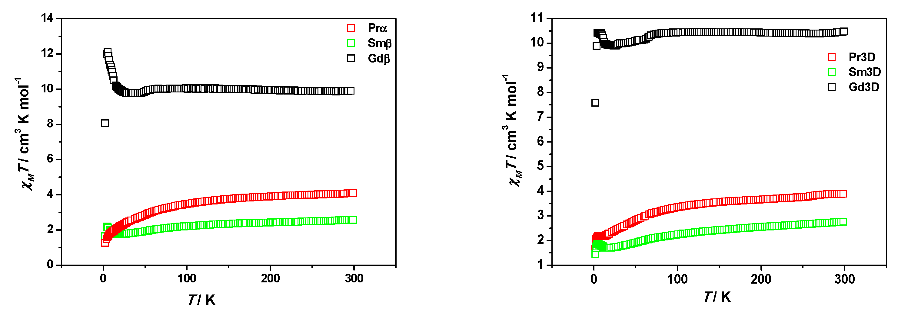
3.3. Magnetic Properties

The temperature dependence of the magnetic susceptibility of Prα, Smβ, Gdβ, Pr3D and Sm3D was measured between 300 and 2 K at 1 T. The plots of the χMT vs. temperature are displayed in Figure 8. Compounds with identical lanthanide present approximately the same χMT values at room temperature despite their different crystal structure. Those values (4.10, 2.56, 10.40, 3.89 and 2.76 emu mol1 K for Prα, Smβ, Gdβ, Pr3D and Sm3D, respectively) are slightly higher than the value expected from the sum of independent Ru25+ and Ln3+ ions (3,48, 1.97 and 9.76 emu mol1 K, respectively, for Pr3+, Sm3+ and Gd3+ with Ru25+).
The χMT values for Prα, Smβ, Pr3D and Sm3D descend smoothly until ≈80 K. Below this temperature a sharper decrease is observed until 2 K for Prα and until 18, 12.6 and 12.7 K for Smβ, Sm3D and Pr3D, when the χMT values increase and a maximum in the curves is observed at 5 K. However, there is almost no variation in the χMT values for Gdβ until 60 K, even a slight increase can be detected. Then, the χMT values decrease until 30 K and at lower temperatures they increase to reach a maximum of 12.10 emu mol1 K at 5.4 K. Finally, χMT values abruptly descend. This is the same pattern observed for [Gd(H2O)4][Ru2(CO3)4(H2O)2]·2.5H2O (Gd3D) [21] although the χMT maximum is 10.43 emu mol1 K at 4.6 K (Figure 8).
The decrease of χMT has been observed in other heteronuclear tetracarbonatodiruthenium(II,III) derivatives in which the Ru25+ centers are the sole magnetic species [7,9,13,14,19] and it has been ascribed to a large zero field splitting (ZFS) associated with the Ru25+ species [7]. In Prα, Smβ, Pr3D and Sm3D this decrease is due to the sum of the ZFS of the Ru25+ units and the depopulation of the MJ sublevels of the Ln(III) ions produced by the splitting of the ground state by the ligand field [22].
The Gdβ compound does not present an important temperature dependence of χMT until low temperatures and, therefore, the contribution of Gd(III) to χMT at high temperatures comes basically from the 7 unpaired electrons of the lanthanide ion that arise a 8S7/2 ground state, without first order spin-orbit coupling [23].
Intramolecular exchange coupling in lanthanide compounds is usually very weak due to the radially contracted nature of 4f orbitals [24]. Therefore, the increase in χMT values at low temperatures could be ascribed to a canted ferrimagnetism produced by the diruthenium species. Actually, this phenomenon has been reported for several tetracarbonatodiruthenium compounds without other magnetic centers [7,9,13,14,19,21]. Interestingly, it was only observed for compounds in which two Ru25+ species are connected by a carbonate ligand in the same fashion found in Smβ, Gdβ, Pr3D and Sm3D. However, a continuous lowering of χMT values with temperature was observed when the axial position of the diruthenium species was occupied by other ligands. This is also the case for Prα.
The field dependence of the magnetization at 2 K between −5 and 5 T of compounds Smβ and Gdβ (Figures S11 and S12) shows almost saturation of the magnetization for Gdβ while the value of the magnetization is far from saturation at 5 T for Smβ. These measurements suggest the existence of predominant ferromagnetic interactions in Gdβ as in Mn4(H2O)16H[Ru2(CO3)4]2[Ru2(CO3)4(H2O)2]·11H2O and predominant canted ferrigmagnetism in Smβ as in KxH1−x[M(H2O)4][Ru2(CO3)4]·zH2O (M = Mg, Mn, Fe, Co, Ni) [18] and other tetracarbonatodiruthenium compounds without other magnetic centers [7,9,13,19,21]. In fact, Ru–O–Gd–O–Ru fragments with Ru–Gd distances of 4.352 and 4.445 Å are found in the structure of Gdβ.
The magnetic behavior of Gdβ has been fitted with Equation (1) [21], considering the sum of the contribution of the lanthanide ions following the free ion approximation, the contribution of diruthenium species taking into account a ZFS parameter (D) and a Weiss constant (θ) to consider intermolecular interactions. A TIP has also been added:
χ = χ R u + χ L n + T I P
where
χ R u = N g R u 2 μ B 2 3 k ( T θ ) [ 1 + 9 e 2 D k T 4 ( 1 + e 2 D k T ) + 2 + 3 k T 2 D ( 1 e 2 D k T ) 1 + e 2 D k T ]
and
χ L n = N g L n 2 μ B 2 3 k ( T θ ) J ( J + 1 )
where N, g µB and k have the usual meanings.
The magnetic behavior of Smβ and Sm3D were fitted following a similar approach but considering the presence of excited states that can be thermally populated in the lanthanide ion. Thus, a spin-orbit parameter (λ) was considered for the Sm3+ ions as follows [23]:
χ S m = N μ B 2 3 k T x [ a 1 x + b 1 + ( a 2 x + b 2 ) e 7 x / 2 + ( a 3 x + b 3 ) e 8 x + ( a 4 x + b 4 ) e 27 x / 2 + ( a 5 x + b 5 ) e 20 x + ( a 6 x + b 6 ) e 55 x / 2 ] / [ 3 + 4 e 7 x / 2 + 5 e 8 x + 6 e 27 x / 2 + 7 e 20 x + 8 e 55 x / 2 ] w i t h a 1 = 2.143 b 1 = 7.347 a 2 = 42.92 b 2 = 1.641 a 3 = 283.7 b 3 = 0.6571 a 4 = 620.6 b 4 = 1.9400 a 5 = 1122 b 5 = 2.835 a 6 = 1813 b 6 = 3.556
where x = λ/kT.
The equation to simulate the magnetic contribution of Pr3+ ions in Prα or Pr3D should consider the depopulation of the MJ sublevels which requires too many Hamiltonian Crystal Field parameters [22]. Therefore, we have used as an approximation the same model above employed for the Gd3+ derivatives.
The best data obtained from the fits are shown in Table 2 and the figures can be found in the SI (Figures S13–S18). The fits were made with the χMT values from room temperature until the minimum of the χMT vs. T curves. The gRu and D values obtained from the fits are within the normal range observed for diruthenium(II,III) compounds and are close to those for K3[Ru2(CO3)4]·4H2O, which were estimated to be 2.20 and 70 cm1 [7]. However, the D value obtained for Gdβ was lower (39 cm1) than expected. For this reason, a new fit was done with a fixed D value of 70 cm1. In these cases, a higher θ and a lower g values were obtained.

4. Conclusions

The use of different solvents allows one to control the dimensionality of coordination polymers made from the reaction between K3[Ru2(CO3)4]·4H2O and Ln(NO3)3·xH2O (Ln3+ = Pr, Sm and Gd). The use of neat water leads to the formation of 3D coordination polymers while a H2O/DMSO mixture leads to the formation of 2D structures. A different 2D phase can be obtained depending of the reaction method. Thus, slow diffusion of the reagents gives a net made by triconnected Ln3+ nodes and two different Ru25+ units that are bi- and tetraconnected when the Ln3+ ion is Pr3+ (α-structure, Prα). A grid-like net formed by biconnected Ln3+ nodes and two different tetraconnected Ru25+ is obtained when the Ln3+ ions are Sm or Gd (β structure, Smβ and Gdβ). Direct mixing of the reagents leads to the α-structure in all cases.
The magnetic behavior of the complexes is consistent with the sum of the individual contributions of diruthenium and lanthanide species. The increase in χMT at low temperatures is associated with a weak canted ferrimagnetism from the diruthenium species and weak ferromagnetic interaction between Ru25+ and lanthanide ions.

Supplementary Materials

The following are available online at https://www.mdpi.com/2073-4360/11/3/426/s1. Figure S1: IR spectrum of Prα. Figure S2: IR spectra of Smβ and Gdβ. Figure S3: View of the structure of Sm3D showing the different coordination environments. Figure S4: View along the b axis of a 1 × 1 × 1 packing of the structure of Sm3D. Figure S5: View along the c axis of a 1 × 1 × 1 packing of the structure of Sm3D. Figure S6: Experimental powder X-ray diffraction pattern obtained for Pr3D and calculated powder X-ray diffractogram simulated from the single crystal data of Sm3D. Figure S7: Simplification of the 2D net of Prα and simplification of the 3D net of [Ln(OH2)4][Ru2(CO3)4(OH2)]·xH2O (Ln = Nd, Eu, Gd, Yb), Pr3D and Sm3D. Figure S8: Thermogram of Prα. Figure S9: Thermogram of Smβ. Figure S10: Thermogram of Gdβ. Figure S11: Magnetization versus magnetic field between −5 T to 5 T for Smβ. Figure S12: Magnetization versus magnetic field between −5 T to 5 T for Gdβ. Figures S13–S18: Fits of the temperature dependence of the molar susceptibility χM and χMT for all the compounds.

Author Contributions

R.J.-A. and J.-L.P. conceived and designed the experiments; D.G.-M. and M.C. performed the experiments; Á.M.-H. contributed to the synthesis of some compounds; M.C. and P.D.-M. solved and analyzed the crystal structures; M.C., R.G.-P., S.H. and R.J.-A. analyzed the magnetic data; M.C., S.H. and R.J.-A. wrote the manuscript.

Funding

This work was financially supported by the Spanish Ministerio de Economía y Competitividad (project CTQ2015-63858-P, MINECO/FEDER) and Comunidad de Madrid (project B2017/BMD-3770-CM).

Conflicts of Interest

The authors declare no conflict of interest.

References

  1. Lindsay, A.J.; Wilkinson, G.; Motevalli, M.; Hursthouse, M.B. Reactions of Tetra-μ-carboxylato-diruthenium(II,II) Compounds. X-Ray Crystal Structures of Ru2(μ-O2CCF3)4(thf)2, Ru2(μ-O2CR)4(NO)2 (R = Et or CF3), and {Na3[Ru2(μ-O2CO)4]·6H2O}n. J. Chem. Soc. Dalton Trans. 1987, 0, 2723–2736. [Google Scholar] [CrossRef]
  2. Cotton, F.A.; Labella, L.; Shang, M. Further Study of Tetracarbonato Diruthenium(II,III) Compounds. Inorg. Chem. 1992, 31, 2385–2389. [Google Scholar] [CrossRef]
  3. Norman, J.G.; Renzoni, G.E.; Case, D.A. Electronic Structure of Ru2(O2CR)4+ and Rh2(O2CR)4+ Complexes. J. Am. Chem. Soc. 1979, 101, 5256–5267. [Google Scholar] [CrossRef]
  4. Cotton, F.A.; Walton, R.A. Multiple Bonds between Metal Atoms, 2nd ed.; Wiley: New York, NY, USA, 1982. [Google Scholar]
  5. Cotton, F.A.; Murillo, C.A.; Walton, R.A. Multiple Bonds between Metal Atoms, 3rd ed.; Springer: New York, NY, USA, 2005. [Google Scholar]
  6. Liddle, S.T. (Ed.) Molecular Metal-Metal Bonds: Compounds, Synthesis, Properties; Wiley-VCH: Weinheim, Germany, 2015. [Google Scholar]
  7. Kennon, B.S.; Miller, J.S. Observation of Magnetic Ordering for Layered (2-D) Potassium Diruthenium Tetracarbonate, K3[Ru2II/III(O2CO)4]: A Rare Second Row Transition Metal-based Magnet. Inorg. Chem. 2010, 49, 5542–5545. [Google Scholar] [CrossRef] [PubMed]
  8. Kennon, B.S.; Her, J.-H.; Stephens, P.W.; Shum, W.W.; Miller, J.S. Structure and Magnetic Ordering of KxH1-xNi(OH2)4[Ru2(CO3)4]·zH2O. Inorg. Chem. 2007, 46, 9033–9035. [Google Scholar] [CrossRef] [PubMed]
  9. Kennon, B.S.; Her, J.-H.; Stephens, P.W.; Miller, J.S. Diruthenium Tetracarbonate Trianion, [Ru2II/III(O2CO)4]3−, Based Molecule-Based Magnets: Three-Dimensional Network Structure and Two-Dimensional Magnetic Ordering. Inorg. Chem. 2009, 48, 6117–6123. [Google Scholar] [CrossRef] [PubMed]
  10. Liu, B.; Jia, Y.-Y.; Jin, J.; Liu, X.-M.; Xue, G.-L. Layer structural bimetallic metamagnets obtained from the aggregation of Ru2(CO3)43− and Co2+ in existence of halogen. CrystEngCommun 2013, 15, 4280–4287. [Google Scholar] [CrossRef]
  11. Jia, Y.-Y.; Liu, B.; Liu, X.-M.; Yang, J.-H. Syntheses, structures and magnetic properties of two heterometallic carbonates: K2Li[Cu(H2O)2Ru2(CO3)4X2]·5H2O (X = Cl, Br). CrystEngCommun 2013, 15, 7936–7942. [Google Scholar] [CrossRef]
  12. Liu, B.; Wang, D.; Jin, J.; Jia, Y.-Y.; Liu, X.-M.; Xue, G.-L. Heterometallic Co(II)–Ru2(II,III) carbonates: From discrete ionic crystals to three-dimensional network. CrystEngCommun 2013, 15, 5726–5734. [Google Scholar] [CrossRef]
  13. Liu, B.; Jia, Y.-Y.; Jin, J.; Wang, X.-M.; Liu, D.; Xue, G.-L. Cadmium diruthenium(II,III) carbonates showing diverse magnetism behavior arising from variety configuration of [Ru2(CO3)4]n3n− layer. Dalton Trans. 2013, 42, 10208–10213. [Google Scholar] [CrossRef] [PubMed]
  14. Liu, B.; Jia, Y.-Y.; Yang, H.-Q.; Yang, J.-H.; Xue, G.-L. Incorporation of M(H2O)62+ between layers {M(H2O)2Ru2(CO3)4Cl2}n2n− (M = Zn, Mn): Syntheses, structures and magnetic properties. Dalton Trans. 2013, 42, 16742–16748. [Google Scholar] [CrossRef] [PubMed]
  15. Hou, X.-F.; Jia, Y.-Y.; Yang, J.-H.; Cao, Z.; Liu, B. Isomeric chain structures of {[Mn(H2O)4]2Ru2(CO3)4Br2}nn−: Syntheses, structural diversity and magnetic properties. Dalton Trans. 2014, 43, 13316–13324. [Google Scholar] [CrossRef] [PubMed]
  16. Wang, D.; Liu, B.; Jin, J.; Liu, X.-M.; Jia, Y.-Y.; Xue, G.-L. X-ray single-crystal structure and magnetic properties of KMn(H2O)5Ru2(CO3)4·5H2O: A layered soft magnet. Inorg. Chem. Commun. 2013, 33, 138–141. [Google Scholar] [CrossRef]
  17. Jia, Y.-Y.; Chang, Z.-G.; Yang, J.-H.; Liu, B. Spin glass ordering in heterometallic carbonates with pillared layer structure: K4[Mn(H2O)4{Ru2(CO3)4}2]·4H2O. Inorg. Chim. Acta 2015, 424, 162–166. [Google Scholar] [CrossRef]
  18. Liu, B.; Jin, J.; Liu, X.-M.; Hu, H.-M.; Ding, T.; Zhang, N.; Jia, Y.-Y.; Xue, G.-L. A diruthenium soft ferromagnet showing Tc = 3.0 K: Mn4(H2O)16H[Ru2(CO3)4]2[Ru2(CO3)4(H2O)2]·11H2O. Dalton Trans. 2012, 41, 4748–4750. [Google Scholar] [CrossRef] [PubMed]
  19. Yang, J.-H.; Cheng, R.-M.; Jia, Y.-Y.; Jin, J.; Yang, B.-B.; Cao, Z.; Liu, B. Chlorine and Temperature Directed Self-assembly of Mg-Ru2(II,III) Carbonates and Particle Size Dependent Magnetic Properties. Dalton Trans. 2016, 45, 2945–2954. [Google Scholar] [CrossRef] [PubMed]
  20. Her, J.-H.; Kennon, B.S.; Shum, W.W.; Stephens, P.W.; Miller, J.S. Structure and magnetic properties of LnIII[Ru2(CO3)4]·8H2O. Inorg. Chim. Acta 2008, 361, 3462–3464. [Google Scholar] [CrossRef]
  21. Delgado-Martínez, P.; González-Prieto, R.; Herrero, S.; Jiménez-Aparicio, R.; Perles, J.; Priego, J.L.; Torres, M.R.; Sufrate, B. Preparation of Crystalline Phases of 3D Coordination Polymers Based on Tetracarbonatodiruthenium Units and Lanthanide(III) Ions—Magnetic Characterization. Eur. J. Inorg. Chem. 2017, 3161–3168. [Google Scholar] [CrossRef]
  22. Oyarzabal, I.; Artetxe, B.; Rodríguez-Diéguez, A.; García, J.A.; Seco, J.M.; Colacio, E. A family of acetato-diphenoxo triply bridged dimetallic ZnIILnIII complexes: SMM behavior and luminescent properties. Dalton Trans. 2016, 45, 9712–9726. [Google Scholar] [CrossRef] [PubMed]
  23. Kahn, O. Molecular Magnetism; VCH: New York, NY, USA, 1991. [Google Scholar]
  24. Woodruff, D.N.; Winpenny, R.E.P.; Layfield, R.A. Lanthanide Single-Molecule Magnets. Chem. Rev. 2013, 113, 5110–5148. [Google Scholar] [CrossRef] [PubMed]
Figure 1. Representation of the [Ru2(CO3)4]3− anion.
Polymers 11 00426 g001
Figure 2. Calculated powder X-ray diffractograms for the 2D structures simulated from the single crystal data of Prα (α structure, green) and Smβ (β structure, black). Experimental powder X-ray diffraction pattern obtained for a bulk sample of Prα (pink), Smβ (red) and Gdβ (blue).
Polymers 11 00426 g002
Figure 3. Theoretical powder X-ray diffractogram for the 2D α structure simulated from the single crystal data of Prα (green). Experimental powder X-ray diffraction pattern obtained for the bulk sample prepared by direct mixing of a 15 mL water solution of 0.095 mmol (0.06 g) of K3[Ru2(CO3)4]·4H2O into a solution of 0.11 mmol of the corresponding Ln(NO3)3·6H2O in 15 mL of DMSO (Pink: Ln = Pr. Red: Ln = Sm. Blue: Ln = Gd).
Polymers 11 00426 g003
Figure 4. Representation (50% probability ellipsoids) of the coordination environments of the Ru25+ (left and center) and Pr3+ (right) units that form the structure of Prα. Ruthenium: turquoise and black; praseodymium: pale green; oxygen: red; carbon: gray; sulfur: yellow; hydrogen: white. Ellipsoids of the hydrogen atoms are omitted for clarity.
Polymers 11 00426 g004
Figure 5. (Top): Ball and stick representation of the 2D structure of Prα. Ruthenium: turquoise and black; praseodymium: pale green; oxygen: red; carbon: gray; sulfur: yellow. Hydrogen atoms are omitted for clarity. (Bottom): Simplification of the 2D net. Turquoise: [Ru2(CO3)4(DMSO)2]3− units; black: [Ru2(CO3)4(OH2)2]3− units; pale green: [Pr(DMSO)2(OH2)3]3+ units.
Polymers 11 00426 g005
Figure 6. Representation (50% probability ellipsoids) of the coordination environments of the Ru25+ (left and center) and Gd3+ (right) units that form the structure of Gdβ. Ruthenium: turquoise and black; gadolinium: pale green; oxygen: red; carbon: gray; sulfur: yellow; hydrogen: white. Ellipsoids of the hydrogen atoms are omitted for clarity.
Polymers 11 00426 g006
Figure 7. (Top): Ball and stick representation of the 2D structure of Gdβ. Ruthenium: turquoise and black; gadolinium: pale green; oxygen: red; carbon: gray; sulfur: yellow. Hydrogen atoms are omitted for clarity. (Bottom): Simplification of the 2D net. Turquoise: [Ru2(CO3)4]3− units; black: [Ru2(CO3)4(DMSO)2]3− units; pale green: [Gd(OH2)5]3+· units.
Polymers 11 00426 g007
Figure 8. Plots of the χMT vs. temperature for compounds Prα, Smβ, Gdβ (left) and Pr3D, Sm3D and [Gd(H2O)4][Ru2(CO3)4(H2O)2]·2.5H2O (Gd3D) [21] (right).
Polymers 11 00426 g008
Table 1. Crystal and refinement data for Prα, Smβ, Gdβ and Sm3D.
Crystallographic ParametersPrαSmβGdβSm3D
FormulaPrRu2C10H26O19
S3·5H2O
SmRu2C6H16O18
S·3H2O
GdRu2C6H16O18
S·2H2O
SmRu2C4H12O18
2H2O
fw979.62814.80803.67736.67
Space groupP-1P-1P-1C2/c
a8.4508(4)9.7951(5)9.7687(6)25.063(2)
b12.4403(6)9.8502(5)9.8237(6)9.8420(8)
c15.5262(8)12.8759(6)12.8403(7)14.0568(12)
α/°78.243(1)75.608(1)75.657(1)90
β/°89.339(1)70.296(1)70.560(1)95.092(2)
γ/°72.147(1)73.885(1)73.951(1)90
V31518.81(13)1107.0(10)1100.07(11)3453.7(5)
Z2228
d calc/g·cm−32.1422.4472.4262.833
μ/mm−12.8574.1514.5175.185
R indices (all data)R1 = 0.0743
wR2 = 0.0974
R1 = 0.0504
wR2 = 0.1100
R1 = 0.0553
wR2 = 0.1241
R1 = 0.0585
wR2 = 0.0983
GooF on F21.0821.0391.0731.052
Table 2. Magnetic parameters obtained for the fit of the magnetic data.
CompoundgRugLn1D [cm−1]λ [cm−1]θ [K]TIP [emu/mol]σ 2
Gdβ2.282.0039 0.922.17 × 10−41.12 × 10⁻²
Gdβ 22.102.0070 1.984.14 × 10−127.11 × 10⁻³
Smβ2.180.29732561.735.27 × 10−115.12 × 10⁻⁵
Sm3D2.170.29772701.117.76 × 10−44.88 × 10⁻⁵
Prα2.070.8078 −5.732.20 × 10−39.14 × 10⁻³
Pr3D2.080.8073 −7.093.27 × 10−37.95 × 10⁻⁴
1 These values were fixed in the fits. 2 Fixed D value.

