Article

Effect of Microgravity on Synthesis of Nano Ceria

1
William G. Lowrie Department of Chemical and Biomolecular Engineering, the Ohio State University, 151 W. Woodruff Avenue, Columbus, OH 43210, USA
2
Mechanical and Aerospace Engineering, the Ohio State University, 201 W. 19th Street, N350 Scott Laboratory, Columbus, OH 43210, USA
*
Author to whom correspondence should be addressed.
Catalysts 2015, 5(3), 1306-1320; https://doi.org/10.3390/catal5031306
Submission received: 19 May 2015 / Revised: 8 July 2015 / Accepted: 14 July 2015 / Published: 20 July 2015
(This article belongs to the Special Issue Feature Papers to Celebrate the Landmarks of Catalysts)

Abstract

:
Cerium oxide (CeO2) was prepared using a controlled-precipitation method under microgravity at the International Space Station (ISS). For comparison, ceria was also synthesized under normal-gravity conditions (referred as control). The Brunauer-Emmett-Teller (BET) surface area, pore volume and pore size analysis results indicated that the ceria particles grown in space had lower surface area and pore volume compared to the control samples. Furthermore, the space samples had a broader pore size distribution ranging from 30–600 Å, whereas the control samples consisted of pore sizes from 30–50 Å range. Structural information of the ceria particles were obtained using TEM and XRD. Based on the TEM images, it was confirmed that the space samples were predominantly nano-rods, on the other hand, only nano-polyhedra particles were seen in the control ceria samples. The average particle size was larger for ceria samples synthesized in space. XRD results showed higher crystallinity as well as larger mean crystal size for the space samples. The effect of sodium hydroxide concentration on synthesis of ceria was also examined using 1 M and 3 M solutions. It was found that the control samples, prepared in 1 M and 3 M sodium hydroxide solutions, did not show a significant difference between the two. However, when the ceria samples were prepared in a more basic medium (3 M) under microgravity, a decrease in the particle size of the nano-rods and appearances of nano-polyhedra and spheres were observed.

Graphical Abstract

1. Introduction

Cerium oxide (CeO2) is a widely used catalyst support due to its high oxygen storage capacity and high oxygen ion conductivity. The high oxygen mobility of ceria leads to a change in its oxidation state between Ce3+ and Ce4+, which facilitates its use as an oxygen buffer for catalytic redox reactions [1,2]. In addition, ceria has been shown to have catalytic activity itself for various reactions, such as oxidation and reforming [3,4]. Moreover, the high thermal stability of ceria is attractive for reactions which are conducted at high temperatures [2]. Therefore, ceria has been extensively utilized in three-way catalysts [5,6,7] and fuel cells [8,9], as well as investigated for its activity in catalytic reactions such as CO oxidation [10,11], steam reforming [12,13,14,15,16,17], and water gas shift (WGS) [18,19].
A variety of methods to synthesize ceria have been reported until now, including precipitation [20], spray pyrolysis [21,22], thermal hydrolysis [23], micro-emulsion [24], combustion [25], sonochemical [26], hydrothermal [27], solvothermal [28,29], and sol-gel [30] techniques. It is a well-established fact that changing the synthesis parameters, such as temperature, pressure, pH, and crystallization time, produces different particle sizes and morphologies, which, in turn, alter the physical and chemical properties of ceria, as well as the catalytic performance of the ceria-supported catalysts. For example, an increase in oxygen vacancies with decreased particle size of ceria has been reported in many studies [31,32,33]. Deshpande et al. [32] obtained 17% to 44% increase in Ce3+ concentration as ceria particles size was reduced from 30 nm to 3 nm, which was prepared by micro-emulsion. This was attributed to the generation of oxygen vacancies of ceria [32]. Flytzani-Stephanopoulos and colleagues synthesized nano-polyhedra, rods and cubic ceria particles using hydrolysis method and examined its catalytic activity for various reactions [19,34,35,36,37]. They demonstrated that different shapes of ceria nanoparticles exposing different crystal planes on the catalyst surface significantly influenced the Au dispersion on the ceria support.
Although there are many studies about the ceria synthesis process, very little is understood regarding the effect of microgravity on the characteristics of ceria. It is expected that gravitational forces would affect the crystallization process and lead to different chemical properties of a solid especially when it is prepared in a heterogeneous solution. This can be attributed to the gravity-driven disturbance in the solution (convection), sedimentation of the solid phase, and the frequency of collisions between particles during the reaction. In the literature, several microgravity studies have been conducted on synthesis of zeolites [38,39,40,41]. Coker et al. [38] obtained larger average crystal dimensions, less surface area of the Zeolite Socony Mobil (ZSM)-5 under microgravity conditions. The formation of larger crystals of ZSM-5 zeolite was explained by lack of convection and sedimentation of zeolite particles during crystallization. Sano and coworkers [40] prepared ZSM-5 zeolite in space and observed significantly better uniformity of shape and size. Tsuchida et al. calculated the formation rates for colloidal crystallization of silica spheres. It was shown that rate constants were significantly smaller in microgravity [42]. Smith et al. reported that formation of silica crystals to be slower under microgravity environment due to diffusion-limited crystallization where mass transfer dominates the aggregation of particles rather than reaction kinetics [43]. The effect of microgravity on synthesis of microporous tin(IV) sulfides was examined by Ahari and coworkers [44]. Their results indicated a higher degree of crystallinity, as well as smoother crystal surface of the sample. Additionally, the Langmuir isotherm showed 60% larger pore volume [44]. Similarly, a significant reduction of number of pores were reported from aluminum-copper alloy by Fujii et al. [45].
Previously, we have reported on synthesis of ceria with different morphologies (polyhedra, rods, cubes) and different particle sizes (3 nm–25 nm). These materials were examined both as supports, as well as catalysts, in ethanol steam reforming to understand the effect of particle size and morphology [3,46]. In this article, we report the effect of microgravity on the synthesis of ceria.
Ceria samples were synthesized using cerium nitrate and sodium hydroxide solutions. Solutions with different concentrations were taken separately to the International Space Station and mixed under microgravity. The samples were launched on 22 January 2011, and the experiment was performed on 21 February 2011. After staying at microgravity for 23 days, the samples were returned to Earth and delivered to our laboratories. Similar synthesis experiments were also performed on Earth as control experiments.
Brunauer-Emmett-Teller (BET) surface area, pore volume and pore size distribution were measured using nitrogen physisorption technique for both sets of samples. Transmission electron microscopy (TEM) images were taken to obtain particle size and shape. X-ray Diffraction (XRD) was also used to determine crystallite size and exposed surface planes.

2. Results and Discussion

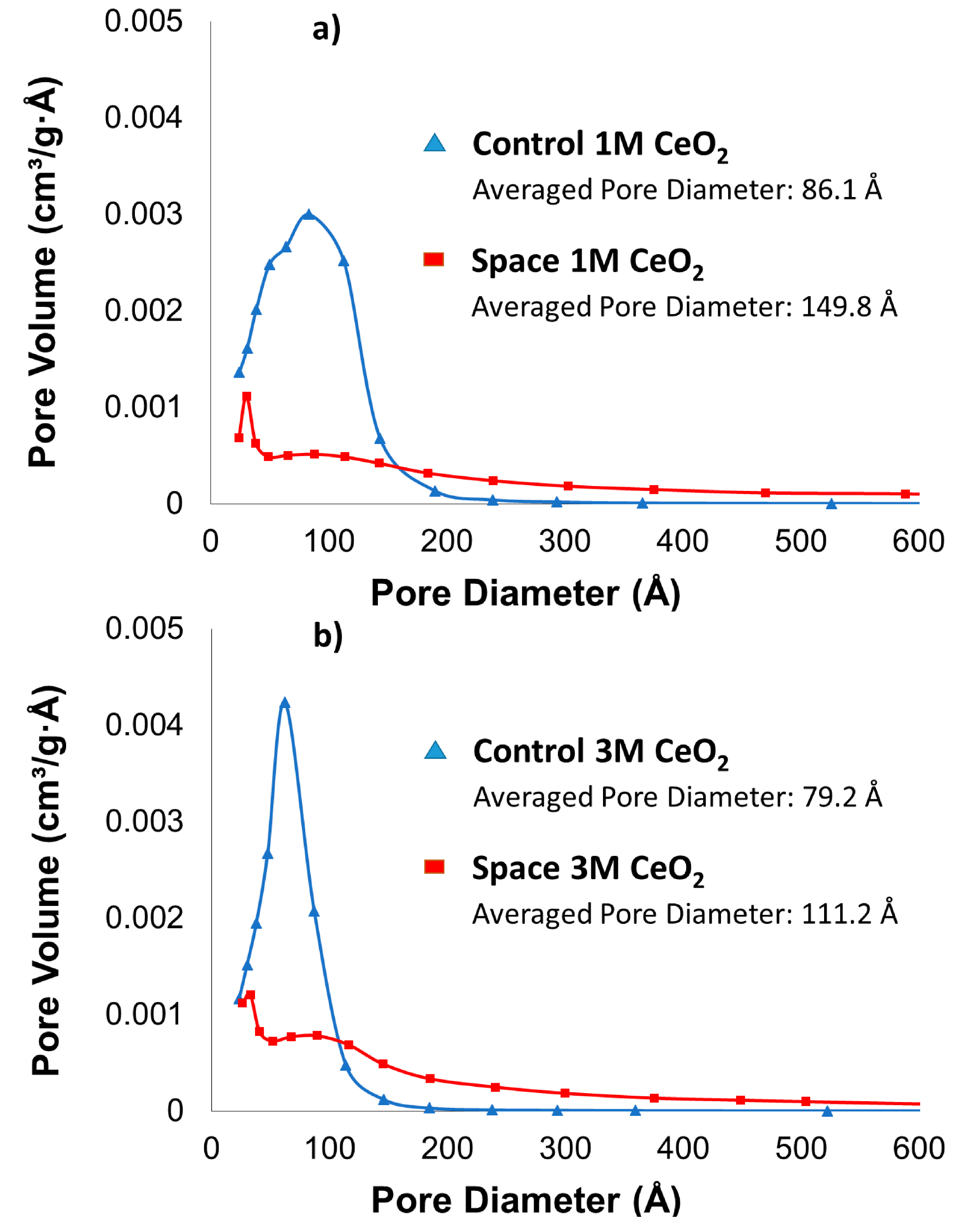
2.1. BET Surface Area, Pore Size and Pore Volume

BET surface analysis was conducted on the ceria samples to determine the effect of microgravity on their surface characteristics. Table 1 compares the surface areas and pore volumes of the various samples tested. The control samples exhibited higher surface area and higher pore volume than the space samples. Regarding the effect of pH on the pore volume and surface area, higher surface areas and pore volumes were observed with increase in basicity of the medium for space samples whereas the opposite trend was exhibited by the control samples.
Table 1. Surface area and pore volume of CeO2 sample measured using N2 physisorption.
SamplesBET Surface Area (m2/g)Pore Volume (cm3/g)
Control 1 M1380.3
Control 3 M1180.23
Space 1 M470.18
Space 3 M690.19
There was a considerable difference between control and space ceria in terms of the pore size distribution, as observed in Figure 1. The control ceria had a narrower pore size distribution with most of the pores being the 50–100 Å range and no pores larger than ~200 Å. On the other hand, for the space samples, a wide range of pore diameters ranging from 30–600 Å were observed. The lower pore volume and surface area can be due to larger particles size of the space samples compared to control samples. This was further confirmed in the TEM section. With respect to the effect of NaOH concentration, broader pore size distribution was obtained in case of 1 M control ceria than 3 M. The highest pore volume was obtained at a pore diameter of 83 Å and 62 Å for 1 M and 3 M, respectively. For space-grown ceria, there were no significant changes in the pore size distribution.
Figure 1. Barrett-Joyner-Halenda (BJH) Pore size distribution of CeO2 sample measured using N2 physisorptionprepared (a) with 1M NaOH (b) with 3M NaOH.
Catalysts 05 01306 g001

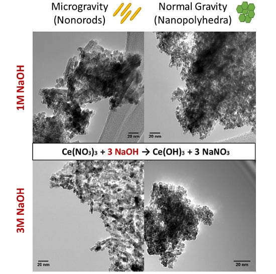
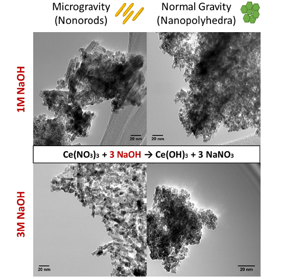
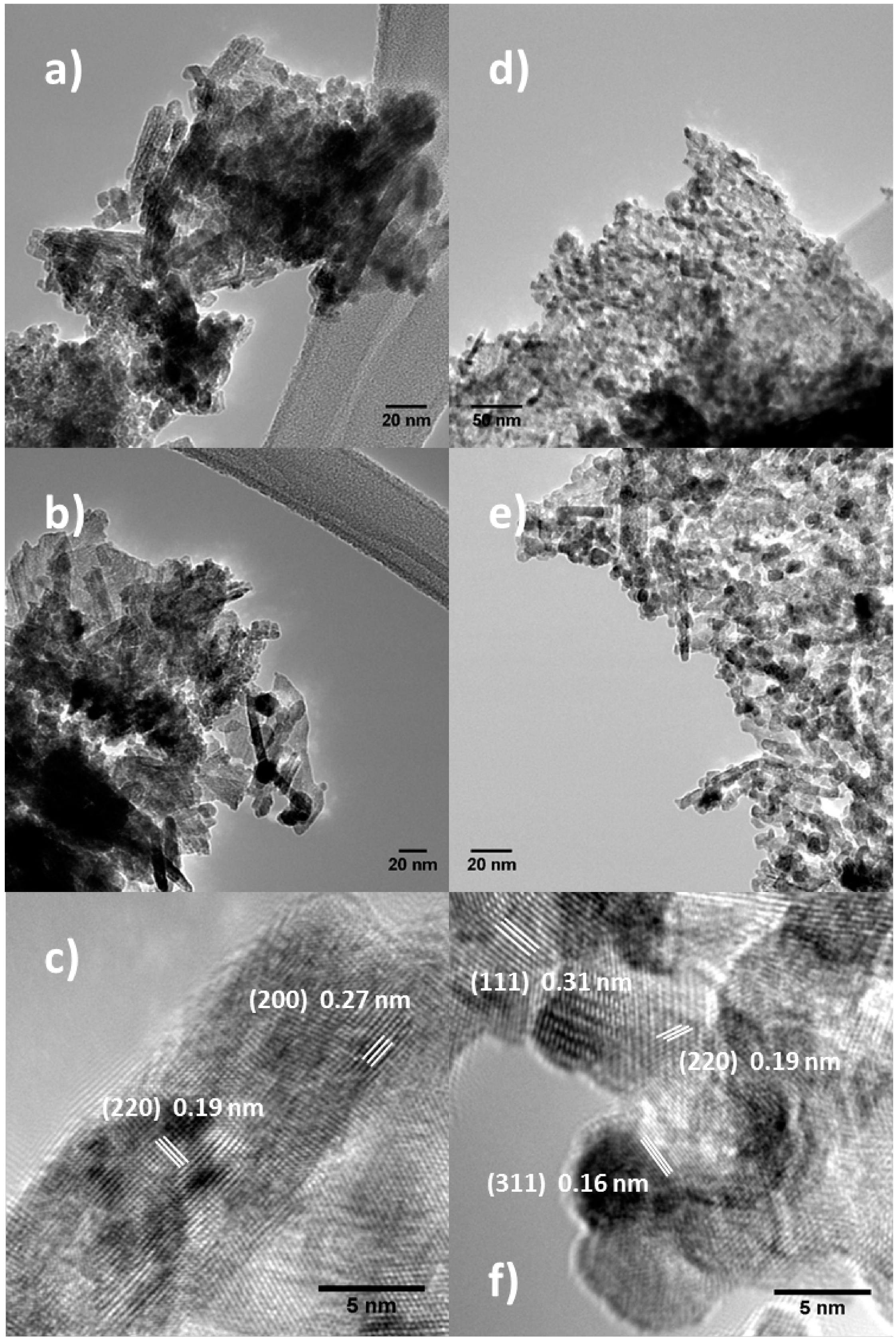
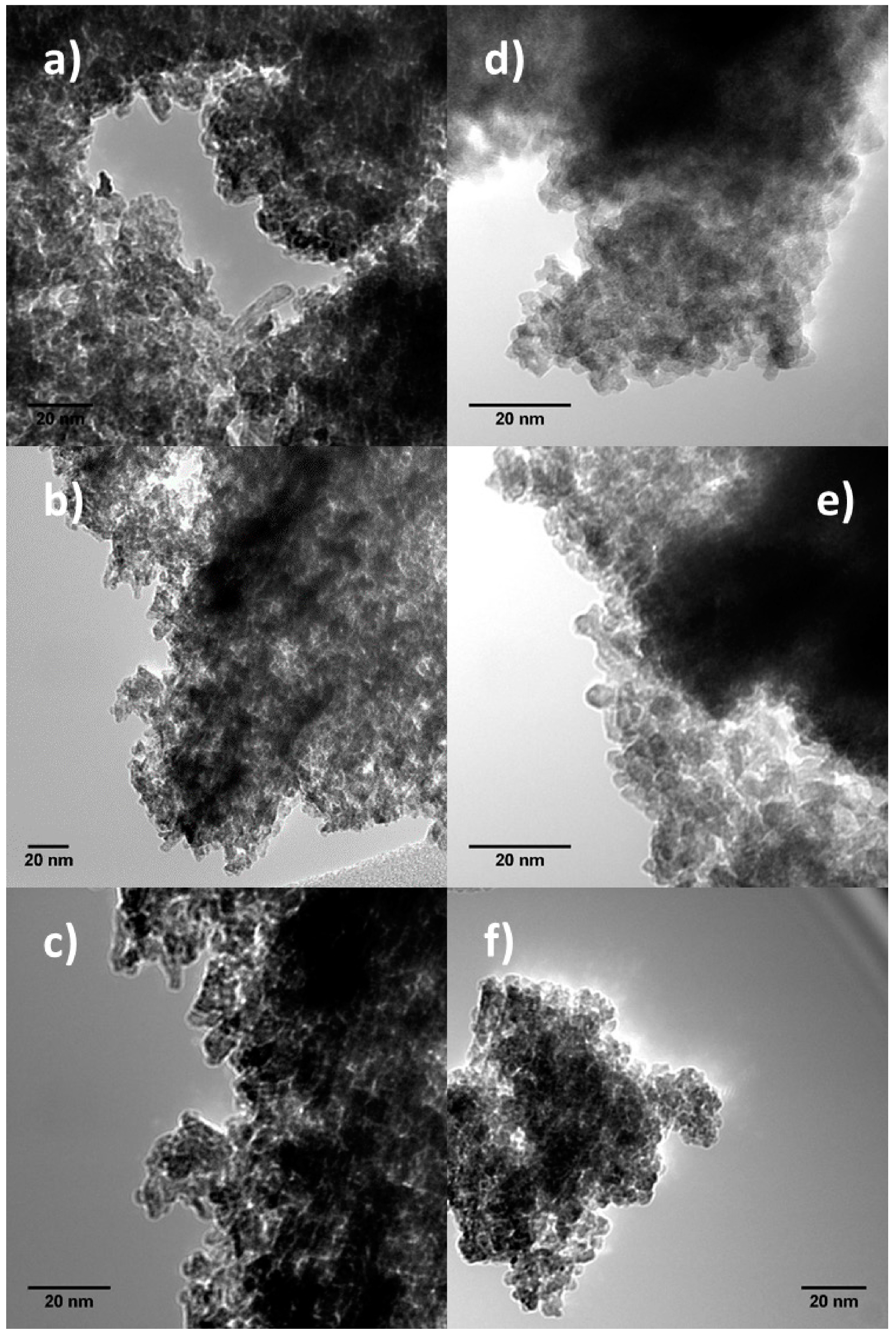
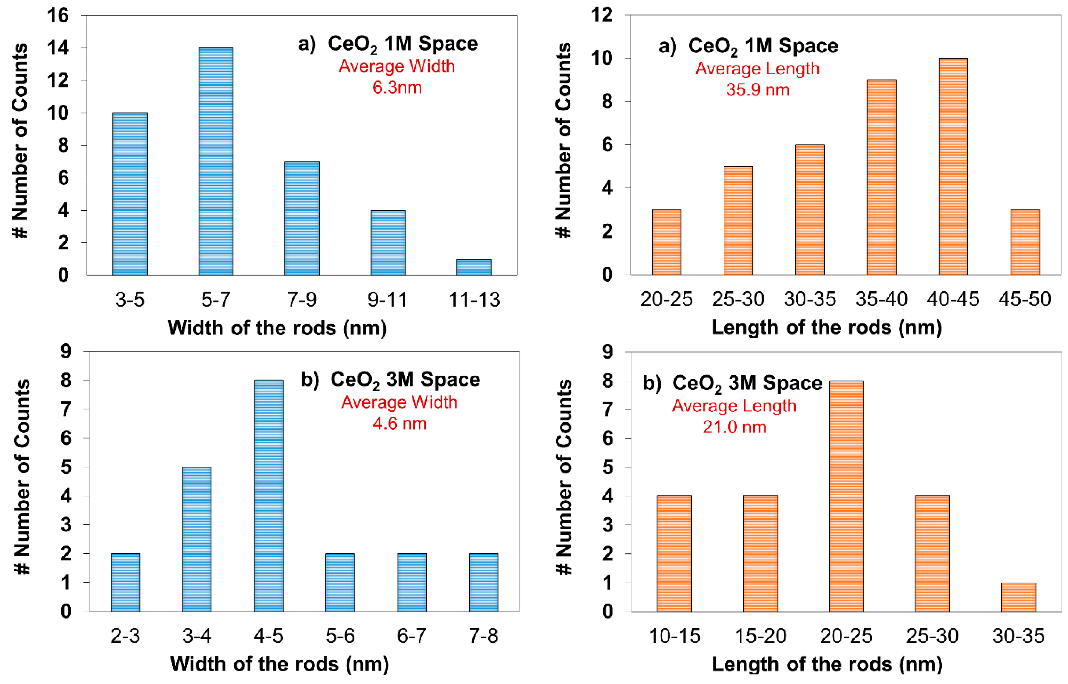
2.2. Transmission Electron Microscopy (TEM)

TEM images were obtained to confirm the ceria morphology and particle size (Figure 2 and Figure 3). The particle size measurement was done by random sampling using multiple micrographs. Additionally, different batches of space and control samples are used for analysis.
In Figure 2a–c, TEM images of control ceria prepared with 1 M NaOH solution are shown. These samples consisted of polyhedron-shaped particles with an average particle size of 3.8 nm, fairly uniform particle size and shape throughout the sample. Similarly, control samples prepared at more basic conditions (Figure 2d–f) contained smaller nanopolyhedra with an average particle size of 2.9 nm.
TEM images of the space samples are presented in Figure 3. The samples had mostly nano-rods, with 2–13 nm in width and 10–50 nm in length. Samples prepared with 1 M-solution were seen to give larger and more uniform particles on the average. The 3 M-space samples had some nano-spheres as well as rods. The particle size distributions for all four ceria samples are shown in Figure 4 and Figure 5.
Figure 2. TEM image of control CeO2 sample: 1 M (ac) and 3 M (df).
Catalysts 05 01306 g002
These results are significant in showing that ceria nanorods can be synthesized at room temperature and atmospheric pressure. To the best of our knowledge, there have not been any publications for the synthesis of nanorods under such conditions. In most cases, studies have concluded that ceria nanorods are formed at much higher temperatures and pressures [27,46,47,48,49]. In order to understand the formation of ceria nanorods under microgravitiy condition, two crystal growth processes, Oswald ripening and oriented attachment, can be considered. These crystal growth mechanisms have been utilized to demonstrate the increase in particle size and change in morphology of the ceria particles [50,51,52]. The Ostwald ripening process follows the dissolution and reprecipitation of solution ions (Ce4+, OH) from the ceria nuclei minimizing the surface energy state [53]. On the other hand, the oriented attachment mechanism predominantly depends on the collision between nuclei during reaction. This process allows ceria nano-crystal to grow into a certain direction [53]. Under microgravity condition, the lack of sedimentation of ceria particles in the solution may increase the residence time for nucleation of ceria crystals [40]. Additionally, the diffusion and/or natural convection of ceria particles can be somewhat enhanced. These factors can significantly affect the crystal growth through oriented attachment, which results in more frequent collision between nuclei, thus forming larger particles and nanorods. There have been several articles showing evidence that oriented attachment crystal growth is the main route for the formation of nanorods of ceria [51], as well as various metal oxides [54].
Figure 3. TEM image of Space CeO2 sample: 1 M (ac) and 3 M (df).
Catalysts 05 01306 g003
Figure 4. Particle size distribution histogram of Control CeO2 sample: (a) 1 M; (b) 3 M.
Catalysts 05 01306 g004
Figure 5. Width and length distribution histogram of Space CeO2 sample: (a) 1 M; (b) 3 M.
Catalysts 05 01306 g005
In the literature, numerous studies on understanding the effect of pH on ceria particles size and shape have been conducted, however, depending on the synthesis method and choice of ceria precursor, different results have been reported. Tok et al. [50] observed similar behavior where smaller grains were seen in a more basic environment. This was because the Ce(OH)4 precipitate is basic; thus, the solubility of Ce(OH)4 in a more basic medium limits further agglomeration to form larger particles [50]. However, an opposite trend was reported by Yang and coworkers [51], where changing the concentration of NaOH from 1 M to 5 M, increased the particle size of the ceria cubes from 20 nm to 25–40 nm. Mai et al. [52] prepared ceria using cerium nitrate precursor at different NaOH concentrations of 1, 3, 6 and 9 M, at a fixed reaction temperature of 100 °C and reaction time of 24 h. It was observed that polyhedra particles were formed using 1 M NaOH, but as the solution became more basic, only rods were obtained.
In this study, the effect of pH on the ceria morphology and particle size was more apparent in the case of the space samples. It was found that the length of the nanorods were longer for the 1 M-space samples whereas smaller dimensions were observed for the 3 M-space samples. Moreover, the rods were prominent the former, while the number of rods were smaller in the 3 M-space samples, with nano-polyhedra and spheres also being observed. It should also be noted that the control ceria samples were under reaction condition of 25 °C and 1 atm. For example, Wu et al. [53] prepared ceria powders in acidic, basic and neutral reaction mediums indicating that the effect of pH on particles size was insignificant at room temperature. The similarities between the particles size and shape in the 1 M and 3 M-control samples are in good agreement with their results. Therefore, it can be concluded that under microgravity, the pH affects the ceria morphology and particle size, whereas the effect is less prominent under normal gravity at room temperature.
Lastly, identification of the diffracted planes were conducted. D-spacing was measured using Image J software. Figure 5c shows a ceria rod including (220) and (200) planes with 0.19 and 0.27 nm d-spacing [34,46,52]. Figure 5f indicates ceria nanopolyhedra and spheres with (311) and (220) planes [54].

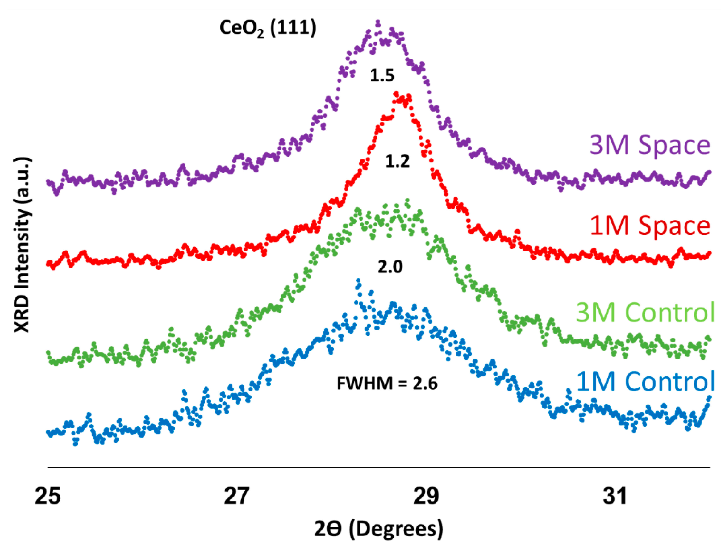
2.3. X-ray Diffraction (XRD) Analysis

The XRD patterns of the ceria samples are shown in Figure 6. All patterns exhibited five distinct peaks which ensured that pure ceria was formed. Each peak was assigned to a particular crystal plane. The peak at a 2θ value of 28.5°, which showed the highest intensity, was identified as (111) plane of the cubic cerianite phase (ICDD 81-792). Others at 2θ values of 33°, 47.5°, 56°, and 69.4° were identified as (200), (220), (311), and (400) planes of ceria. For comparison, sharper diffraction lines were obtained for space samples, which suggest higher crystallinity of the structure.
Figure 6. XRD pattern of the CeO2 sample.
Catalysts 05 01306 g006
The full width at half maximum (FWHM) values are shown in Figure 7. These values were used to calculate the mean crystal size which is listed in Table 2. It appears that the (111) peak is broader for control samples, resulting smaller crystal size than those that were prepared in space. Generally, the crystal size estimated from XRD is in good agreement with the particle size observed in TEM. In the control samples, however, an increase of crystal size from 31.6 to 39.6 Å was obtained with an increasing NaOH concentration, whereas almost no change in terms of particle size was concluded from TEM results. In contrast, the effect of pH on the crystal size was clear for space samples, where a significant decrease of crystal size from 67.9 to 53.5 Å was seen, which follows the same trend as that of the TEM results.
Table 2. Mean crystal sizes of CeO2 sample.
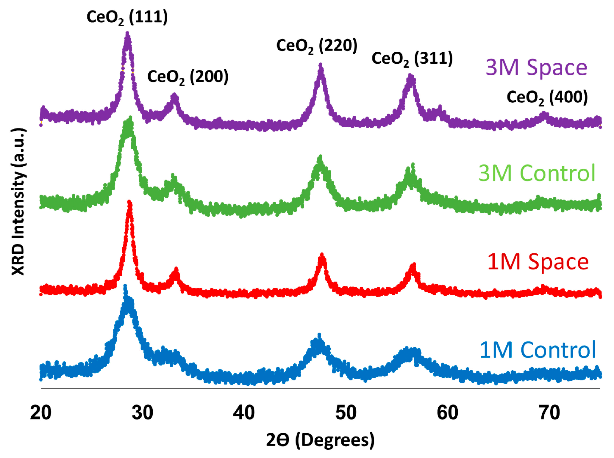
Mean Crystal Size (nm)
1 M Control3.2
3 M Control4.0
1 M Space6.8
3 M Space5.3
Figure 7. Comparison of FWHM of (111) plane for CeO2 sample.
Catalysts 05 01306 g007

3. Experimental Methods

3.1. Catalyst Preparation

A CubeLab container (Nanoracks, Houston, TX, USA), shown in Figure 8, with the dimensions of 10 cm × 10 cm × 20 cm was utilized to protect the experimental containers during take-off and landing on route to and back from the International Space Station (ISS). Therefore, the catalyst preparation apparatus had to fit into a single CubeLab container.
Figure 8. Schematic and dimensions of CubeLab and NanoRack Modules.
Catalysts 05 01306 g008
Ceria nanoparticles were prepared by precipitation method, where 2.1 g Ce(NO3)3·6H2O (Sigma-Aldrich, 99.999%, St. Louis, MO, USA) was dissolved in 13.5 mL water. In separate containers, caustic solutions with 66.5 mL of 1 M and 3 M NaOH were prepared. The solutions were loaded into concentric cylindrical rods. The innermost tube contained the cerium nitrate solution. Surrounding the inner glass tube was the sodium hydroxide solution. The astronauts were instructed to bend the assembly and thereby break the innermost glass tubing, allowing the solutions to mix. The assembly was then shaken to allow for a more thorough mixing. It should be noted that the liquid stays spread out through the length of the tube in microgravity, thereby showing different precipitation behavior. The concentric tubes and the protective cover layers around the catalyst preparation assembly are shown in Figure 9.
Figure 9. Schematic for the catalyst preparation assembly.
Catalysts 05 01306 g009
Twenty-four rods containing different molarities of NaOH solutions were loaded into the Cubelab. Another batch of 24 rods were prepared and kept in our laboratories as control. The CubeLab sample holder was then tested for vibration, vacuum, and off-gassing [55], verification as per the NASA space-flight qualification requirements and flight safety regulations, and was sent to the International Space Station (ISS). The samples were not exposed to air during this process. It should be noted that ISS was kept at 25 °C and 1 atm [55]. On approximately the same day, the control samples were treated under similar reaction conditions and the solutions were mixed in our laboratories. No further treatment was done until the samples prepared at ISS were delivered to our laboratories.
Following the delivery of the ISS samples, the samples were tested for ruptures. No serious containment breaches were found. Following the inspection, all samples including the control were filtered and washed with deionized and distilled water until a pH of 7 is reached. The samples were then dried in an oven at 90 °C overnight and were ready for characterization. Throughout the text, the ceria samples prepared in normal-gravity and microgravity are referred to as control and space samples, respectively.

3.2. Surface Analysis

BET surface area and pore volume measurements were carried out on a Micromeritics ASAP 2020 (Norcross, GA, USA) accelerated surface area and porosimetry instrument. The samples were degassed at 130 °C for 12 h prior to analysis under vacuum (<2 μm·Hg). Adsorption/desorption isotherms were collected at liquid nitrogen temperature where the desorption branch of the isotherm was used to calculate Barrett-Joyner-Halenda (BJH) pore size distributions.

3.3. Transmission Electron Microscopy (TEM)

A Phillips Tecnai F20 TEM (Hillsboro, OR, USA) equipped with a Field Emission Gun (FEG) and operated at 200 kV was used for imaging the particles. All exposures were collected in the bright field. The samples were suspended in ethanol and were sonicated for ~10 min to improve dispersion. Following sonication, the resulting mixture was immediately deposited on a 200-mesh copper grid coated with lacey carbon in order to prevent agglomeration of the particles. Image J open-source processing and analysis software was used to estimate the particle sizes.

3.4. X-ray Diffraction (XRD)

XRD powder patterns were collected using a Rigaku diffractometer (Tokyo, Japan) with Cu Kα radiation with λ = 1.5418 Å operated at 40 kV and 25 mA. The patterns were collected in the 2θ range of 20°–75°. International Center for Diffraction Data (ICDD) database was used for identification of the crystalline phases. The crystal size of the samples were calculated by Scherrer equation L = Kλ/βcosθ where K is the Scherrer constant, λ is the wavelength of the X-ray, β is the full width half maximum (FWHM), and θ is the diffraction angle.

4. Conclusions

The effect of microgravity on ceria synthesis was investigated. Ceria was synthesized by mixing cerium nitrate and sodium hydroxide solutions at room temperature, under both microgravity (space) and normal-gravity (control) for comparison. The ceria samples that were prepared in space showed lower surface area and pore volume compared to the Earth samples. BJH pore size analysis indicated that the space ceria samples consisted of larger pores with a broader pore size distribution. The morphology of the ceria particles were observed from TEM images. It was concluded that the space samples were mostly nano-rods, whereas control samples contained nano-polyhedra. The average particle size observed was larger for space ceria. From XRD results, space ceria samples were more crystalline than the control samples. The mean crystal size obtained using Scherer equation was larger in case of space samples than control. The difference in particle size and shape between space and control ceria is thought to be due to improved oriented attachment crystal growth process where more collision of ceria particle occurs. This can be attributed to lack of sedimentation of the ceria particles under microgravity condition. Lastly, the effect of pH was examined by changing the concentration of sodium hydroxide solution (1 M and 3 M). The ceria samples synthesized under normal-gravity at room temperature showed minimal differences between 1 M and 3 M. However, a significant decrease in particle size was observed for space samples which were prepared in a more basic medium, with a number of nano-polyhedra and spheres also being present.

Acknowledgment

We gratefully acknowledge the funding from the US Department of Energy for the Grant DE-FG36-05GO15033.

Author Contributions

Ilgaz I. Soykal worked primarily on characterization of the samples, data analysis and co-wrote the manuscript. Hyuntae Sohn worked on sample characterization and co-wrote the manuscript. Burcu Bayram helped design the experiments. She was the liaison between the Department of Chemical and Biomolecular Engineering and Mechanical and Aerospace Engineering. Preshit Gawade worked on sample preparation and synthesis. Michael P. Snyder, Stephen E. Levine and Hayrani Oz coordinated the CubeLab experimental rack and organized the portion of the experimentation that took place in the International Space Station. Umit S. Ozkan is the Project Leader leading the project. She conceived and designed the experiments, oversaw characterization and co-wrote the manuscript.

Conflicts of Interest

The authors declare no conflict of interest.

References

  1. Sun, C.; Li, H.; Chen, L. Nanostructured ceria-based materials: Synthesis, properties, and applications. Energy Environ. Sci. 2012, 5, 8475–8505. [Google Scholar] [CrossRef]
  2. Trovarelli, A. Catalytic Properties of Ceria and CeO2-Containing Materials. Catal. Rev. Sci. Eng. 1996, 38, 439–520. [Google Scholar] [CrossRef]
  3. Soykal, I.I.; Sohn, H.; Ozkan, U.S. Effect of Support Particle Size in Steam Reforming of Ethanol over Co/CeO2 Catalysts. ACS Catal. 2012, 2, 2335–2348. [Google Scholar] [CrossRef]
  4. Soykal, I.I.; Sohn, H.; Singh, D.; Miller, J.T.; Ozkan, U.S. Reduction Characteristics of Ceria under Ethanol Steam Reforming Conditions: Effect of the Particle Size. ACS Catal. 2014, 4, 585–592. [Google Scholar] [CrossRef]
  5. Kaspar, J.; Fornasiero, P.; Graziani, M. Use of CeO2-based oxides in the three-way catalysis. Catal. Today 1999, 50, 285–298. [Google Scholar] [CrossRef]
  6. Diwell, A.; Rajaram, R.; Shaw, H.; Truex, T. The Role of Ceria in Three-Way Catalysts. Stud. Surf. Sci. Catal. 1991, 71, 139–152. [Google Scholar]
  7. Whittington, B.; Jiang, C.; Trimm, D. Vehicle exhaust catalysis: I. The relative importance of catalytic oxidation, steam reforming and water-gas shift reactions. Catal. Today 1995, 26, 41–45. [Google Scholar] [CrossRef]
  8. Murray, E.P.; Tsai, T.; Barnett, S. A direct-methane fuel cell with a ceria-based anode. Nature 1999, 400, 649–651. [Google Scholar] [CrossRef] [PubMed]
  9. Steele, B.C.; Heinzel, A. Materials for fuel-cell technologies. Nature 2001, 414, 345–352. [Google Scholar] [CrossRef] [PubMed]
  10. Carrettin, S.; Concepcion, P.; Corma, A.; Nieto, J.M.L.; Puntes, V.F. Nanocrystalline CeO2 Increases the Activity of Au for CO Oxidation by Two Orders of Magnitude. Angew. Chem. 2004, 43, 2538–2540. [Google Scholar] [CrossRef] [PubMed]
  11. Diagne, C.; Idriss, H.; Pearson, K.; Gómez-García, M.A.; Kiennemann, A.R. Efficient hydrogen production by ethanol reforming over Rh catalysts. Effect of addition of Zr on CeO2 for the oxidation of CO to CO2. C.R. Chim. 2004, 7, 617–622. [Google Scholar] [CrossRef]
  12. Biswas, P.; Kunzru, D. Steam reforming of ethanol on Ni–CeO2–ZrO2 catalysts: Effect of doping with copper, cobalt and calcium. Catal. Lett. 2007, 118, 36–49. [Google Scholar] [CrossRef]
  13. Cai, W.; Wang, F.; van Veen, A.C.; Provendier, H.; Mirodatos, C.; Shen, W. Autothermal reforming of ethanol for hydrogen production over an Rh/CeO2 catalyst. Catal. Today 2008, 138, 152–156. [Google Scholar] [CrossRef]
  14. Cai, W.; Zhang, B.; Li, Y.; Xu, Y.; Shen, W. Hydrogen production by oxidative steam reforming of ethanol over an Ir/CeO2 catalyst. Catal. Commun. 2007, 8, 1588–1594. [Google Scholar] [CrossRef]
  15. Frusteri, F.; Freni, S.; Chiodo, V.; Donato, S.; Bonura, G.; Cavallaro, S. Steam and auto-thermal reforming of bio-ethanol over MgO and CeO2/Ni supported catalysts. Int. J. Hydrogen Energy 2006, 31, 2193–2199. [Google Scholar] [CrossRef]
  16. Laosiripojana, N.; Assabumrungrat, S. Catalytic steam reforming of ethanol over high surface area CeO2: The role of CeO2 as an internal pre-reforming catalyst. Appl. Catal. 2006, 66, 29–39. [Google Scholar] [CrossRef]
  17. Song, H.; Ozkan, U.S. Changing the Oxygen Mobility in Co/Ceria Catalysts by Ca Incorporation: Implications for Ethanol Steam Reforming. J. Phys. Chem. A 2010, 114, 3796–3801. [Google Scholar] [CrossRef] [PubMed]
  18. Jacobs, G.; Chenu, E.; Patterson, P.M.; Williams, L.; Sparks, D.; Thomas, G.; Davis, B.H. Water-gas shift: Comparative screening of metal promoters for metal/ceria systems and role of the metal. Appl. Catal. A 2004, 258, 203–214. [Google Scholar] [CrossRef]
  19. Yi, N.; Si, R.; Saltsburg, H.; Flytzani-Stephanopoulos, M. Active gold species on cerium oxide nanoshapes for methanol steam reforming and the water gas shift reactions. Energy Environ. Sci. 2010, 3, 831–837. [Google Scholar] [CrossRef]
  20. Anupriya, K.; Vivek, E.; Subramanian, B. Facile synthesis of ceria nanoparticles by precipitation route for UV blockers. J. Alloys Compd. 2014, 590, 406–410. [Google Scholar] [CrossRef]
  21. Shih, S.J.; Tzeng, W.L.; Kuo, W.L. Fabrication of ceria particles using glycine nitrate spray pyrolysis. Surf. Coat. Technol. 2014, 259, 302–309. [Google Scholar] [CrossRef]
  22. Christensen, J.; Deiana, D.; Grunwaldt, J.D.; Jensen, A. Ceria Prepared by Flame Spray Pyrolysis as an Efficient Catalyst for Oxidation of Diesel Soot. Catal. Lett. 2014, 144, 1661–1666. [Google Scholar] [CrossRef]
  23. Hirano, M.; Inagaki, M. Preparation of monodispersed cerium(IV) oxide particles by thermalhydrolysis: Influence of the presence of urea and Gd doping on their morphology and growth. J. Mater. Chem. 2000, 10, 473–477. [Google Scholar] [CrossRef]
  24. Bumajdad, A.; Zaki, M.I.; Eastoe, J.; Pasupulety, L. Microemulsion-Based Synthesis of CeO2 Powders with High Surface Area and High-Temperature Stabilities. Langmuir 2004, 20, 11223–11233. [Google Scholar] [CrossRef] [PubMed]
  25. Charojrochkul, S.; Nualpaeng, W.; Laosiripojana, N.; Assabumrungrat, S. Materials Challenges and Testing for Supply of Energy and Resources; Springer: Berlin, Germany, 2012; pp. 189–202. [Google Scholar]
  26. Derakhshandeh, P.G.; Soleimannejad, J.; Janczak, J. Sonochemical synthesis of a new nano-sized cerium(III) coordination polymer and its conversion to nanoceria. Ultrason. Sonochem. 2015, 26, 273–280. [Google Scholar] [CrossRef] [PubMed]
  27. Cui, M.Y.; He, J.X.; Lu, N.P.; Zheng, Y.Y.; Dong, W.J.; Tang, W.H.; Chen, B.Y.; Li, C.R. Morphology and size control of cerium carbonate hydroxide and ceria micro/nanostructures by hydrothermal technology. Mater. Chem. Phys. 2010, 121, 314–319. [Google Scholar] [CrossRef]
  28. Zawadzki, M. Preparation and characterization of ceria nanoparticles by microwave-assisted solvothermal process. J. Alloys Compd. 2008, 454, 347–351. [Google Scholar] [CrossRef]
  29. Zhang, D.; Niu, F.; Li, H.; Shi, L.; Fang, J. Uniform ceria nanospheres: Solvothermal synthesis, formation mechanism, size-control and catalytic activity. Powder Technol. 2011, 207, 35–41. [Google Scholar] [CrossRef]
  30. Abbasi, Z.; Haghighi, M.; Fatehifar, E.; Rahemi, N. Comparative synthesis and physicochemical characterization of CeO2 nanopowder via redox reaction, precipitation and sol-gel methods used for total oxidation of toluene. Asia Pac. J. Chem. Eng. 2012, 7, 868–876. [Google Scholar] [CrossRef]
  31. Zhou, X.D.; Huebner, W. Size-induced lattice relaxation in CeO2 nanoparticles. Appl. Phys. Lett. 2001, 79, 3512–3514. [Google Scholar] [CrossRef]
  32. Deshpande, S.; Patil, S.; Kuchibhatla, S.V.N.T.; Seal, S. Size dependency variation in lattice parameter and valency states in nanocrystalline cerium oxide. Appl. Phys. Lett. 2005, 87, 133113. [Google Scholar] [CrossRef]
  33. Zhang, F.; Chan, S.W.; Spanier, J.E.; Apak, E.; Jin, Q.; Robinson, R.D.; Herman, I.P. Cerium oxide nanoparticles: Size-selective formation and structure analysis. Appl. Phys. Lett. 2002, 80, 127–129. [Google Scholar] [CrossRef]
  34. Si, R.; Flytzani-Stephanopoulos, M. Shape and Crystal-Plane Effects of Nanoscale Ceria on the Activity of Au–CeO2 Catalysts for the Water-Gas Shift Reaction. Angew. Chem. Int. Ed. 2008, 47, 2884–2887. [Google Scholar] [CrossRef] [PubMed]
  35. Yi, N.; Si, R.; Saltsburg, H.; Flytzani-Stephanopoulos, M. Steam reforming of methanol over ceria and gold-ceria nanoshapes. Appl. Catal. B 2010, 95, 87–92. [Google Scholar] [CrossRef]
  36. Lee, Y.; He, G.; Akey, A.J.; Si, R.; Flytzani-Stephanopoulos, M.; Herman, I.P. Raman Analysis of Mode Softening in Nanoparticle CeO2−δ and Au–CeO2−δ during CO Oxidation. J. Am. Chem. Soc. 2011, 133, 12952–12955. [Google Scholar] [CrossRef] [PubMed]
  37. Boucher, M.B.; Goergen, S.; Yi, N.; Flytzani-Stephanopoulos, M. “Shape effects” in metal oxide supported nanoscale gold catalysts. Phys. Chem. Chem. Phys. 2011, 13, 2517–2527. [Google Scholar] [CrossRef] [PubMed]
  38. Coker, E.N.; Jansen, J.C.; DiRenzo, F.; Fajula, F.; Martens, J.A.; Jacobs, P.A.; Microporous, A.S., Jr. Zeolite ZSM-5 synthesized in space: Catalysts with reduced external surface activity. Microporous Mesoporous Mater. 2001, 46, 223–236. [Google Scholar] [CrossRef]
  39. Coker, E.N.; Jansen, J.C.; Martens, J.A.; Jacobs, P.A.; DiRenzo, F.; Fajula, F.; Microporou, A.S., Jr. The synthesis of zeolites under micro-gravity conditions: A review. Microporous Mesoporous Mater. 1998, 23, 119–136. [Google Scholar] [CrossRef]
  40. Sano, T.; Mizukami, F.; Kawamura, M.; Takaya, H.; Mouri, T.; Inaoka, W.; Toida, Y.; Watanabe, M.; Toyoda, K. Crystallization of ZSM-5 zeolite under microgravity. Zeolites 1992, 12, 801–805. [Google Scholar] [CrossRef]
  41. Warzywoda, J.; Baç, N.; Rossetti, G.A., Jr.; van der Puil, N.; Jansen, J.C.; Bekkum, H.V.; Sacco, A., Jr. Synthesis of high-silica ZSM-5 in microgravity. Microporous Mesoporous Mater. 2000, 38, 423–432. [Google Scholar] [CrossRef]
  42. Tsuchida, A.; Taguchi, K.; Takyo, E.; Yoshimi, H.; Kiriyama, S.; Okubo, T.; Ishikawa, M. Microgravity experiments on colloidal crystallization of silica spheres in the presence of sodium chloride. Colloid Polym. Sci. 2000, 278, 872–877. [Google Scholar] [CrossRef]
  43. Smith, D.D.; Sibille, L.; Cronise, R.J.; Hunt, A.J.; Oldenburg, S.J.; Wolfe, D.; Halas, N.J. Effect of Microgravity on the Growth of Silica Nanostructures. Langmuir 2000, 16, 10055–10060. [Google Scholar] [CrossRef]
  44. Ahari, H.; Bedard, R.L.; Bowes, C.L.; Coombs, N.; Dag, O.; Jiang, T.; Ozin, G.A.; Petrov, S.; Sokolov, I.; Verma, A.; et al. Effect of microgravity on the crystallization of a self-assembling layered material. Nature 1997, 388, 857–860. [Google Scholar]
  45. Fujii, H.; Nogi, K. Formation and disappearance of pores in aluminum alloy molten pool under microgravity. Sci. Technol. Adv. Mater. 2004, 5, 219–223. [Google Scholar] [CrossRef]
  46. Soykal, I.I.; Bayram, B.; Sohn, H.; Gawade, P.; Miller, J.T.; Ozkan, U.S. Ethanol steam reforming over Co/CeO2 catalysts: Investigation of the effect of ceria morphology. Appl. Catal. A 2012, 449, 47–58. [Google Scholar] [CrossRef]
  47. Ho, C.M.; Yu, J.C.; Kwong, T.; Mak, A.C.; Lai, S.Y. Morphology-Controllable Synthesis of Mesoporous CeO2 Nano- and Microstructures. Chem. Mater. 2005, 17, 4514–4522. [Google Scholar] [CrossRef]
  48. Huang, P.X.; Wu, F.; Zhu, B.L.; Gao, X.P.; Zhu, H.Y.; Yan, T.Y.; Huang, W.P.; Wu, S.H.; Song, D.Y. CeO2 Nanorods and Gold Nanocrystals Supported on CeO2 Nanorods as Catalyst. J. Phys. Chem. B 2005, 109, 19169–19174. [Google Scholar] [CrossRef] [PubMed]
  49. Liu, L.; Yao, Z.; Deng, Y.; Gao, F.; Liu, B.; Dong, L. Morphology and Crystal-Plane Effects of Nanoscale Ceria on the Activity of CuO/CeO2 for NO Reduction by CO. ChemCatChem 2011, 3, 978–989. [Google Scholar] [CrossRef]
  50. Tok, A.I.Y.; Boey, F.Y.C.; Dong, Z.; Sun, X.L. Hydrothermal synthesis of CeO2 nano-particles. J. Mater. Process. Technol. 2007, 190, 217–222. [Google Scholar] [CrossRef]
  51. Yang, Z.; Zhou, K.; Liu, X.; Tian, Q.; Lu, D.; Yang, S. Single-crystalline ceria nanocubes: Size-controlled synthesis, characterization and redox property. Nanotechnology 2007, 18, 185606. [Google Scholar] [CrossRef]
  52. Mai, H.X.; Sun, L.D.; Zhang, Y.W.; Si, R.; Feng, W.; Zhang, H.P.; Liu, H.C.; Yan, C.H. Shape-Selective Synthesis and Oxygen Storage Behavior of Ceria Nanopolyhedra, Nanorods, and Nanocubes. J. Phys. Chem. B 2005, 109, 24380–24385. [Google Scholar] [CrossRef] [PubMed]
  53. Wu, N.C.; Shi, E.W.; Zheng, Y.Q.; Li, W.J. Effect of pH of Medium on Hydrothermal Synthesis of Nanocrystalline Cerium(IV) Oxide Powders. J. Am. Ceram. Soc. 2002, 85, 2462–2468. [Google Scholar] [CrossRef]
  54. Li, J.G.; Ikegami, T.; Wang, Y.; Mori, T. Reactive Ceria Nanopowders via Carbonate Precipitation. J. Am. Ceram. Soc. 2002, 85, 2376–2378. [Google Scholar] [CrossRef]
  55. Levine, S.; Snyder, M. Microgravity Cube Lab Experiment Design: Setting New Precedents for Micro-Gravity Testing. In Proceedings of the AIAA SPACE 2012 Conference & Exposition, Pasadena, CA, USA, 11–13 September 2012.

