Review

Analysis of Pre-Analytic Factors Affecting the Success of Clinical Next-Generation Sequencing of Solid Organ Malignancies

by
Hui Chen
1,
Rajyalakshmi Luthra
2,*,
Rashmi S. Goswami
2,†,
Rajesh R. Singh
2 and
Sinchita Roy-Chowdhuri
1
1
Department of Pathology, The University of Texas MD Anderson Cancer Center, 1515 Holcombe Blvd, Houston, TX 77030, USA
2
Department of Hematopathology, The University of Texas MD Anderson Cancer Center, 1515 Holcombe Blvd, Houston, TX 77030, USA
*
Author to whom correspondence should be addressed.
Current affiliation: Department of Laboratory Hematology, Toronto General Hospital, University Health Network, 200 Elizabeth Street, 11 Eaton South, Toronto, ON M5G 2C4, Canada
Cancers 2015, 7(3), 1699-1715; https://doi.org/10.3390/cancers7030859
Submission received: 3 July 2015 / Revised: 20 August 2015 / Accepted: 21 August 2015 / Published: 28 August 2015
(This article belongs to the Special Issue Next Generation Sequencing Approaches in Cancer)

Abstract

:
Application of next-generation sequencing (NGS) technology to routine clinical practice has enabled characterization of personalized cancer genomes to identify patients likely to have a response to targeted therapy. The proper selection of tumor sample for downstream NGS based mutational analysis is critical to generate accurate results and to guide therapeutic intervention. However, multiple pre-analytic factors come into play in determining the success of NGS testing. In this review, we discuss pre-analytic requirements for AmpliSeq PCR-based sequencing using Ion Torrent Personal Genome Machine (PGM) (Life Technologies), a NGS sequencing platform that is often used by clinical laboratories for sequencing solid tumors because of its low input DNA requirement from formalin fixed and paraffin embedded tissue. The success of NGS mutational analysis is affected not only by the input DNA quantity but also by several other factors, including the specimen type, the DNA quality, and the tumor cellularity. Here, we review tissue requirements for solid tumor NGS based mutational analysis, including procedure types, tissue types, tumor volume and fraction, decalcification, and treatment effects.

1. Introduction

The advent of next-generation sequencing (NGS) in routine clinical molecular diagnostics is facilitating alignment of cancer patients with appropriate targeted therapies through comprehensive mutation profiling [1,2,3,4,5,6,7,8,9,10,11,12,13,14,15]. Molecular oncology testing, especially of solid tumors is challenging because formalin fixation of surgical specimens compromises DNA integrity through chemical crosslinking of proteins and nucleic acids [16,17] and the yield of nucleic acid is often low from limited tissue specimens obtained through procedures such as fine-needle aspiration (FNA) and core-needle biopsy (CNB). These factors play a key role in selecting target enrichment method and NGS platform for mutation testing in solid tumors.
The two main types of NGS platforms used in molecular oncology clinical laboratories, the Ion Torrent PGM (Thermo Scientific) and MiSeq (Illumina) use multiplex PCR-based and/or hybridization capture–based target enrichment technology to generate the DNA library [1,2,8] (Table 1). Both have sequential quality control checkpoints at critical processing steps to ensure high-quality data is generated for the final analysis. The quality monitoring checkpoints include pre-analytical tissue qualification, DNA quantification, library preparation, clonal amplification, sequencing, and post-analytical data analysis [18] (Figure 1).
The amount of input DNA required for NGS depends on the platform, gene panel size and target enrichment method. For instance, testing for hotspots in 50 genes using the multiplex PCR based Ion AmpliSeq Cancer Hotspot Panel (Life Technologies) on the Ion Torrent PGM requires 10 ng of input DNA [2] whereas 250 ng is recommended for the similar hotspot TruSeq panel using MiSeq technology (Table 1). The sequencing error rate is slightly higher for the pH based Ion Torrent semiconductor detection technology (AQ20, 1 misaligned base per 100 bases) than for the fluorescence-based detection systems used in MiSeq (Q30, 1 incorrect call per 1000 bases) [19,20]; however, the low DNA input requirements of the Ion Torrent technology allows for testing of extremely small tumor samples, including FNA and CNB specimens of deep-seated organs [3].
Analyzing RNA sequences from clinical specimens also poses challenges including, potential degradation of RNA by RNases, RNA fragmentation from FFPE tissue, and low quantitative yield from small biopsy specimens. Despite these challenges, targeted RNA-Seq, such as anchored multiplex PCR on clinical FFPE samples has been successfully performed and reported [21]. In contrast to a DNA-based assay like NGS mutational analysis, where DNA quantity is critical for success, the success of RNA-Seq depends on the quality of RNA as determined by qPCR using internal housekeeping genes.
Table 1. Common commercial targeted and comprehensive sequencing panels for solid tumors.
Platform/panel parametersIon AmpliSeqTM Cancer Hotspot Panel v2Ion AmpliSeqTM Comprehensive Cancer PanelTruSeq Amplicon Cancer PanelTruSightTM Tumor Sequencing Panel
NGS platform, ManufacturerIon Torrent, Life TechnologiesIon Proton, Life TechnologiesMiSeq or NextSeq, IlluminaMiSeq or NextSeq, Illumina
Sample typeFFPE, cytologyFFPE, cytologyFFPE, cytology *FFPE, cytology
DNA input10 ng40 ng250 ng30–300 ng
Genes in the panel504094826
Library preparationMultiplexed PCRMultiplexed PCRMultiplexed probe-based captureMultiplexed probe-based capture
Amplicon size111–187 bp125–175 bp170–190 bp165–195 bp
Number of amplicons207~16,000212174
Sequencing techniqueSemiconductor sequencingSemiconductor sequencingFluorescence based sequencing-by-synthesisFluorescence based sequencing-by-synthesis
Sequencing quality cutoffAQ 20AQ 20Q30Q30
* only specimens with high cellularity meeting minimum DNA threshold.
Figure 1. Illustration of sequential quality monitoring checkpoints (QC) for successful next-generation sequencing analysis based mutation analysis. The first checkpoint is pre-analytical tissue qualification; the important factors in tissue qualification are summarized here. Other checkpoints include DNA quantity, library preparation, clonal amplification, and sequencing and data analysis.
Cancers 07 00859 g001
Pre-analytical tissue selection plays a key role in the subsequent success of NGS mutational analysis. To ensure selection of appropriate tissue, most institutions have designated surgical pathologists and/or cytopathologists to select appropriate surgical tissue sections or cytologic preparations that best meet the requirements of the NGS platform. The pathologists circle tumor-rich areas of the tissue sections on the slides and quantify the tumor fraction to meet adequacy criteria for NGS mutational analysis. In the case of patients for whom multiple specimens or tumor blocks and/or cytologic specimens are available for biomarker testing, the most recent specimen from the metastasis or recurrence is usually recommended for testing. In this review, we discuss the pre-analytical tissue factors that are associated with the NGS performance, focusing on DNA-based mutational analysis (Table 1 and Table 2). Since NGS mutational analysis on solid tumors is a relatively new area for clinical laboratories; currently very few guidelines are available and they mainly focus on analytical wet bench processes, bioinformatics and reporting [22]. This review will provide the guideline regarding pre-analytical tissue factors for solid tumor NGS mutational analysis.
Table 2. Pre-analytical factors associated with success of next-generation sequencing (NGS) mutational analysis of solid tumor specimens.
Associated FactorsComments
Tissue processing and storageFormalin-fixed paraffin-embedded tissue (FFPE)
  • Standard for histologic evaluation and long term storage (in years)
  • DNA fragmented due to formalin fixation
  • Tumor fraction in unstained FFPE section can be approximated from mirror H&E section
Frozen tissue
  • Intact DNA suitable for molecular testing
  • Long term storage at −80 °C
  • Presence of tumor in tissue and tumor fraction cannot be accurately evaluated
Tumor size and cellularityTumor size
  • Large and cellular lesions compatible with most NGS platforms
  • Extremely small lesions may work on NGS platforms requiring low DNA input
Tumor cellularity
  • High cellularity and larger tumors compatible with most NGS platforms
  • Low-cellularity tumors require more unstained tumor sections for DNA extraction
  • Tumor cellularity depends on nature of tumor
Type of procedureSurgical resection and excision
  • Usually associated with large tumor size and high DNA yield
Superficial biopsy
  • Variable size and cellularity
Imaging-guided biopsy
  • Usually associated with small or extremely small tumor and low DNA yield
Tumor type and tumor siteTumor type
  • Nature of tumor (solid vs cystic vs sclerotic vs mucinous)
  • Primary and metastatic bone tumors (decalcification procedures)
Tumor site
  • Diagnostic material accessibility (superficial vs deep vs imaging guided)
  • Bone (decalcification procedures)
Tumor fractionTumor fraction determinants
  • Tumor type, primary vs metastasis, and associated stromal and inflammatory cells
Tumor fraction requirement for NGS
  • Depends on technical sensitivity of NGS platform
DNA yield and DNA inputDNA yield
  • Depends on selected tumor size and cellularity
DNA input
  • DNA input at manufacturer recommendation associated with high NGS success; may be able to use less DNA
Tumor viabilityViable tumor
  • Best for histologic evaluation and PCR-based sequencing
Nonviable tumor from apoptosis
  • May be compatible with some PCR-based sequencing using short targeted sequences
Nonviable or necrotic tumor from autolysis
  • Incompatible with PCR based sequencing
Decalcification of boneStrong acid–based decalcification
  • Rapid
  • Good for histologic evaluation
  • Not suitable for PCR-based sequencing
Weak acid–based decalcification
  • Slow
  • Very good for histologic evaluation
  • Compatible with PCR-based sequencing
Chelating agent–based decalcification
  • Extremely slow
  • Best for histologic evaluation
  • Good for PCR-based sequencing
Other factorsBlood
  • Heme in red blood cells may inhibit PCR reaction
Mucin
  • May lower tumor cellularity
NGS, next-generation sequencing; H&E, hematoxylin and eosin.

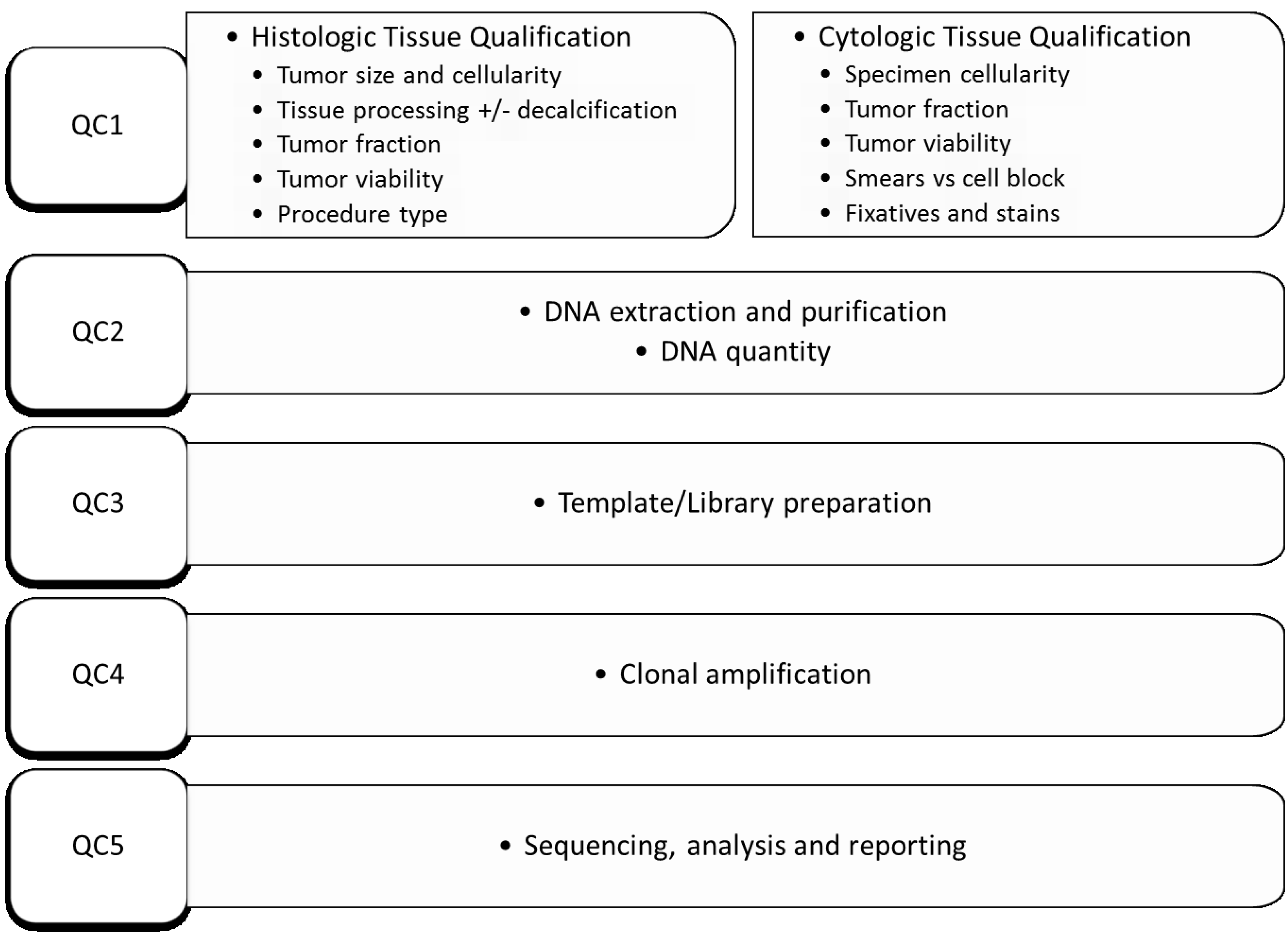
2. Histologic Specimens

2.1. Tissue Processing and Storage

Tissues procured via biopsy and surgery are routinely processed through formalin fixation after gross examination and tissue sectioning. The selected tissue sections are embedded in paraffin blocks, which are then used to prepare thin, 4-5-micron sections for hematoxylin and eosin (H&E) stain and ancillary studies such as special staining, immunohistochemistry staining, in situ hybridization, and molecular testing. The H&E staining and ancillary studies of the formalin-fixed paraffin-embedded tissue (FFPE) sections are reviewed by pathologists to provide the final pathological diagnosis. Pathology reporting of malignant neoplasms includes tumor grade, stage, size, biomarker status, nodal involvement, and response to neoadjuvant therapy if indicated, and tumor block for future biomarker testing at The University of MD Anderson Cancer Center (MD Anderson).
Formalin, the standard tissue fixative in most anatomical pathology laboratories, is an aqueous 37% solution of formaldehyde, commonly used as a neutral buffered formalin solution at 10% concentration (approximately 4% formaldehyde). It preserves tissue in relatively close approximation to its in vivo morphology by fixing both cytoskeletal and soluble proteins [23] Thus, formalin fixation provides excellent preservation of morphology. However, reversible cross links form between proteins and nucleic acids, in addition to random breaks that occur, resulting in highly fragmented nuclei acids [17]. Furthermore, FFPE derived nucleic acid is only compatible with PCR based assays designed for short target sequences.
On the other hand, FFPE tissue is the standard substrate for most clinical molecular laboratory testing, since it represents the majority of tissue sample processed in a clinical pathology laboratory. The FFPE tissue blocks can be stored at room temperatures for years at a low cost and can be used for both histologic examination as well as for selected ancillary studies. Using sequential cuts from the FFPE tissue block, the tumor fraction in unstained sections can be estimated using a representative mirror H&E stained section. In contrast, the collection and storage of frozen tissue is relatively costly and resource intensive. Although frozen tissue provides unfragmented and high quality DNA, the presence of tumor and tumor fraction cannot always be accurately evaluated.

2.2. Tumor Size and Cellularity

The size of the selected tumor-rich areas and number of viable tumor cells within those areas determine the DNA yield, a critical quality control step for NGS. The expected average DNA yield from a single nucleated cell is approximately 6 pg. To obtain the 10 ng recommended DNA for the Ion AmpliSeq Cancer Hotspot Panel, approximately 2000 whole cells are required. In a typical large sample of excised tumor, a single unstained FFPE section with at least 60–100 mm2 of selected tumor-rich tissue would be required to provide the 2000 cells necessary to yield adequate DNA for NGS mutational analysis. In contrast, extremely small tumor sections with <10 mm2 of selected tumor-rich tissue often fall short of providing adequate DNA, even if tissue is extracted from multiple (10 to 20) unstained FFPE sections [24].
In our experience, as expected, the NGS failure rate due to inadequate DNA quantity is usually much higher in tumors from which the selected tumor tissue sections are extremely small than in tumors from which the selected sections are larger [24]. Increasing the tissue size by selecting a larger area may increase the DNA yield, but this increase often is at the expense of decreasing the tumor fraction by including more non-tumor components, such as stromal cells. When the estimated tumor fraction is less than the analytical sensitivity of the NGS platform (approximately 5%), the risk of false-negative calls is high and the unequivocal determination of low-frequency variant calls is difficult.
Hypocellular tumors require that larger areas be selected and thus greater numbers of unstained sections are needed to ensure adequacy of tumor extraction and cellularity for DNA purification. Common hypocellular specimens include mucinous tumors with scant tumor cells floating in large pools of mucin and treated tumors with residual tumor cells in hyalinized fibrotic stroma (Table 2).

2.3. Type of Procedure

Solid tumor specimens tested by NGS are from surgical resections, excisions, or CNB or FNA procedures. The surgical resection and excision specimens generally provide large tumor sections and yield more DNA than the smaller CNB and FNA specimens and are associated with higher NGS success rates [24]. However, a large fraction of diagnostic samples are obtained by minimally invasive procedures such as FNA and CNB. Not only do these samples tend to be smaller and yield less DNA to begin with, but often they get smaller with subsequent recutting of deeper sections used for molecular biomarker testing. Consequently, when compared side by side, the NGS success rate is lower in small samples (CNB and FNA specimens) than in larger samples (resections and excisions), when the manufacturer-recommended DNA input amount is strictly followed [24]. However, if the input DNA threshold is lowered, a subset of cases with high-quality DNA but low DNA concentration can be sequenced successfully on the Ion Torrent platform, allowing uniform NGS success rates across all procedure types (resection, excision, CNB, and FNA) [24,25].

2.4. Tumor Type and Tumor Site

Deep-seated tumors require image-guided FNA/biopsy by an interventional radiologist. They can often pose difficulties in terms of accessibility of lesion, acquisition of adequate material, and risk of bleeding and other complications [26]. With recent advances in imaging technology, our ability to detect smaller lesions is improving. Often the interventional radiologist has to use a small caliber (18 to 20 gauge) needle to sample these sub-centimeter lesions. The tumor tissue obtained by these smaller needles is generally scant and thus the DNA yield is low. In small and/or technically challenging lesions, the interventional radiologist often will forego the CNB and perform an FNA with a smaller (20 to 23 gauge) needle to minimize potential complications associated with the procedure [26]. This underscores the need for novel molecular techniques such as NGS that can provide highly multiplexed and massively parallel sequencing for multiple genes using very small quantities of input DNA. A study performed at our center that reviewed various types of solid tumors sampled by various techniques (resection, excision, CNB, and FNA) showed that NGS success rates were similar across all tumor sites and types after adjusting for tumor size and absence of decalcification [24].

2.5. Tumor Fraction

The tumor fraction requirement for NGS is dependent on the analytical sensitivity of the platform. The lowest limit of detection (i.e., analytical sensitivity) for low-frequency variants is approximately 5%–10% in deep sequencing coverage of 250 to 500× [2,5,8,27,28]. Since a majority of tumors are heterozygous, and somatic mutations usually affect one of two alleles in oncogenes, the minimum tumor fraction within selected areas is 10%–20%. Because ultra-deep sequencing can detect variants at <1% frequency, the minimum tumor fraction can drop below 10% [29]. In specimens matched for tumor size with tumor fractions >20%, the NGS success rates on the Ion Torrent platform are similar across various tumor fractions [24,30].

2.6. DNA Yield and DNA Input

The DNA input is a critical quality control step to achieve the highest success rate for NGS mutational analysis of solid tumors. However, it should be noted that strict adherence to the manufacturer-recommended DNA input, such as 10 ng for Ion AmpliSeq Cancer Hotspot panel can exclude a small subset of samples with borderline DNA yield that is of sufficiently high quality but falls short of the recommended DNA input threshold. A validation study at MD Anderson showed that NGS can be performed successfully in solid tumor samples with DNA input less than the manufacturer’s recommendation, improving the overall NGS success rate [24,25]. This allows a larger number of extremely small tumor samples, such as those obtained from minimally invasive procedures like FNAs and CNBs, to be analyzed successfully with NGS.

2.7. Tumor Viability

Viable tumor tissue is preferred for molecular testing [31]. However, tumor necrosis is not infrequently observed in tumors such as colorectal adenocarcinoma, squamous cell carcinoma, and glioblastoma, as well as in tumors that have undergone neoadjuvant therapy. Cell death can be caused by either apoptosis or necrosis. Necrosis typically leads to autolysis of cellular components, including karyolysis, which is incompatible with PCR-based sequencing tests. In contrast, apoptosis, also known as programmed cell death, is associated with nuclear and DNA fragmentation, which is presumed to be compatible with PCR-based sequencing tests if the target sequences are short. Many chemotherapy drugs such as antimetabolites and alkylating agents exert their antitumor effects through apoptosis. Therefore, non-viable tumor in post-treatment samples, which may appear dead morphologically, can yield fragmented DNA that can still be used for molecular testing. A study on areas of necrosis in colorectal adenocarcinomas generated results comparable to those of viable tumor on NGS mutational analysis by the Ion AmpliSeq Cancer Hotspot Panel [32]. However, the findings are preliminary and further studies are needed to evaluate the effect of tumor necrosis, both in treated tumors and in other necrotic tumor types.

2.8. Decalcification of Bone Specimens

Mineralized bone specimens are hard and brittle after routine histologic processing, making production of optimal sections for histologic evaluation and ancillary studies extremely difficult [33]. A decalcification process removes calcium ions from bone for routine histologic processing and evaluation. Decalcifying reagents typically contain a strong acid (nitric acid or hydrochloric acid), a weak acid (picric, acetic, or formic acid), or a chelating agent (EDTA) [34]. Adequate tissue fixation is required prior to decalcification to protect the cellular and stromal components of bone and attached soft tissue from damage.
Strong acid–based decalcification procedures use acids of extremely low pH to achieve the most rapid removal of calcium ion from bone [35,36]; however, prolonged fixation in strong acid rapidly macerates the tissue and prevents nuclear staining (basic staining). In addition, strong acids tend to degrade DNA, making the decalcified bone sections suboptimal for PCR-based molecular testing [34].
Weak acids, although slower than the strong acid agents in the decalcifying process, are much gentler in action and less likely to interfere with nuclear staining [33]. Thus, formic acid–based decalcification is compatible with histologic evaluation, immunohistochemical studies, and PCR-based sequencing analysis. The slow decalcification induced by the formic acid-based decalcification protocol, for example, allows repeated monitoring of the specimen by radiographic methods to evaluate decalcification status, which permits optimization of results. The process is halted with a neutralizing buffer once decalcification is noted in the specimen, grossly or radiographically. A study at our institution showed the success rate of NGS performed on the Ion Torrent platform reached more than 70% in bony specimens processed with a formic acid–based decalcification procedure; only slightly lower than that achieved in solid tumors from non-bony sites without decalcification [24].
Chelating agents work very slowly, and the decalcifying process with these agents may take weeks [34]. EDTA captures calcium ions from apatite crystal to slowly reduce its size. Although the EDTA decalcifying process is very gentle and preserves morphology and tissue features for immunohistochemistry, in situ hybridization, and PCR-based molecular testing [34], the extremely slow process limits its use in clinical laboratories that must respond to needs for time-sensitive diagnostic information critical for patient management.
Therefore, bony samples that have undergone strong acid-based decalcification are compatible with histologic evaluation but are not suitable for PCR-based sequencing procedures, whereas samples that undergo formic acid- or EDTA-based decalcification procedures take longer to process but are suitable for both morphologic analysis and PCR-based sequencing, including NGS. Ultimately the choice of decalcifying agent will depend on the specific laboratory and balancing the priorities for turnaround time, high-quality histologic sections, and reliable molecular results by the laboratory.

2.9. Other Factors: Blood and Mucin

Although anucleated blood cells such as red blood cells (RBCs) do not reduce the tumor fraction, heme in RBCs can act as an inhibitor in the PCR reaction [37]. Therefore, special care is needed during selection of tumor areas to enrich the tumor fraction while minimizing entrapped RBCs in bloody specimens. Mucin and hyalinized stroma in tumor samples do not affect the PCR reaction per se; however, large quantities of these in the tissue section can reduce the overall tumor cellularity and dilute the sample. Therefore, appropriate numbers of unstained tumor sections are usually required to ensure sufficient tumor cells for DNA extraction.

3. Cytology Specimens

As previously mentioned, it is becoming increasingly common to perform NGS mutational analysis on cytology specimens. FNA procedures, which are minimally invasive and generally well tolerated by the patient, can provide adequate material for diagnosis as well as ancillary studies, including NGS mutational analysis [3,25,38,39]. Cytologic specimens offer a number of advantages in terms of molecular testing. At many institutions, rapid on-site evaluation (ROSE) guides the interventional radiologist in acquiring an adequate specimen, which can be assessed immediately for diagnosis as well as ancillary studies. Direct smears, cytospin preparations, and liquid-based cytologic preparations (LBC) are not formalin-fixed and provide whole cells with whole nuclei, unlike FFPE sections which are typically 4 or 5 microns in thickness, and therefore yield higher quality/quantity DNA [38]. FNAs typically offer wider sampling of the lesion since, unlike CNB specimens, the aspiration is not unidirectional. In addition, FNA specimens usually contain fewer background benign cells (e.g., stromal cells) and therefore have a higher tumor fraction than other types of specimens [38]. Finally, cytologic bone samples are not decalcified, and therefore DNA quality is not compromised by acid or harsh fixatives [38,40]. However, cytology specimens are historically underutilized for molecular testing [41], primarily because most molecular testing requests are directed reflexively to the histology specimens, and cytology specimens are typically tested only when there is no concurrent core biopsy or the core biopsy is depleted/insufficient for testing [25]. The other issue that complicates testing of cytology specimens is that molecular laboratories are not always validated to test all the different types of cytologic specimen preparations (direct smears, cytospin preparations, LBC, and FFPE cell blocks), and most laboratories prefer to treat all specimens as they do traditional FFPE histology blocks [40]. Some of the pre-analytical factors affecting NGS mutational analysis in cytology specimens are discussed below.

3.1. Specimen Cellularity

In cytology, as in histology specimens, the overall cellularity plays an important role in NGS mutational analysis. For routine NGS mutational analysis on an Ion Torrent PGM system requiring 10 ng input DNA for a hotspot panel, as already stated, the goal is for a specimen with a total of at least 2000 cells. The specimen cellularity for FNAs depends largely on the quality of the procedure (i.e., skill and expertise of the proceduralist) and the nature of the lesion (i.e., size, location and cellular cohesion of the tumor). Small sub-centimeter lesions as well as deep-seated tumors that are technically challenging to aspirate typically have a lower cellular yield. In these cases, the cellularity may be a function of the skill and experience of the aspirator. In addition, the nature of the tumor may also play a role in the cellularity. For example, sclerotic or cystic tumors tend to be paucicellular, while tumors with largely discohesive cells, like melanoma or neuroendocrine tumors, usually yield highly cellular aspirates (Table 3).

3.2. Type of Preparation

While specimen cellularity may depend largely on the FNA procedure, the cytologic preparation also plays an important role in the choice of a substrate for NGS testing. Cytology offers a multitude of preparations for NGS mutational analysis, including direct smears and cytospins, FFPE cell blocks, and LBC. Cell block preparations, although easiest to use and validate, in our experience and others, often fall short of providing adequate material [38,42,43]. The biggest disadvantage of cell block preparations is the inability to assess them immediately for adequacy. While implementing dedicated FNA passes for the needle rinse may enhance cellularity for the cell block, it still does not guarantee an adequate sample for ancillary studies. LBC preparations share the same disadvantages in being not immediately assessable and having variable neoplastic cellularity; however, the overall ease of specimen collection and transportation, combined with an optimally preserved sample that largely eliminates background blood and debris, make LBC an attractive sample preparation for NGS testing [44]. Most institutions that perform NGS testing from LBC preparations employ some form of pre-analytical measure through visualization of neoplastic cells in the LBC preparation to avoid false-negative calls [45,46,47]. In contrast, direct smears provide the only source of reliable testing material in cases with limited tissue and where the cell block is inadequate for testing. DNA obtained from direct smears do not have formalin fixation artifacts [17,25,40] and provide whole cells with intact nuclei (Table 3). However, utilizing direct smears for NGS mutational analysis requires sacrificing the slide, which may be a medicolegal issue in certain states and institutions, as well as additional in-house validation for performing the NGS assay.
Table 3. Pre-analytical factors in cytologic specimens associated with success of NGS mutational analysis.
Associated FactorsComments
Specimen cellularityDepends on multiple factors, including
  • Size and location of tumor
  • Skill and experience of aspirator
  • Nature of tumor (e.g., solid vs cystic vs sclerotic)Type of tumor (discohesive cells such as melanoma, neuroendocrine carcinoma usually have higher yield)
Type of preparationDirect smears
  • Immediate assessment
  • 3-dimensional clusters with whole nuclei
  • Usually better quality DNA than cell blocks
  • No formalin fixation artifact
  • Requires in-house validation
  • Requires sacrificing of the slide
Cell blocks
  • Variable cellularity; no immediate assessment
  • Formalin-fixed cells
  • 4- to 5-micron sections with partial nuclei
  • Easy to validateLiquid-based preparations
Liquid-based preparations
  • Lack of immediate assessment
  • Ease of specimen collection and transportation
  • Optimal preservation of nucleic acids
  • Requires in-house validation
Type of fixative and stainsDirect smears
  • Air-dried methanol fixed, Diff-Quik stained
  • Ethanol fixed, Papanicolaou (Pap) stained
Type of glass slideNon-frosted vs positively charged vs fully frosted slides
Tumor fractionTumor fraction determinants
  • Tumor type, primary vs metastasis
  • Specimen type, fluids and EBUS-guided lymph node FNAs often have low tumor fraction
Tumor fraction requirement for NGS
  • Depends on technical sensitivity of NGS platform
DNA yieldDepends on the overall cellularity of sample
  • Samples with high tumor fraction but scant cellularity may not have adequate DNA yield for PCR amplification
Input DNAHigh-quality/low-DNA-concentration samples can benefit from lowering the threshold of input DNA
NGS, next-generation sequencing; EBUS, endobronchial ultrasound; FNA, fine needle aspiration.

3.3. Type of Fixative and Stains

Studies by several groups have shown that both methanol-fixed Diff-Quik-stained smears as well as ethanol-fixed Papanicolaou (Pap)-stained smears can be used for molecular testing [44,48,49,50]. However, interlaboratory variation in fixation and staining techniques may play a role in DNA yield and molecular testing success. In our experience there is no significant difference in NGS mutational analysis findings between different cytologic fixatives and stains [3].

3.4. Type of Slide

A variety of glass slides are commercially available for preparation of cytologic direct smears and cytospins. These include the standard non-frosted or frosted tip slides that do not have any specialized coating; the fully frosted slides that have a rough surface to enhance cellular adhesion; the positively charged slides that electrostatically enhance cellular adhesion; and coated slides that use silane or lysine allowing covalent bonding of cells to the slide surface. It is reasonable to assume the type of slide used for specimen preparation plays a role in the cellularity seen on the smears. In our experience fully frosted slides show enhanced cellular retention, with lower cell loss during fixation and processing, than the frosted tip or positively charged slides. However, glass slides that enhance cellular adherence, can also pose difficulties during tissue extraction. A recent study by our group showed that the frosted tip slides yield higher DNA quantities than the fully frosted slides, presumably due to the difficulty in dislodging cells from the rough surface of the fully frosted slides [25]. Therefore, the choice of glass slide type can be an important factor in the success of NGS (Table 3).

3.5. Tumor Fraction

As mentioned earlier, the tumor fraction requirement for NGS is dependent on the analytical sensitivity of the platform which is approximately 5%–10% in deep sequencing coverage of 250 to 500×. Assuming most tumors are heterozygous, the minimum tumor fraction within tumor enriched areas needs to be approximately 10%–20%. In cytology, some specimens have an intrinsically low tumor fraction due to the abundant benign cells in the background. Examples would include pleural and peritoneal fluids with contaminating mesothelial cells and histiocytes and samples obtained through endobronchial ultrasound guided (EBUS) FNA with background lymphocytes and bronchial cells (Table 3).
Cytology specimens like histology specimens are enriched for tumor by identifying areas on the slide with a higher tumor fraction for tissue extraction. Cell block sections are treated the same as histology specimens: the selected tumor-rich area of the hematoxylin and eosin (H&E)-stained section is circled and the tumor fraction estimated by the cytopathologist. Unstained sections are then matched up with the H&E slide and the corresponding areas are manually macro/microdissected using a scalpel blade. For direct smear slides, the selected tumor-rich area is circled on the slide and then the back of the slide is etched with a diamond-tip pen in the area corresponding to the circled tumor-rich area. The slide is then dipped in xylene to remove the coverslip. The process of removing the coverslip from archival slides does not affect the DNA yield [40,48]. Alternatively, representative slides can be triaged at the time of rapid on-site evaluation and left uncoverslipped for DNA extraction and molecular analysis. The circled/etched areas on the slides can then be macro/microdissected using a scalpel blade.

3.6. DNA Yield and DNA Input

As previously mentioned, adhering to the manufacturer-recommended DNA input can exclude a small subset of samples with borderline DNA yield that falls short of the recommended DNA input threshold but is of sufficiently high quality for NGS success. A study from MD Anderson showed that the overall NGS success rates were significantly improved in cytology samples using DNA input less than the manufacturer’s recommendation [25].

4. Summary

The success of NGS mutational analysis in solid tumors is affected not only by the input DNA quantity but also by a constellation of factors including the DNA quality, the tumor fraction, and the nucleic acid extraction method. To meet the nucleic acid requirement for the testing platform, dedicated pathologists select viable and cellular tumor sections/smears to optimize the tumor-to-stroma ratio and to minimize the admixed inflammatory cells, blood, mucin, and necrotic tumor cells. Sections of decalcified bone may be chosen with caution if a weak acid- or EDTA-based decalcification protocol has been properly administered. Pre-analytical qualification of tissues from solid tumor specimens would ensure a high rate of success in subsequent NGS steps to achieve overall high-quality NGS mutational analysis results.

Acknowledgments

We thank team members from the molecular diagnostic laboratory and the tissue qualification laboratory and Thomas Huynh at MD Anderson for their technical assistance and helpful discussions. RSG is supported by a Post-Doctoral Fellowship Award through the Canadian Institutes of Health Research (www.cihr-irsc.gc.ca).

Author Contributions

All the co-authors contributed equally to the content. H.C. and S.R.-C. contributed to the majority of writing.

Conflicts of Interest

The authors declare no conflict of interest.

References

  1. Frampton, G.M.; Fichtenholtz, A.; Otto, G.A.; Wang, K.; Downing, S.R.; He, J.; Schnall-Levin, M.; White, J.; Sanford, E.M.; An, P.; et al. Development and validation of a clinical cancer genomic profiling test based on massively parallel DNA sequencing. Nat. Biotechnol. 2013, 31, 1023–1031. [Google Scholar] [CrossRef] [PubMed]
  2. Singh, R.R.; Patel, K.P.; Routbort, M.J.; Reddy, N.G.; Barkoh, B.A.; Handal, B.; Kanagal-Shamanna, R.; Greaves, W.O.; Medeiros, L.J.; Aldape, K.D.; et al. Clinical validation of a next-generation sequencing screen for mutational hotspots in 46 cancer-related genes. J. Mol. Diagn. 2013, 15, 607–622. [Google Scholar] [CrossRef] [PubMed]
  3. Kanagal-Shamanna, R.; Portier, B.P.; Singh, R.R.; Routbort, M.J.; Aldape, K.D.; Handal, B.A.; Rahimi, H.; Reddy, N.G.; Barkoh, B.A.; Mishra, B.M.; et al. Next-generation sequencing-based multi-gene mutation profiling of solid tumors using fine needle aspiration samples: Promises and challenges for routine clinical diagnostics. Mod. Pathol. 2014, 27, 314–327. [Google Scholar] [CrossRef] [PubMed]
  4. Luthra, R.; Patel, K.P.; Reddy, N.G.; Haghshenas, V.; Routbort, M.J.; Harmon, M.A.; Barkoh, B.A.; Kanagal-Shamanna, R.; Ravandi, F.; Cortes, J.E.; et al. Next-generation sequencing-based multigene mutational screening for acute myeloid leukemia using MiSeq: Applicability for diagnostics and disease monitoring. Haematologica 2014, 99, 465–473. [Google Scholar] [CrossRef] [PubMed]
  5. Singh, R.R.; Patel, K.P.; Routbort, M.J.; Aldape, K.; Lu, X.; Manekia, J.; Abraham, R.; Reddy, N.G.; Barkoh, B.A.; Veliyathu, J.; et al. Clinical massively parallel next-generation sequencing analysis of 409 cancer-related genes for mutations and copy number variations in solid tumours. Br. J. Cancer 2014, 111, 2014–2023. [Google Scholar] [CrossRef] [PubMed]
  6. Bennett, N.C.; Farah, C.S. Next-generation sequencing in clinical oncology: Next steps towards clinical validation. Cancers 2014, 6, 2296–2312. [Google Scholar] [CrossRef] [PubMed]
  7. Chang, F.; Li, M.M. Clinical application of amplicon-based next-generation sequencing in cancer. Cancer Genet. 2013, 206, 413–419. [Google Scholar] [CrossRef] [PubMed]
  8. Cheng, D.T.; Mitchell, T.; Zehir, A.; Shah, R.H.; Benayed, R.; Syed, A.; Chandramohan, R.; Liu, Z.Y.; Won, H.H.; Scott, S.N.; et al. MSK-IMPACT: A Hybridization Capture-Based Next-Generation Sequencing Clinical Assay for Solid Tumor Molecular Oncology. J. Mol. Diagn. 2015, 17, 251–264. [Google Scholar] [CrossRef] [PubMed]
  9. Desai, A.N.; Jere, A. Next-generation sequencing: Ready for the clinics? Clin. Genet. 2012, 81, 503–510. [Google Scholar] [CrossRef] [PubMed]
  10. Guan, Y.F.; Li, G.R.; Wang, R.J.; Yi, Y.T.; Yang, L.; Jiang, D.; Zhang, X.P.; Peng, Y. Application of next-generation sequencing in clinical oncology to advance personalized treatment of cancer. Chin. J. Cancer 2012, 31, 463–470. [Google Scholar] [CrossRef] [PubMed]
  11. Hadd, A.G.; Houghton, J.; Choudhary, A.; Sah, S.; Chen, L.; Marko, A.C.; Sanford, T.; Buddavarapu, K.; Krosting, J.; Garmire, L.; et al. Targeted, high-depth, next-generation sequencing of cancer genes in formalin-fixed, paraffin-embedded and fine-needle aspiration tumor specimens. J. Mol. Diagn. 2013, 15, 234–247. [Google Scholar] [CrossRef] [PubMed]
  12. McCutcheon, J.N.; Giaccone, G. Next-Generation Sequencing: Targeting Targeted Therapies. Clin. Cancer Res. 2015. [Google Scholar] [CrossRef] [PubMed]
  13. Pant, S.; Weiner, R.; Marton, M.J. Navigating the rapids: The development of regulated next-generation sequencing-based clinical trial assays and companion diagnostics. Front. Oncol. 2014, 4, 78. [Google Scholar] [CrossRef] [PubMed]
  14. Salto-Tellez, M.; de Castro, G.D. Next-generation sequencing: A change of paradigm in molecular diagnostic validation. J. Pathol. 2014, 234, 5–10. [Google Scholar] [CrossRef] [PubMed]
  15. Young, G.; Wang, K.; He, J.; Otto, G.; Hawryluk, M.; Zwirco, Z.; Brennan, T.; Nahas, M.; Donahue, A.; Yelensky, R.; et al. Clinical next-generation sequencing successfully applied to fine-needle aspirations of pulmonary and pancreatic neoplasms. Cancer Cytopathol. 2013, 121, 688–694. [Google Scholar] [CrossRef] [PubMed]
  16. Srinivasan, M.; Sedmak, D.; Jewell, S. Effect of fixatives and tissue processing on the content and integrity of nucleic acids. Am. J. Pathol. 2002, 161, 1961–1971. [Google Scholar] [CrossRef]
  17. Williams, C.; Ponten, F.; Moberg, C.; Söderkvist, P.; Uhlén, M.; Pontén, J.; Sitbon, G.; Lundeberg, J. A high frequency of sequence alterations is due to formalin fixation of archival specimens. Am. J. Pathol. 1999, 155, 1467–1471. [Google Scholar] [CrossRef]
  18. Gargis, A.S.; Kalman, L.; Berry, M.W.; Bick, D.P.; Dimmock, D.P.; Hambuch, T.; Lu, F.; Lyon, E.; Voelkerding, K.V.; Zehnbauer, B.A.; et al. Assuring the quality of next-generation sequencing in clinical laboratory practice. Nat. Biotechnol. 2012, 30, 1033–1036. [Google Scholar] [CrossRef] [PubMed]
  19. English, A.C.; Salerno, W.J.; Hampton, O.A.; Gonzaga-Jauregui, C.; Ambreth, S.; Ritter, D.I.; Beck, C.R.; Davis, C.F.; Dahdouli, M.; Ma, S.; et al. Assessing structural variation in a personal genome-towards a human reference diploid genome. BMC Genomics 2015, 16, 286. [Google Scholar] [CrossRef] [PubMed]
  20. Wang, M.; Beck, C.R.; English, A.C.; Meng, Q.; Buhay, C.; Han, Y.; Doddapaneni, H.V.; Yu, F.; Boerwinkle, E.; Lupski, J.R.; et al. PacBio-LITS: A large-insert targeted sequencing method for characterization of human disease-associated chromosomal structural variations. BMC Genomics 2015, 16, 214. [Google Scholar] [CrossRef] [PubMed]
  21. Zheng, Z.; Liebers, M.; Zhelyazkova, B.; Cao, Y.; Panditi, D.; Lynch, K.D.; Chen, J.; Robinson, H.E.; Shim, H.S.; Chmielecki, J.; et al. Anchored multiplex PCR for targeted next-generation sequencing. Nat. Med. 2014, 20, 1479–1484. [Google Scholar] [CrossRef] [PubMed]
  22. Aziz, N.; Zhao, Q.; Bry, L.; Driscoll, D.K.; Funke, B.; Gibson, J.S.; Grody, W.W.; Hegde, M.R.; Hoeltge, G.A.; Leonard, D.G.; et al. College of American Pathologists’ laboratory standards for next-generation sequencing clinical tests. Arch. Pathol. Lab. Med. 2015, 139, 481–493. [Google Scholar] [CrossRef] [PubMed]
  23. Frankel, A. Formalin fixation in the “-omics” era: A primer for the surgeon-scientist. ANZ J. Surg. 2012, 82, 395–402. [Google Scholar] [CrossRef] [PubMed]
  24. Goswami, R.S.; Luthra, R.; Singh, R.R.; Patel, K.P.; Routbort, M.J.; Aldape, K.D.; Yao, H.; Dang, H.D.; Barkoh, B.A.; Manekia, J.; et al. Identification of Factors Affecting the Success of Next-Generation Sequencing Testing in Solid Tumors. Am. J. Clin. Pathol 2015, in press. [Google Scholar]
  25. Roy-Chowdhuri, S.; Goswami, R.S.; Chen, H.; Patel, K.P.; Roubort, M.J.; Singh, R.R.; Broaddus, R.R.; Barkoh, B.A.; Manekia, J.; Yao, H.; et al. Factors affecting the success of next-generation sequencing in cytology specimens. Cancer Cytopathol. 2015. [Google Scholar] [CrossRef] [PubMed]
  26. Gupta, S.; Madoff, D.C. Image-guided percutaneous needle biopsy in cancer diagnosis and staging. Tech. Vasc. Interv. Radiol. 2007, 10, 88–101. [Google Scholar] [CrossRef] [PubMed]
  27. Rossle, M.; Sigg, M.; Ruschoff, J.H.; Wild, P.J.; Moch, H.; Weber, A.; Rechsteiner, M.P. Ultra-deep sequencing confirms immunohistochemistry as a highly sensitive and specific method for detecting BRAF V600E mutations in colorectal carcinoma. Virchows Arch. 2013, 463, 623–631. [Google Scholar] [CrossRef] [PubMed]
  28. Portier, B.P.; Kanagal-Shamanna, R.; Luthra, R.; Singh, R.; Routbort, M.J.; Handal, B.; Reddy, N.; Barkoh, B.A.; Zuo, Z.; Medeiros, L.J.; et al. Quantitative assessment of mutant allele burden in solid tumors by semiconductor-based next-generation sequencing. Am. J. Clin. Pathol. 2014, 141, 559–572. [Google Scholar] [CrossRef] [PubMed]
  29. Rechsteiner, M.; von Teichman, A.; Ruschoff, J.H.; Fankhauser, N.; Pestalozzi, B.; Schraml, P.; Weber, A.; Wild, P.; Zimmermann, D.; Moch, H.; et al. KRAS, BRAF, and TP53 deep sequencing for colorectal carcinoma patient diagnostics. J. Mol. Diagn. 2013, 15, 299–311. [Google Scholar] [CrossRef] [PubMed]
  30. Roy-Chowdhuri, S. BRAF p.V600E Mutation Detection is Independent of Estimated Sample Tumor Percentage in a Large Cohort of Melanoma Samples Evaluated by Next-Generation Sequencing. J. Mol. Diagn. 2014, 16, 768. [Google Scholar]
  31. Long, E.; Ilie, M.; Hofman, V.; Lassalle, S.; Butori, C.; Alsubaie, S.; Hofman, P. Role of the surgical pathologist for tissue management in oncology. Bull. Cancer 2013, 100, 837–845. [Google Scholar] [PubMed]
  32. Jabbar, K. Impact of tumor necrosis on success of clinical next-generation sequencing. Mod. Pathol. 2015, 28, S501. [Google Scholar]
  33. Mack, S.A.; Maltby, K.M.; Hilton, M.J. Demineralized murine skeletal histology. Methods Mol. Biol. 2014, 1130, 165–183. [Google Scholar] [PubMed]
  34. Singh, V.M.; Salunga, R.C.; Huang, V.J.; Tran, Y.; Erlander, M.; Plumlee, P.; Peterson, M.R. Analysis of the effect of various decalcification agents on the quantity and quality of nucleic acid (DNA and RNA) recovered from bone biopsies. Ann. Diagn. Pathol. 2013, 17, 322–326. [Google Scholar] [CrossRef] [PubMed]
  35. Castania, V.A.; Silveira, J.W.; Issy, A.C.; Pitol, D.L.; Castania, M.L.; Neto, A.D.; del Bel, E.A.; Defino, H.L.A. Advantages of a combined method of decalcification compared to EDTA. Microsc. Res. Tech. 2015, 78, 111–118. [Google Scholar] [CrossRef] [PubMed]
  36. Gertych, A.; Mohan, S.; Maclary, S.; Mohanty, S.; Wawrowsky, K.; Mirocha, J.; Balzer, B.; Knudsen, B.S. Effects of tissue decalcification on the quantification of breast cancer biomarkers by digital image analysis. Diagn. Pathol. 2014, 9, 213. [Google Scholar] [CrossRef] [PubMed]
  37. Akane, A.; Matsubara, K.; Nakamura, H.; Takahashi, S.; Kimura, K. Identification of the heme compound copurified with deoxyribonucleic acid (DNA) from bloodstains, a major inhibitor of polymerase chain reaction (PCR) amplification. J. Forensic Sci. 1994, 39, 362–372. [Google Scholar] [CrossRef] [PubMed]
  38. Knoepp, S.M.; Roh, M.H. Ancillary techniques on direct-smear aspirate slides: A significant evolution for cytopathology techniques. Cancer Cytopathol. 2013, 121, 120–128. [Google Scholar] [CrossRef] [PubMed]
  39. Schmitt, F.C. Molecular cytopathology and flow cytometry: Pre-analytical procedures matter. Cytopathology 2011, 22, 355–357. [Google Scholar] [CrossRef] [PubMed]
  40. Bellevicine, C.; Malapelle, U.; de Luca, C.; Iaccarino, A.; Troncone, G. EGFR analysis: Current evidence and future directions. Diagn. Cytopathol. 2014, 42, 984–992. [Google Scholar] [CrossRef] [PubMed]
  41. Clark, D.P. Seize the opportunity: Underutilization of fine-needle aspiration biopsy to inform targeted cancer therapy decisions. Cancer 2009, 117, 289–297. [Google Scholar] [CrossRef] [PubMed]
  42. Cai, G.; Wong, R.; Chhieng, D.; Levy, G.H.; Gettinger, S.N.; Herbst, R.S.; Puchalski, J.T.; Homer, R.J.; Hui, P. Identification of EGFR mutation, KRAS mutation, and ALK gene rearrangement in cytological specimens of primary and metastatic lung adenocarcinoma. Cancer Cytopathol. 2013, 121, 500–507. [Google Scholar] [CrossRef] [PubMed]
  43. Pang, B.; Dettmer, M.; Ong, C.W.; Dhewar, A.N.; Gupta, S.; Lim, G.L.; Nga, M.E.; Seet, J.E.; Qasim, A.; Chin, T.M.; et al. The positive impact of cytological specimens for EGFR mutation testing in non-small cell lung cancer: A single South East Asian laboratory’s analysis of 670 cases. Cytopathology 2012, 23, 229–236. [Google Scholar] [CrossRef] [PubMed]
  44. Dejmek, A.; Zendehrokh, N.; Tomaszewska, M.; Edsjo, A. Preparation of DNA from cytological material: Effects of fixation, staining, and mounting medium on DNA yield and quality. Cancer Cytopathol. 2013, 121, 344–353. [Google Scholar] [CrossRef] [PubMed]
  45. Nikiforov, Y.E.; Carty, S.E.; Chiosea, S.I.; Coyne, C.; Duvvuri, U.; Ferris, R.L.; Gooding, W.E.; Hodak, S.P.; LeBeau, S.O.; Ohori, N.P.; et al. Highly accurate diagnosis of cancer in thyroid nodules with follicular neoplasm/suspicious for a follicular neoplasm cytology by ThyroSeq v2 next-generation sequencing assay. Cancer 2014, 120, 3627–3634. [Google Scholar] [CrossRef] [PubMed]
  46. Nikiforova, M.N.; Wald, A.I.; Roy, S.; Durso, M.B.; Nikiforov, Y.E. Targeted next-generation sequencing panel (ThyroSeq) for detection of mutations in thyroid cancer. J. Clin. Endocrinol Metab. 2013, 98, E1852–E1860. [Google Scholar] [CrossRef] [PubMed]
  47. Gailey, M.P.; Stence, A.A.; Jensen, C.S.; Ma, D. Multiplatform comparison of molecular oncology tests performed on cytology specimens and formalin-fixed, paraffin-embedded tissue. Cancer Cytopathol. 2015, 123, 30–39. [Google Scholar] [CrossRef] [PubMed]
  48. Lozano, M.D.; Zulueta, J.J.; Echeveste, J.I.; Gúrpide, A.; Seijo, L.M.; Martín-Algarra, S.; del Barrio, A.; Pio, R.; Idoate, M.A.; Labiano, T.; et al. Assessment of epidermal growth factor receptor and K-ras mutation status in cytological stained smears of non-small cell lung cancer patients: Correlation with clinical outcomes. Oncologist 2011, 16, 877–885. [Google Scholar] [CrossRef] [PubMed]
  49. Bozzetti, C.; Negri, F.V.; Azzoni, C.; Naldi, N.; Nizzoli, R.; Bortesi, B.; Zobbi, V.; Bottarelli, L.; Tiseo, M.; Silini, E.M.; et al. Epidermal growth factor receptor and Kras gene expression: Reliability of mutational analysis on cytological samples. Diagn. Cytopathol. 2013, 41, 595–598. [Google Scholar] [CrossRef] [PubMed]
  50. Betz, B.L.; Roh, M.H.; Weigelin, H.C.; Placido, J.B.; Schmidt, L.A.; Farmen, S.; Arenberg, D.A.; Kalemkerian, G.P.; Knoepp, S.M. The application of molecular diagnostic studies interrogating EGFR and KRAS mutations to stained cytologic smears of lung carcinoma. Am. J. Clin. Pathol. 2011, 136, 564–571. [Google Scholar] [CrossRef] [PubMed]

