Review

MALAT-1 Is a Key Regulator of Epithelial–Mesenchymal Transition in Cancer: A Potential Therapeutic Target for Metastasis

by
Mohamed Ali Hussein
1,2,†,
Kamyab Valinezhad
3,†,
Eman Adel
2 and
Gnanasekar Munirathinam
3,*
1
Department of Pharmaceutical Services, Children’s Cancer Hospital Egypt, Cairo 57357, Egypt
2
Department of Biology, School of Sciences and Engineering, American University in Cairo, New Cairo 11835, Egypt
3
Department of Biomedical Sciences, College of Medicine, University of Illinois, Rockford, IL 61107, USA
*
Author to whom correspondence should be addressed.
These authors contributed equally to this work.
Cancers 2024, 16(1), 234; https://doi.org/10.3390/cancers16010234
Submission received: 12 December 2023 / Revised: 29 December 2023 / Accepted: 1 January 2024 / Published: 4 January 2024
(This article belongs to the Section Cancer Pathophysiology)

Abstract

:

Simple Summary

Metastasis-associated lung adenocarcinoma transcript-1 (MALAT-1) is overexpressed in several cancers and exerts its effect by controlling gene expression and stimulating cell proliferation, migration, and metastasis and playing a dynamic role in mediating the epithelial-to-mesenchymal transition (EMT), which leads to the acquisition of stem cell-like properties and chemoresistance. MALAT-1 modulates EMT by interacting with various intracellular signaling pathways, including phosphoinositide 3-kinase (PI3K)/Akt and Wnt/β-catenin. It also sponges microRNAs, consequently increasing the gene expression of several essential genes regulating cancer progression and metastasis, rendering it a good candidate for therapeutic intervention. Several innovative approaches have been exploited to target MALAT-1, such as short hairpin RNAs (shRNAs), antisense oligonucleotides (ASOs), and natural products.

Abstract

Metastasis-associated lung adenocarcinoma transcript-1 (MALAT-1) is a long intergenic non-coding RNA (lncRNA) located on chr11q13. It is overexpressed in several cancers and controls gene expression through chromatin modification, transcriptional regulation, and post-transcriptional regulation. Importantly, MALAT-1 stimulates cell proliferation, migration, and metastasis and serves a vital role in driving the epithelial-to-mesenchymal transition (EMT), subsequently acquiring cancer stem cell-like properties and developing drug resistance. MALAT-1 modulates EMT by interacting with various intracellular signaling pathways, notably the phosphoinositide 3-kinase (PI3K)/Akt and Wnt/β-catenin pathways. It also behaves like a sponge for microRNAs, preventing their interaction with target genes and promoting EMT. In addition, we have used bioinformatics online tools to highlight the disparities in the expression of MALAT-1 between normal and cancer samples using data from The Cancer Genome Atlas (TCGA). Furthermore, the intricate interplay of MALAT-1 with several essential targets of cancer progression and metastasis renders it a good candidate for therapeutic interventions. Several innovative approaches have been exploited to target MALAT-1, such as short hairpin RNAs (shRNAs), antisense oligonucleotides (ASOs), and natural products. This review emphasizes the interplay between MALAT-1 and EMT in modulating cancer metastasis, stemness, and chemoresistance in different cancers.

1. Introduction

Metastasis-associated lung adenocarcinoma transcript-1 (MALAT-1) is a long non-coding intergenic RNA of 12,820 bases on chr11q13. It is initially transcribed as a precursor transcript, followed by enzymatic processing by RNase P to form the mature long non-coding RNA [1,2]. A triple helical structure at the 3′ end stabilizes the structure of MALAT-1 and compensates for the missing poly-A tail. It regulates gene expression by various mechanisms, including modulating gene transcription through the repression of promoters of target genes, regulating RNA-binding proteins or activating mesenchymal transcription factors, modifying the chromatin, and regulating post-transcriptional processing (Figure 1) [3,4,5,6]. In addition, it is involved in DNA repair and cell death [7]. Targeting MALAT-1 induces DNA damage and sensitizes cancer to chemotherapy treatment [8].
MALAT-1 induces cancer proliferation, invasion, migration, and metastasis to distant sites. Recently, several studies highlighted the immunomodulatory role of MALAT-1 and how it can enable cancer cells to escape immune surveillance by exerting an immunosuppressive effect and regulating the expression of several molecules associated with the tumor microenvironment [9,10]. In the triple-negative breast cancer (TNBC) cell model, MALAT-1 knockdown results in a marked induction in MHC class I chain-related proteins A/B expression and the repression of the checkpoint molecules PD-L1 and B7-H4 [11]. In addition, MALAT1 also modulates its suppressive effect by negatively modulating Myeloid-derived suppressor cells (MDSCs) and decreasing peripheral blood mononuclear cells (PBMCs) in cancer patients [12]. Indeed, MALAT-1 knockdown using MALAT-1 antisense oligonucleotides (ASO) in an immune-competent mouse model results in a decrease in MDSC as well as immunosuppressive tumor-associated macrophages (TAM). In contrast, an increase in cytotoxic CD8+ T cells was also observed, which opened new avenues in understanding the conspicuous role of MALAT-1 in modulating carcinogenesis [13]. Various studies have reported MALAT-1 overexpression in numerous cancers such as esophageal squamous cell carcinoma (ESCC), gastric cancer (GC), non-small cell lung cancer (NSCLC), colorectal cancer (CRC), pancreatic cancer, breast cancer (BC), and hepatocellular carcinoma (HCC) [14,15,16,17,18,19,20,21,22,23,24]. Using the online portal of the University of Alabama at Birmingham (UALCAN) (https://ualcan.path.uab.edu/) (Accessed on 26 December 2023), we analyzed MALAT-1 gene expression across different cancers using data from The Cancer Genome Atlas (TCGA) [25,26]. The expression levels of MALAT-1 between samples of different cancers and normal patients show that high expression of MALAT-1 is a prevalent event in various tumors such as ESCC, HCC, cholangiocarcinoma, cervical cancer (CC), sarcoma, and melanoma (Figure 2). Moreover, plausible differences in the expression of MALAT-1 between normal and cancerous samples were observed in other cancers, such as bladder carcinoma, thyroid carcinoma, and stomach adenocarcinoma (Figure 2). Contradictory to the disparity in MALAT-1 expression between normal and cancerous samples that are generally observed, samples having approximately the same MALAT-1 expression levels also exist. This ambiguity implies that MALAT-1 has a pleiotropic effect in cancer cells.
MALAT-1 is a promising diagnostic marker for detecting endometrial, breast, NSCLC, bladder, and nasopharyngeal carcinoma (NPC) [27,28,29,30,31]. However, there is a discrepancy in reporting the diagnostic accuracy of MALAT-1 [29,30,31]. A pooled analysis including 17 studies with 3255 subjects demonstrated that MALAT-1 exhibits moderate accuracy in detecting and diagnosing cancer, and it was strongly associated with the metastasis of early-stage NSCLC [32]. A wealth of evidence has revealed that the role of MALAT-1 is pivotal in modulating EMT, driving cells to become cancer stem cells (CSCs) or acquire stem cell–like properties, develop chemoresistance, and metastasize to distant places in the body [17,33,34,35,36]. Albeit the role of MALAT-1 in cancer has been extensively studied, molecular mechanisms that regulate MALAT-1 are scarcely reported. Several reports examined the expression level of MALAT-1 and its significance as a diagnostic, prognostic, and, recently, as a novel drug target. However, the underlying mechanisms that induce MALAT-1 to play its role are still largely unknown.
In EMT, epithelial cells display migratory and invasive features and become mesenchymal cells via the downregulation of E-cadherin, desmosomes, and claudin, and the upregulation of mesenchymal markers such as N-cadherin, fibronectin, and vimentin [37,38,39,40,41,42]. EMT increases the invasiveness and plasticity of cancer cells, which results in cancer cell dissemination to distant sites through the basement membrane, thereby inducing metastasis. Tumor-associated stroma upregulates the expression of various growth factors such as PDGF, EGF, HGF, and TGF-β, which in turn induces the activation of a series of transcription factors, including SNAI1, Slug, Twist, Zinc finger E-box binding homeobox1 (ZEB1), Goosecoid, and FOXC2, consequently initiating the EMT process [43,44,45,46,47,48]. Several reports demonstrate that EMT is crucial in stimulating cancer progression and metastasis and acquiring drug-resistant properties by modulating alternative cell signaling pathways [49,50,51,52,53]. Essential player proteins like Akt, ERK, MAPK, PI3K, β-catenin, and SMAD are essential in modulating EMT by central cell-signaling pathways [54]. Furthermore, microRNAs also have a crucial role in the cellular signaling circuitry that controls the EMT process. MALAT-1 serves as a competitive endogenous RNA (ceRNA) for tumor-suppressive microRNA and consequently downregulates their gene expression.
This review will dissect the mechanisms by which MALAT-1 modulates EMT to enhance cancer proliferation, metastasis, stemness, and chemoresistance (Table 1). We will also thoroughly discuss the interaction between MALAT-1 and microRNA (Table 2). Different signaling pathways will also be explored. In this review, we provide an overview of the interplay between MALAT-1 and EMT and shed light on the importance of MALAT-1 in enabling cancer invasiveness, metastasis, stemness, and chemoresistance through several mechanisms.

2. Methodology for Searching the Literature and Study Selection Criteria

In this review, the literature search was conducted using different controlled search terms with several conjunctions: (NEAT2 [TIAB] OR “metastasis-associated lung adenocarcinoma transcript 1” [TIAB] OR “metastasis-associated lung adenocarcinoma transcript-1” [TIAB] OR MALAT-1 [TIAB]) AND (“Epithelial-Mesenchymal Transition” [TIAB] OR “Epithelial-Mesenchymal Transitions” [TIAB] OR “Epithelial-Mesenchymal Transformation” [TIAB] OR “Epithelial-Mesenchymal Transformation” [TIAB] OR “Epithelial-Mesenchymal Transformations” [TIAB] OR EMT [TIAB]). Our search was executed using several databases, including PubMed, Web of Science, and Scopus. Associated citations of every article were examined. We included all articles published in English that discussed the mechanistic role of MALAT-1 in modulating EMT in different cancers.

3. MALAT-1 Modulates EMT and Promotes Cancer Metastasis, Stemness, and Chemoresistance

3.1. MALAT-1 Induces Cancer Progression and Metastasis by Modulating EMT

The overexpression of MALAT-1 enhances the ability of tumor cells to migrate, invade, metastasize, and escape the cytotoxic effect of chemotherapy. One study reported that MALAT-1 inhibition decreases the progression and metastasis of CC in vitro and in vivo [56]. MALAT-1 knockdown upregulates the epithelial markers and downregulates the mesenchymal markers, contributing to an active EMT process [56]. In addition, the EMT regulator SNAI1 was markedly downregulated, indicating that SNAI1 regulates the expression of E-cadherin in response to MALAT-1 knockdown [56].
Furthermore, MALAT-1 downregulation inhibits cancer cell proliferation, invasion, and metastasis in GC cells in vitro and in vivo. MALAT-1 silencing upregulates E-cadherin while downregulating vimentin and reverses the EMT process, indicating that MALAT-1 has a role in inducing GC cells to undergo EMT [58]. Jiao et al. [21] demonstrated that MALAT-1 was highly expressed in cell lines and tissue samples of pancreatic cancer, and its knockdown was shown to induce apoptosis and suppress cell migration and invasion in pancreatic cancer [21]. Furthermore, MALAT-1 knockdown increased the expression of the epithelial marker E-cadherin and decreased the expression of the mesenchymal markers N-cadherin, SNAI1, Slug, and metalloproteinases (MMP2 and MMP9), indicating that EMT was inhibited [21].
Additionally, in lung cancer, Shen et al. [17] demonstrated that MALAT-1 expression was markedly higher in lung tumor samples with brain metastasis than those without brain metastasis, indicating EMT in metastatic samples [17]. MALAT-1 silencing inhibited the invasion and metastasis of a highly invasive subline of brain-metastatic lung cancer cells in vitro and in vivo. Intriguingly, E-cadherin and vimentin expression increased in this study, concluding that MALAT-1 promoted brain metastasis of lung cancer cells by inducing EMT [17]. Furthermore, we analyzed MALAT-1 RNA-Seq expression data using the TNM plot online portal (https://tnmplot.com/analysis/) (Accessed on 26 December 2023) in normal, tumor, and metastatic samples of CC, head and neck squamous cell carcinoma (HNSCC), pheochromocytoma and paraganglioma, thyroid carcinoma, and breast invasive carcinoma (Figure 3) [60]. Indeed, the analysis illustrated that MALAT-1 expression is significantly higher in metastatic samples than in tumor and normal samples, affirming the fundamental role of MALAT-1 in inducing cancer metastasis. Intriguingly, the graph in Figure 3 shows a difference in the distribution between normal and tumor samples and a remarkably high expression in metastatic samples, implying that MALAT-1 plays an essential role in inducing metastasis.

3.2. MALAT-1 Promotes Chemoresistance via Modulating EMT

Several studies have discussed the role of MALAT-1 in facilitating chemoresistance in cancer [33,34,35,59]. Li et al. [59] revealed that a higher level of MALAT-1 in glioblastoma cells is associated with drug resistance through the upregulation of ZEB1 [59]. Knockdown of MALAT-1 markedly enhanced the sensitivity of multi-resistant glioblastoma cells to Temozolomide (TMZ) by decreasing the expression level of the resistance genes MDR1, MRP5, and LRP1 as well as the mesenchymal markers ZEB1, α-SMA, and fibronectin. These results affirm the pivotal role of MALAT-1 in modulating chemoresistance in cancer [59].
Another study investigates the role of MALAT-1 in modulating chemoresistance in CRC. Xiong et al. [33] established Oxymatrine-resistant CRC cells and showed that chemo-resistant cell lines possess many characteristics associated with EMT. The cells lose their polarity, downregulate E-cadherin, and upregulate vimentin [33]. MALAT-1 was also shown to be highly expressed in Oxymatrine-resistant CRC cells. Therefore, MALAT-1 silencing resulted in a significant upregulation of epithelial markers and downregulation of mesenchymal markers. Hence, MALAT-1 knockdown may reverse the EMT process and reduce chemotherapy resistance [33].
Moreover, another study conducted by Wu et al. [34] illustrated that the expression of MALAT-1 was higher in Trastuzumab-resistant HER2+ cells and metastatic TNBC cells than in normal cells. MALAT-1 silencing was associated with a more robust response to Trastuzumab treatment and decreased cell proliferation and invasion [34]. The overexpression of MALAT-1 was correlated with higher expression of EMT markers. Thus, MALAT-1 was proposed to induce resistance to Cisplatin through EMT [35]. MALAT-1 silencing reduces Cisplatin resistance in OSCC by reversing EMT and enhancing cell apoptosis [35]. In addition, another study reported that MALAT-1 knockdown in diffuse large B cell lymphoma cells resistant to Adriamycin induces autophagy-related death, accompanied by enhanced chemosensitivity [57]. Moreover, MALAT-1 knockdown promotes chemosensitivity to Gemcitabine in pancreatic cancer [36]. MALAT-1 tethered EZH2 to the CDH1 promoter and inhibited miR-218, resulting in resistance to oxaliplatin in CRC [61].
EMT acquisition has induced resistance to EGFR-TKIs in advanced NSCLC [62]. In addition, MALAT-1 has also been shown to induce resistance to the newly developed EGFR-TKI targeted therapy, Gefitinib [63]. In lung adenocarcinoma A549 cells that are resistant to Gefitinib, MALAT-1 expression was significantly higher than in normal cells [63]. MiR-200a has been reported to increase sensitivity to Gefitinib treatment in NSCLC [64]. MALAT-1 was shown to sponge miR-200a in Gefitinib-resistant A549 cells that inhibit EMT by targeting ZEB1 and ZEB2 [63,65]. Therefore, knockdown of MALAT-1 by shMALAT-1 significantly decreases cell proliferation and resistance to Gefitinib [63]. Collectively, the role of MALAT-1 in promoting chemoresistance is monumental and involves interference with several mechanisms that facilitate the development of resistance, including DNA damage and repair pathways, drug efflux, cell cycle, apoptosis, autophagy, stemness, and EMT reviewed extensively by Hou et al. [66]. We mainly focused on discussing the role of MALAT-1 in modulating EMT-induced chemoresistance. Therefore, MALAT-1 may be a potential target for pharmacological intervention to overcome chemotherapy resistance and enhance the response to the current treatment options.

3.3. MALAT-1 Drives Cancer Cells toward More Stem Cell-like Features by Inducing EMT

Tumor heterogeneity is mainly caused by a minor subpopulation called CSCs, which is responsible for tumor plasticity, angiogenesis, invasion, and resistance to cancer treatment. CSC populations respond to the adjacent microenvironment by interacting with transcriptional, post-transcriptional, and metabolic factors [67,68]. Various lncRNAs have been associated with initiating, maintaining, and regulating CSCs, including HOTAIR, MALAT-1, HOTTIP, and H19 [69]. Jiao et al. [36] examined MALAT-1 expression in the pancreatic CSC population and showed that it was overexpressed in CSCs. Knockdown of MALAT-1 resulted in a decrease in the CD133+ subpopulation [36]. In addition, the induction of EMT using TGF-β increased the expression of MALAT-1 in the CD133+ subpopulation compared to the CD133- subpopulation, indicating that MALAT-1 was upregulated in pancreatic CSCs. MALAT-1 knockdown decreases sphere formation, colony formation, and tumor size [36].
Furthermore, another study demonstrated that MALAT-1 promoted CSC-like phenotypes in the MCF7 cell line. MALAT-1 was markedly overexpressed in the CD133+ subpopulation compared to the rest of the MCF7 cells [55]. MALAT-1 silencing subsequently leads to a decrease in the CD133+ CSC population. In addition, the sphere formation rate, proliferation, colony formation, migration, and invasion of CSCs also decreased [55]. In conclusion, MALAT-1 promotes cell proliferation, migration, invasion, metastasis, and chemoresistance by promoting EMT, which may give rise to CSCs. However, further studies are required to decipher the mechanisms underlying inducing a CSC-like phenotype.

4. MALAT-1 Regulates EMT by Competitively Inhibiting microRNAs, Enabling Cancer Invasion, Metastasis, and Chemoresistance

Non-coding RNAs (ncRNAs) are roughly divided into small ncRNAs fewer than 200 nucleotides long and long ncRNAs with more than 200. The category of small ncRNAs can be further divided into microRNAs (miRNAs), small interfering RNAs (siRNAs), and PIWI-interacting RNAs (piRNAs) [70]. A myriad of interactions has been shown to exist among small ncRNAs and long ncRNAs that regulate cell growth and survival [71]. MicroRNA (miRNA) is a group of non-coding RNAs with small nucleotide lengths ranging from 17–25 nucleotides. MiRNAs mainly regulate gene expression by directly interfering with mRNA [70]. Several miRNAs were deregulated in cancers such as glioblastoma, lung cancer, leukemia, BC, HCC, and thyroid carcinoma [72,73,74,75,76,77]. MiRNA profiling revealed that miRNAs are considered a signature for various types of cancer, such as lung adenocarcinoma, HCC, glioblastoma, papillary thyroid carcinoma, BC, and lymphocytic leukemia [72,73,74,75,76,77]. Therefore, profiling miRNA expression in different cancer types is extensively studied. Due to their dysregulation in different cancers, miRNAs are shown to be useful as a prognostic and diagnostic marker [78,79].
Many studies investigate the interplay between MALAT-1 and microRNAs. In normal cells, lncRNAs and miRNAs are famous for their crucial role in interfering with mRNA [80,81,82]. However, studies suggest that lncRNA and microRNA interactions are paramount in regulating cancer cells [83,84,85]. The study of the lncRNA–miRNA–mRNA axis has grown extensively in the last decade, paving the way for an advanced understanding of cancer biology [85]. The interaction between lncRNAs and miRNAs results in mRNA sequestering and degradation. A review article by Yoon et al. [86] extensively discusses the interaction between lncRNAs and miRNAs [86]. In these instances, we discuss the reports that addressed the interplay between lncRNAs and miRNAs and how this interaction influences the EMT process in cancer (Table 2).
Table 2. Summary of interaction between MALAT-1 and MicroRNAs modulating EMT, CSC properties, metastasis, and chemoresistance.
CancerMicroRNATarget GeneEffectMechanismCell LinesIn VivoReferences
CCmiR-202-3pPeriostinDownregulate EMT and cancer metastasis↑ E-cadherin,
↓ N-cadherin,
↓ vimentin,
↓ Periostin
H8, HeLa, and SiHa-[87]
CRCmiR-218EZH2/CDH1Downregulate EMT and chemoresistance↑ E-cadherinT29, SW480, SW620, and FHC-[61]
EECmiR-200cTGF-βDownregulate EMT and cancer metastasis↑ E-cadherin, ↓ ZEB1,
↓ N-cadherin, ↓ β-catenin, ↓ vimentin.
L-952, HEC-1-B, and JECBALB/c nude mice[88]
ECmiR-1-3pCORO1C/TPM3Downregulate EMT and cancer metastasis↑ E-cadherin, ↓ N-cadherin.KYSE-510, and TE-7BALB/c mice[89]
HNSCCmiR-30aTGF-β/STAT3Downregulate EMT and cancer metastasis↓ Twist, ↓ MMP2/9,
↓ STAT3,
↑ E-cadherin,
↓ N-cadherin,
↓ vimentin,
SCC15, SCC25, CAL27, and HaCaTBALB/c-nu mice[90]
HCCmiR-142-3pSMAD5Downregulate cancer cell growth, migration, and invasion↓ vimentin, ↑ E-cadherin, ↓ SMAD5, ↓ Ki-67Bel-7402, Huh-7, SMMC-7721, HL7702, and HepG2,NOD/SCID mouse[91]
miR-125a-3pFOXM1Decrease cell proliferation, migration, and invasion↓ FOXM1Huh-6, HCCLM3, SK-HEP1, HuH-7, and PLC, L02Female athymic nude mice[92]
miR-22SNAI1Downregulate EMT↓ SNAI1,
↑ E-cadherin
HepG2, Hep3B, HuH7, and PLC/PRF5BALB/c nude mice[93]
Lung adenocarcinomamiR-429RhoAinhibit cell growth and metastasis↓ N-cadherin,
↑ E-cadherin, ↓ vimentin, ↓ Cyclin D1, ↓ MMP-9
BEAS-2B, HBE, A549, H1299, SPC-A-1, and PG49 HPAEpiC-[94]
miR-204SlugDownregulate EMT and cancer metastasis↑ E-cadherin,
↓ N-cadherin,
↓ vimentin,
↓ Slug
A549, H1299, H460, H446, and BEAS-2BBALB/c-nu/nu mice[95]
NPCmiR-124Capn4Downregulate EMT and cancer metastasis↑ E-cadherin, ↓ N-cadherin, ↓ vimentin, ↓ Capn4HNEpC, C666-1, HONE-1,5-8F, and CNE-2-[96]

4.1. MALAT-1 Regulates EMT by Competitively Inhibiting microRNAs in Hepatocellular Carcinoma

In HCC, Yu et al. [97] demonstrated that miR-142-3p is a potential MALAT-1-binding miRNA. The expression level of miR-142-3p in HCC tissues was lower than in adjacent normal tissues. MiR-142-3p has a tumor suppressor role in CC, suppressing proliferation and invasion capacities by inhibiting frizzled class receptor 7 (FZD7) [97]. MALAT-1 knockdown increased the expression of miR-142-3p in HCC cell lines. MiR-142-3p inhibited cell growth and promoted cell death by targeting SMAD5, a signal transducer protein belonging to the family SMAD [98]. SMAD5 works by activating different genes in the cell in response to signals from activated TGF-β. Hence, it induces cell proliferation, invasion, and EMT [98]. SMAD5 was more highly expressed in HCC cell lines than in adjacent cells. MALAT-1 modulates EMT by sponging miR-142-3p, resulting in the upregulation of SMAD5 expression and subsequently activating tumor invasion and metastasis [91].
Moreover, MALAT-1 binds and blocks miR-125a-3p, which targets FoxM1. FoxM1 is a transcription factor that increases cancer progression by inducing migration, invasion, metastasis, and EMT in cancer cells [99]. Knockdown of MALAT-1 upregulates the epithelial marker E-cadherin and downregulates the mesenchymal markers N-cadherin, vimentin, FoxM1, and SNAI1 [92]. MALAT-1 competitively binds to miR-125a-3p and activates FoxM1, inducing cancer proliferation and invasion [92]. Furthermore, one study reports that MALAT-1 sponges the tumor suppressor miR-22 by interacting with zeste homolog 2 (EZH2) enhancers to suppress miR-22 and E-cadherin. MALAT-1 sponging of miR-22 increased the expression of SNAI1 and activated EMT [93].

4.2. MALAT-1 Regulates EMT via Competitively Inhibiting microRNAs in Lung Cancers

A study conducted by Xiao and his colleagues demonstrated that MALAT-1 knockdown in PG49 and A549 cells significantly inhibits cell proliferation [94]. In addition, the expression of EMT markers were downregulated, while the expression of epithelial markers were upregulated [94]. MiR-429 is a tumor suppressor, and its expression was inversely correlated with MALAT-1 expression in lung adenocarcinoma. RhoA is a member of the Rho family of small GTPases participating in several biological processes, such as cell morphology, cell polarity, and cell invasion in cancer [100,101,102]. MiR-429 has been shown to downregulate MALAT-1 by binding to the RhoA 3′-UTR [94]. Therefore, MALAT-1 competes with miR-429 and inhibits its effect on RhoA, increasing cell proliferation, migration, and invasion [94]. Another study concluded that MALAT-1 competitively binds to miR-204 and suppresses its inhibitory effect on Slug. Slug is a transcription factor that regulates EMT by binding to the E-box sequence in the E-cadherin promoter [103]. MALAT-1 modulates EMT by increasing the Slug level and enhancing cancer proliferation and metastasis [95].
Furthermore, Tang et al. [104] demonstrated that MALAT-1 negatively correlates with mir-206. The overexpression of mir-206 was associated with inhibiting cell growth, migration, invasion, and metastasis. Silencing MALAT-1 reduces cancer migration, invasion, and metastatic properties [104]. Wu et al. [105] demonstrated that MALAT-1 binds to Ago2 and forms a complex that competitively inhibits miR-124. MALAT-1 knockdown upregulates the expression of E-cadherin, downregulates the expression of vimentin, and increases miR-124, which enhances cell apoptosis. This indicates that MALAT-1 regulates EMT by working as a competitive endogenous RNA and increasing NSCLC, thereby increasing cancer invasion and metastasis [105].

4.3. MALAT-1 Regulates EMT by Competitively Inhibiting microRNAs in Genitourinary Cancers

In CC, Han et al. [106] elucidated that the expression level of periostin was significantly high in both CC tissue samples and cell lines. Periostin is essential in modulating cell adhesion and migration [106]. Periostin knockdown inhibits CC cell proliferation, growth, and EMT. MALAT-1 was shown to be positively correlated with periostin level. In contrast, miR-202-3p was negatively associated with periostin levels. MiR-202-3p decreased periostin expression by binding to its 3′-UTR [87]. MALAT-1 sequesters miR-202-3p and leads to markedly increased expression of periostin. MALAT-1 silencing results in the upregulation of epithelial markers and downregulation of EMT markers, suppressing cell proliferation, invasion, and metastasis. This concludes that MALAT-1 regulates EMT via the MALAT-1/miR-202-3p/periostin axis [87].
Moreover, in endometrioid endometrial carcinoma (EEC), Li et al. [88] examined the expression of MALAT-1 and miR-200c in EEC tissue samples and cell lines [88]. They found a negative correlation between MALAT-1 and miR-200c. MiR-200c is significantly upregulated, while MALAT-1 is downregulated in EEC tissues and cell lines. By inhibiting miR-200c, the MALAT-1 expression level was increased and vice versa. MALAT-1 sponges miR-200c by binding to the 3′-UTR sequence of MALAT-1 [88]. MiR-200c is a tumor suppressor that decreases EEC’s invasion and migration in vitro. Silencing of MALAT-1 resulted in the upregulation of miR-200c expression and downregulation of the EMT markers such as β-catenin, ZEB1, and N-cadherin, which inhibit cell migration and metastasis [88]. In addition, MALAT-1 knockdown inhibits the effect of growth factor β (TGF-β) in inducing EMT in EEC in vitro. These results suggest that MALAT1 modulates EMT via the MALAT1/miR-200c /TGF-β axis [88].

4.4. MALAT-1 Regulates EMT by Competitively Inhibiting microRNAs in GIT Cancers

In Esophageal cancer (EC), Li et al. [89] observed that MALAT-1 expression was significantly higher in tumor tissues than in normal tissues. In contrast, the miR-1-3p expression level was downregulated considerably [89]. Furthermore, MALAT-1 knockdown decreased EMT markers and increased miR-1-3p expression levels, inhibiting the downstream effector CORO1C/TPM3. Silencing miR-1-3p upregulates the expression levels of MALAT-1, TPM3, and CORO1C and restores cell viability, migration, and invasion abilities, indicating that MALAT-1 regulates EMT via the miR-1-3p/CORO1C/TPM3 axis [89]. Furthermore, Li et al. [61] elucidated that MALAT-1 is overexpressed while miR-218 is downregulated. MALAT-1 tethered EZH2 to the CDH1 promoter and inhibited miR-218, inducing EMT, metastasis, and drug resistance [61]. Yes-associated protein 1 (YAP1) has been shown to have an oncogenic role in several cancers [107]. YAP1 has been shown to induce MALAT-1 expression, which promotes the expression of Twist, Slug, and VEGFA by sponging miR-126-5p, indicating that MALAT-1 promotes tumor metastasis by modulating EMT in CRC [108].

4.5. MALAT-1 Regulates EMT by Competitively Inhibiting microRNAs in Head and Neck Cancers

In HNSCC, Wang et al. [90] treated cancer cells with TGF-β and observed increases in cell invasion and migration capacities by increasing N-cadherin, vimentin, and Twist while inhibiting E-cadherin [90]. The study also reported that the signal transducers and activators of transcription (STAT3) expression were significantly increased in HNSCC cell lines after treatment with TGF-β [90]. STAT3 is a transcriptional activator that binds to MALAT-1 and induces cell proliferation, invasion, and EMT [109]. In addition, there is a mutual expression between STAT3 and MALAT-1, as STAT3 binds to the promoter region and activates MALAT-1 in HNSCC [90]. The administration of TGF-β inhibitor decreases the EMT phenotype and STAT3 activation, showing that the upregulation of TGF-β promotes EMT and STAT3 activation. MALAT-1 knockdown led to an increased E-cadherin level. In contrast, the levels of N-cadherin, Twist, vimentin, MMP2, and MMP9 were decreased. Mir-30a exerts an antitumor effect by decreasing cell invasion and inhibiting EMT. MALAT-1 competitively inhibits mir-30a and enhances EMT, subsequently increasing cell proliferation, invasion, and metastasis by sponging mir-30a [90]. In NPC, Shi et al. [96] showed that MALAT-1 and Capn4 were overexpressed, whereas miR-124 expression was decreased [96]. Capn4 is a small subunit of the calpain regulatory system, which is overexpressed in cancer and serves as a tumor promoter [110]. The author demonstrates that MALAT-1 knockdown inhibits cell proliferation, invasion, and EMT. A dual-luciferase reporter assay showed that MALAT-1 competitively inhibited mir-124. MiR-124 is a tumor suppressor that binds to the 3′-UTR of Capn4 and inhibits its effect. MALAT-1 acts as an endogenous competitor of miR-124 by sponging miR-124. Therefore, this decreases miR-124-induced suppression of Capn4 [96]. Previous studies revealed the interplay between MALAT-1 and different microRNAs in regulating EMT and inducing cancer invasion and metastasis. However, further studies are needed to demonstrate the link between lncRNA–miRNA–mRNA and how they affect EMT. Additionally, the interplay between miRNA and lncRNA varies among different cancer types. Further studies would allow us to gain a clearer understanding of the role of MALAT-1 in promoting EMT.

5. MALAT-1 Promotes EMT via Modulation of Different Signaling Pathways

As mentioned, MALAT-1 regulates EMT, metastasis, and chemoresistance in different cancers. Here, we discuss in detail the foremost pathways that intricately interact with MALAT-1 to modulate this process (Figure 4).

5.1. PI3K/Akt Signaling Pathway

The phosphatidylinositol-3-kinase (PI3K/Akt) signaling pathway is paramount in controlling cancer cell growth, cell cycle, migration, invasion, and metastasis [111,112,113]. PI3K Activation leads to the phosphorylation of the downstream kinase Akt, which interacts with an intricate network inside the cell [114,115,116]. Studies show that MALAT-1 regulates PI3K/Akt in several cancers, such as osteosarcoma, gastric, breast, and cervical [34,117,118,119].
To study the role of MALAT-1 in regulating the PI3K/Akt signaling pathway, Wu et al. [34] examined the interaction between MALAT-1 and the PI3K/Akt signaling pathway in BC and showed that MALAT-1 induces EMT, promoting cell invasion, metastasis, and chemoresistance. The downregulation of MALAT-1 was associated with an enhanced response to chemotherapy, decreased cell growth, and invasion [34]. The EMT markers Slug, SNAI1, Twist, and Nanog were highly expressed in Trastuzumab-resistant HER2+ cell lines compared to their parental-resistant cell lines. However, these markers significantly decreased after MALAT-1 knockdown. MALAT-1 knockdown decreased the EMT transition phenotype and cell invasion in different types of BC, including TNBC, Trastuzumab-resistant HER2+ cells, and other subtypes [34]. The transcription factor FOXO1 modulated MALAT-1 in osteosarcoma [120]. It has been shown that FOXO1 modulates the effect of MALAT-1 by interacting with PI3K/Akt [34].
Paradoxically, Xu et al. [20] demonstrated that the level of MALAT-1 was significantly under-expressed in BC tissues than in adjacent normal tissues, indicating that MALAT-1 may act as a tumor suppressor instead of being identified as an oncogene [20]. MALAT-1 increased morphological features such as the spindle-like shape of the cell, invasiveness, and fibroblastic characteristics. EMT marker cadherin 2 (CDH2) was significantly upregulated in BC cell lines, while CDH1 was downregulated [20]. Furthermore, N-cadherin was significantly increased, and E-cadherin was decreased. In addition, knockdown of MALAT-1 markedly increased the expression levels of phosphorylated Akt (pS473) compared to control cells. These results conclude that MALAT-1 may regulate EMT through the PI3K/Akt pathway [20].
Furthermore, a study by Wang et al. [121] on cholangiocarcinoma concluded that MALAT-1 expression was significantly high in cholangiocarcinoma cells and tissue samples. MALAT-1 silencing inhibited cell viability, migration, invasion, and EMT. The epithelial marker E-cadherin was significantly increased, while the EMT marker vimentin was decreased [121]. This indicates that MALAT-1 promotes cholangiocarcinoma cell invasion and metastasis by modulating EMT-related proteins [121]. Silencing MALAT-1 downregulates the phosphorylated level of PI3Kand Akt, while the total protein level remains the same, indicating that MALAT-1 may modulate cholangiocarcinoma cell migration and invasion by activating the PI3K/Akt signaling pathway [121].
MALAT-1 knockdown inhibited cell growth and metastasis by inhibiting EMT in ovarian cancer. N-cadherin, vimentin, and SNAI1 were downregulated compared to E-cadherin [122]. The Akt protein expression level was higher than its phosphorylated form, confirming that the total protein level was not affected; meanwhile, p-Akt was significantly reduced, indicating that MALAT-1 silencing modulated EMT by downregulating the PI3K/Akt signaling pathway in ovarian cancer [122]. One study reports that in OSCC, overexpressed MALAT-1 activates the PI3K/Akt/mTOR signaling pathway, decreasing cell sensitivity to the chemotherapeutic agent Cisplatin [35].

5.2. Wnt/β-Catenin Signaling Pathway

In OSCC, MALAT-1 is overexpressed, and its silencing induces E-cadherin while decreasing N-cadherin and Vimentin expression. The expression of transcription factors such as ZEB1, Twist-1, and Slug was also decreased. MALAT-1 silencing decreased metastasis in vitro by suppressing the expression of MMP2, MMP9, and VEGF [123]. In addition, the authors also showed that MALAT-1 modulates the β-catenin and NF-κB signaling pathways.
The downregulation of MALAT-1 decreased the expression of β-catenin, phosphorylated β-catenin, NF-κB-p65 subunit, and the activated form of NF-κB in both the nucleus and cytoplasm. This concludes that MALAT-1 modulates EMT by interacting with the β-catenin/NF-κB signaling pathway [123]. Furthermore, in bladder cancer, the knockdown of MALAT-1 markedly reduced β-catenin accumulation in the nucleus [124]. One study reported that in HCC, targeting MALAT-1 via shRNAs downregulates the expression of MALAT-1 and Wnt, indicating that MALAT-1 induces cell stemness and decreases differentiation through the Wnt pathway [125].
Another study reports that gallic acid decreases the expression of MALAT-1 in HCC cells. Gallic acid attenuated cell invasion and metastasis by inhibiting EMT [126]. An immunoblot assay revealed that upon treating HCC cells with gallic acid, the expression level of E-cadherin increased; in contrast, the expression levels of EMT markers N-cadherin, fibronectin, and vimentin along with the EMT transcription factors Twist, SNAI, and Zeb decreased [126]. Moreover, gallic acid inhibits the expression level of β-catenin at both mRNA and protein levels and many downstream effectors such as CCND1, VEGF, survivin, and Oct3/4. MALAT-1 has been shown to reverse the effect of gallic acid on β-catenin expression and localization, indicating that MALAT-1 mediates gallic acid’s inhibition of EMT via the Wnt/β-catenin pathway [126].

5.3. Other Signaling Pathways

One study on Kazakh’s esophageal squamous cell carcinoma investigated the interaction between MALAT-1 and TGF-β1. It showed that the combination of MALAT-1 knockdown and the TGF-β1 inhibitor significantly upregulates the expression level of E-cadherin compared to using TGF-β1 inhibitor alone, indicating that MALAT-1 inhibited TGF-β1-induced EMT [127]. Yu et al. [128] investigated the role of TGF-β1 in inducing MALAT-1 overexpression and EMT in bladder cancer. TGF-β1 treatment upregulated the EMT-related markers N-cadherin and fibronectin and downregulated the epithelial marker E-cadherin [128]. Upon knocking down MALAT-1, the expression of the epithelial-related marker E-cadherin was enhanced in bladder cancer. The authors also demonstrated that MALAT-1 inhibits E-cadherin epigenetically by binding to Suz12, inhibiting the effect of TGF-β1 in inducing EMT through the MALAT-1/Suz12 pathway [128].
Mingjiu et al. [129] showed that MALAT-1 expression was upregulated in EC cell lines. MALAT-1 knockdown inhibits cell proliferation, migration, invasion, and metastasis [129]. The expression levels of EZH2, Notch1, Hes1, MMP9, and vimentin decreased while the expression level of E-cadherin significantly increased. EC cell lines were co-transfected with shMALAT-1 and pcDNA3. The expression levels of EZH2, Notch1, Hes1, MMP9, and vimentin proteins were recovered, indicating the role of MALAT-1 in regulating EMT by modulating the EZH2–Notch1 signaling pathway [129]. More studies are warranted to decipher the direct interaction between MALAT-1 and different signaling pathways and whether dual inhibition will provide therapeutic advantages in preclinical models.

6. Drug Targeting

MALAT-1 has been shown to work as an oncogene and increase cancer invasiveness and metastasis. Therefore, MALAT-1 has tremendous potential as a candidate for therapeutic intervention. In general, several approaches have been exploited to target lncRNAs, including small molecules, deoxy ribozymes, ribozymes, nanobodies, small interfering RNAs (siRNAs), short hairpin RNAs (shRNAs), RNA decoys, antisense oligonucleotides (ASOs), aptamers, and mixmers [130]. Despite its location inside the nucleus, which renders its selective targeting problematic, multiple methods to target MALAT-1 in a preclinical model have been used, including shRNAs, ASO, and small molecules [130]. Chang et al. [125] demonstrated that shRNA-mediated knockdown of MALAT-1 resulted in downregulating the mesenchymal markers vimentin and Twist1. In addition, β-catenin and c-Myc were downregulated, indicating that targeting MALAT-1 suppresses HCC stemness and metastatic properties [125]. In osteosarcoma, small interfering RNAs target MALAT-1 to reduce tumor size, suppress tumor proliferation capability, and abrogate its pro-angiogenic effects [131]. Another approach used nanostructure conjugate ASO to increase nuclear delivery and ASO stability. One study used ASO and the nucleus-targeting TAT peptide conjugated to Au nanoparticles (ASO-Au-TAT NPs) to target MALAT-1 in lung cancer. The study shows that ASO-Au-TAT NPs decrease MALAT-1 expression and inhibit apoptosis [132]. Drug targeting MALAT-1 is still not a fully discovered area due to the difficulty of targeting and possible interactions with nonspecific RNA.
Natural products can also be used to target MALAT-1. In prostatic cancer, Lu et al. [116] have elucidated that Quercetin-treated prostatic cancer cell lines inhibit MALAT-1 in a dose- and time-dependent manner. Quercetin inhibited cell growth, invasion, and migration through MALAT-1 inhibition [116]. Quercetin markedly inhibited the phosphorylation of Akt, increased the expression of E-cadherin, and decreased N-cadherin [116]. Quercetin treatment inhibited EMT through MALAT-1 modulation of the PI3K/Akt signaling pathway [116]. Chen et al. [133] demonstrated that the pentacyclic triterpene Betulinic acid (BA) suppresses the expression level of the MALAT-1 gene and induces autophagy apoptosis in HCC [133]. In addition, the natural polyphenolic phytoalexin drug Resveratrol and its derivatives (3,5,4′-trimethoxystilbene and triacetyl Resveratrol) were shown to inhibit the expression of MALAT-1 and the EMT process in several cancers [134,135,136]. Yang et al. [52] revealed that the natural product Resveratrol extracted from several plants exhibits an anticancer effect by targeting MALAT-1 [52]. Resveratrol decreases the expression level of MALAT-1 and the EMT marker vimentin in GC cell lines. Resveratrol inhibits the metastatic and invasion capabilities of GC mediated by MALAT-1, confirming the genuine implication of MALAT-1 in modulating EMT in different cancers [52]. Moreover, in cutaneous squamous cell carcinoma (CSCC), the flavonoid dihydromyricetin (DHM) decreases the expression of MALAT-1. The overexpression of MALAT-1 inhibits transcription factor EB (TFEB) and subsequently inhibits autophagy in CSCC [137]. Natural products seem promising in targeting MALAT-1, devoid of the complexity associated with ASOs and siRNAs. However, more investigation is warranted to decipher how different natural products inhibit MALAT-1.
In silico studies have shown that RNA can target specific druggable pockets resulting from the higher-order structure and resembling proteins, paving the way for further studies in vitro and in vivo [138]. However, discussing drug docking is beyond the scope of this review. We will summarize the promising in silico small molecules targeting the triple helix or element for nuclear expression (ENE) of MALAT-1. Abulwerdi et al. [139] used the compound microarray strategy and found that small molecules can target the ‘3 terminal stability ENE that constitute the helical configuration responsible for MALAT-1 stability and function. The authors show that two molecules with benzimidazole and imidazole scaffolds have a promising result in targeting MALAT-1 [139].
Moreover, they used an organoid model of mammary cancer to investigate the biological role of these molecules. These compounds significantly decrease MALAT-1 and organoid branching [139]. Another study used silico models to test in silico compounds against the MALAT-1 triple helix and ENE core and found that 12 MALAT-1 targeting compounds (MTC) can target MALAT-1 at different binding sites [138]. In addition, another study revealed that the innovative small molecule of diphenylfuran scaffold binds to the MALAT-1 triple helix, disturbing the stability of MALAT-1 and inducing degradation in vitro [140]. The novel bifacial peptide nucleic acids (bPNAs) are a polypeptide oligomer, which resembles DNA and RNA, which binds to the U-rich internal loop in the ENE and disrupts the binding of MALAT-1′ oligo-A tail to the U-rich internal loop, exposing MALAT-1 to exonuclease activity and subsequently decreasing its expression [141]. In addition, several innovative CRISPR-Cas9 systems have been recently developed to target MALAT-1 [142,143]. Together, these results open a new era for targeting RNA implicated in a wide range of diseases by designing small-molecule probes targeting specific binding sites found in the higher structure of RNA. However, further in vitro and in vivo studies are warranted.

7. Conclusions and Future Prospective

Multiple lines of evidence insinuate that MALAT-1 plays a crucial role in cancer by regulating EMT. It is significantly overexpressed in several cancer types and functions by enhancing cell proliferation, invasiveness, metastasis, and chemoresistance. The salient features of MALAT-1 include inducing EMT by activating many critical pathways in cancers, such as β-catenin, PI3K, Wnt, TGF-β, and Ezh2-Notch1. In addition, MALAT-1 competes with a wide range of different miRNAs and interferes with transcription factors that drive the EMT process, leading to increased cancer invasiveness and chemoresistance. MALAT-1 is a promising candidate for drug targeting as it possesses properties that render it efficiently targetable compared to complex pathways with intricate interactions within the cell. More studies are needed to elaborate on the role of MALAT-1 in modulating CSCs and targeting MALAT-1 in different cancer types using different strategies.

Author Contributions

Conceptualization, M.A.H. and G.M.; methodology, M.A.H. and G.M.; writing—original draft preparation, M.A.H., K.V. and E.A.; writing—review and editing, M.A.H., K.V. and G.M.; supervision, G.M. All authors have read and agreed to the published version of the manuscript.

Funding

This work received no external funding.

Conflicts of Interest

The authors declare no conflict of interest. Figure 1 and Figure 4 were created with BioRender.com (Accessed on 27 December 2023).

Abbreviations

ASOsAntisense oligonucleotides
CCCervical cancer
CDH1Cadherin 1
CRCColorectal cancer
EECEndometrioid endometrial carcinoma
EMTEpithelial to mesenchymal transitions
ESCCEsophageal Squamous Cell Carcinoma
HCCHepatocellular carcinoma
HNSCCHead and neck squamous cell carcinoma
lncRNALong non-coding RNA
MALAT-1Metastasis-associated lung adenocarcinoma transcript 1
NEAT2Nuclear enriched abundant transcript 2
NPCNasopharyngeal carcinoma
NSCLCNon-small cell lung cancer
ntNucleotides
OSCCOral squamous cell carcinoma
TMZTemozolomide

References

  1. Ji, P.; Diederichs, S.; Wang, W.; Boing, S.; Metzger, R.; Schneider, P.M.; Tidow, N.; Brandt, B.; Buerger, H.; Bulk, E.; et al. MALAT-1, a novel noncoding RNA, and thymosin beta4 predict metastasis and survival in early-stage non-small cell lung cancer. Oncogene 2003, 22, 8031–8041. [Google Scholar] [CrossRef] [PubMed]
  2. Li, Z.X.; Zhu, Q.N.; Zhang, H.B.; Hu, Y.; Wang, G.; Zhu, Y.S. MALAT1: A potential biomarker in cancer. Cancer Manag. Res. 2018, 10, 6757–6768. [Google Scholar] [CrossRef] [PubMed]
  3. Dueva, R.; Akopyan, K.; Pederiva, C.; Trevisan, D.; Dhanjal, S.; Lindqvist, A.; Farnebo, M. Neutralization of the Positive Charges on Histone Tails by RNA Promotes an Open Chromatin Structure. Cell Chem. Biol. 2019, 26, 1436–1449.e1435. [Google Scholar] [CrossRef] [PubMed]
  4. Blank-Giwojna, A.; Postepska-Igielska, A.; Grummt, I. lncRNA KHPS1 Activates a Poised Enhancer by Triplex-Dependent Recruitment of Epigenomic Regulators. Cell Rep. 2019, 26, 2904–2915.e2904. [Google Scholar] [CrossRef] [PubMed]
  5. Seila, A.C.; Calabrese, J.M.; Levine, S.S.; Yeo, G.W.; Rahl, P.B.; Flynn, R.A.; Young, R.A.; Sharp, P.A. Divergent transcription from active promoters. Science 2008, 322, 1849–1851. [Google Scholar] [CrossRef] [PubMed]
  6. Hartford, C.C.R.; Lal, A. When Long Noncoding Becomes Protein Coding. Mol. Cell Biol. 2020, 40, e00528-19. [Google Scholar] [CrossRef] [PubMed]
  7. Hu, Y.; Lin, J.; Fang, H.; Fang, J.; Li, C.; Chen, W.; Liu, S.; Ondrejka, S.; Gong, Z.; Reu, F.; et al. Targeting the MALAT1/PARP1/LIG3 complex induces DNA damage and apoptosis in multiple myeloma. Leukemia 2018, 32, 2250–2262. [Google Scholar] [CrossRef] [PubMed]
  8. Huang, J.; Lin, C.; Dong, H.; Piao, Z.; Jin, C.; Han, H.; Jin, D. Targeting MALAT1 induces DNA damage and sensitize non-small cell lung cancer cells to cisplatin by repressing BRCA1. Cancer Chemother. Pharmacol. 2020, 86, 663–672. [Google Scholar] [CrossRef]
  9. Anbiyaee, O.; Moalemnia, A.; Ghaedrahmati, F.; Shooshtari, M.K.; Khoshnam, S.E.; Kempisty, B.; Halili, S.A.; Farzaneh, M.; Morenikeji, O.B. The functions of long non-coding RNA (lncRNA)-MALAT-1 in the pathogenesis of renal cell carcinoma. BMC Nephrol. 2023, 24, 380. [Google Scholar] [CrossRef]
  10. Hou, Z.H.; Xu, X.W.; Fu, X.Y.; Zhou, L.D.; Liu, S.P.; Tan, D.M. Long non-coding RNA MALAT1 promotes angiogenesis and immunosuppressive properties of HCC cells by sponging miR-140. Am. J. Physiol. Cell Physiol. 2020, 318, C649–C663. [Google Scholar] [CrossRef]
  11. Mekky, R.Y.; Ragab, M.F.; Manie, T.; Attia, A.A.; Youness, R.A. MALAT-1: Immunomodulatory lncRNA hampering the innate and the adaptive immune arms in triple negative breast cancer. Transl. Oncol. 2023, 31, 101653. [Google Scholar] [CrossRef] [PubMed]
  12. Zhou, Q.; Tang, X.; Tian, X.; Tian, J.; Zhang, Y.; Ma, J.; Xu, H.; Wang, S. LncRNA MALAT1 negatively regulates MDSCs in patients with lung cancer. J. Cancer 2018, 9, 2436–2442. [Google Scholar] [CrossRef] [PubMed]
  13. Adewunmi, O.; Shen, Y.; Zhang, X.H.; Rosen, J.M. Targeted Inhibition of lncRNA Malat1 Alters the Tumor Immune Microenvironment in Preclinical Syngeneic Mouse Models of Triple-Negative Breast Cancer. Cancer Immunol. Res. 2023, 11, 1462–1479. [Google Scholar] [CrossRef] [PubMed]
  14. Hu, L.; Wu, Y.; Tan, D.; Meng, H.; Wang, K.; Bai, Y.; Yang, K. Up-regulation of long noncoding RNA MALAT1 contributes to proliferation and metastasis in esophageal squamous cell carcinoma. J. Exp. Clin. Cancer Res. 2015, 34, 7. [Google Scholar] [CrossRef] [PubMed]
  15. Okugawa, Y.; Toiyama, Y.; Hur, K.; Toden, S.; Saigusa, S.; Tanaka, K.; Inoue, Y.; Mohri, Y.; Kusunoki, M.; Boland, C.R.; et al. Metastasis-associated long non-coding RNA drives gastric cancer development and promotes peritoneal metastasis. Carcinogenesis 2014, 35, 2731–2739. [Google Scholar] [CrossRef] [PubMed]
  16. Djebali, S.; Davis, C.A.; Merkel, A.; Dobin, A.; Lassmann, T.; Mortazavi, A.; Tanzer, A.; Lagarde, J.; Lin, W.; Schlesinger, F.; et al. Landscape of transcription in human cells. Nature 2012, 489, 101–108. [Google Scholar] [CrossRef] [PubMed]
  17. Shen, L.; Chen, L.; Wang, Y.; Jiang, X.; Xia, H.; Zhuang, Z. Long noncoding RNA MALAT1 promotes brain metastasis by inducing epithelial-mesenchymal transition in lung cancer. J. Neurooncol. 2015, 121, 101–108. [Google Scholar] [CrossRef] [PubMed]
  18. Zheng, H.T.; Shi, D.B.; Wang, Y.W.; Li, X.X.; Xu, Y.; Tripathi, P.; Gu, W.L.; Cai, G.X.; Cai, S.J. High expression of lncRNA MALAT1 suggests a biomarker of poor prognosis in colorectal cancer. Int. J. Clin. Exp. Pathol. 2014, 7, 3174–3181. [Google Scholar]
  19. Lai, M.C.; Yang, Z.; Zhou, L.; Zhu, Q.Q.; Xie, H.Y.; Zhang, F.; Wu, L.M.; Chen, L.M.; Zheng, S.S. Long non-coding RNA MALAT-1 overexpression predicts tumor recurrence of hepatocellular carcinoma after liver transplantation. Med. Oncol. 2012, 29, 1810–1816. [Google Scholar] [CrossRef]
  20. Xu, S.; Sui, S.; Zhang, J.; Bai, N.; Shi, Q.; Zhang, G.; Gao, S.; You, Z.; Zhan, C.; Liu, F.; et al. Downregulation of long noncoding RNA MALAT1 induces epithelial-to-mesenchymal transition via the PI3K-AKT pathway in breast cancer. Int. J. Clin. Exp. Pathol. 2015, 8, 4881–4891. [Google Scholar]
  21. Jiao, F.; Hu, H.; Yuan, C.; Wang, L.; Jiang, W.; Jin, Z.; Guo, Z.; Wang, L. Elevated expression level of long noncoding RNA MALAT-1 facilitates cell growth, migration and invasion in pancreatic cancer. Oncol. Rep. 2014, 32, 2485–2492. [Google Scholar] [CrossRef] [PubMed]
  22. Dong, Y.; Liang, G.; Yuan, B.; Yang, C.; Gao, R.; Zhou, X. MALAT1 promotes the proliferation and metastasis of osteosarcoma cells by activating the PI3K/Akt pathway. Tumour Biol. 2015, 36, 1477–1486. [Google Scholar] [CrossRef] [PubMed]
  23. Tee, A.E.; Ling, D.; Nelson, C.; Atmadibrata, B.; Dinger, M.E.; Xu, N.; Mizukami, T.; Liu, P.Y.; Liu, B.; Cheung, B.; et al. The histone demethylase JMJD1A induces cell migration and invasion by up-regulating the expression of the long noncoding RNA MALAT1. Oncotarget 2014, 5, 1793–1804. [Google Scholar] [CrossRef] [PubMed]
  24. Glover, A.R.; Zhao, J.T.; Ip, J.C.; Lee, J.C.; Robinson, B.G.; Gill, A.J.; Soon, P.S.; Sidhu, S.B. Long noncoding RNA profiles of adrenocortical cancer can be used to predict recurrence. Endocr. Relat. Cancer 2015, 22, 99–109. [Google Scholar] [CrossRef] [PubMed]
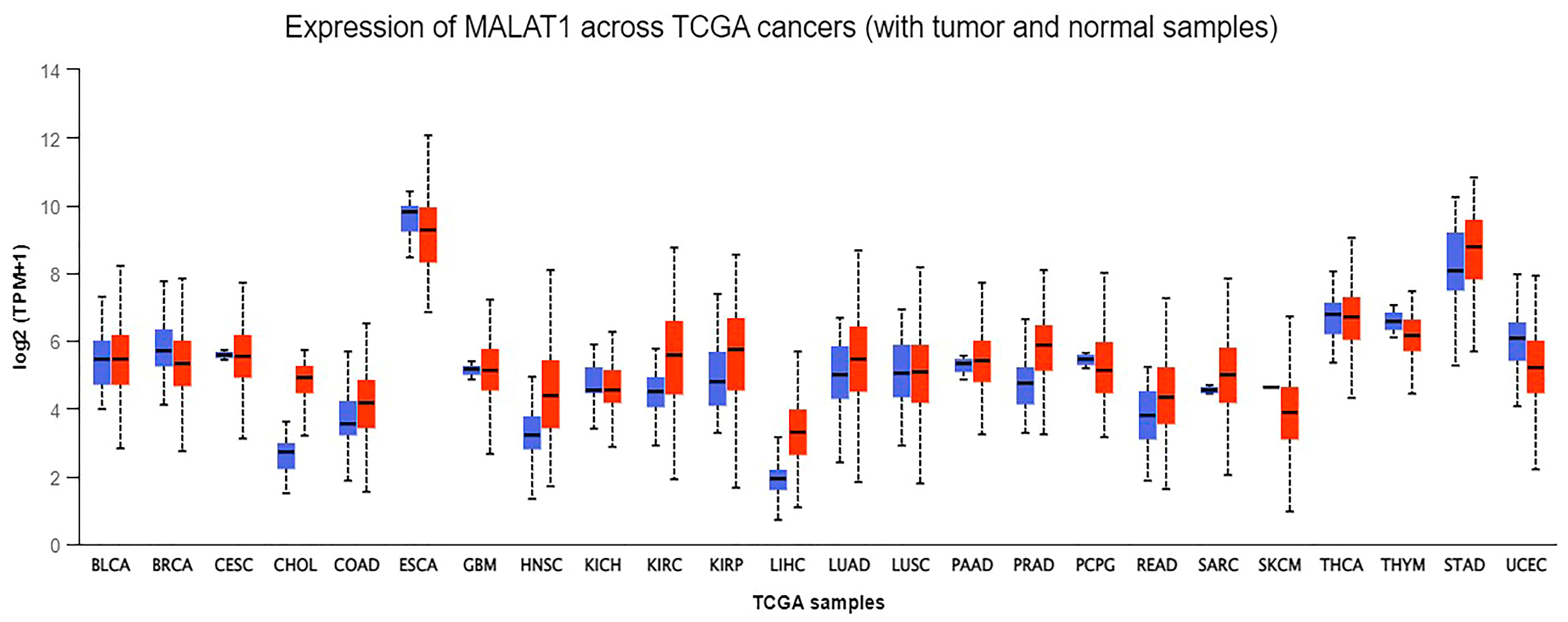
  25. (UALCAN), The University of Alabama at Birmingham. MALAT-1 Gene Expression across Different Cancers Using Data from The Cancer Genome Atlas (TCGA). Available online: http://ualcan.path.uab.edu/analysis.html (accessed on 20 May 2022).
  26. Chandrashekar, D.S.; Karthikeyan, S.K.; Korla, P.K.; Patel, H.; Shovon, A.R.; Athar, M.; Netto, G.J.; Qin, Z.S.; Kumar, S.; Manne, U.; et al. UALCAN: An update to the integrated cancer data analysis platform. Neoplasia 2022, 25, 18–27. [Google Scholar] [CrossRef] [PubMed]
  27. Peng, H.; Wang, J.; Li, J.; Zhao, M.; Huang, S.K.; Gu, Y.Y.; Li, Y.; Sun, X.J.; Yang, L.; Luo, Q.; et al. A circulating non-coding RNA panel as an early detection predictor of non-small cell lung cancer. Life Sci. 2016, 151, 235–242. [Google Scholar] [CrossRef] [PubMed]
  28. Duan, W.; Du, L.; Jiang, X.; Wang, R.; Yan, S.; Xie, Y.; Yan, K.; Wang, Q.; Wang, L.; Zhang, X.; et al. Identification of a serum circulating lncRNA panel for the diagnosis and recurrence prediction of bladder cancer. Oncotarget 2016, 7, 78850–78858. [Google Scholar] [CrossRef]
  29. Huang, S.K.; Luo, Q.; Peng, H.; Li, J.; Zhao, M.; Wang, J.; Gu, Y.Y.; Li, Y.; Yuan, P.; Zhao, G.H.; et al. A Panel of Serum Noncoding RNAs for the Diagnosis and Monitoring of Response to Therapy in Patients with Breast Cancer. Med. Sci. Monit. 2018, 24, 2476–2488. [Google Scholar] [CrossRef]
  30. He, B.; Zeng, J.; Chao, W.; Chen, X.; Huang, Y.; Deng, K.; Huang, Z.; Li, J.; Dai, M.; Chen, S.; et al. Serum long non-coding RNAs MALAT1, AFAP1-AS1 and AL359062 as diagnostic and prognostic biomarkers for nasopharyngeal carcinoma. Oncotarget 2017, 8, 41166–41177. [Google Scholar] [CrossRef]
  31. Eissmann, M.; Gutschner, T.; Hammerle, M.; Gunther, S.; Caudron-Herger, M.; Gross, M.; Schirmacher, P.; Rippe, K.; Braun, T.; Zornig, M.; et al. Loss of the abundant nuclear non-coding RNA MALAT1 is compatible with life and development. RNA Biol. 2012, 9, 1076–1087. [Google Scholar] [CrossRef]
  32. Zhao, Y.; Yu, Y.Q.; You, S.; Zhang, C.M.; Wu, L.; Zhao, W.; Wang, X.M. Long Non-Coding RNA MALAT1 as a Detection and Diagnostic Molecular Marker in Various Human Cancers: A Pooled Analysis Based on 3255 Subjects. Onco Targets Ther. 2020, 13, 5807–5817. [Google Scholar] [CrossRef] [PubMed]
  33. Xiong, Y.; Wang, J.; Zhu, H.; Liu, L.; Jiang, Y. Chronic oxymatrine treatment induces resistance and epithelialmesenchymal transition through targeting the long non-coding RNA MALAT1 in colorectal cancer cells. Oncol. Rep. 2018, 39, 967–976. [Google Scholar] [CrossRef] [PubMed]
  34. Wu, Y.; Sarkissyan, M.; Ogah, O.; Kim, J.; Vadgama, J.V. Expression of MALAT1 Promotes Trastuzumab Resistance in HER2 Overexpressing Breast Cancers. Cancers 2020, 12, 1918. [Google Scholar] [CrossRef] [PubMed]
  35. Wang, R.; Lu, X.; Yu, R. lncRNA MALAT1 Promotes EMT Process and Cisplatin Resistance of Oral Squamous Cell Carcinoma via PI3K/AKT/m-TOR Signal Pathway. Onco Targets Ther. 2020, 13, 4049–4061. [Google Scholar] [CrossRef] [PubMed]
  36. Jiao, F.; Hu, H.; Han, T.; Yuan, C.; Wang, L.; Jin, Z.; Guo, Z.; Wang, L. Long noncoding RNA MALAT-1 enhances stem cell-like phenotypes in pancreatic cancer cells. Int. J. Mol. Sci. 2015, 16, 6677–6693. [Google Scholar] [CrossRef] [PubMed]
  37. Iderzorig, T.; Kellen, J.; Osude, C.; Singh, S.; Woodman, J.A.; Garcia, C.; Puri, N. Comparison of EMT mediated tyrosine kinase inhibitor resistance in NSCLC. Biochem. Biophys. Res. Commun. 2018, 496, 770–777. [Google Scholar] [CrossRef] [PubMed]
  38. Lee, J.M.; Dedhar, S.; Kalluri, R.; Thompson, E.W. The epithelial-mesenchymal transition: New insights in signaling, development, and disease. J. Cell Biol. 2006, 172, 973–981. [Google Scholar] [CrossRef] [PubMed]
  39. Thiery, J.P.; Sleeman, J.P. Complex networks orchestrate epithelial-mesenchymal transitions. Nat. Rev. Mol. Cell Biol. 2006, 7, 131–142. [Google Scholar] [CrossRef]
  40. Puram, S.V.; Tirosh, I.; Parikh, A.S.; Patel, A.P.; Yizhak, K.; Gillespie, S.; Rodman, C.; Luo, C.L.; Mroz, E.A.; Emerick, K.S.; et al. Single-Cell Transcriptomic Analysis of Primary and Metastatic Tumor Ecosystems in Head and Neck Cancer. Cell 2017, 171, 1611–1624.e1624. [Google Scholar] [CrossRef]
  41. Jang, M.H.; Kim, H.J.; Kim, E.J.; Chung, Y.R.; Park, S.Y. Expression of epithelial-mesenchymal transition-related markers in triple-negative breast cancer: ZEB1 as a potential biomarker for poor clinical outcome. Hum. Pathol. 2015, 46, 1267–1274. [Google Scholar] [CrossRef]
  42. Dong, J.; Hu, Y.; Fan, X.; Wu, X.; Mao, Y.; Hu, B.; Guo, H.; Wen, L.; Tang, F. Single-cell RNA-seq analysis unveils a prevalent epithelial/mesenchymal hybrid state during mouse organogenesis. Genome Biol. 2018, 19, 31. [Google Scholar] [CrossRef] [PubMed]
  43. Thiery, J.P. Epithelial-mesenchymal transitions in tumour progression. Nat. Rev. Cancer 2002, 2, 442–454. [Google Scholar] [CrossRef] [PubMed]
  44. Jechlinger, M.; Grunert, S.; Beug, H. Mechanisms in epithelial plasticity and metastasis: Insights from 3D cultures and expression profiling. J. Mammary Gland. Biol. Neoplasia 2002, 7, 415–432. [Google Scholar] [CrossRef] [PubMed]
  45. Shi, Y.; Massague, J. Mechanisms of TGF-beta signaling from cell membrane to the nucleus. Cell 2003, 113, 685–700. [Google Scholar] [CrossRef] [PubMed]
  46. Niessen, K.; Fu, Y.; Chang, L.; Hoodless, P.A.; McFadden, D.; Karsan, A. Slug is a direct Notch target required for initiation of cardiac cushion cellularization. J. Cell Biol. 2008, 182, 315–325. [Google Scholar] [CrossRef] [PubMed]
  47. Medici, D.; Hay, E.D.; Olsen, B.R. Snail and Slug promote epithelial-mesenchymal transition through beta-catenin-T-cell factor-4-dependent expression of transforming growth factor-beta3. Mol. Biol. Cell 2008, 19, 4875–4887. [Google Scholar] [CrossRef]
  48. Kokudo, T.; Suzuki, Y.; Yoshimatsu, Y.; Yamazaki, T.; Watabe, T.; Miyazono, K. Snail is required for TGFbeta-induced endothelial-mesenchymal transition of embryonic stem cell-derived endothelial cells. J. Cell Sci. 2008, 121, 3317–3324. [Google Scholar] [CrossRef]
  49. Patel, M.; Eckburg, A.; Gantiwala, S.; Hart, Z.; Dein, J.; Lam, K.; Puri, N. Resistance to Molecularly Targeted Therapies in Melanoma. Cancers 2021, 13, 1115. [Google Scholar] [CrossRef]
  50. Kalluri, R.; Weinberg, R.A. The basics of epithelial-mesenchymal transition. J. Clin. Investig. 2009, 119, 1420–1428. [Google Scholar] [CrossRef]
  51. Garside, V.C.; Chang, A.C.; Karsan, A.; Hoodless, P.A. Co-ordinating Notch, BMP, and TGF-beta signaling during heart valve development. Cell Mol. Life Sci. 2013, 70, 2899–2917. [Google Scholar] [CrossRef]
  52. Micalizzi, D.S.; Farabaugh, S.M.; Ford, H.L. Epithelial-mesenchymal transition in cancer: Parallels between normal development and tumor progression. J. Mammary Gland. Biol. Neoplasia 2010, 15, 117–134. [Google Scholar] [CrossRef] [PubMed]
  53. Wells, A.; Yates, C.; Shepard, C.R. E-cadherin as an indicator of mesenchymal to epithelial reverting transitions during the metastatic seeding of disseminated carcinomas. Clin. Exp. Metastasis 2008, 25, 621–628. [Google Scholar] [CrossRef] [PubMed]
  54. Tse, J.C.; Kalluri, R. Mechanisms of metastasis: Epithelial-to-mesenchymal transition and contribution of tumor microenvironment. J. Cell Biochem. 2007, 101, 816–829. [Google Scholar] [CrossRef] [PubMed]
  55. Zeng, L.; Cen, Y.; Chen, J. Long non-coding RNA MALAT-1 contributes to maintenance of stem cell-like phenotypes in breast cancer cells. Oncol. Lett. 2018, 15, 2117–2122. [Google Scholar] [CrossRef] [PubMed]
  56. Sun, R.; Qin, C.; Jiang, B.; Fang, S.; Pan, X.; Peng, L.; Liu, Z.; Li, W.; Li, Y.; Li, G. Down-regulation of MALAT1 inhibits cervical cancer cell invasion and metastasis by inhibition of epithelial-mesenchymal transition. Mol. Biosyst. 2016, 12, 952–962. [Google Scholar] [CrossRef] [PubMed]
  57. Li, L.J.; Chai, Y.; Guo, X.J.; Chu, S.L.; Zhang, L.S. The effects of the long non-coding RNA MALAT-1 regulated autophagy-related signaling pathway on chemotherapy resistance in diffuse large B-cell lymphoma. Biomed. Pharmacother. 2017, 89, 939–948. [Google Scholar] [CrossRef]
  58. Chen, D.; Liu, L.; Wang, K.; Yu, H.; Wang, Y.; Liu, J.; Guo, Y.; Zhang, H. The role of MALAT-1 in the invasion and metastasis of gastric cancer. Scand. J. Gastroenterol. 2017, 52, 790–796. [Google Scholar] [CrossRef]
  59. Li, H.; Yuan, X.; Yan, D.; Li, D.; Guan, F.; Dong, Y.; Wang, H.; Liu, X.; Yang, B. Long Non-Coding RNA MALAT1 Decreases the Sensitivity of Resistant Glioblastoma Cell Lines to Temozolomide. Cell Physiol. Biochem. 2017, 42, 1192–1201. [Google Scholar] [CrossRef]
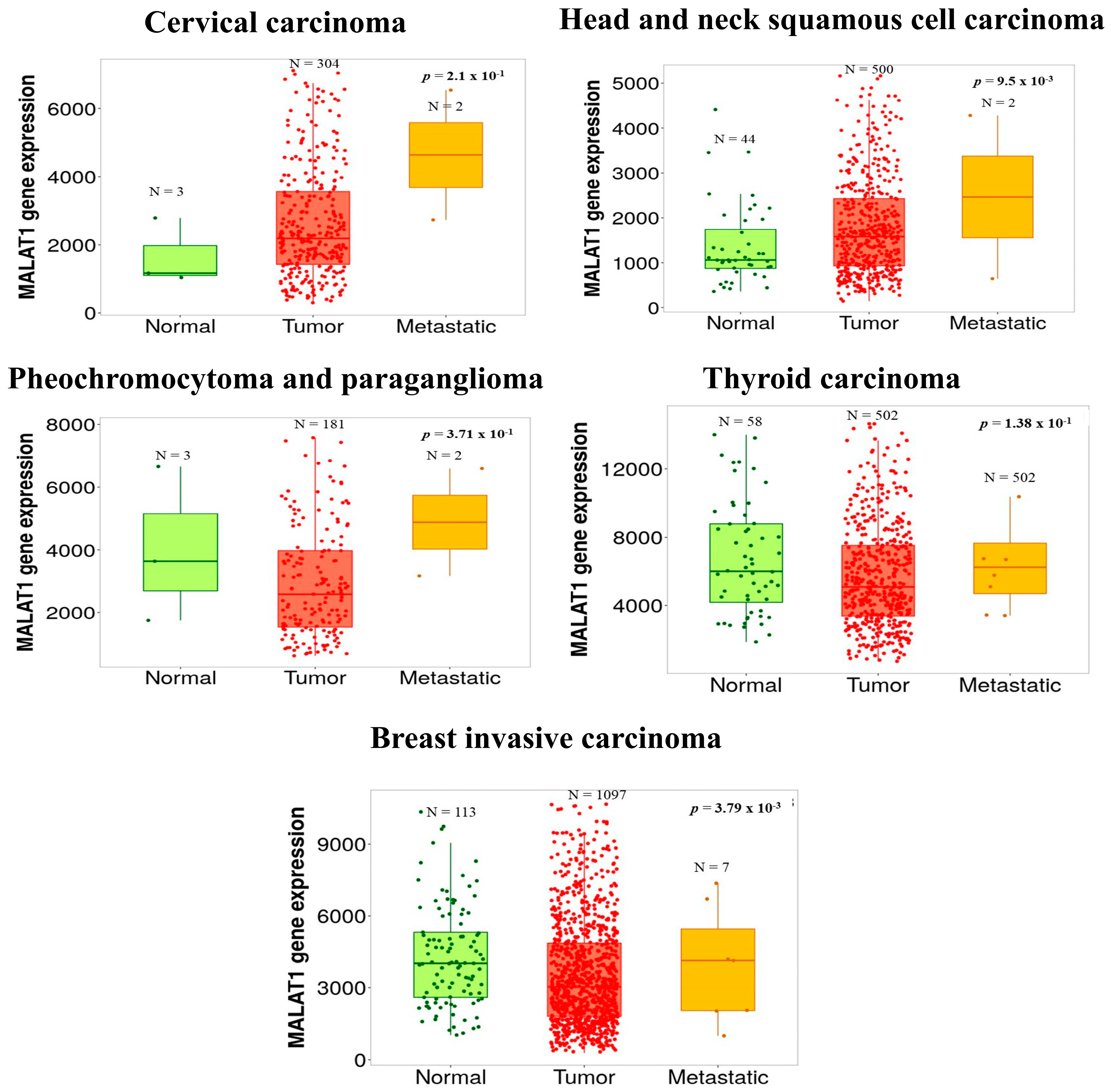
  60. Bartha, A.; Gyorffy, B. TNMplot.com: A Web Tool for the Comparison of Gene Expression in Normal, Tumor and Metastatic Tissues. Int. J. Mol. Sci. 2021, 22, 2622. [Google Scholar] [CrossRef]
  61. Li, P.; Zhang, X.; Wang, H.; Wang, L.; Liu, T.; Du, L.; Yang, Y.; Wang, C. MALAT1 Is Associated with Poor Response to Oxaliplatin-Based Chemotherapy in Colorectal Cancer Patients and Promotes Chemoresistance through EZH2. Mol. Cancer Ther. 2017, 16, 739–751. [Google Scholar] [CrossRef]
  62. Zhou, J.; Wang, J.; Zeng, Y.; Zhang, X.; Hu, Q.; Zheng, J.; Chen, B.; Xie, B.; Zhang, W.M. Implication of epithelial-mesenchymal transition in IGF1R-induced resistance to EGFR-TKIs in advanced non-small cell lung cancer. Oncotarget 2015, 6, 44332–44345. [Google Scholar] [CrossRef] [PubMed]
  63. Feng, C.; Zhao, Y.; Li, Y.; Zhang, T.; Ma, Y.; Liu, Y. LncRNA MALAT1 Promotes Lung Cancer Proliferation and Gefitinib Resistance by Acting as a miR-200a Sponge. Arch Bronconeumol. 2019, 55, 627–633. [Google Scholar] [CrossRef] [PubMed]
  64. Zhen, Q.; Liu, J.; Gao, L.; Liu, J.; Wang, R.; Chu, W.; Zhang, Y.; Tan, G.; Zhao, X.; Lv, B. MicroRNA-200a Targets EGFR and c-Met to Inhibit Migration, Invasion, and Gefitinib Resistance in Non-Small Cell Lung Cancer. Cytogenet. Genome Res. 2015, 146, 1–8. [Google Scholar] [CrossRef] [PubMed]
  65. Bracken, C.P.; Gregory, P.A.; Kolesnikoff, N.; Bert, A.G.; Wang, J.; Shannon, M.F.; Goodall, G.J. A double-negative feedback loop between ZEB1-SIP1 and the microRNA-200 family regulates epithelial-mesenchymal transition. Cancer Res. 2008, 68, 7846–7854. [Google Scholar] [CrossRef] [PubMed]
  66. Hou, J.; Zhang, G.; Wang, X.; Wang, Y.; Wang, K. Functions and mechanisms of lncRNA MALAT1 in cancer chemotherapy resistance. Biomark. Res. 2023, 11, 23. [Google Scholar] [CrossRef] [PubMed]
  67. Huang, T.; Song, X.; Xu, D.; Tiek, D.; Goenka, A.; Wu, B.; Sastry, N.; Hu, B.; Cheng, S.Y. Stem cell programs in cancer initiation, progression, and therapy resistance. Theranostics 2020, 10, 8721–8743. [Google Scholar] [CrossRef] [PubMed]
  68. Nassar, D.; Blanpain, C. Cancer Stem Cells: Basic Concepts and Therapeutic Implications. Annu. Rev. Pathol. 2016, 11, 47–76. [Google Scholar] [CrossRef]
  69. Castro-Oropeza, R.; Melendez-Zajgla, J.; Maldonado, V.; Vazquez-Santillan, K. The emerging role of lncRNAs in the regulation of cancer stem cells. Cell Oncol. 2018, 41, 585–603. [Google Scholar] [CrossRef]
  70. Choudhuri, S. Small noncoding RNAs: Biogenesis, function, and emerging significance in toxicology. J. Biochem. Mol. Toxicol. 2010, 24, 195–216. [Google Scholar] [CrossRef]
  71. Gutschner, T.; Diederichs, S. The hallmarks of cancer: A long non-coding RNA point of view. RNA Biol. 2012, 9, 703–719. [Google Scholar] [CrossRef]
  72. Yanaihara, N.; Caplen, N.; Bowman, E.; Seike, M.; Kumamoto, K.; Yi, M.; Stephens, R.M.; Okamoto, A.; Yokota, J.; Tanaka, T.; et al. Unique microRNA molecular profiles in lung cancer diagnosis and prognosis. Cancer Cell 2006, 9, 189–198. [Google Scholar] [CrossRef] [PubMed]
  73. Iorio, M.V.; Ferracin, M.; Liu, C.G.; Veronese, A.; Spizzo, R.; Sabbioni, S.; Magri, E.; Pedriali, M.; Fabbri, M.; Campiglio, M.; et al. MicroRNA gene expression deregulation in human breast cancer. Cancer Res. 2005, 65, 7065–7070. [Google Scholar] [CrossRef] [PubMed]
  74. Murakami, Y.; Yasuda, T.; Saigo, K.; Urashima, T.; Toyoda, H.; Okanoue, T.; Shimotohno, K. Comprehensive analysis of microRNA expression patterns in hepatocellular carcinoma and non-tumorous tissues. Oncogene 2006, 25, 2537–2545. [Google Scholar] [CrossRef] [PubMed]
  75. Ciafre, S.A.; Galardi, S.; Mangiola, A.; Ferracin, M.; Liu, C.G.; Sabatino, G.; Negrini, M.; Maira, G.; Croce, C.M.; Farace, M.G. Extensive modulation of a set of microRNAs in primary glioblastoma. Biochem. Biophys. Res. Commun. 2005, 334, 1351–1358. [Google Scholar] [CrossRef] [PubMed]
  76. Pallante, P.; Visone, R.; Ferracin, M.; Ferraro, A.; Berlingieri, M.T.; Troncone, G.; Chiappetta, G.; Liu, C.G.; Santoro, M.; Negrini, M.; et al. MicroRNA deregulation in human thyroid papillary carcinomas. Endocr. Relat. Cancer 2006, 13, 497–508. [Google Scholar] [CrossRef] [PubMed]
  77. Calin, G.A.; Liu, C.G.; Sevignani, C.; Ferracin, M.; Felli, N.; Dumitru, C.D.; Shimizu, M.; Cimmino, A.; Zupo, S.; Dono, M.; et al. MicroRNA profiling reveals distinct signatures in B cell chronic lymphocytic leukemias. Proc. Natl. Acad. Sci. USA 2004, 101, 11755–11760. [Google Scholar] [CrossRef] [PubMed]
  78. Iqbal, M.A.; Arora, S.; Prakasam, G.; Calin, G.A.; Syed, M.A. MicroRNA in lung cancer: Role, mechanisms, pathways and therapeutic relevance. Mol. Aspects Med. 2019, 70, 3–20. [Google Scholar] [CrossRef]
  79. Lee, Y.S.; Dutta, A. MicroRNAs in cancer. Annu. Rev. Pathol. 2009, 4, 199–227. [Google Scholar] [CrossRef]
  80. Meng, X.; Li, A.; Yu, B.; Li, S. Interplay between miRNAs and lncRNAs: Mode of action and biological roles in plant development and stress adaptation. Comput. Struct. Biotechnol. J. 2021, 19, 2567–2574. [Google Scholar] [CrossRef]
  81. Tang, X.J.; Wang, W.; Hann, S.S. Interactions among lncRNAs, miRNAs and mRNA in colorectal cancer. Biochimie 2019, 163, 58–72. [Google Scholar] [CrossRef]
  82. Zhang, Y.; Li, Y.; Wang, Q.; Zhang, X.; Wang, D.; Tang, H.C.; Meng, X.; Ding, X. Identification of an lncRNAmiRNAmRNA interaction mechanism in breast cancer based on bioinformatic analysis. Mol. Med. Rep. 2017, 16, 5113–5120. [Google Scholar] [CrossRef] [PubMed]
  83. Hu, C.Y.; Wu, K.Y.; Lin, T.Y.; Chen, C.C. The Crosstalk of Long Non-Coding RNA and MicroRNA in Castration-Resistant and Neuroendocrine Prostate Cancer: Their Interaction and Clinical Importance. Int. J. Mol. Sci. 2021, 23, 392. [Google Scholar] [CrossRef] [PubMed]
  84. Sun, B.; Liu, C.; Li, H.; Zhang, L.; Luo, G.; Liang, S.; Lu, M. Research progress on the interactions between long non-coding RNAs and microRNAs in human cancer. Oncol. Lett. 2020, 19, 595–605. [Google Scholar] [CrossRef] [PubMed]
  85. Lopez-Urrutia, E.; Bustamante Montes, L.P.; Ladron de Guevara Cervantes, D.; Perez-Plasencia, C.; Campos-Parra, A.D. Crosstalk Between Long Non-coding RNAs, Micro-RNAs and mRNAs: Deciphering Molecular Mechanisms of Master Regulators in Cancer. Front. Oncol. 2019, 9, 669. [Google Scholar] [CrossRef] [PubMed]
  86. Yoon, J.H.; Abdelmohsen, K.; Gorospe, M. Functional interactions among microRNAs and long noncoding RNAs. Semin. Cell Dev. Biol. 2014, 34, 9–14. [Google Scholar] [CrossRef] [PubMed]
  87. Han, X.; Wang, Q.; Wang, Y.; Hu, B.; Dong, X.; Zhang, H.; Wang, W. Long non-coding RNA metastasis-associated lung adenocarcinoma transcript 1/microRNA-202-3p/periostin axis modulates invasion and epithelial-mesenchymal transition in human cervical cancer. J. Cell Physiol. 2019, 234, 14170–14180. [Google Scholar] [CrossRef] [PubMed]
  88. Li, Q.; Zhang, C.; Chen, R.; Xiong, H.; Qiu, F.; Liu, S.; Zhang, M.; Wang, F.; Wang, Y.; Zhou, X.; et al. Disrupting MALAT1/miR-200c sponge decreases invasion and migration in endometrioid endometrial carcinoma. Cancer Lett. 2016, 383, 28–40. [Google Scholar] [CrossRef] [PubMed]
  89. Li, Q.; Dai, Z.; Xia, C.; Jin, L.; Chen, X. Suppression of long non-coding RNA MALAT1 inhibits survival and metastasis of esophagus cancer cells by sponging miR-1-3p/CORO1C/TPM3 axis. Mol. Cell Biochem. 2020, 470, 165–174. [Google Scholar] [CrossRef]
  90. Wang, Y.; Wu, C.; Zhang, C.; Li, Z.; Zhu, T.; Chen, J.; Ren, Y.; Wang, X.; Zhang, L.; Zhou, X. TGF-beta-induced STAT3 overexpression promotes human head and neck squamous cell carcinoma invasion and metastasis through malat1/miR-30a interactions. Cancer Lett. 2018, 436, 52–62. [Google Scholar] [CrossRef]
  91. Yu, Q.; Xiang, L.; Chen, Z.; Liu, X.; Ou, H.; Zhou, J.; Yang, D. MALAT1 functions as a competing endogenous RNA to regulate SMAD5 expression by acting as a sponge for miR-142-3p in hepatocellular carcinoma. Cell Biosci. 2019, 9, 39. [Google Scholar] [CrossRef]
  92. Liu, S.; Qiu, J.; He, G.; Liang, Y.; Wang, L.; Liu, C.; Pan, H. LncRNA MALAT1 acts as a miR-125a-3p sponge to regulate FOXM1 expression and promote hepatocellular carcinoma progression. J. Cancer 2019, 10, 6649–6659. [Google Scholar] [CrossRef]
  93. Chen, S.; Wang, G.; Tao, K.; Cai, K.; Wu, K.; Ye, L.; Bai, J.; Yin, Y.; Wang, J.; Shuai, X.; et al. Long noncoding RNA metastasis-associated lung adenocarcinoma transcript 1 cooperates with enhancer of zeste homolog 2 to promote hepatocellular carcinoma development by modulating the microRNA-22/Snail family transcriptional repressor 1 axis. Cancer Sci. 2020, 111, 1582–1595. [Google Scholar] [CrossRef] [PubMed]
  94. Xiao, H.; Zhu, Q.; Zhou, J. Long non-coding RNA MALAT1 interaction with miR-429 regulates the proliferation and EMT of lung adenocarcinoma cells through RhoA. Int. J. Clin. Exp. Pathol. 2019, 12, 419–430. [Google Scholar] [PubMed]
  95. Li, J.; Wang, J.; Chen, Y.; Li, S.; Jin, M.; Wang, H.; Chen, Z.; Yu, W. LncRNA MALAT1 exerts oncogenic functions in lung adenocarcinoma by targeting miR-204. Am. J. Cancer Res. 2016, 6, 1099–1107. [Google Scholar] [PubMed]
  96. Shi, B.; Wang, Y.; Yin, F. MALAT1/miR-124/Capn4 axis regulates proliferation, invasion and EMT in nasopharyngeal carcinoma cells. Cancer Biol. Ther. 2017, 18, 792–800. [Google Scholar] [CrossRef] [PubMed]
  97. Deng, B.; Zhang, Y.; Zhang, S.; Wen, F.; Miao, Y.; Guo, K. MicroRNA-142-3p inhibits cell proliferation and invasion of cervical cancer cells by targeting FZD7. Tumour Biol. 2015, 36, 8065–8073. [Google Scholar] [CrossRef]
  98. Wu, J.W.; Hu, M.; Chai, J.; Seoane, J.; Huse, M.; Li, C.; Rigotti, D.J.; Kyin, S.; Muir, T.W.; Fairman, R.; et al. Crystal structure of a phosphorylated Smad2. Recognition of phosphoserine by the MH2 domain and insights on Smad function in TGF-beta signaling. Mol. Cell 2001, 8, 1277–1289. [Google Scholar] [CrossRef]
  99. Raychaudhuri, P.; Park, H.J. FoxM1: A master regulator of tumor metastasis. Cancer Res. 2011, 71, 4329–4333. [Google Scholar] [CrossRef]
  100. Etienne-Manneville, S.; Hall, A. Rho GTPases in cell biology. Nature 2002, 420, 629–635. [Google Scholar] [CrossRef]
  101. Hanna, S.; El-Sibai, M. Signaling networks of Rho GTPases in cell motility. Cell Signal 2013, 25, 1955–1961. [Google Scholar] [CrossRef]
  102. Lin, Y.; Zheng, Y. Approaches of targeting Rho GTPases in cancer drug discovery. Expert. Opin. Drug Discov. 2015, 10, 991–1010. [Google Scholar] [CrossRef]
  103. An, N.; Zheng, B. MiR-203a-3p Inhibits Pancreatic Cancer Cell Proliferation, EMT, and Apoptosis by Regulating SLUG. Technol. Cancer Res. Treat. 2020, 19, 1533033819898729. [Google Scholar] [CrossRef] [PubMed]
  104. Tang, Y.; Xiao, G.; Chen, Y.; Deng, Y. LncRNA MALAT1 promotes migration and invasion of non-small-cell lung cancer by targeting miR-206 and activating Akt/mTOR signaling. Anticancer. Drugs 2018, 29, 725–735. [Google Scholar] [CrossRef]
  105. Wu, J.; Weng, Y.; He, F.; Liang, D.; Cai, L. LncRNA MALAT-1 competitively regulates miR-124 to promote EMT and development of non-small-cell lung cancer. Anticancer. Drugs 2018, 29, 628–636. [Google Scholar] [CrossRef]
  106. Gillan, L.; Matei, D.; Fishman, D.A.; Gerbin, C.S.; Karlan, B.Y.; Chang, D.D. Periostin secreted by epithelial ovarian carcinoma is a ligand for alpha(V)beta(3) and alpha(V)beta(5) integrins and promotes cell motility. Cancer Res. 2002, 62, 5358–5364. [Google Scholar] [PubMed]
  107. Seton-Rogers, S. Oncogenes: All eyes on YAP1. Nat. Rev. Cancer 2014, 14, 514–515. [Google Scholar] [CrossRef] [PubMed]
  108. Sun, Z.; Ou, C.; Liu, J.; Chen, C.; Zhou, Q.; Yang, S.; Li, G.; Wang, G.; Song, J.; Li, Z.; et al. YAP1-induced MALAT1 promotes epithelial-mesenchymal transition and angiogenesis by sponging miR-126-5p in colorectal cancer. Oncogene 2019, 38, 2627–2644. [Google Scholar] [CrossRef] [PubMed]
  109. Levy, D.E.; Lee, C.K. What does Stat3 do? J. Clin. Investig. 2002, 109, 1143–1148. [Google Scholar] [CrossRef]
  110. Zhao, C.; Yuan, G.; Jiang, Y.; Xu, J.; Ye, L.; Zhan, W.; Wang, J. Capn4 contributes to tumor invasion and metastasis in gastric cancer via activation of the Wnt/beta-catenin/MMP9 signalling pathways. Exp. Cell Res. 2020, 395, 112220. [Google Scholar] [CrossRef]
  111. Lunardi, A.; Webster, K.A.; Papa, A.; Padmani, B.; Clohessy, J.G.; Bronson, R.T.; Pandolfi, P.P. Role of aberrant PI3K pathway activation in gallbladder tumorigenesis. Oncotarget 2014, 5, 894–900. [Google Scholar] [CrossRef]
  112. Carnero, A.; Blanco-Aparicio, C.; Renner, O.; Link, W.; Leal, J.F. The PTEN/PI3K/AKT signalling pathway in cancer, therapeutic implications. Curr. Cancer Drug Targets 2008, 8, 187–198. [Google Scholar] [CrossRef] [PubMed]
  113. Carnero, A. The PKB/AKT pathway in cancer. Curr. Pharm. Des. 2010, 16, 34–44. [Google Scholar] [CrossRef] [PubMed]
  114. Ye, B.; Jiang, L.L.; Xu, H.T.; Zhou, D.W.; Li, Z.S. Expression of PI3K/AKT pathway in gastric cancer and its blockade suppresses tumor growth and metastasis. Int. J. Immunopathol. Pharmacol. 2012, 25, 627–636. [Google Scholar] [CrossRef] [PubMed]
  115. Aoki, M.; Fujishita, T. Oncogenic Roles of the PI3K/AKT/mTOR Axis. Curr. Top. Microbiol. Immunol. 2017, 407, 153–189. [Google Scholar] [CrossRef]
  116. Chen, H.; Zhou, L.; Wu, X.; Li, R.; Wen, J.; Sha, J.; Wen, X. The PI3K/AKT pathway in the pathogenesis of prostate cancer. Front. Biosci. 2016, 21, 1084–1091. [Google Scholar] [CrossRef]
  117. Dai, Q.; Zhang, T.; Li, C. LncRNA MALAT1 Regulates the Cell Proliferation and Cisplatin Resistance in Gastric Cancer via PI3K/AKT Pathway. Cancer Manag. Res. 2020, 12, 1929–1939. [Google Scholar] [CrossRef] [PubMed]
  118. Chen, Y.; Huang, W.; Sun, W.; Zheng, B.; Wang, C.; Luo, Z.; Wang, J.; Yan, W. LncRNA MALAT1 Promotes Cancer Metastasis in Osteosarcoma via Activation of the PI3K-Akt Signaling Pathway. Cell Physiol. Biochem. 2018, 51, 1313–1326. [Google Scholar] [CrossRef]
  119. Wang, N.; Hou, M.S.; Zhan, Y.; Shen, X.B.; Xue, H.Y. MALAT1 promotes cisplatin resistance in cervical cancer by activating the PI3K/AKT pathway. Eur. Rev. Med. Pharmacol. Sci. 2018, 22, 7653–7659. [Google Scholar] [CrossRef]
  120. Wang, J.; Sun, G. FOXO1-MALAT1-miR-26a-5p Feedback Loop Mediates Proliferation and Migration in Osteosarcoma Cells. Oncol. Res. 2017, 25, 1517–1527. [Google Scholar] [CrossRef]
  121. Wang, C.; Mao, Z.P.; Wang, L.; Wu, G.H.; Zhang, F.H.; Wang, D.Y.; Shi, J.L. Long non-coding RNA MALAT1 promotes cholangiocarcinoma cell proliferation and invasion by activating PI3K/Akt pathway. Neoplasma 2017, 64, 725–731. [Google Scholar] [CrossRef]
  122. Jin, Y.; Feng, S.J.; Qiu, S.; Shao, N.; Zheng, J.H. LncRNA MALAT1 promotes proliferation and metastasis in epithelial ovarian cancer via the PI3K-AKT pathway. Eur. Rev. Med. Pharmacol. Sci. 2017, 21, 3176–3184. [Google Scholar] [PubMed]
  123. Zhou, X.; Liu, S.; Cai, G.; Kong, L.; Zhang, T.; Ren, Y.; Wu, Y.; Mei, M.; Zhang, L.; Wang, X. Long Non Coding RNA MALAT1 Promotes Tumor Growth and Metastasis by inducing Epithelial-Mesenchymal Transition in Oral Squamous Cell Carcinoma. Sci. Rep. 2015, 5, 15972. [Google Scholar] [CrossRef] [PubMed]
  124. Ying, L.; Chen, Q.; Wang, Y.; Zhou, Z.; Huang, Y.; Qiu, F. Upregulated MALAT-1 contributes to bladder cancer cell migration by inducing epithelial-to-mesenchymal transition. Mol. Biosyst. 2012, 8, 2289–2294. [Google Scholar] [CrossRef] [PubMed]
  125. Chang, H.L.; Bamodu, O.A.; Ong, J.R.; Lee, W.H.; Yeh, C.T.; Tsai, J.T. Targeting the Epigenetic Non-Coding RNA MALAT1/Wnt Signaling Axis as a Therapeutic Approach to Suppress Stemness and Metastasis in Hepatocellular Carcinoma. Cells 2020, 9, 1020. [Google Scholar] [CrossRef] [PubMed]
  126. Shi, C.J.; Zheng, Y.B.; Pan, F.F.; Zhang, F.W.; Zhuang, P.; Fu, W.M. Gallic Acid Suppressed Tumorigenesis by an LncRNA MALAT1-Wnt/beta-Catenin Axis in Hepatocellular Carcinoma. Front. Pharmacol. 2021, 12, 708967. [Google Scholar] [CrossRef] [PubMed]
  127. Liu, Q.; Zheng, S.; Chen, Y.; Liu, T.; Han, X.; Zhang, X.; Shen, T.; Lu, X. TGF-beta1-Induced Upregulation of MALAT1 Promotes Kazakh’s Esophageal Squamous Cell Carcinoma Invasion by EMT. J. Cancer 2020, 11, 6892–6901. [Google Scholar] [CrossRef]
  128. Fan, Y.; Shen, B.; Tan, M.; Mu, X.; Qin, Y.; Zhang, F.; Liu, Y. TGF-beta-induced upregulation of malat1 promotes bladder cancer metastasis by associating with suz12. Clin. Cancer Res. 2014, 20, 1531–1541. [Google Scholar] [CrossRef]
  129. Chen, M.; Xia, Z.; Chen, C.; Hu, W.; Yuan, Y. LncRNA MALAT1 promotes epithelial-to-mesenchymal transition of esophageal cancer through Ezh2-Notch1 signaling pathway. Anticancer. Drugs 2018, 29, 767–773. [Google Scholar] [CrossRef]
  130. Fathi Dizaji, B. Strategies to target long non-coding RNAs in cancer treatment: Progress and challenges. Egypt. J. Med. Human. Genet. 2020, 21, 41. [Google Scholar] [CrossRef]
  131. Zhang, Z.C.; Tang, C.; Dong, Y.; Zhang, J.; Yuan, T.; Tao, S.C.; Li, X.L. Targeting the long noncoding RNA MALAT1 blocks the pro-angiogenic effects of osteosarcoma and suppresses tumour growth. Int. J. Biol. Sci. 2017, 13, 1398–1408. [Google Scholar] [CrossRef]
  132. Gong, N.; Teng, X.; Li, J.; Liang, X.J. Antisense Oligonucleotide-Conjugated Nanostructure-Targeting lncRNA MALAT1 Inhibits Cancer Metastasis. ACS Appl. Mater. Interfaces 2019, 11, 37–42. [Google Scholar] [CrossRef] [PubMed]
  133. Chen, F.; Zhong, Z.; Tan, H.Y.; Guo, W.; Zhang, C.; Cheng, C.S.; Wang, N.; Ren, J.; Feng, Y. Suppression of lncRNA MALAT1 by betulinic acid inhibits hepatocellular carcinoma progression by targeting IAPs via miR-22-3p. Clin. Transl. Med. 2020, 10, e190. [Google Scholar] [CrossRef] [PubMed]
  134. Ji, Q.; Liu, X.; Fu, X.; Zhang, L.; Sui, H.; Zhou, L.; Sun, J.; Cai, J.; Qin, J.; Ren, J.; et al. Resveratrol inhibits invasion and metastasis of colorectal cancer cells via MALAT1 mediated Wnt/beta-catenin signal pathway. PLoS ONE 2013, 8, e78700. [Google Scholar] [CrossRef] [PubMed]
  135. Yang, Z.; Xie, Q.; Chen, Z.; Ni, H.; Xia, L.; Zhao, Q.; Chen, Z.; Chen, P. Resveratrol suppresses the invasion and migration of human gastric cancer cells via inhibition of MALAT1-mediated epithelial-to-mesenchymal transition. Exp. Ther. Med. 2019, 17, 1569–1578. [Google Scholar] [CrossRef] [PubMed]
  136. Guo, K.; Feng, Y.; Zheng, X.; Sun, L.; Wasan, H.S.; Ruan, S.; Shen, M. Resveratrol and Its Analogs: Potent Agents to Reverse Epithelial-to-Mesenchymal Transition in Tumors. Front. Oncol. 2021, 11, 644134. [Google Scholar] [CrossRef] [PubMed]
  137. Tan, M.; Jiang, B.; Wang, H.; Ouyang, W.; Chen, X.; Wang, T.; Dong, D.; Yi, S.; Yi, J.; Huang, Y.; et al. Dihydromyricetin induced lncRNA MALAT1-TFEB-dependent autophagic cell death in cutaneous squamous cell carcinoma. J. Cancer 2019, 10, 4245–4255. [Google Scholar] [CrossRef] [PubMed]
  138. Francois-Moutal, L.; Miranda, V.G.; Mollasalehi, N.; Gokhale, V.; Khanna, M. In Silico Targeting of the Long Noncoding RNA MALAT1. ACS Med. Chem. Lett. 2021, 12, 915–921. [Google Scholar] [CrossRef]
  139. Abulwerdi, F.A.; Xu, W.; Ageeli, A.A.; Yonkunas, M.J.; Arun, G.; Nam, H.; Schneekloth, J.S., Jr.; Dayie, T.K.; Spector, D.; Baird, N.; et al. Selective Small-Molecule Targeting of a Triple Helix Encoded by the Long Noncoding RNA, MALAT1. ACS Chem. Biol. 2019, 14, 223–235. [Google Scholar] [CrossRef]
  140. Donlic, A.; Zafferani, M.; Padroni, G.; Puri, M.; Hargrove, A.E. Regulation of MALAT1 triple helix stability and in vitro degradation by diphenylfurans. Nucleic Acids Res. 2020, 48, 7653–7664. [Google Scholar] [CrossRef]
  141. Miao, S.; Bhunia, D.; Devari, S.; Liang, Y.; Munyaradzi, O.; Rundell, S.; Bong, D. Bifacial PNAs Destabilize MALAT1 by 3’ A-Tail Displacement from the U-Rich Internal Loop. ACS Chem. Biol. 2021, 16, 1600–1609. [Google Scholar] [CrossRef]
  142. Liu, X.M.; Zhou, J.; Mao, Y.; Ji, Q.; Qian, S.B. Programmable RNA N(6)-methyladenosine editing by CRISPR-Cas9 conjugates. Nat. Chem. Biol. 2019, 15, 865–871. [Google Scholar] [CrossRef] [PubMed]
  143. Qi, F.; Tan, B.; Ma, F.; Zhu, B.; Zhang, L.; Liu, X.; Li, H.; Yang, J.; Cheng, B. A Synthetic Light-switchable System based on CRISPR Cas13a Regulates the Expression of LncRNA MALAT1 and Affects the Malignant Phenotype of Bladder Cancer Cells. Int. J. Biol. Sci. 2019, 15, 1630–1636. [Google Scholar] [CrossRef] [PubMed]
Figure 1. MALAT-1 is an RNA gene encoded by chromosome 11q13. MALAT-1 is initially transcribed as a precursor immature transcript, which the enzyme RNAase P processes to produce mature MALAT-1. MALAT-1 decreases gene expression in many ways, including interfering with a transcription factor, interfering with RNA Pol II, therefore inhibiting transcription, interfering with mRNA splicing, interfering with epigenetic regulation leading to gene silencing and competing with miRNAs, and preventing mRNA transcriptions. Figure 1 created with BioRender.com (Accessed on 27 December 2023).
Cancers 16 00234 g001
Figure 2. Depicts the comparison of MALAT-1 gene expression. MALAT-1 gene expression between tumor samples (denoted in red color) and non-cancerous samples (blue color) across different cancers through the UALCAN database online portal (https://ualcan.path.uab.edu/) (Accessed on 26 December 2023). The tumor sample shows high expression in various tumors such as esophageal carcinoma, CC, HCC, sarcoma, and melanoma compared to normal patients’ samples.
Cancers 16 00234 g002
Figure 3. Illustrates the boxplot of the MALAT-1 expression. The analysis of MALAT-1 expression data through the online TNM plot portal (https://tnmplot.com/analysis/) (Accessed on 26 December 2023) compares the different expressions between normal, tumor, and metastatic samples in CC, HNSCC, pheochromocytoma and paraganglioma, thyroid carcinoma, and breast invasive carcinoma. Our results revealed that MALAT-1 expression is highly upregulated in metastatic samples compared to the tumor and normal samples.
Cancers 16 00234 g003
Figure 4. Emphasizes the different mechanisms implicated in MALAT-1 modulating EMT and metastasis. The PI3K/Akt pathway activates the protein kinase Akt to translocate to the nucleus and phosphorylates the transcription factor FOXO1, inhibiting EMT, thereby inducing chemoresistance. The activation of Wnt/β-catenin stimulates β-catenin migration to the nucleus and upregulates the expression of EMT transcription factors ZEB1/2, SNAI1/2, Twist1, and Slug, which in turn increase MALAT-1 gene expression. NF-kB activation leads to dislocation of the P65 subunit to the nucleus, which activates SNAI1/2, ZEB1/2, Twist1, and Slug, leading to the overexpression of MALAT-1 and activates VEGF and metalloproteinases MMP2 and MMP9, which in turn facilitate tumor invasion and metastasis. Notch and TGF-β activation bind to EZH2 and Suz12 and modulates post-transcription suppression of the epithelial markers. Figure 4 created with BioRender.com (Accessed on 27 December 2023).
Cancers 16 00234 g004
Table 1. Various mechanisms of MALAT-1 modulate EMT, chemoresistance, and CSC properties in different cancers.
Cancer TypePhenotypeEffectMechanismCell LinesIn VivoReferences
BCCancer stem cell-like propertiesModulate stem cell-like properties in BC↓ CD133+, ↓ ALDH+, and
↓ Sox2
MCF7-[55]
CCEMTInhibit invasion and metastasis↑ E-cadherin, ↑ ZO-1,
↓ vimentin,
↓ β-catenin, and
↓ Snail1
H8, CC, CaSki, HeLa, SiHaFemale BALB/c nude mice[56]
CRCChemoresistanceReverse EMT and reverse Oxymatrine resistance↑ E-cadherin, and ↑ vimentinHT29, SW480, HT29 Oxymatrine resistant-[33]
Diffuse large B-cell lymphomaChemoresistanceEnhance drug sensitivity by inducing autophagy in DLBCL↑ LC3-II, ↑ LC3-I,
and ↓ p62
IM-9I, Ly3, Ly8, Pfeiffer, Farage, Raji, Daud, Ly10, Ly1BALB/c-nu/nu nude mice[57]
GCEMTInhibit invasion and metastasis↑ E-cadherin, and ↓ vimentinSGC-7901, BGC823, AGS, MKN4, SGC7901M, SGC7901NMFemale nude mice[58]
GlioblastomaChemoresistanceIncrease sensitivity to Temozolomide↓ MDR1, ↓ MRP5, ↓ LRP, ↓ ZEB,
↑ E-cadherin, ↑ ZO-1, ↓ SMA, and ↓ Fibronectin
U251, U87, U251/TMZ, U87/TMZNude mouse[59]
Lung cancer with brain metastasesEMTInhibit invasion and metastasis↑ E-cadherin, and ↑ vimentinH1915-L10, H1915-H10Athymic BALB/c-nu/
nu mice
[17]
Oral squamous cell carcinoma (OSCC)ChemoresistanceReverse EMT and increase Cisplatin chemosensitivity↓ p-PI3K, ↓ PI3K
↓ p-AKT, ↓ AKT,
↓ pm-TOR, ↓ mTOR, and ↑ E-cadherin
CAL-27, SCC-9BALB/c nude mice[35]
Pancreatic cancerEMTpromote apoptosis, inhibit tumor invasion and migration↑ p21,
↑ p53, ↓ CDC2, ↓ Snail, ↓ Slug, ↑ E-cadherin, ↓ N-cadherin, ↓ vimentin, ↓ MMP-2, and ↓ MMP-9
BxPC-3, AsPC-1, PANC-1, CFPAC-1,
CAPAN-1, SW1990, HS-766T
-[21]
Cancer stem cells like phenotype and chemoresistanceModulate stem cell-like phenotype and enhance drug sensitivity↓ CD133+, and ↓ Sox2AsPC-1, CFPAC-1,BALB/c nude mice[36]
TNBCChemoresistanceReverse EMT and reverse Trastuzumab resistance↓ Snail, ↓ Slug, ↓ Twist, ↓ NanogSKBR3, BT474, MCF7, T47D, MDA-MB231, MCF-12A, JIMT1Female athymic nude mice[34]
Disclaimer/Publisher’s Note: The statements, opinions and data contained in all publications are solely those of the individual author(s) and contributor(s) and not of MDPI and/or the editor(s). MDPI and/or the editor(s) disclaim responsibility for any injury to people or property resulting from any ideas, methods, instructions or products referred to in the content.

