Prognostic and Predictive Value of PBRM1 in Clear Cell Renal Cell Carcinoma
by
Lucía Carril-Ajuria
1, 
María Santos
2,
Juan María Roldán-Romero
2,
Cristina Rodriguez-Antona
2,3,* and
Guillermo de Velasco
1,* 1
Department of Medical Oncology, University Hospital 12 de Octubre, 28041 Madrid, Spain
2
Hereditary Endocrine Cancer Group, Human Cancer Genetics Programme, Spanish National Cancer Research Centre (CNIO), 28029 Madrid, Spain
3
Centro de Investigación Biomédica en Red de Enfermedades Raras (CIBERER), 28029 Madrid, Spain
*
Authors to whom correspondence should be addressed.
Cancers 2020, 12(1), 16; https://doi.org/10.3390/cancers12010016
Submission received: 30 November 2019
/
Revised: 13 December 2019
/
Accepted: 15 December 2019
/
Published: 19 December 2019
(This article belongs to the Special Issue Renal Cell Carcinoma)
Abstract
:Renal cell carcinoma (RCC) is the most frequent kidney solid tumor, the clear cell RCC (ccRCC) being the major histological subtype. The probability of recurrence and the clinical behavior of ccRCC will greatly depend on the different clinical and histopathological features, already incorporated to different scoring systems, and on the genomic landscape of the tumor. In this sense, ccRCC has for a long time been known to be associated to the biallelic inactivation of Von Hippel-Lindau (VHL) gene which causes aberrant hypoxia inducible factor (HIF) accumulation. Recently, next generation-sequencing technologies have provided the bases for an in-depth molecular characterization of ccRCC, identifying additional recurrently mutated genes, such as PBRM1 (≈40–50%), SETD2 (≈12%), or BAP1 (≈10%). PBRM1, the second most common mutated gene in ccRCC after VHL, is a component of the SWI/SNF chromatin remodeling complex. Different studies have investigated the biological consequences and the potential role of PBRM1 alterations in RCC prognosis and as a drug response modulator, although some results are contradictory. In the present article, we review the current evidence on PBRM1 as potential prognostic and predictive marker in both localized and metastatic RCC.
1. Introduction
Kidney cancer is the 12th most frequent solid tumor worldwide with around 400,000 new cases and 175,000 deaths from renal cell carcinoma (RCC) estimated in 2018 [1]. RCC accounts for almost 90% of all kidney tumors, the clear cell (ccRCC) being the most common histologic subtype [2].
Approximately 70% of the RCC will present with localized disease, but around one third of them will relapse after surgical excision [3,4]. However, the probability of recurrence will depend greatly on different clinical and histopathological features, which have been incorporated to up to 20 different scoring systems characterized by substantial heterogeneity [5,6,7].
Regarding the molecular characteristics of RCC, the inactivation of the Von Hippel–Lindau (VHL) gene is by far the most common oncogenic driver event in ccRCC. VHL is an E3 ligase substrate that labels the transcription factors hypoxia inducible factor (HIF) 1alpha and HIF2alpha with ubiquitin for proteosomal degradation. The accumulation of HIF triggers the uncontrolled expression of angiogenesis promoting genes and gives rise to highly vascularized tumors [8].
Recent advances in next-generation sequencing (NGS) and their implementation in large cohort of cancer patients (e.g., The Cancer Genome Atlas (TCGA) projects), have led to the identification of additional genes frequently mutated in ccRCC, such as PBRM1 (≈40–50%), SETD2 (12%), BAP1 (10%), and KDM5C (5%) [9,10,11]. These are genes coding for proteins participating in the regulation of chromatin remodeling and histone methylation. Although VHL is the initiating event in ccRCC, the acquisition of additional mutations, some in subclonal cancer cell populations, is a common characteristic during ccRCC tumor development and metastasis. In this context, multiregion sequencing-based studies have highlighted the potential relevance of PBRM1 alterations for tumor growth and metastatic capacity, which are determinant for clinical outcome [10].
The advances in systemic therapy of advanced RCC (mRCC) in the last 20 years have been remarkable. This progress has led to a significant improvement in the median overall survival (OS), from 13 months in the cytokine therapy era to ≈30 months using combination of targeted therapies and immune checkpoint blockade [12,13,14]. Well established prognostic scoring systems for mRCC, such as the Memorial Sloan Kettering Cancer Center (MSKCC) or the International Metastatic RCC Database Consortium (IMDC), include clinical parameters such as performance status or time from diagnosis to treatment, as well as laboratory parameters [15]. However, other clinical variables which have a prognostic value in mRCC are not included in those models [16]. Likewise, in the current therapeutic context there is a need of updating the scoring systems with molecular parameters, to better personalize treatment strategies to achieve survival improvement and avoid unnecessary toxicities.
2. Bromodomain-Containing Protein BAF180 Function
The SWI/SNF complexes are multiprotein machineries able to mobilize nucleosomes and alter chromatin structure through the hydrolysis of ATP molecules. By controlling chromatin accessibility, these epigenetic remodeling complexes act as transcriptional regulators, mediating the binding of transcription factors, coactivators, and corepressors to target promoters and enhancers in the DNA. A wide variety of cellular functions are modulated by SWI/SNF complexes including the balance between self-renewal and cell differentiation, cell cycle, metabolism, and DNA repair processes [17]. SWI/SNF components are recurrently mutated across human malignancies. It is estimated that about 20% of all human tumors harbor alterations in genes encoding SWI/SNF complex subunits [18]. This mutation frequency is comparable to that observed for other canonical cancer genes such as TP53, KRAS, or PTEN [19]. Interestingly, mutations in particular SWI/SNF components are tumor type-dependent, which may indicate that these complexes carry out not only universal but also tissue-specific functions. For instance, SMARCB1 is found mutated in almost 95% of malignant rhabdoid tumors and PBRM1 is mutated in ≈40% of ccRCC [20]. Other components of the SWI/SNF complex, such as ARID1A, are highly mutated in several cancer types, but rarely altered in ccRCC (e.g., ARID1A is mutated in 49% of ovarian clear cell carcinomas, 39% of endometrial cancers, and only in 3% of ccRCC tumors).
The gene PBRM1 encodes the bromodomain-containing protein BAF180 that serves as the DNA-targeting subunit of the pBAF SWI/SNF complex [21]. BAF180 harbors six bromodomains that bind acetylated residues on histone tails. These domains are capable of recognizing acetylating patterns and target the complete complex to specific chromatin regions. This protein is involved in various DNA repair mechanisms [22] and it is also important for cohesion between centromeres, which is necessary for maintaining genomic stability [23].
How mutations in the gene PBRM1 promote carcinogenesis and tumor progression is still unknown. PBRM1 is considered a tumor suppressor gene and this role is supported by in vitro experiments in ccRCC derived cell-lines, which show that PBRM1 gene silencing results in increased proliferation, migration, and colony formation [20]. Consistently, Macher-Goeppinger et al. found that re-expression of PBRM1 into A704 cells, that harbor a homozygous truncating mutation in this gene, results in diminished colony formation [24]. The role of PBRM1 as tumor suppressor is also supported by the fact that ≈80% of the somatic mutations found in the gene result in loss of function (LOF) of the protein, not only in ccRCC but also in other tumor types, including breast and pancreatic cancers. Furthermore, BAF180 has been demonstrated to modulate the activity of p53 [24], inducing the transcription of some p53-targets such as p21 and repressing other such as 14–3–3σ (Figure 1). PBRM1 inactivation can therefore disturb p53-dependent chromatin regulation and enable ccRCC tumors to escape from p53-mediated surveillance [25]. Even if PBRM1 has mainly been described as a tumor suppressor gene, Murakami et al. recently demonstrated that its role may be context dependent, and that PBRM1 may also act as an oncogene under specific conditions [26]. They showed that the inactivation of PBRM1 in 786-O cells, a ccRCC cell line expressing BAF180, resulted in a reduction of cell survival and proliferation. This effect seemed to be dependent on HIF1α/HIF2α expression. In this regard, PBRM1 acts as a coactivator of both transcription factors, which play opposite roles in established ccRCC tumors. In this particular context, HIF1α acts as a tumor suppressor gene [27], while HIF2α favors tumor development. In in vitro models in which both transcription factors are expressed, PBRM1 showed tumor suppressive activity. By contrast, when HIF1α was not expressed, PBRM1 induced expression of HIF2α oncogenic targets, which resulted in increased cell survival and proliferation. In summary, in ccRCC the function of PBRM1 as a tumor-suppressor or oncogene is context dependent.
3. PBRM1 Mutations in Clear Cell Renal Cell Carcinoma
Hereditary cases of RCC account for 3–5% of all renal cancers. These cases are associated with RCC syndromes and are characterized by young age of onset and multi or bilateral tumors. They are related to mutations in specific susceptibility genes such as VHL, MET, FLCN, FH, and SDHB, which increase the risk of developing specific RCC subtypes [28,29]. Other minority inherited forms of RCC are associated with BAP1 tumor predisposition syndrome and constitutional chromosome 3 translocations. Nevertheless, some cases of inherited RCC tumors are not explained by any of the genes mentioned above. Recently, a PBRM1 mutational screening in a cohort of 35 French unrelated patients with a familial history of RCC revealed one patient with a germline PBRM1 truncating mutation (p.Asp1333Glyfs). In the family there were three additional cases with ccRCC and the truncating mutation co-segregated with the disease [30]. To our knowledge, this case is the only familial form of RCC explained by a germline mutation in PBRM1. This finding could eventually support the inclusion of this gene in genetic screenings for RCC syndromes.
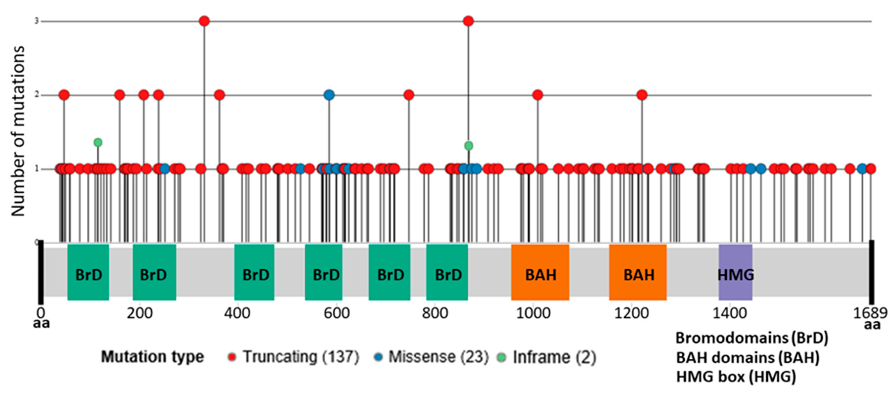
The somatic genomic landscape of ccRCC tumors is characterized by VHL inactivation, which is found altered in ≈90% of the cases. Inactivation is typically achieved through the loss of the chromosome arm 3p (encompassing the genes VHL, PBRM1, BAP1, and SETD2) in a process of unbalanced translocation followed by the somatic inactivation of the remaining VHL allele. However, even if VHL biallelic inactivation is a critical founder event, it is not sufficient for tumor development and additional mutations and epigenetic changes are necessary. Extensive sequencing projects by TCGA have identified PBRM1 as the second most frequently altered gene in ccRCC (49% of explored cases, 163 point mutations, eight deep deletions, and one fusion in a total of 354 cases) [20,31] Even though ccRCC is the tumor type with the highest mutational rate in PBRM1 in the TCGA Pan-Can Project [32], somatic PBRM1 mutations were first described in breast cancer [29] and are also observed in other cancer types such as cholangiocarcinoma (22%) and uterine (10%). Notably, the high incidence of mutations in PBRM1 in renal cancer seems to be limited to clear cell histology and mutations are rare or not observed in other renal cancer subtypes, such as papillary (4%) or chromophobe (0%). In agreement with its role as a tumor suppressor, most mutations observed in ccRCC are truncating (137 out of 163 mutations) while missense mutations are less frequent (23 out of 163). Mutations in PBRM1 are found distributed along the entire gene, affecting Bromodomains, Bromo-Adjacent Homology (BAH) and High Mobility Group (HMG) domains, with some relevant hotspots of truncating mutations [33] (Figure 2). PBRM1 gene (at 3p21.1) is also affected by copy number alterations, as a consequence of the characteristic chromosome 3p deletion in ccRCC tumors.
Intratumoral heterogeneity (ITH) is a common feature of primary ccRCC tumors, leading to somatic point mutations and copy number alterations that are only present in a subset of tumor cells within the same tumor [10,34]. Multiregion sequencing showed that renal tumors are characterized by harboring only a small number of clonal mutations, shared by all tumor cells, and a majority of subclonal alterations found at varying frequencies across the tumor. Mutations in PBRM1 are clonal in ≈75% of cases and subclonal in the remaining 25% of ccRCC primary tumors. Truncal PBRM1 mutations define particular evolutionary trajectories in the tumor, that impact the prognosis and therapeutic response. Three out of the seven evolutionary patterns described in primary ccRCC tumors presented somatic mutations in PBRM1 as a truncal event that precede subclonal mutations in SETD2, genes from the PI3K pathway or copy number alterations [35] (Figure 3). These three clonal PBRM1 driven evolutionary patterns are enriched in tumors with high ITH and a large number of different subclones. Tumors following these evolutionary trajectories are characterized by a slow growth rate, are associated with advanced disease and with longer progression free survival (PFS). Consistently, Joseph RW et al. found that the loss of PBRM1 expression in 1330 ccRCC tumor samples was associated with an increased risk of metastasis development, without an effect on OS [36]. Several studies have described mutual exclusivity between PBRM1 and BAP1 mutations in RCC primary tumors [37,38]. Still, truncal PBRM1 mutations are observed in an additional ccRCC evolutionary subtype characterized by multiple driver clonal mutations in genes that include, in addition to PBRM1, BAP1, SETD2, or PTEN. These tumors have low ITH and high genomic instability and are associated with a rapid progression to multiple sites. BAP1 clonal mutations are characteristic of an additional ccRCC evolutionary pattern that harbors a high genomic instability and a low ITH.
Turajlic et al. proposed that tumor clones with mutations in PBRM1 are preferably selected for metastasis development [35]. However, mutations in additional genes seem to be needed for RCC progression [39]. Metastatic dissemination of primary tumors harboring VHL and PBRM1 mutations as the two only clonal events, is mainly an attenuated process that leads to initial solitary or oligometastases. Metastatic potential in these ccRCC tumors is acquired gradually over time and it is limited to certain subpopulations in the primary tumor that act as a reservoir of metastases. Therefore, mutations in PBRM1 are a key factor not only for primary tumor development but also for tumor metastatic dissemination, in which additional subclonal mutations, acquired after PBRM1 loss, seem to play a decisive role. Detection of clonal and subclonal events in PBRM1, SETD2, PI3K pathway genes, together with somatic copy number alterations is therefore essential to establish a reliable map of the complexity of tumor progression driven by PBRM1 loss.
4. Prognostic Value of PBRM1 Mutations in Localized and Advanced Disease
Different studies have tried to elucidate the impact of PBRM1 status in RCC but with controversial results [36,40,41,42,43,44,45,46,47,48,49,50,51]. Two small retrospective studies were conducted to compare the different prognostic implications of both BAP1 and PBRM1 mutations in localized ccRCC. The study conducted by Kapur et al. showed no survival differences depending on the PBRM1 mutational status, but identified two molecular subgroups with different prognostic implications and different gene expression [40]. The PBRM1-mutant group showed superior median OS than the BAP1-mutant group (HR = 2.7; PBRM1-MT 10.6 months vs. BAP1-MT 4.6 months). Likewise, Gossage et al. reported worse relapse free survival (RFS) in those tumors carrying BAP1 mutation when compared to PBRM1 mutant tumors, although no differences in OS were found (Table 1) [41].
According to recent studies focused exclusively on PBRM1 status as a prognostic factor, it seems that the PBRM1 LOF is associated with a different prognostic value in localized and advanced disease.
4.1. Localized Disease
Most studies assessing the prognostic value of PBRM1 in RCC have focused on localized disease. It is noteworthy that only one of these studies has included other histological subtypes aside from ccRCC [42]. The loss of PBRM1 function has also been correlated with advanced tumor and clinical stage, as well as with more aggressive features, such as lymphovascular invasion and poor differentiation [42,43]. In a systematic review including seven studies (Table 2) PBRM1 LOF in localized disease was associated with poor prognosis, both in terms of RFS and OS [44].
In 2013 the results obtained by Costa et al. showed that PBRM1 LOF was associated with poor prognosis and worse 5 years RFS rates (PBRM1 MT 66.7% vs. PBRM1 WT 87.3%; p = 0.048) and worse 5 years disease-specific survival rates (DSS) (PBRM1 MT 70.6% vs. PBRM1 WT 89.7%; p = 0.017) [43]. Pawlowski et al. conducted a similar study but including different histological subtypes (227 clear cell, 40 papillary and 12 chromophobe). Their results confirmed that PBRM1 LOF is more frequent in ccRCC than in other histological subtypes, and suggested that most of truncating mutations in ccRCC negatively affect PBRM1 expression [42]. The study also showed an association of PBRM1 LOF with late tumor stage and worse OS (PBRM1 negative 80 months vs. PBRM1 positive NR (not reached)). Conversely, a Korean article, which also demonstrated the independent prognostic value of PBRM1 expression, suggested that the negative prognostic impact of PBRM1 LOF was limited to lower-stage tumors (stage I/II; p = 0.001 for cancer specific survival (CSS)/PFS) and not to higher-stage tumors (stage III/IV; p > 0.05), although most of the patients (≈76.6%) were stage I/II [45]. Hakimi et al. investigated the impact of PBRM1 status in 609 patients with ccRCC from two different cohorts (MSKCC, n = 188 and TCGA, n = 421) showing no significant impact on CSS. However, the addition of PBRM1 mutation to BAP1 mutation, which did correlate with a poor prognosis, increased the risk of cancer death (HR 4.18) [46]. The largest study to date was conducted by the Mayo Clinic and included 1330 patients with ccRCC. PBRM1 and BAP1 expression were analyzed by immunohistochemistry (IHC) and they did not find significant differences in CSS according to PBRM1 expression and adjusting by age [36]. The authors did find, however, a significantly higher risk of metastasis in PBRM1 deficient tumors. The study was also able to define, with regards to the prognostic significance, four different ccRCC subtypes depending on PBRM1 and BAP1 status. Thus, there was a progressive worsening of both RFS and CSS from PBRM1+/BAP1+ to PBRM1-/BAP1+ to PBRM1+/BAP1- to PBRM1-/BAP1- [36].
Overall, it seems that in localized ccRCC, the loss of PBRM1 function correlates with aggressive features and advanced stage, as well as with worse prognosis.
4.2. Advanced Disease
There is less data on PBRM1 role in advanced ccRCC compared to localized disease. The first study to assess the prognostic value of PBRM1 expression in stage IV/recurrent RCC was a Korean study including a small population (n = 53) [48]. Patients were divided in two groups using 2.5 IHC score for PBRM1 expression as the cut-off value. Patients with high expression of PBRM1 in the tumor had a significantly worse OS than those with low PBRM1 expression. (HR 2.29, p = 0.039), but there was not statistically significant difference when only the ccRCC subgroup was considered (HR 2.22, p = 0.068) [48]. Consistently, patients harboring tumors with high expression of PBRM1 showed shorter OS when treated with both sunitinib and everolimus (low vs. high expression; OS 45.0 vs. 23.0 months, p = 0.035) [48]. However, the most robust data come from the study conducted by Voss et al. which included 357 advanced ccRCC patients (COMPARZ trial (training cohort), n = 357/RECORD3 trial (validation cohort), n = 258) [51]. They confirmed the prognostic role of BAP1, TP53, and PBRM1 in metastatic renal cell carcinoma patients treated with tyrosine kinase inhibitors (TKI). Remarkably, PBRM1 mutations were associated with a better prognosis, both in terms of PFS (HR 0.67, p = 0.004) and OS (HR 0.63, p = 0.002) [51], whereas BAP1 mutations were associated with worse prognosis, and a significant worse OS (HR 1.51). Conversely, Tennenbaum et al. did not find an association between PBRM1 mutational status (determined by NGS) and OS in 167 advanced ccRCC patients from the MSKCC and TCGA cohorts [50].
Other studies which included cohorts with both localized and advanced disease have shown contradictory results (Table 2) [47,49].
Overall, the studies published to date suggest that PBRM1 LOF has a different role in localized and advanced disease, constituting a poor prognostic factor in localized disease and a good prognostic factor in advanced disease. The inactivation of BAP1 and PBRM1 mainly contribute to different pathways of tumor evolution, being almost mutually exclusive events conferring different prognosis. As suggested, ccRCC could be initiated by a focal mutation in VHL followed by 3p loss, predisposing to BAP1 or PBRM1 inactivation. The mutation of the remaining allele of PBRM1 or BAP1 would lead to tumorigenesis, and depending on which gene is mutated, to different tumor aggressiveness. In advanced RCC, BAP1 mutation leads to a more aggressive disease and PBRM1 mutation would be associated with a better prognosis [35].
5. Predictive Value of PBRM1 Mutations
Antiangiogenics have constituted the cornerstone of mRCC systemic treatment for years. However, during the last years the treatment landscape of mRCC has rapidly changed with the development of new drugs, such as cabozantinib, a multikinase inhibitor, or immune-checkpoint inhibitors, as well as the investigation of different treatment combinations. With such a wide range of treatment options, efforts are now focused on identifying predictive biomarkers of response that will help to establish the best treatment sequence for each tumor and patient. There are studies that suggest the correlation of certain mutations with treatment outcomes [53,54,55]. According to some retrospective studies and preclinical data it seems that the mutational status of PBRM1 could have a predictive role [48,56,57,58]
5.1. Targeted Therapy
Data on PBRM1 predictive value for targeted therapy is scarce and mainly derive from retrospective studies (Table 3) [48,56], aside from the prospective IMMOTION150 trial [59]. According to the results of the IMMOTION150 study, molecular profiles, related with angiogenesis, immune infiltration, and myeloid inflammation, could constitute predictors of response to immunotherapy and antiangiogenics. Interestingly, the tumors with high angiogenesis were enriched for PBRM1 mutations. In addition, when comparing the treatment outcomes, there was a clear benefit in sunitinib PFS, compared to atezolizumab monotherapy and to atezolizumab plus bevacizumab, in tumors with PBRM1 mutations. These results suggest an association between angiogenesis and PBRM1 mutations, indicating that PBRM1 mutated patients may benefit more from antiangiogenics than from immunotherapy.
Similarly, Fay et al. performed whole-exome sequencing in pretreatment specimens of 27 mRCC patients who had experienced an “extreme response” (extreme responders or primary refractory patients) to first line VEGF targeted therapy (sunitinib or pazopanib). In agreement with IMMOTION150 study, PBRM1 mutations were significantly more frequent in patients benefiting from antiangiogenic treatment (54% vs. 7%, respectively) [60].
The retrospective study conducted by Kim et al. investigated the correlation of PBRM1 expression and treatment outcomes in 53 clear cell mRCC patients [48]. Lines of therapy were not specified. High PBRM1 expression correlated with worse outcomes in patients treated with either sunitinib or everolimus. When analyzing each drug separately, the effect of PBRM1 expression was stronger in patients treated with everolimus (median PFS in low and high PBRM1 expression groups was 3.0 months and 1.9 months, respectively; p = 0.10) although it did not reach statistical significance. PBRM1 expression did not correlate with PFS in patients treated with sunitinib (low vs. high median PFS 7.3 vs. 9.0 months, p = 0.83).
Later on, Hsieh et al. investigated the association between PBRM1 mutational status, analyzed by NGS, and treatment outcomes in 220 clear cell mRCC patients included in the RECORD3 trial [56]. The RECORD3 trial was a phase 2 randomized study comparing sunitinib with everolimus in the first line setting with a crossover design. In this study, patients harboring PBRM1 mutation had better outcomes than those with PBRM1 wild type tumors in the everolimus-sunitinib sequence group (MT vs. WT PFS 12.8 vs. 5.5 months; HR 0.53, p = 0.004), whereas no differences were seen in the sunitinib-everolimus sequence group (p = 0.4).
5.2. Immunotherapy
Immune checkpoint inhibitors (ICI) have become a new standard of treatment in RCC. Therefore, there is a need to identify which patients will benefit most from ICI or antiangiogenics in order to better determine the treatment sequence and improve survival outcomes. Although PD-L1 expression is a well established biomarker of response to ICI in some tumors (e.g., non-small cell lung cancer), in RCC it is not useful [59,61,62]. Furthermore, neither the tumor mutational burden or the neoantigen expression have been shown to be suitable for patient selection [63].
The PBAF loss was shown in preclinical in vivo studies to increase sensitivity to T-cell mediated cytotoxicity compared to those with intact PBAF [64]. Recently, Miao et al. as part of a prospective trial (CA 209-009), analyzed by whole exome sequencing 35 pretreated tumors [57]. They tried to correlate the tumor genome profile with the clinical benefit from anti-PD1 therapy and discovered there was a correlation between PBRM1 mutations and clinical benefit. However, when they tried to compare the type of tumor-immune microenvironment in ccRCC tumors according to the mutational status of PBRM1, in three different cohorts (TCGA, an independent cohort of untreated ccRCC tumors and patient tumors of their study), tumors harboring PBRM1 mutations in all three cohorts showed a lower expression of immune inhibitory ligands than those with intact PBRM1.
More recently, a post hoc analysis of the CHECKMATE025 trial which compared nivolumab and sunitinib in ccRCC patients previously treated with antiangiogenics, analyzed the correlation between PBRM1 status and outcomes (PFS and OS) across-treatment in 382 of the 803 patients included in the trial [58]. The results showed a significant correlation between PRBM1 status and response to anti-PD1 therapy, with a significant benefit both in PFS and OS in those patients harboring PBRM1 mutations (HR 0.67, 95% CI, 0.47–0.96; p = 0.03, and HR 0.65, 95% CI, 0.44–0.96; p = 0.03 respectively) compared to those with PBRM1 wild type tumors. There was no evidence of an association between PBRM1 status and clinical outcomes in the everolimus cohort [58].
On the other hand, as discussed above, in the clinical trial IMMOTION150 the group of tumors with high angiogenesis showed an enrichment in PBRM1 mutations and these patients seemed to perform worse when treated with ICI alone (atezolizumab) than when treated with antiangiogenics alone (sunitinib) or in combination with immunotherapy (atezolizumab plus bevacizumab) [59].
Altogether, it seems that PBRM1 mutations are associated with specific molecular tumor expression profiles, and that they may have a predictive role. However, its predictive value is still unclear and needs to be elucidated in future larger randomized trials.
6. Future Perspectives and Conclusions
Cancer is a combination of evolutionary inherited and environmental factors. Most recurrently mutated genes in RCC include PBRM1. Evolutionary information from the TRACERX study has shown that RCC tumors with PBRM1 mutations may have a less aggressive behavior, however, additional events may be synergistic and confer a worse prognosis. This may support that PBRM1 gene could have dual roles in oncogenesis under different cellular contexts (i.e., localized disease or metastatic disease). This duality in tumor suppressors genes (e.g., TP53) have been shown in other tumors [65]. For a gene with both oncogenic and tumor-suppressor potentials, it is possible that one single mutation event would unleash its oncogenic power and abolish its tumor-suppressor function. Theoretically, such mutation events would be easy to target. Indeed, systematic drug sensitivity analyses have proven drug vulnerabilities associated with loss of tumor suppressor genes. This could lead to expanding precision cancer medicine in tumors with a basis on mutations of tumor suppressor genes [66].
In summary, the definition of RCC genetic components that contribute to cancer development is an area of great interest, as it will help to understand cancer evolution and to generate better targeted therapies for the patients. PBRM1, the second most commonly mutated gene in ccRCC, seems to play a relevant role shaping tumor aggressiveness and pathway dependencies. This knowledge has important clinical implications and, together with additional genomic markers, will have the potential to personalize the clinical management of RCC patients.
Author Contributions
Conceptualization, C.R.-A. and G.d.V.; Methodology, L.C.-A., M.S. and J.M.R.-R.; Original Draft Preparation, all authors; Writing-Review & Editing, all authors; Supervision, C.R.-A. and G.d.V. All authors have read and agreed to the published version of the manuscript.
Funding
This research was funded by Agencia Estatal de Investigación, Ministerio de Ciencia, Innovación y Universidades, co-funded by the European Regional Development Fund ERDF (grant number RTI2018-095039-B-I00) and from the Carlos III Institute of Health, Ministry of Economy and Competitiveness (Spain), reference FSI17/1728.
Conflicts of Interest
Guillermo de Velasco reports consulting and advisory services and speaking/writing engagements from Pfizer, Novartis, Bayer, Roche, Ipsen, Astellas, Bristol-Myers Squibb, and MSD, and research fees from Ipsen, Roche, and Pfizer, outside the submitted work. Cristina Rodriguez-Antona reports research projects funded by Pfizer.
References
- Ferlay, J.; Colombet, M.; Soerjomataram, I.; Mathers, C.; Parkin, D.M.; Piñeros, M.; Znaor, A.; Bray, F. Estimating the global cancer incidence and mortality in 2018: GLOBOCAN sources and methods. Int. J. Cancer 2019, 144, 1941–1953. [Google Scholar] [CrossRef] [Green Version]
- Moch, H.; Cubilla, A.L.; Humphrey, P.A.; Reuter, V.E.; Ulbright, T.M. The 2016 WHO classification of tumors of the urinary system and male genital organs-part A: Renal, penile, and testicular tumors. Eur. Urol. 2016, 70, 93–105. [Google Scholar] [CrossRef]
- TNM. Classification of Malignant Tumors, 8th Edition | Wiley [Internet]. Wiley.com. Available online: https://www.wiley.com/en-es/TNM+Classification+of+Malignant+Tumors%2C+8th+Edition-p-9781119263579 (accessed on 1 November 2019).
- Janzen, N.K.; Kim, H.L.; Figlin, R.A.; Belldegrun, A.S. Surveillance after radical or partial nephrectomy for localized renal cell carcinoma and management of recurrent disease. Urol. Clin. N. Am. 2003, 30, 843–852. [Google Scholar] [CrossRef]
- Meskawi, M.; Sun, M.; Trinh, Q.-D.; Bianchi, M.; Hansen, J.; Tian, Z.; Rink, M.; Ismail, S.; Shariat, S.F.; Montorsi, F.; et al. A review of integrated staging systems for renal cell carcinoma. Eur. Urol. 2012, 62, 303–314. [Google Scholar] [CrossRef]
- Patard, J.-J.; Kim, H.L.; Lam, J.S.; Dorey, F.J.; Pantuck, A.J.; Zisman, A.; Ficarra, V.; Han, K.R.; Cindolo, L.; De La Taille, A.; et al. Use of the University of California Los Angeles integrated staging system to predict survival in renal cell carcinoma: An international multicenter study. J. Clin. Oncol. 2004, 22, 3316–3322. [Google Scholar] [CrossRef] [PubMed]
- Leibovich, B.C.; Blute, M.L.; Cheville, J.C.; Lohse, C.M.; Frank, I.; Kwon, E.D.; Weaver, A.L.; Parker, A.S.; Zincke, H. Prediction of progression after radical nephrectomy for patients with clear cell renal cell carcinoma: A stratification tool for prospective clinical trials. Cancer 2003, 97, 1663–1671. [Google Scholar] [CrossRef] [PubMed]
- Gnarra, J.R.; Tory, K.; Weng, Y.; Schmidt, L.; Wei, M.H.; Li, H.; Latif, F.; Liu, S.; Chen, F.; Duh, F.M.; et al. Mutations of the VHL tumor suppressor gene in renal carcinoma. Nat. Genet. 1994, 7, 85–90. [Google Scholar] [CrossRef] [PubMed]
- Brugarolas, J. Molecular genetics of clear-cell renal cell carcinoma. J. Clin. Oncol. 2014, 32, 1968–1976. [Google Scholar] [CrossRef] [PubMed] [Green Version]
- Gerlinger, M.; Horswell, S.; Larkin, J.; Rowan, A.J.; Salm, M.P.; Varela, I.; Fisher, R.; McGranahan, N.; Matthews, N.; Santos, C.R.; et al. Genomic architecture and evolution of clear cell renal cell carcinomas defined by multiregion sequencing. Nat. Genet. 2014, 46, 225–233. [Google Scholar] [CrossRef]
- Linehan, W.M.; Ricketts, C.J. The Cancer Genome Atlas of renal cell carcinoma: Findings and clinical implications. Nat. Rev. Urol. 2019, 16, 539–552. [Google Scholar] [CrossRef]
- Ljungberg, B.; Albiges, L.; Abu-Ghanem, Y.; Bensalah, K.; Dabestani, S.; Fernández-Pello, S.; Rachel, H.G.; Fabian, H.; Milan, H.; Markus, A.K.; et al. European association of urology guidelines on renal cell carcinoma: The 2019 update. Eur. Urol. 2019, 75, 799–810. [Google Scholar] [CrossRef] [PubMed]
- Choueiri, T.K.; Motzer, R.J. Systemic therapy for metastatic renal-cell carcinoma. N. Engl. J. Med. 2017, 376, 354–366. [Google Scholar] [CrossRef] [PubMed]
- de Velasco, G.; Bex, A.; Albiges, L.; Powles, T.; Rini, B.I.; Motzer, R.J.; Heng, D.Y.C.; Escudier, B. Sequencing and combination of systemic therapy in metastatic renal cell carcinoma. Eur. Urol. Oncol. 2019, 2, 505–514. [Google Scholar] [CrossRef]
- Heng, D.Y.C.; Xie, W.; Regan, M.M.; Warren, M.A.; Golshayan, A.R.; Sahi, C.; Eigl, B.J.; Ruether, J.D.; Cheng, T.; North, S.; et al. Prognostic factors for overall survival in patients with metastatic renal cell carcinoma treated with vascular endothelial growth factor-targeted agents: Results from a large, multicenter study. J. Clin. Oncol. 2009, 27, 5794–5799. [Google Scholar] [CrossRef]
- Graham, J.; Dudani, S.; Heng, D.Y.C. Prognostication in kidney cancer: Recent advances and future directions. J. Clin. Oncol. 2018, 36, 3567–3573. [Google Scholar] [CrossRef]
- Hargreaves, D.C.; Crabtree, G.R. ATP-dependent chromatin remodeling: Genetics, genomics and mechanisms. Cell Res. 2011, 21, 396–420. [Google Scholar] [CrossRef]
- Kadoch, C.; Hargreaves, D.C.; Hodges, C.; Elias, L.; Ho, L.; Ranish, J.; Crabtree, G.R. Proteomic and bioinformatic analysis of mammalian SWI/SNF complexes identifies extensive roles in human malignancy. Nat. Genet. 2013, 45, 592–601. [Google Scholar] [CrossRef]
- Masliah-Planchon, J.; Bièche, I.; Guinebretière, J.-M.; Bourdeaut, F.; Delattre, O. SWI/SNF chromatin remodeling and human malignancies. Annu. Rev. Pathol. Mech. Dis. 2015, 10, 145–171. [Google Scholar] [CrossRef]
- Varela, I.; Tarpey, P.; Raine, K.; Huang, D.; Ong, C.K.; Stephens, P.; Davies, H.; Jones, D.; Lin, M.L.; Teague, J.; et al. Exome sequencing identifies frequent mutation of the SWI/SNF complex gene PBRM1 in renal carcinoma. Nature 2011, 469, 539–542. [Google Scholar] [CrossRef] [Green Version]
- Chowdhury, B.; Porter, E.G.; Stewart, J.C.; Ferreira, C.R.; Schipma, M.J.; Dykhuizen, E.C. PBRM1 regulates the expression of genes involved in metabolism and cell adhesion in renal clear cell carcinoma. PLoS ONE 2016, 11, e0153718. [Google Scholar] [CrossRef] [Green Version]
- Kakarougkas, A.; Ismail, A.; Chambers, A.L.; Riballo, E.; Herbert, A.D.; Künzel, J.; Löbrich, M.; Jeggo, P.A.; Downs, J.A. Requirement for PBAF in transcriptional repression and repair at DNA breaks in actively transcribed regions of chromatin. Mol. Cell 2014, 55, 723–732. [Google Scholar] [CrossRef] [PubMed] [Green Version]
- Brownlee, P.M.; Chambers, A.L.; Cloney, R.; Bianchi, A.; Downs, J.A. BAF180 promotes cohesion and prevents genome instability and aneuploidy. Cell Rep. 2014, 6, 973–981. [Google Scholar] [CrossRef] [PubMed] [Green Version]
- Macher-Goeppinger, S.; Keith, M.; Tagscherer, K.E.; Singer, S.; Winkler, J.; Hofmann, T.G.; Pahernik, S.; Duensing, S.; Hohenfellner, M.; Kopitz, J.; et al. PBRM1 (BAF180) protein is functionally regulated by p53-induced protein degradation in renal cell carcinomas. J Pathol. 2015, 237, 460–471. [Google Scholar] [CrossRef]
- Burrows, A.E.; Smogorzewska, A.; Elledge, S.J. Polybromo-associated BRG1-associated factor components BRD7 and BAF180 are critical regulators of p53 required for induction of replicative senescence. Proc. Natl. Acad. Sci. USA 2010, 107, 14280–14285. [Google Scholar] [CrossRef] [Green Version]
- Murakami, A.; Wang, L.; Kalhorn, S.; Schraml, P.; Rathmell, W.K.; Tan, A.C.; Nemenoff, R.; Stenmark, K.; Jiang, B.H.; Reyland, M.E.; et al. Context-dependent role for chromatin remodeling component PBRM1/BAF180 in clear cell renal cell carcinoma. Oncogenesis 2017, 6, e287. [Google Scholar] [CrossRef] [Green Version]
- Gordan, J.D.; Bertout, J.A.; Hu, C.-J.; Diehl, J.A.; Simon, M.C. HIF-2alpha promotes hypoxic cell proliferation by enhancing c-myc transcriptional activity. Cancer Cell 2007, 11, 335–347. [Google Scholar] [CrossRef] [Green Version]
- Linehan, W.M.; Srinivasan, R.; Schmidt, L.S. The genetic basis of kidney cancer: A metabolic disease. Nat. Rev. Urol. 2010, 7, 277–285. [Google Scholar] [CrossRef]
- Xia, W.; Nagase, S.; Montia, A.G.; Kalachikov, S.M.; Keniry, M.; Su, T.; Memeo, L.; Hibshoosh, H.; Parsons, R. BAF180 is a critical regulator of p21 induction and a tumor suppressor mutated in breast cancer. Cancer Res. 2008, 68, 1667–1674. [Google Scholar] [CrossRef] [Green Version]
- Benusiglio, P.R.; Couvé, S.; Gilbert-Dussardier, B.; Deveaux, S.; Le Jeune, H.; Da Costa, M.; Fromont, G.; Memeteau, F.; Yacoub, M.; Coupier, I.; et al. A germline mutation in PBRM1 predisposes to renal cell carcinoma. J. Med. Genet. 2015, 52, 426–430. [Google Scholar] [CrossRef]
- Ricketts, C.J.; De Cubas, A.A.; Fan, H.; Smith, C.C.; Lang, M.; Reznik, E.; Bowlby, R.; Gibb, E.A.; Akbani, R.; Beroukhim, R.; et al. The cancer genome atlas comprehensive molecular characterization of renal cell carcinoma. Cell Rep. 2018, 23, 313.e5–326.e5. [Google Scholar] [CrossRef] [Green Version]
- Bailey, M.H.; Tokheim, C.; Porta-Pardo, E.; Sengupta, S.; Bertrand, D.; Weerasinghe, A.; Colaprico, A.; Wendl, M.C.; Kim, J.; Reardon, B.; et al. Comprehensive characterization of cancer driver genes and mutations. Cell 2018, 173, 371.e18–385.e18. [Google Scholar] [CrossRef] [Green Version]
- cBioPortal for Cancer Genomics [Internet]. Available online: http://www.cbioportal.org/ (accessed on 27 November 2019).
- Gerlinger, M.; Rowan, A.J.; Horswell, S.; Math, M.; Larkin, J.; Endesfelder, D.; Gronroos, E.; Martinez, P.; Matthews, N.; Stewart, A.; et al. Intratumor heterogeneity and branched evolution revealed by multiregion sequencing. N. Engl. J. Med. 2012, 366, 883–892. [Google Scholar] [CrossRef] [Green Version]
- Turajlic, S.; Xu, H.; Litchfield, K.; Rowan, A.; Chambers, T.; Lopez, J.I.; Nicol, D.; O’Brien, T.; Larkin, J.; Horswell, S.; et al. Tracking cancer evolution reveals constrained routes to metastases: TRACERx renal. Cell 2018, 173, 581.e12–594.e12. [Google Scholar] [CrossRef]
- Joseph, R.W.; Kapur, P.; Serie, D.J.; Parasramka, M.; Ho, T.H.; Cheville, J.C.; Frenkel, E.; Parker, A.S.; Brugarolas, J. Clear cell renal cell carcinoma subtypes identified by BAP1 and PBRM1 expression. J. Urol. 2016, 195, 180–187. [Google Scholar] [CrossRef] [Green Version]
- Peña-Llopis, S.; Vega-Rubín-de-Celis, S.; Liao, A.; Leng, N.; Pavía-Jiménez, A.; Wang, S.; Yamasaki, T.; Zhrebker, L.; Sivanand, S.; Spence, P.; et al. BAP1 loss defines a new class of renal cell carcinoma. Nat. Genet. 2012, 44, 751–759. [Google Scholar] [CrossRef]
- Peña-Llopis, S.; Christie, A.; Xie, X.-J.; Brugarolas, J. Cooperation and antagonism among cancer genes: The renal cancer paradigm. Cancer Res. 2013, 73, 4173–4179. [Google Scholar] [CrossRef] [Green Version]
- Gu, Y.-F.; Cohn, S.; Christie, A.; McKenzie, T.; Wolff, N.; Do, Q.N.; Madhuranthakam, A.J.; Pedrosa, I.; Wang, T.; Dey, A.; et al. Modeling renal cell carcinoma in mice: Bap1 and Pbrm1 inactivation drive tumor grade. Cancer Discov. 2017, 7, 900–917. [Google Scholar] [CrossRef] [Green Version]
- Kapur, P.; Peña-Llopis, S.; Christie, A.; Zhrebker, L.; Pavía-Jiménez, A.; Rathmell, W.K.; Xie, X.J.; Brugarolas, J. Effects on survival of BAP1 and PBRM1 mutations in sporadic clear-cell renal-cell carcinoma: A retrospective analysis with independent validation. Lancet Oncol. 2013, 14, 159–167. [Google Scholar] [CrossRef] [Green Version]
- Gossage, L.; Murtaza, M.; Slatter, A.F.; Lichtenstein, C.P.; Warren, A.; Haynes, B.; Marass, F.; Roberts, I.; Shanahan, S.J.; Claas, A.; et al. Clinical and pathological impact of VHL, PBRM1, BAP1, SETD2, KDM6A, and JARID1c in clear cell renal cell carcinoma. Genes Chromosomes Cancer 2014, 53, 38–51. [Google Scholar] [CrossRef]
- Pawłowski, R.; Mühl, S.M.; Sulser, T.; Krek, W.; Moch, H.; Schraml, P. Loss of PBRM1 expression is associated with renal cell carcinoma progression. Int. J. Cancer 2013, 132, E11–E17. [Google Scholar] [CrossRef]
- da Costa, W.H.; Rezende, M.; Carneiro, F.C.; Rocha, R.M.; da Cunha, I.W.; Carraro, D.M.; Guimaraes, G.C.; de Cassio Zequi, S. Polybromo-1 (PBRM1), a SWI/SNF complex subunit is a prognostic marker in clear cell renal cell carcinoma. BJU Int. 2014, 113, E157–E163. [Google Scholar] [CrossRef] [Green Version]
- Wang, Z.; Peng, S.; Guo, L.; Xie, H.; Wang, A.; Shang, Z.; Niu, Y. Prognostic and clinicopathological value of PBRM1 expression in renal cell carcinoma. Clin. Chim. Acta 2018, 486, 9–17. [Google Scholar] [CrossRef]
- Nam, S.J.; Lee, C.; Park, J.H.; Moon, K.C. Decreased PBRM1 expression predicts unfavorable prognosis in patients with clear cell renal cell carcinoma. Urol. Oncol. 2015, 33, 340.e9–340.e16. [Google Scholar] [CrossRef]
- Hakimi, A.A.; Ostrovnaya, I.; Reva, B.; Schultz, N.; Chen, Y.-B.; Gonen, M.; Liu, H.; Takeda, S.; Voss, M.H.; Tickoo, S.K.; et al. Adverse outcomes in clear cell renal cell carcinoma with mutations of 3p21 epigenetic regulators BAP1 and SETD2: A report by MSKCC and the KIRC TCGA research network. Clin. Cancer Res. 2013, 19, 3259–3267. [Google Scholar] [CrossRef] [Green Version]
- Jiang, W.; Dulaimi, E.; Devarajan, K.; Parsons, T.; Wang, Q.; O’Neill, R.; Solomides, C.; Peiper, S.C.; Testa, J.R.; Uzzo, R.; et al. Intratumoral heterogeneity analysis reveals hidden associations between protein expression losses and patient survival in clear cell renal cell carcinoma. Oncotarget 2017, 8, 37423–37434. [Google Scholar] [CrossRef] [Green Version]
- Kim, J.-Y.; Lee, S.-H.; Moon, K.C.; Kwak, C.; Kim, H.H.; Keam, B.; Kim, T.M.; Heo, D.S. The impact of PBRM1 expression as a prognostic and predictive marker in metastatic renal cell carcinoma. J. Urol. 2015, 194, 1112–1119. [Google Scholar] [CrossRef]
- Carlo, M.I.; Manley, B.; Patil, S.; Woo, K.M.; Coskey, D.T.; Redzematovic, A.; Arcila, M.; Ladanyi, M.; Lee, W.; Chen, Y.B.; et al. Genomic alterations and outcomes with VEGF-targeted therapy in patients with clear cell renal cell carcinoma. Kidney Cancer 2017, 1, 49–56. [Google Scholar] [CrossRef] [Green Version]
- Tennenbaum, D.M.; Manley, B.J.; Zabor, E.; Becerra, M.F.; Carlo, M.I.; Casuscelli, J.; Redzematovic, A.; Khan, N.; Arcila, M.E.; Voss, M.H.; et al. Genomic alterations as predictors of survival among patients within a combined cohort with clear cell renal cell carcinoma undergoing cytoreductive nephrectomy. Urol. Oncol. 2017, 35, 532.e7–532.e13. [Google Scholar] [CrossRef]
- Voss, M.H.; Reising, A.; Cheng, Y.; Patel, P.; Marker, M.; Kuo, F.; Chan, T.A.; Choueiri, T.K.; Hsieh, J.J.; Hakimi, A.A.; et al. Genomically annotated risk model for advanced renal-cell carcinoma: A retrospective cohort study. Lancet Oncol. 2018, 19, 1688–1698. [Google Scholar] [CrossRef]
- Kim, S.H.; Park, W.S.; Park, E.Y.; Park, B.; Joo, J.; Joung, J.Y.; Seo, H.K.; Lee, K.H.; Chung, J. The prognostic value of BAP1, PBRM1, pS6, PTEN, TGase2, PD-L1, CA9, PSMA, and Ki-67 tissue markers in localized renal cell carcinoma: A retrospective study of tissue microarrays using immunohistochemistry. PLoS ONE 2017. [Google Scholar] [CrossRef] [Green Version]
- Voss, M.H.; Hakimi, A.A.; Pham, C.G.; Brannon, A.R.; Chen, Y.-B.; Cunha, L.F.; Akin, O.; Liu, H.; Takeda, S.; Scott, S.N.; et al. Tumor genetic analyses of patients with metastatic renal cell carcinoma and extended benefit from mTOR inhibitor therapy. Clin. Cancer Res. 2014, 20, 1955–1964. [Google Scholar] [CrossRef] [Green Version]
- Kwiatkowski, D.J.; Choueiri, T.K.; Fay, A.P.; Rini, B.I.; Thorner, A.R.; de Velasco, G.; Tyburczy, M.E.; Hamieh, L.; Albiges, L.; Agarwal, N.; et al. Mutations in TSC1, TSC2, and MTOR are associated with response to rapalogs in patients with metastatic renal cell carcinoma. Clin. Cancer Res. 2016, 22, 2445–2452. [Google Scholar] [CrossRef] [Green Version]
- Ho, T.H.; Choueiri, T.K.; Wang, K.; Karam, J.A.; Chalmers, Z.; Frampton, G.; Elvin, J.A.; Johnson, A.; Liu, X.; Lin, Y.; et al. Correlation between molecular subclassifications of clear cell renal cell carcinoma and targeted therapy response. Eur. Urol. Focus 2016, 2, 204–209. [Google Scholar] [CrossRef]
- Hsieh, J.J.; Chen, D.; Wang, P.I.; Marker, M.; Redzematovic, A.; Chen, Y.B.; Selcuklu, S.D.; Weinhold, N.; Bouvier, N.; Huberman, K.H.; et al. Genomic biomarkers of a randomized trial comparing first-line everolimus and sunitinib in patients with metastatic renal cell carcinoma. Eur. Urol. 2017, 71, 405–414. [Google Scholar] [CrossRef] [Green Version]
- Miao, D.; Margolis, C.A.; Gao, W.; Voss, M.H.; Li, W.; Martini, D.J.; Norton, C.; Bossé, D.; Wankowicz, S.M.; Cullen, D.; et al. Genomic correlates of response to immune checkpoint therapies in clear cell renal cell carcinoma. Science 2018, 359, 801–806. [Google Scholar] [CrossRef] [Green Version]
- Braun, D.A.; Ishii, Y.; Walsh, A.M.; Van Allen, E.M.; Wu, C.J.; Shukla, S.A.; Choueiri, T.K. Clinical validation of PBRM1 alterations as a marker of immune checkpoint inhibitor response in renal cell carcinoma. JAMA Oncol. 2019, 5, 1631–1633. [Google Scholar] [CrossRef]
- McDermott, D.F.; Huseni, M.A.; Atkins, M.B.; Motzer, R.J.; Rini, B.I.; Escudier, B.; Fong, L.; Joseph, R.W.; Pal, S.K.; Reeves, J.A.; et al. Clinical activity and molecular correlates of response to atezolizumab alone or in combination with bevacizumab versus sunitinib in renal cell carcinoma. Nat. Med. 2018, 24, 749–757. [Google Scholar] [CrossRef]
- Fay, A.P.; de Velasco, G.; Ho, T.H.; Van Allen, E.M.; Murray, B.; Albiges, L.; Signoretti, S.; Hakimi, A.A.; Stanton, M.L.; Bellmunt, J.; et al. Whole-exome sequencing in two extreme phenotypes of response to VEGF-targeted therapies in patients with metastatic clear cell renal cell carcinoma. J. Natl. Compr. Cancer Netw. JNCCN 2016, 14, 820–824. [Google Scholar] [CrossRef] [Green Version]
- Motzer, R.J.; Tannir, N.M.; McDermott, D.F.; ArénFrontera, O.; Melichar, B.; Choueiri, T.K.; Plimack, E.R.; Barthélémy, P.; Porta, C.; George, S.; et al. Nivolumab plus ipilimumab versus sunitinib in advanced renal-cell carcinoma. N. Engl. J. Med. 2018, 378, 1277–1290. [Google Scholar] [CrossRef]
- Motzer, R.J.; Escudier, B.; McDermott, D.F.; George, S.; Hammers, H.J.; Srinivas, S.; Tykodi, S.S.; Sosman, J.A.; Procopio, G.; Plimack, E.R.; et al. Nivolumab versus everolimus in advanced renal-cell carcinoma. N. Engl. J. Med. 2015, 373, 1803–1813. [Google Scholar] [CrossRef]
- Maia, M.C.; Almeida, L.; Bergerot, P.G.; Dizman, N.; Pal, S.K. Relationship of tumor mutational burden (TMB) to immunotherapy response in metastatic renal cell carcinoma (mRCC). J. Clin. Oncol. 2018, 36 (Suppl. 6), 662. [Google Scholar] [CrossRef]
- Pan, D.; Kobayashi, A.; Jiang, P.; Ferrari de Andrade, L.; Tay, R.E.; Luoma, A.M.; Tsoucas, D.; Qiu, X.; Lim, K.; Rao, P.; et al. A major chromatin regulator determines resistance of tumor cells to T cell-mediated killing. Science 2018, 359, 770–775. [Google Scholar] [CrossRef] [Green Version]
- Soussi, T.; Wiman, K.G. TP53: An oncogene in disguise. Cell Death Differ. 2015, 22, 1239–1249. [Google Scholar] [CrossRef]
- Ding, H.; Zhao, J.; Zhang, Y.; Yu, J.; Liu, M.; Li, X.; Xu, L.; Lin, M.; Liu, C.; He, Z.; et al. Systematic analysis of drug vulnerabilities conferred by tumor suppressor loss. Cell Rep. 2019, 27, 3331.e6–3344.e6. [Google Scholar] [CrossRef] [Green Version]
Figure 1.
Role of PBRM1 in established clear cell renal cell carcinoma (ccRCC) tumors.

Figure 2.
PBRM1 mutations in clear cell RCC. From The Cancer Genome Atlas (TCGA) PanCancer Atlas Project.
Figure 2.
PBRM1 mutations in clear cell RCC. From The Cancer Genome Atlas (TCGA) PanCancer Atlas Project.

Figure 3.
Branched clear cell RCC evolutionary model. Based on Turajlic et al. [35].
Figure 3.
Branched clear cell RCC evolutionary model. Based on Turajlic et al. [35].


Table 1.
Value of BAP1 versus PBRM1 in localized disease.
| Author [Ref.] | Year | Country | Gender M(%)/F(%) | Age (y) | N | BAP1 MT(%) | PBRM1 MT(%) | Tech | Survival Efficacy Parameters | |
|---|---|---|---|---|---|---|---|---|---|---|
| RFS (HR/ p) | OS (HR/ p) | |||||||||
| Kapur [40] | 2013 | USA | 80(55)/65(45) | 62 | 145 | 21(14) | 78(54) | NGS | NA | BAP1 MT: 4.6; PBRM1 MT: 10.6 (2.7/0.044) |
| Gossage [41] | 2014 | UK | 83(63)/49(37) | 62 | 132 | 14(11) | 42(33) | NGS | BAP1 MT: 1.2 a; PBRM1 MT: 4.9 a (n.sp./0.059) | n.sp. (n.sp./NS) |
M: male patients. F: female patients. Age: median age. y: years. N: number of patients. MT: mutant patients. Tech: Technique. NGS: next-generation sequencing. RFS: recurrence-free-survival in years. HR: hazard ratio. OS: overall survival in years. NS: non statistically significant. NA: not assessed. n.sp.: not specified. a 75th percentile for survival.
Table 2.
Current evidence on PBRM1 prognosis value.
| Disease Type | Author [Ref.] | Year | Country | Gender M(%)/F(%) | Age (y) | N | PBRM1 -/Low (%) | Tech | Ab (Dilution) | PBRM1 Cut-off | Survival Efficacy Parameters | |
|---|---|---|---|---|---|---|---|---|---|---|---|---|
| RFS/PFS (HR/p) | OS/CSS (HR/p) | |||||||||||
| Loc | da Costa [43] | 2013 | Brazil | 66(59)/46(41) | 56 | 112 | 34(30) | IHC | PB1 Ab b (1:25) | Presence | PBRM1 -: 66.7% vs. PBRM1 +: 87.3% d (n.sp./0.048) | PBRM1 -: 70.6 vs. PBRM1 +: 89.7% f (n.sp./0.017) |
| Hakimi [46] | 2013 | USA | 408(67)/200(33) | 61 | 609 | 198(33) | NGS | - | - | NA | n.sp. (n.sp./NS) | |
| Pawlowski [42] | 2013 | Switzerland | NA | NA | 279 | 175(64) a. | IHC | PB1Ab c (1:25) | >5% | NA | PBRM1 -: 80 vs. PBRM1 +: not reach (n.sp./0.025) | |
| Nam [45] | 2015 | Korea | 485(74)/172(26) | NA | 657 | NA | IHC | PB1 Ab c (1:100) | >50% | PBRM1 LE: 109.5 vs. PBRM1 HE: 156.2 (n.sp./<0.001) | PBRM1 LE: 145.5 vs. PBRM1 HE: 171.7 (n.sp./<0.001) | |
| Joseph [36] | 2016 | USA | 823(62)/435(33) | 64 | 1330 | 674(51) | IHC | PB1 Ab c | Presence | PBRM1 -: higher mtx. risk (1.46/0.001) | n.sp. (n.sp/NS) | |
| Kim [52] | 2017 | Korea | 244(70)/107(30) | 54 | 351 | 208(59) | IHC | PB1 Ab | Score > 2 | n.sp. (0.81/0.64) | n.sp. (1.86/0.1) | |
| Loc and Mtx | Jiang [47] | 2017 | USA | 118(74)/42(26) | 60 | 160 | 49(31) | IHC | PB1 Abc (1:50) | >5% | n.sp. (0.79/0.21) | n.sp. (0.42/2.45e–05) |
| Carlo [49] | 2017 | USA | 77(73)/28(27) | 57 | 105 | 53(51) | NGS | - | - | PBRM1 MT: 12.0 vs. PBRM1 WT: 6.9 e (n.sp./0.01) | PBRM1 MT: not reach vs. PBRM1 WT: 36.3 (n.sp./0.12) | |
| Mtx | Tennenbaum [50] | 2017 | USA | 116(69)/51(31) | 60 | 167 | 64(38) | NGS | - | - | NA | n.sp. (0.87/0.49) |
| Kim [48] | 2015 | Korea | 44(83)/9(17) | 62 | 53 | 28(52) | IHC | PB1 Ab c (1:100) | Score >2.5 | NA | PBRM1 LE: 46 vs. PBRM1 HE: 24 (n.sp./0.022) | |
| Voss [51] | 2018 | USA | 279(74)/98(26) | 61 | 357 | 160(45) | NGS | - | - | PBRM1 MT: 11.1 vs. PBRM1 WT: 8.2 (0.67/0.004) | PBRM1 MT: 35.5 vs. PBRM1 WT: 23.8 (0.63/0.002) | |
Loc: localized. Mtx: metastatic. M: male patients. F: female patients. Age: median age. y: years. N: number of patients. PBRM1 -: patients not expressing PBRM1. PBRM1 LE: patients with low PBRM1 expression. PBRM1 HE: patients with high PBRM1 expression. Tech: technology. Ab: antibody. IHC: immunohistochemistry. NGS: next-generation sequencing. RFS: recurrence-free survival in months. PFS: progression-free survival in months. OS: overall survival in months. CSS: cancer-specific-survival. MT: mutant patients. WT: wild-type patients. n.sp.: not specified. NA: not assessed. HR: hazard ratio. a: Out of the 175 patients negative for PBRM1 expression 155(68%) were clear cell, 16(40%) papillary and 4(30%) chromophobe histology. b: Anti-BAF180 antibody (Sigma-Aldrich, MO, USA). c: PB1/BAF180 antibody (Montgomery TX, Texas, USA). d: 5-year recurrence free survival rate. e: Time to treatment failure in months. f: 5-year cancer-specific survival rate.
Table 3.
Current evidence supporting PBRM1 predictive value for everolimus in metastatic disease.
| Author [Ref.] | Year | Country | Gender M(%)/F(%) | Age (y) | N | BAP1 MT (%) | PBRM1 MT (%) | Tech | Ab(Dilution) | PBRM1 Cut-off | Everolimus PFS | Sunitinib PFS |
|---|---|---|---|---|---|---|---|---|---|---|---|---|
| PBRM1 MT or LE vs. PBRM1 WT or HE (HR/ p) | PBRM1 MT or LE vs. PBRM1 WT or HE (HR/ p) | |||||||||||
| Hsieh [56] | 2016 | USA | 168 (76)/52(24) | 62 | 220 | 42(19) | 101(46) | NGS | - | - | 12.8 vs. 5.5 (0.53/ 0.004) | 11.0 vs. 8.3 (0.79/ 0.4) |
| Kim [48] | 2015 | Korea | 44(83)/9(17) | 62 | 53 | NA | 25(47) | IHC | PB1 Ab a (1:100) | 2.5 | 3.0 vs. 1.9 (NA/ 0.1) | 7.3 vs. 9 (NA/ 0.8) |
M: male patients. F: female patients. Age: median age. y: years. N: number of patients. MT: mutant patients. NA: not assessed. Tech: technique. NGS: next-generation sequencing. IHC: immunohistochemistry. PFS: progression-free-survival in months. HR: hazard ratio. NA: not assessed. a PB1/BAF180 AB Montgomery, TX.
© 2019 by the authors. Licensee MDPI, Basel, Switzerland. This article is an open access article distributed under the terms and conditions of the Creative Commons Attribution (CC BY) license (http://creativecommons.org/licenses/by/4.0/).
Share and Cite
MDPI and ACS Style
Carril-Ajuria, L.; Santos, M.; Roldán-Romero, J.M.; Rodriguez-Antona, C.; de Velasco, G. Prognostic and Predictive Value of PBRM1 in Clear Cell Renal Cell Carcinoma. Cancers 2020, 12, 16. https://doi.org/10.3390/cancers12010016
AMA Style
Carril-Ajuria L, Santos M, Roldán-Romero JM, Rodriguez-Antona C, de Velasco G. Prognostic and Predictive Value of PBRM1 in Clear Cell Renal Cell Carcinoma. Cancers. 2020; 12(1):16. https://doi.org/10.3390/cancers12010016
Chicago/Turabian StyleCarril-Ajuria, Lucía, María Santos, Juan María Roldán-Romero, Cristina Rodriguez-Antona, and Guillermo de Velasco. 2020. "Prognostic and Predictive Value of PBRM1 in Clear Cell Renal Cell Carcinoma" Cancers 12, no. 1: 16. https://doi.org/10.3390/cancers12010016
Note that from the first issue of 2016, this journal uses article numbers instead of page numbers. See further details here.