Analysing Tumour Growth Delay Data from Animal Irradiation Experiments with Deviations from the Prescribed Dose


 ,
,
Abstract
:1. Introduction
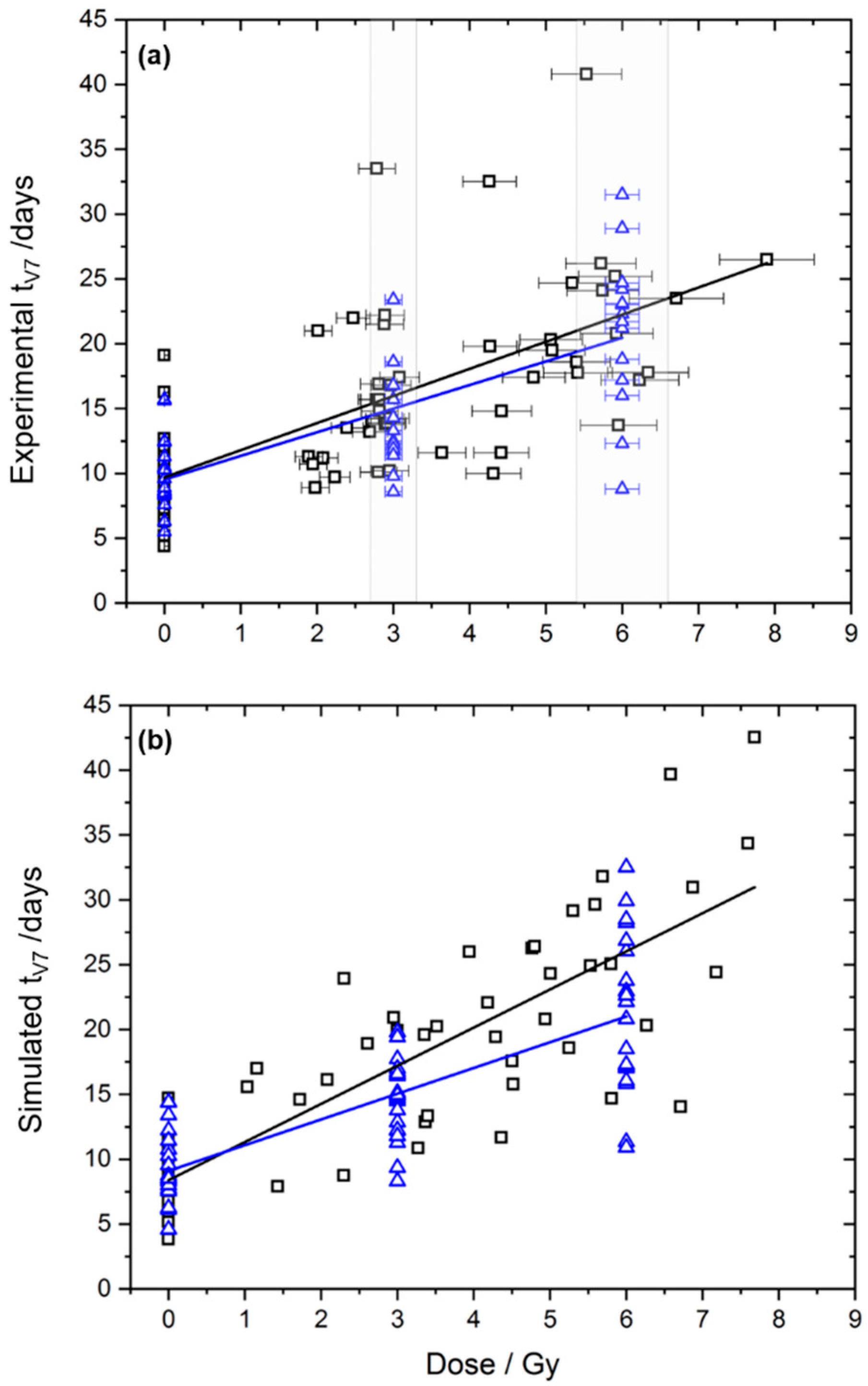
2. Results
3. Discussion
4. Materials and Methods
4.1. Input Data Sets
4.1.1. Experimental Data
4.1.2. Simulated Data
4.2. Methods for Analysing Tumour Growth Data
4.2.1. Conventional Analysis
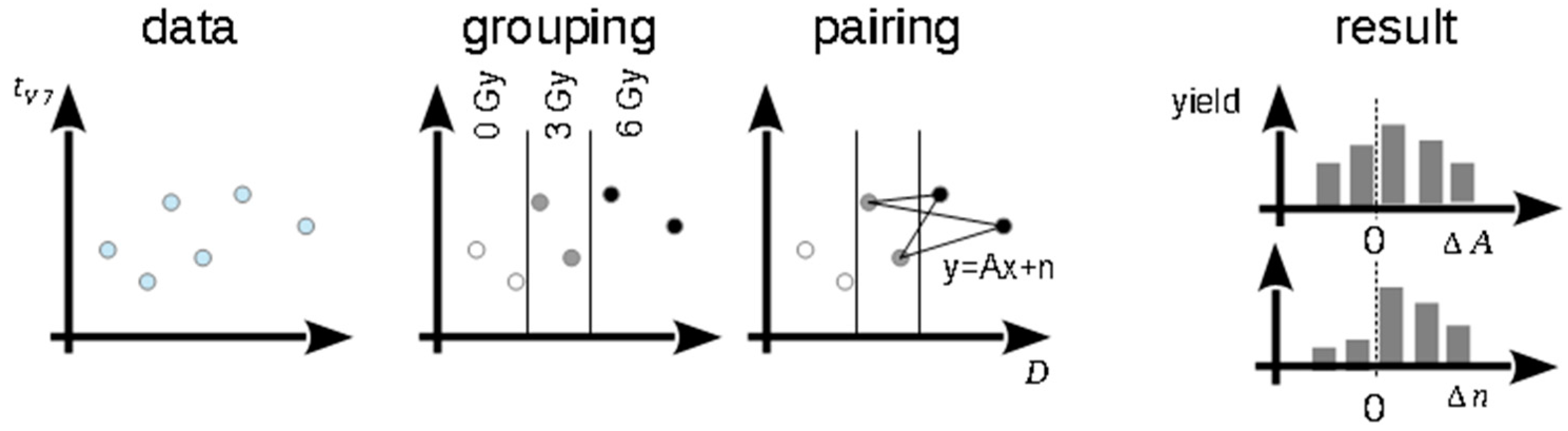
4.2.2. Monte-Carlo-Based Method
4.2.3. Linear Regression
4.2.4. Cox Regression
5. Conclusions
Supplementary Materials
Author Contributions
Funding
Acknowledgments
Conflicts of Interest
References
- Borghesi, M.; Macchi, A. Laser-Driven Particle Acceleration Towards Radiobiology and Medicine, 1st ed.; Giulietti, A., Ed.; Springer International Publishing: New York, NY, USA, 2016; pp. 203–218. ISBN 978-3-319-31563-8. [Google Scholar]
- Schültke, E.; Balosso, J.; Breslin, T.; Cavaletti, G.; Djonov, V.; Esteve, F.; Grotzer, M.; Hildebrandt, G.; Valdman, A.; Laissue, J. Microbeam radiation therapy—grid therapy and beyond: a clinical perspective. Br. J. Radiol. 2017, 90, 20170073. [Google Scholar] [CrossRef] [PubMed]
- Bourhis, J.; Montay-Gruel, P.; Jorge, P.G.; Bailat, C.; Petit, B.; Ollivier, J.; Jeanneret-Sozzi, W.; Ozsahin, M.; Bochud, F.; Moeckli, R.; et al. Clinical translation of FLASH radiotherapy: Why and how? Radiother. Oncol. 2019. [Google Scholar] [CrossRef] [PubMed]
- Baumann, M.; Bentzen, S.M.; Doerr, W.; Joiner, M.C.; Saunders, M.; Tannock, I.F.; Thames, H.D. The translational research chain: is it delivering the goods? Int. J. Radiat. Oncol. 2001, 49, 345–351. [Google Scholar] [CrossRef]
- Karsch, L.; Beyreuther, E.; Enghardt, W.; Gotz, M.; Masood, U.; Schramm, U.; Zeil, K.; Pawelke, J. Towards ion beam therapy based on laser plasma accelerators. Acta Oncol. 2017, 56, 1359–1366. [Google Scholar] [CrossRef] [PubMed]
- Daido, H.; Nishiuchi, M.; Pirozhkov, A.S. Review of laser-driven ion sources and their applications. Rep. Prog. Phys. 2012, 75, 56401. [Google Scholar] [CrossRef] [PubMed]
- Laschinsky, L.; Baumann, M.; Beyreuther, E.; Enghardt, W.; Kaluza, M.; Karsch, L.; Lessmann, E.; Naumburger, D.; Nicolai, M.; Richter, C.; et al. Radiobiological effectiveness of laser accelerated electrons in comparison to electron beams from a conventional linear accelerator. J. Radiat. Res. 2012, 53, 395–403. [Google Scholar] [CrossRef] [PubMed]
- Yogo, A.; Sato, K.; Nishikino, M.; Mori, M.; Teshima, T.; Numasaki, H.; Murakami, M.; Demizu, Y.; Akagi, S.; Nagayama, S.; et al. Application of laser-accelerated protons to the demonstration of DNA double-strand breaks in human cancer cells. Appl. Phys. Lett. 2009, 94, 181502. [Google Scholar] [CrossRef]
- Yogo, A.; Maeda, T.; Hori, T.; Sakaki, H.; Ogura, K.; Nishiuchi, M.; Sagisaka, A.; Kiriyama, H.; Okada, H.; Kanazawa, S.; et al. Measurement of relative biological effectiveness of protons in human cancer cells using a laser-driven quasimonoenergetic proton beamline. Appl. Phys. Lett. 2011, 98, 053701. [Google Scholar] [CrossRef]
- Schmid, T.E.; Dollinger, G.; Hable, V.; Greubel, C.; Zlobinskaya, O.; Michalski, D.; Molls, M.; Röper, B. Relative biological effectiveness of pulsed and continuous 20MeV protons for micronucleus induction in 3D human reconstructed skin tissue. Radiother. Oncol. 2010, 95, 66–72. [Google Scholar] [CrossRef] [PubMed]
- Brüchner, K.; Beyreuther, E.; Baumann, M.; Krause, M.; Oppelt, M.; Pawelke, J. Establishment of a small animal tumour model for in vivo studies with low energy laser accelerated particles. Radiat. Oncol. 2014, 9, 57. [Google Scholar] [CrossRef] [PubMed]
- Oppelt, M.; Baumann, M.; Bergmann, R.; Beyreuther, E.; Brüchner, K.; Hartmann, J.; Karsch, L.; Krause, M.; Laschinsky, L.; Leßmann, E.; et al. Comparison study of in vivo dose response to laser-driven versus conventional electron beam. Radiat. Environ. Biophys. 2015, 54, 155–166. [Google Scholar] [CrossRef] [PubMed]
- Heitjan, D.F.; Manni, A.; Santen, R.J. Statistical analysis of in vivo tumor growth experiments. Cancer Res. 1993, 53, 6042–6050. [Google Scholar] [PubMed]
- Rofstad, E.K.; Brustad, T. Tumour growth delay following single dose irradiation of human melanoma xenografts. Correlations with tumour growth parameters, vascular structure and cellular radiosensitivity. Br. J. Cancer 1985, 51, 201–210. [Google Scholar] [CrossRef] [PubMed] [Green Version]
- Meyer, K.; Krueger, S.A.; Kane, J.L.; Wilson, T.G.; Hanna, A.; Dabjan, M.; Hege, K.M.; Wilson, G.D.; Grills, I.; Marples, B. Pulsed Radiation Therapy With Concurrent Cisplatin Results in Superior Tumor Growth Delay in a Head and Neck Squamous Cell Carcinoma Murine Model. Int. J. Radiat. Oncol. 2016, 96, 161–169. [Google Scholar] [CrossRef] [PubMed]
- Beyreuther, E.; Brüchner, K.; Krause, M.; Schmidt, M.; Szabó, R.; Pawelke, J. An optimized small animal tumour model for experimentation with low energy protons. PLoS ONE 2017, 12, e0177428. [Google Scholar] [CrossRef] [PubMed]
- Rygaard, K.; Spang-Thomsen, M. Quantitation and Gompertzian analysis of tumor growth. Breast Cancer Res. Treat. 1997, 46, 303–312. [Google Scholar] [CrossRef] [PubMed]
- Stuschke, M.; Budach, V.; Bamberg, M.; Budach, W. Methods for Analysis of Censored Tumor Growth Delay Data. Radiat. Res. 1990, 122, 172. [Google Scholar] [CrossRef] [PubMed]
- Hanfelt, J.J. Statistical approaches to experimental design and data analysis of in vivo studies. Breast Cancer Res. Treat. 1997, 46, 279–302. [Google Scholar] [CrossRef] [PubMed]
- Zlobinskaya, O.; Siebenwirth, C.; Greubel, C.; Hable, V.; Hertenberger, R.; Humble, N.; Reinhardt, S.; Michalski, D.; Röper, B.; Multhoff, G.; et al. The Effects of Ultra-High Dose Rate Proton Irradiation on Growth Delay in the Treatment of Human Tumor Xenografts in Nude Mice. Radiat. Res. 2014, 181, 177–183. [Google Scholar] [CrossRef] [PubMed] [Green Version]
- West, B.T.; Welch, K.B.; Galecki, A.T. Linear Mixed Models: A Practical Guide Using Statistical Software, 2nd ed.; Chapman and Hall/CRC: London, UK, 2014; ISBN 978-0-429-18656-1. [Google Scholar]


| Parameter | Laser-Driven Electrons | Linac Electrons | p Value | ||||||
|---|---|---|---|---|---|---|---|---|---|
| Conventional Analysis | |||||||||
| mean | sd | N | mean | sd | N | ||||
| tV7(0 Gy) | 9.66 | 2.87 | 41 | 9.90 | 2.64 | 20 | 0.75 | ||
| tV7(3 Gy) | 16.46 | 5.41 | 17 | 13.88 | 3.97 | 13 | 0.14 | ||
| tV7(6 Gy) | 22.22 | 7.64 | 10 | 20.98 | 6.05 | 14 | 0.68 | ||
| Monte-Carlo-Based Method | |||||||||
| Mean | sd | Mean | sd | p Value | |||||
| (0 Gy vs. 3 Gy) | 9.67 | 0.52 | 9.89 | 0.71 | 0.83 | ||||
| (0 Gy vs. 3 Gy) | 2.13 | 0.41 | 1.33 | 0.42 | 0.18 | ||||
| (3 Gy vs. 6 Gy) | 9.39 | 4.95 | 6.75 | 2.66 | 0.63 | ||||
| (3 Gy vs. 6 Gy) | 2.24 | 1.04 | 2.38 | 0.65 | 0.90 | ||||
| Linear Regression | |||||||||
| Value | sd | Value | sd | ||||||
| bDose | 2.09 | 0.23 | 1.82 | 0.25 | |||||
| b0 | 9.70 | 0.69 | 9.53 | 0.90 | |||||
| value | sd | ||||||||
| bDose | 2.09 | 0.22 | |||||||
| bGroup | −0.17 | 1.17 | 0.89 | ||||||
| bDoseGroup | −0.27 | 0.34 | 0.43 | ||||||
| b0 | 9.70 | 0.67 | |||||||
| ΔR2 | 0.006 | 0.46 | |||||||
| Cox Regression | |||||||||
| Value | sd | Value | sd | ||||||
| βDose | −0.43 | 0.063 | −0.44 | 0.083 | |||||
| Value | sd | ||||||||
| βDose | −0.45 | 0.060 | |||||||
| βGroup | 0.12 | 0.26 | 0.66 | ||||||
| βDoseGroup | 0.053 | 0.078 | 0.50 | ||||||
| Δ2*log-likelihood | 2.08 | 0.35 | |||||||
| Parameter | Laser-Driven Electrons | Linac Electrons | p-Value | Power | ||||||
|---|---|---|---|---|---|---|---|---|---|---|
| Conventional Analysis | ||||||||||
| Value | sd | N | Value | sd | N | |||||
| tV7 (0 Gy) | 8.31 | 2.50 | 20 | 9.38 | 2.69 | 20 | 0.20 | |||
| tV7 (3 Gy) | 17.04 | 4.55 | 4 | 14.49 | 3.07 | 20 | 0.35 | |||
| tV7 (6 Gy) | 26.58 | 8.10 | 7 | 21.28 | 6.09 | 20 | 0.15 | 0.42 | ||
| Monte-Carlo-Based Method | ||||||||||
| Mean | sd | Mean | sd | |||||||
| (0 Gy vs. 3 Gy) | 8.31 | 0.54 | 9.38 | 0.59 | 0.17 | |||||
| (0 Gy vs. 3 Gy) | 3.26 | 0.54 | 1.70 | 0.30 | 0.008 | |||||
| (3 Gy vs. 6 Gy) | 7.65 | 3.72 | 7.68 | 1.86 | 0.99 | |||||
| (3 Gy vs. 6 Gy) | 3.03 | 0.83 | 2.26 | 0.49 | 0.44 | 0.75 | ||||
| Linear Regression | ||||||||||
| Value | sd | Value | sd | |||||||
| bDose | 2.94 | 0.26 | 1.98 | 0.22 | ||||||
| b0 | 8.38 | 0.99 | 9.10 | 0.86 | ||||||
| Value | sd | |||||||||
| bDose | 2.94 | 0.24 | ||||||||
| bGroup | 0.72 | 1.32 | 0.59 | |||||||
| bDoseGroup | −0.96 | 0.34 | 0.006 | |||||||
| b0 | 8.38 | 0.92 | ||||||||
| ΔR2 | 0.041 | 0.001 | 0.93 | |||||||
| Cox Regression | ||||||||||
| Value | sd | Value | sd | |||||||
| βDose | −0.71 | 0.10 | −0.60 | 0.089 | ||||||
| Value | sd | |||||||||
| βDose | −0.73 | 0.080 | ||||||||
| βGroup | −0.04 | 0.30 | 0.99 | |||||||
| βDoseGroup | 0.16 | 0.080 | 0.049 | |||||||
| Δ2*log-likelihood | 9.25 | 0.010 | 0.87 | |||||||
| Laser-Driven Electrons | Linac Electrons | |||||
|---|---|---|---|---|---|---|
| 0 Gy | 3 Gy | 6 Gy | 0 Gy | 3 Gy | 6 Gy | |
| Allocated | 41 | 29 | 18 | 20 | 13 | 14 |
| Out of dose tolerance | - | 12 | 8 | - | - | - |
| Final analysis in [12] | 41 | 17 | 10 | 20 | 13 | 14 |
| Included in present work | 41 | 29 | 18 | 20 | 13 | 14 |
© 2019 by the authors. Licensee MDPI, Basel, Switzerland. This article is an open access article distributed under the terms and conditions of the Creative Commons Attribution (CC BY) license (http://creativecommons.org/licenses/by/4.0/).
Share and Cite
Karsch, L.; Beyreuther, E.; Eger Passos, D.; Pawelke, J.; Löck, S. Analysing Tumour Growth Delay Data from Animal Irradiation Experiments with Deviations from the Prescribed Dose. Cancers 2019, 11, 1281. https://doi.org/10.3390/cancers11091281
Karsch L, Beyreuther E, Eger Passos D, Pawelke J, Löck S. Analysing Tumour Growth Delay Data from Animal Irradiation Experiments with Deviations from the Prescribed Dose. Cancers. 2019; 11(9):1281. https://doi.org/10.3390/cancers11091281
Chicago/Turabian StyleKarsch, Leonhard, Elke Beyreuther, Doreen Eger Passos, Jörg Pawelke, and Steffen Löck. 2019. "Analysing Tumour Growth Delay Data from Animal Irradiation Experiments with Deviations from the Prescribed Dose" Cancers 11, no. 9: 1281. https://doi.org/10.3390/cancers11091281
APA StyleKarsch, L., Beyreuther, E., Eger Passos, D., Pawelke, J., & Löck, S. (2019). Analysing Tumour Growth Delay Data from Animal Irradiation Experiments with Deviations from the Prescribed Dose. Cancers, 11(9), 1281. https://doi.org/10.3390/cancers11091281