Article

APOBEC3B Gene Expression in Ductal Carcinoma In Situ and Synchronous Invasive Breast Cancer

by
Anieta M. Sieuwerts
1,2,†,
Shusma C. Doebar
3,†,
Vanja de Weerd
1,
Esther I. Verhoef
3,
Corine M. Beauford
1,
Marie C. Agahozo
3,
John W.M. Martens
1,2 and
Carolien H.M. van Deurzen
3,*
1
Department of Medical Oncology and Erasmus MC Cancer Institute, 3015 GD Rotterdam, The Netherlands
2
Cancer Genomics Netherlands, Erasmus MC Cancer Institute, 3015 GD Rotterdam, The Netherlands
3
Department of Pathology, Erasmus MC Cancer Institute, 3015 GD Rotterdam, The Netherlands
*
Author to whom correspondence should be addressed.
These authors contributed equally to the manuscript.
Cancers 2019, 11(8), 1062; https://doi.org/10.3390/cancers11081062
Submission received: 24 June 2019 / Revised: 17 July 2019 / Accepted: 25 July 2019 / Published: 27 July 2019
(This article belongs to the Special Issue Treatment Strategies and Survival Outcomes in Breast Cancer)

Abstract

:
The underlying mechanism of the progression of ductal carcinoma in situ (DCIS), a non-obligate precursor of invasive breast cancer (IBC), has yet to be elucidated. In IBC, Apolipoprotein B mRNA Editing Enzyme, Catalytic Polypeptide-Like 3B (APOBEC3B) is upregulated in a substantial proportion of cases and is associated with higher mutational load and poor prognosis. However, APOBEC3B expression has never been studied in DCIS. We performed mRNA expression analysis of APOBEC3B in synchronous DCIS and IBC and surrounding normal cells. RNA was obtained from 53 patients. The tumors were categorized based on estrogen receptor (ER), progesterone receptor (PR), human epidermal growth factor receptor 2 (Her2) and phosphoinositide-3-kinase, catalytic, alpha polypeptide (PIK3CA) mutation status. APOBEC3B mRNA levels were measured by RT-qPCR. The expression levels of paired DCIS and adjacent IBC were compared, including subgroup analyses. The normal cells expressed the lowest levels of APOBEC3B. No differences in expression were found between DCIS and IBC. Subgroup analysis showed that APOBEC3B was the highest in the ER subgroups of DCIS and IBC. While there was no difference in APOBEC3B between wild-type versus mutated PIK3CA DCIS, APOBEC3B was higher in wild-type versus PIK3CA-mutated IBC. In summary, our data show that APOBEC3B is already upregulated in DCIS. This suggests that APOBEC3B could already play a role in early carcinogenesis. Since APOBEC3B is a gain-of-function mutagenic enzyme, patients could benefit from the therapeutic targeting of APOBEC3B in the early non-invasive stage of breast cancer.

1. Introduction

Ductal carcinoma in situ (DCIS) is a non-obligate precursor of invasive breast cancer (IBC) [1]. This is supported by previous studies that reported a high genomic concordance of synchronous DCIS and IBC [2,3,4]. However, despite molecular similarities, recent in-depth genetic studies also reported specific mutations that were either restricted to the in situ or the invasive component [3,5]. Increased insight in the molecular changes during DCIS progression has the potential to reveal novel, potentially targetable drivers of progression.
A major role of Apolipoprotein B mRNA Editing Enzyme, Catalytic Polypeptide-Like 3B (APOBEC3B) has been reported in breast cancer and several other cancers [6,7,8,9]. This enzyme is a member of the APOBEC family of deaminases and is involved in DNA cytosine deaminase activity, which has diverse biological functions, including activities in the innate immune system by restricting virus replication [10]. The upregulation of APOBEC3B is correlated with increased C-to-T transitions and increased mutational load, including known driver mutations in PIK3CA and tumor protein 53 (TP53) [10,11,12]. APOBEC3B mRNA is upregulated in a substantial proportion of IBC cases and an association with poor clinical outcome has been reported in Estrogen receptor (ER)-positive subtypes [13]. In addition, we recently reported higher mRNA levels of APOBEC3B in breast cancer metastasis as compared to the corresponding primary tumor, which implied that breast cancer progression is associated with the upregulation of APOBEC3B [14].
In this study we investigated APOBEC3B mRNA expression levels in synchronous DCIS and IBC and correlated the expression with PIK3CA mutation status in order to increase our understanding regarding the expression levels of this enzyme during progression from the in situ to the invasive stage. We believe this could improve breast cancer care in the future since APOBEC3B is a gain-of-function mutagenic enzyme, so patients could potentially be treated with small molecules at a very early, non-invasive stage.

2. Results

2.1. General Clinicopathological Data

In total, 53 patients were included. Table 1 provides an overview of the clinicopathological data of all patients. The overall median age was 53 years (range 28–102 years). The majority of DCIS and IBC samples were high grade (62.3 and 54.7%, respectively). There was no difference in grade between DCIS and adjacent IBC (Fisher Exact Probability Test p = 0.92). Based on immunohistochemical staining, IBCs were categorized into the following five breast cancer subtype categories: ER+/PR high/Her2− (n = 13), ER+/PR− or low/Her2− (n = 12), ER+/any PR/Her2+ (n = 11), ER−/PR−/Her2+ (n = 8), or ER−/PR−/Her2− (n = 9).

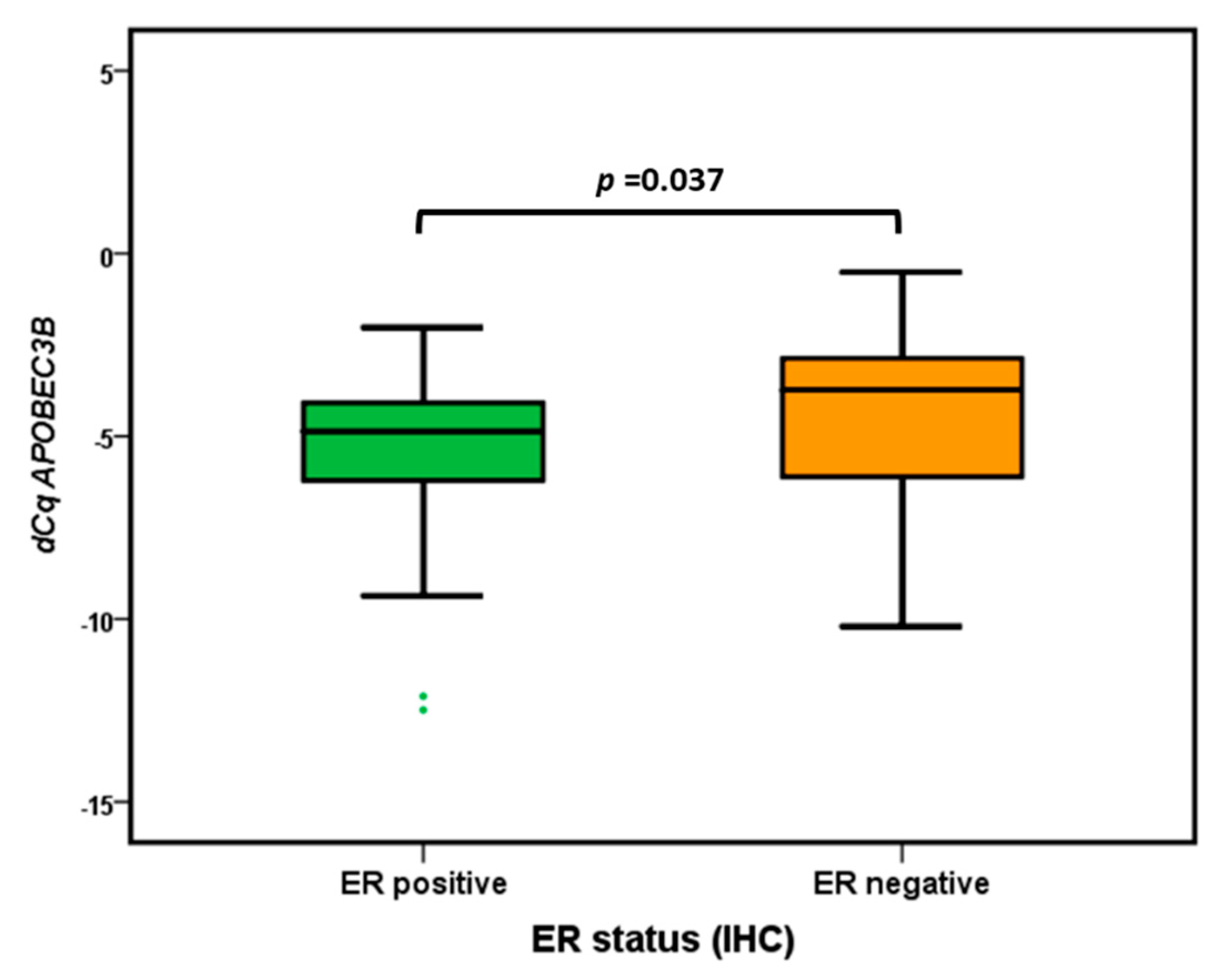
2.2. APOBEC3B Expression in Synchronous Normal, DCIS and IBC Cells

Both the Kruskal-Wallis Test and the Median Test indicated that there was a significant difference (p < 0.001) in APOBEC3B mRNA levels between the normal controls, DCIS and IBC. APOBEC3B mRNA was lower expressed in the normal mammary epithelial tissue adjacent DCIS and IBC (unpaired Mann-Whitney U Test and paired Wilcoxon Signed Ranks Test p < 0.001) (Figure 1). There was no statistically significant difference in APOBEC3B mRNA expression between DCIS and IBC (unpaired Mann–Whitney U Test p = 0.065 (Figure 1), Wilcoxon Signed Ranks Test p = 0.082). (Figure 2).

2.3. APOBEC3B mRNA Subgroup Analysis

Previous studies reported elevated APOBEC3B mRNA levels in breast cancers with otherwise aggressive characteristics, including high histological grade and lack of estrogen expression [7,13,15]. For both DCIS and IBC, there was no correlation between APOBEC3B expression levels and tumor diameter (Spearman Rank Correlation Test p > 0.05) or histological grade (Kruskal-Wallis Test p > 0.05). Our breast cancer subtype analysis showed that the expression of APOBEC3B was the highest in the ER− subgroup (Mann–Whitney U Test, p = 0.037) (Figure 3).

2.4. APOBEC3B Expression in Epithelial Versus Inflammatory Cells

Based on the positive correlation between APOBEC3B and marker for epithelial content (EPCAM) mRNA levels (Spearman Rank Correlation test, p = 0.005 for DCIS, p = 0.001 for IBC), APOBEC3B mRNA was mostly expressed by epithelial cells. Of note, there was no significant difference in the levels of EPCAM mRNA between DCIS and synchronous IBC (Wilcoxon Signed Ranks Test, p = 0.18).
Since inflammatory cells also express APOBEC3B [16], we investigated whether the number of inflammatory cells could have biased our results by comparing Protein Tyrosine Phosphatase Receptor Type C (PTPRC, gene for the common leukocyte antigen CD45) mRNA levels from DCIS and IBC. There was no correlation between APOBEC3B and PTPRC mRNA levels (Spearman Rank Correlation test, p = 0.18 for DCIS and p = 0.29 for IBC). However, IBC expressed slightly higher levels of PTPRC when compared with DCIS (Wilcoxon Signed Ranks Test, p = 0.023).

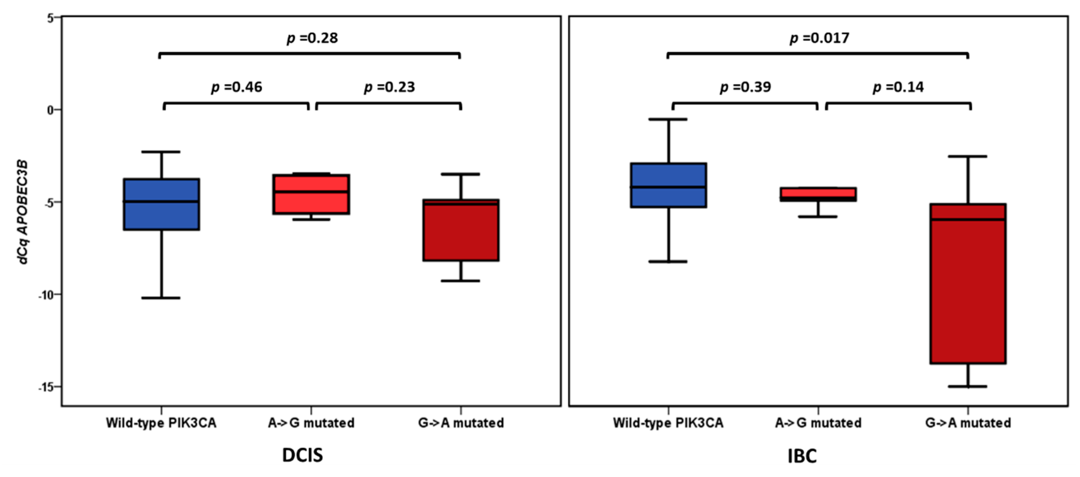
2.5. APOBEC3B Expression and PIK3CA Mutation Status

In a recently published study [17], we detected a PIK3CA somatic hotspot mutation in 24.7% (18 out of 73) patients. For these 18 PIK3CA-positive patients, a significantly higher PIK3CA variant allele frequency (VAF) was detected in the DCIS component (45.8%) when compared with the synchronous IBC component (31.7%) (p = 0.007). For the n = 14 PIK3CA mutation-positive patients (26.4%) included in the current study, a significantly higher PIK3CA VAF was also detected in the DCIS component (52.3%) when compared with the synchronous IBC component (37.2%) (p = 0.027). The correlation of PIK3CA VAF with APOBEC3B showed a negative Spearman Rank correlation in IBC (rs = −0.33, p = 0.001, n = 53). For the DCIS cases, there was no such correlation (rs = 0.02, p = 0.89, n = 53). Analyzing these data irrespective of the degree of the PIK3CA VAF levels revealed that for the 53 patients analyzed in this study, APOBEC3B mRNA levels in IBC were significantly lower in the eight patients with exon 9 (G to A)-mutated PIK3CA when compared with the n = 39 wild-type PIK3CA cases (Mann-Whitney U test p = 0.017). No such difference was observed for the DCIS cases (p = 0.28) (Figure 4). Albeit not statistically significant, APOBEC3B mRNA levels were higher overall in the n = 39 PIK3CA wild-type IBC samples when compared with the PIK3CA wild-type DCIS samples (Mean ± SEM: −4.54 ± 0.36 for IBC versus −5.38 ± 0.35 for DCIS) and lower in the n = 8 G-to-A PIK3CA-mutated IBC samples when compared with G-to-A PIK3CA-mutated DCIS samples (Mean ± SEM: −6.52 ± 1.66 for IBC versus −6.14 ± 0.74 for DCIS). Although the majority of samples with a PIK3CA mutation were ER+, there was no significant interaction effect between ER status and the absence or presence of the two types of tested PIK3CA mutations (p = 0.46 for DCIS and p = 0.20 for IBC).

3. Discussion

APOBEC3B has been identified as an important factor in the evolution of breast cancer [8]. In a recently published pan-tissue, pan-cancer analysis of RNA-seq data specific to the seven APOBEC3 genes in 8951 tumors, 786 cancer cell lines and 6119 normal tissues, APOBEC3B consistently demonstrated its association with proliferative cells and processes, in contrast to other APOBEC3s, especially APOBEC3G and APOBEC3H, which were revealed as more immune cell related [9]. Our current data showed that APOBEC3B mRNA is already upregulated in the in situ stage of breast cancer, which is in line with the high genomic resemblances between DCIS and IBC [18]. In a study we performed earlier, we observed higher mRNA levels of APOBEC3B in breast cancer metastasis as compared to the corresponding primary tumor [14], supporting our hypothesis that, already starting from DCIS, breast cancer progression is associated with deregulated expression of APOBEC3B.
Tumors with upregulated APOBEC3B demonstrate a higher mutational load, which could explain the aggressive behavior of these tumors [7,12]. Two hotspot G-to-A mutations in exon 9 of the—often mutated in breast cancer—PIK3CA gene (E542K and E545K) are thought to be generated by APOBEC3B induced C-to-T (G-to-A) transitions [11]. Whether APOBEC3B is still needed once the mutations are present needs further investigation. In the study of Kosumi et al., APOBEC3B expression in esophageal squamous cell carcinoma was significantly correlated with PIK3CA mutations in exon 9 [10]. However, no correlation was found between APOBEC3B expression and PIK3CA mutations status in a Japanese breast cancer cohort [15]. Although PIK3CA mutations are known to be more prevalent in ER+ cases [19], and thus might have been a confounder in our analysis, we found no significant difference in the distribution of wild-type and mutated PIK3CA in ER+ and ER− cases. In our cohort, APOBEC3B levels were decreased in specifically the G-to-A PIK3CA-mutated IBC samples when compared with wild-type PIK3CA IBC tumors. In the synchronous DCIS counterpart, however, there was no difference in APOBEC3B levels between mutated and PIK3CA wild-type tumors. This might suggest that, in contrast to DCIS, the invasive tumors no longer need APOBEC3B to proliferate and metastasize. Previous studies reported elevated APOBEC3B mRNA levels in breast cancers with otherwise aggressive characteristics, including high histological grade and lack of estrogen expression [7,13,15]. This is consistent with our subgroup analysis, which showed higher APOBEC3B levels in synchronous DCIS and IBC of ER− tumors as compared to ER+ tumors. However, in our study, no significant correlations were found between APOBEC3B levels and histological grade and/or tumor diameter. This could be due to the fact that the majority of our samples were high grade.
This is the first study evaluating APOBEC3B levels within DCIS and co-existing IBC, including different breast cancer subtypes. However, our study has several limitations, such as the relatively small size of our cohort and the analysis of a limited mRNA panel only, with the main focus on APOBEC3B. Since upregulated APOBEC3B is associated with higher mutational load, evaluation of the mutational status of additional markers besides PIK3CA will be interesting. Another limitation is that APOBEC3B is also expressed by inflammatory cells, which could have influenced our data because we performed manual microdissection, and thus contamination with inflammatory cells was not completely avoidable. Although IBC expressed slightly higher levels of PTPRC (the gene for leukocyte antigen CD45) than DCIS, there was no correlation between APOBEC3B and PTPRC mRNA levels. Based on this analysis, it seems unlikely that the number of inflammatory cells biased our data.
Increased insight in molecular mechanisms that contribute to DCIS progression will improve the development of a personalized treatment strategy for patients with DCIS. APOBEC3B could be a potential therapeutic target since it is non-essential, but it has an active enzymatic activity that may be inhibited [7]. Patients with DCIS could therefore benefit from such therapeutic molecules by inhibiting tumor evolution. Concept inhibitors have already been developed for the related enzyme APOBEC3G [20,21]. Additional clinical and pharmaceutical assays are necessary to develop and explore the potential benefit of APOBEC3B inhibitors.

4. Materials and Methods

4.1. Patient Materials

Fifty-three patients with synchronous DCIS and IBC were enrolled. We used coded leftover patient material in accordance with the Code of Conduct of the Federation of Medical Scientific Societies in the Netherlands (http://www.federa.org/codes-conduct). This article is approved by the Medical Ethics Committee of the Erasmus MC (approval number MEC 02.953). According to national guidelines, no informed consent was needed for this study.
Formalin-fixed-paraffin-embedded (FFPE) hematoxylin and eosin (H&E)-stained whole sections of excision specimens were collected and reviewed by two pathologists (Carolien H. M. van Deurzen and Shusma C. Doebar). Histopathological features included the grade of IBC [22], IBC diameter, ER, PR and Her2 status, and grade of DCIS [23]. Tumors were divided into subtypes based on immunohistochemistry (ER, PR and Her2), including the following 5 categories: ER+/PR high/Her2−; ER+/PR− or low/Her2−; ER+/any PR/Her2+; ER−/PR−/Her2+; ER−/PR−/Her2−. ER was considered positive when at least 10% of the tumor cells were positive, irrespective of intensity, according to national guidelines (https://richtlijnendatabase.nl). Low PR was defined as ≤ 20% [24]. Immunohistochemical HER2 expression was scored according to international guidelines [25]. Equivocal cases were evaluated by silver in situ hybridization.

4.2. RT-qPCR

RNA was extracted from tissue areas composed of at least 50% IBC or DCIS cells and analyzed by RT-qPCR as described before [14,17]. In brief, these cells were obtained by microdissection from FFPE tissue, which was performed with a sterile needle under a stereomicroscope. RNA was extracted from these cells using the Qiagen (Hamburg, Germany) AllPrep DNA/RNA FFPE Kit according the manufacturer’s instructions. RNA concentrations were measured with a Nanodrop 2000 system. cDNA was generated from 50 ng/µL cDNA and was generated for 30 min at 48 °C with the RevertAid H minus kit (Thermo Fisher Scientific, Breda, The Netherlands) and gene-specific pre-amplified with Taqman PreAmp Master mix (Thermo Fisher Scientific) for 15 cycles, followed by Taqman probe based real-time PCR according to the manufacturer’s instructions in a MX3000P Real-Time PCR System (Agilent, Amsterdam, The Netherlands). The following intron-spanning gene expression assays (all from Thermo Fisher Scientific) were evaluated: APOBEC3B, assay ID: hs00358981_m1; EPCAM, hs00158980_m1, and PTPRC, hs00236304_m1. Messenger RNA levels were quantified relative to the average expression of 2 reference genes (GUSB, hs9999908_m1 and HMBS, hs00609297_m1) using the delta Cq (average Cq reference genes −Cq target gene) method. According GeNorm and NormFinder, the average of these two reference genes was the most stable expressed across our samples (M-value = 0.59, SD = 0.29). Also, when taking the different groups into account, the inter and intra variation was the lowest for the average of our 2 reference genes (SD = 0.19 for the NormFinder analysis across the control, DCIS and IBC groups and SD = 0.24 for the NormFinder analysis across the ER/PR/Her2 groups). Samples with an average reference gene expression of Cq > 25 were considered to be of insufficient RNA quality and excluded from further analysis, together with their paired samples. A serially diluted RNA pool of FFPE breast tumor samples was included in each experiment to evaluate the linear amplification and efficiencies for all genes included in the panel and absence of amplification in the absence of reverse transcriptase. All gene transcripts were equally efficient amplified (range 94–106%) and were negative in the absence of reverse transcriptase. A summary of the performance of our assays on these serially diluted samples is shown in Supplementary Table S1.

4.3. PIK3CA Mutation Status

PIK3CA mutation status and VAFs were measured as described before [17]. In brief, DNA was extracted from the same micro-dissected FFPE tissues used for RNA extraction using the Qiagen (Hamburg, Germany) AllPrep DNA/RNA FFPE Kit. The SNaPshot Multiplex System for SNP Genotyping (Thermo Fisher Scientific) was used to identify samples positive for PIK3CA hotspot mutations in exon 9 and exon 20. Next, we used digital PCR (dPCR) to validate the SNaPshot results and quantify the relative number of PIK3CA-mutated copies (of E542K, E545K in exon 9 and H1047R and H1047L in exon 20) in both the DCIS and IBC component of those patients with a PIK3CA mutation identified by SNaPshot analysis.

4.4. Statistical Analyses

GeNorm and NormFinder [26,27], present in GenEx qPCR data analysis software (version 6.1, MultiD, Götenborg, Sweden), were used to assess the stability of our reference genes. SPSS version 24 was used for the statistical analyses. Because our APOBEC3B mRNA data were not normally distributed (skewness −1.01 ± 0.33 and −1.75 ± 0.33, kurtosis 0.80 ± 0.64 and 3.60 ± 0.64 for DCIS and IBC, respectively), we only used non-parametric tests. The Wilcoxon Signed Ranks Test was used to compare levels in paired DCIS and IBC and unpaired analyses were performed using the Wilcoxon or Mann-Whitney U Test or the Fisher Exact Probability Test for contingency tabled data. Continuous variables were analyzed by the Spearman Rank Correlation test. p-values ≤ 0.05 were considered statistically significant.

5. Conclusions

In conclusion, our results indicate that APOBEC3B mRNA is similarly upregulated in DCIS and IBC, but declines in PIK3CA-mutated IBC, which suggests that APOBEC3B plays a role in the early stages of breast carcinogenesis. Since APOBEC3B is a gain-of-function mutagenic enzyme, it could be a candidate for therapeutic targeting in an early, non-invasive stage of breast cancer.

Supplementary Materials

The following are available online at https://www.mdpi.com/2072-6694/11/8/1062/s1, Table S1: Performance of the Taqman mRNA assays used in this study.

Author Contributions

Conceptualization, A.M.S., S.C.D. and C.H.M.v.D. Methodology, A.M.S., S.C.D., E.I.V., V.d.W., C.M.B., M.C.A. and C.H.M.v.D.; Formal analysis, A.M.S.; Validation, S.C.D., C.H.M.v.D. Writing—Original Draft Preparation, A.M.S. and S.C.D.; Writing—Review and Editing, A.M.S., S.C.D., J.M.W.M. and C.H.M.v.D.; Visualization, A.M.S.; Supervision, J.W.M.M. and C.H.M.v.D. All co-authors have read and approved of the manuscript and acknowledge their contributions.

Funding

This research was funded by KWF-EMCR-2016-10270 “The role of APOBEC3B in breast cancer therapy resistance”.

Acknowledgments

A.M.S. was supported by Cancer Genomics Netherlands (CGC.nl) funded by the Netherlands Organization for Scientific Research (NWO).

Conflicts of Interest

The authors declare no conflict of interest.

References

  1. Cowell, C.F.; Weigelt, B.; Sakr, R.A.; Ng, C.K.; Hicks, J.; King, T.A.; Reis-Filho, J.S. Progression from ductal carcinoma in situ to invasive breast cancer: Revisited. Mol. Oncol. 2013, 7, 859–869. [Google Scholar] [CrossRef] [PubMed]
  2. Casasent, A.K.; Edgerton, M.; Navin, N.E. Genome evolution in ductal carcinoma in situ: Invasion of the clones. J. Pathol. 2017, 241, 208–218. [Google Scholar] [CrossRef] [PubMed]
  3. Kim, S.Y.; Jung, S.H.; Kim, M.S.; Baek, I.P.; Lee, S.H.; Kim, T.M.; Chung, Y.J.; Lee, S.H. Genomic differences between pure ductal carcinoma in situ and synchronous ductal carcinoma in situ with invasive breast cancer. Oncotarget 2015, 6, 7597–7607. [Google Scholar] [CrossRef] [PubMed]
  4. Casasent, A.K.; Schalck, A.; Gao, R.; Sei, E.; Long, A.; Pangburn, W.; Casasent, T.; Meric-Bernstam, F.; Edgerton, M.E.; Navin, N.E. Multiclonal Invasion in Breast Tumors Identified by Topographic Single Cell Sequencing. Cell 2018, 172, 205–217.e12. [Google Scholar] [CrossRef] [PubMed] [Green Version]
  5. Hernandez, L.; Wilkerson, P.M.; Lambros, M.B.; Campion-Flora, A.; Rodrigues, D.N.; Gauthier, A.; Cabral, C.; Pawar, V.; Mackay, A.; A’Hern, R.; et al. Genomic and mutational profiling of ductal carcinomas in situ and matched adjacent invasive breast cancers reveals intra-tumour genetic heterogeneity and clonal selection. J. Pathol. 2012, 227, 42–52. [Google Scholar] [CrossRef] [PubMed] [Green Version]
  6. Smith, H.C.; Bennett, R.P.; Kizilyer, A.; McDougall, W.M.; Prohaska, K.M. Functions and regulation of the APOBEC family of proteins. Semin. Cell Dev. Biol. 2012, 23, 258–268. [Google Scholar] [CrossRef] [PubMed]
  7. Roberts, S.A.; Lawrence, M.S.; Klimczak, L.J.; Grimm, S.A.; Fargo, D.; Stojanov, P.; Kiezun, A.; Kryukov, G.V.; Carter, S.L.; Saksena, G.; et al. An APOBEC cytidine deaminase mutagenesis pattern is widespread in human cancers. Nat. Genet. 2013, 45, 970–976. [Google Scholar] [CrossRef]
  8. Harris, R.S. Molecular mechanism and clinical impact of APOBEC3B-catalyzed mutagenesis in breast cancer. Breast Cancer Res. 2015, 17, 8. [Google Scholar] [CrossRef]
  9. Ng, J.C.F.; Quist, J.; Grigoriadis, A.; Malim, M.H.; Fraternali, F. Pan-cancer transcriptomic analysis dissects immune and proliferative functions of APOBEC3 cytidine deaminases. Nucleic Acids Res. 2019, 47, 1178–1194. [Google Scholar] [CrossRef] [Green Version]
  10. Kosumi, K.; Baba, Y.; Ishimoto, T.; Harada, K.; Nakamura, K.; Ohuchi, M.; Kiyozumi, Y.; Izumi, D.; Tokunaga, R.; Taki, K.; et al. APOBEC3B is an enzymatic source of molecular alterations in esophageal squamous cell carcinoma. Med. Oncol. 2016, 33, 26. [Google Scholar] [CrossRef]
  11. Henderson, S.; Chakravarthy, A.; Su, X.; Boshoff, C.; Fenton, T.R. APOBEC-mediated cytosine deamination links PIK3CA helical domain mutations to human papillomavirus-driven tumor development. Cell Rep. 2014, 7, 1833–1841. [Google Scholar] [CrossRef] [PubMed]
  12. Burns, M.B.; Lackey, L.; Carpenter, M.A.; Rathore, A.; Land, A.M.; Leonard, B.; Refsland, E.W.; Kotandeniya, D.; Tretyakova, N.; Nikas, J.B.; et al. APOBEC3B is an enzymatic source of mutation in breast cancer. Nature 2013, 494, 366–370. [Google Scholar] [CrossRef] [PubMed]
  13. Sieuwerts, A.M.; Willis, S.; Burns, M.B.; Look, M.P.; Meijer-Van Gelder, M.E.; Schlicker, A.; Heideman, M.R.; Jacobs, H.; Wessels, L.; Leyland-Jones, B.; et al. Elevated APOBEC3B correlates with poor outcomes for estrogen-receptor-positive breast cancers. Horm. Cancer 2014, 5, 405–413. [Google Scholar] [CrossRef]
  14. Sieuwerts, A.M.; Schrijver, W.A.; Dalm, S.U.; de Weerd, V.; Moelans, C.B.; Ter Hoeve, N.; van Diest, P.J.; Martens, J.W.; van Deurzen, C.H. Progressive APOBEC3B mRNA expression in distant breast cancer metastases. PLoS ONE 2017, 12, e0171343. [Google Scholar] [CrossRef] [PubMed]
  15. Tsuboi, M.; Yamane, A.; Horiguchi, J.; Yokobori, T.; Kawabata-Iwakawa, R.; Yoshiyama, S.; Rokudai, S.; Odawara, H.; Tokiniwa, H.; Oyama, T.; et al. APOBEC3B high expression status is associated with aggressive phenotype in Japanese breast cancers. Breast Cancer 2016, 23, 780–788. [Google Scholar] [CrossRef] [PubMed]
  16. Siriwardena, S.U.; Chen, K.; Bhagwat, A.S. Functions and Malfunctions of Mammalian DNA-Cytosine Deaminases. Chem. Rev. 2016, 116, 12688–12710. [Google Scholar] [CrossRef] [PubMed] [Green Version]
  17. Agahozo, M.C.; Sieuwerts, A.M.; Doebar, S.C.; Verhoef, E.; Beaufort, C.M.; Ruigrok-Ritstier, K.; de Weerd, V.; Sleddens, H.; Dinjens, W.N.; Martens, J.; et al. PIK3CA mutations in ductal carcinoma in situ and adjacent invasive breast cancer. Endocr. Relat. Cancer 2019, 26, 471–482. [Google Scholar] [CrossRef] [PubMed]
  18. Aubele, M.; Mattis, A.; Zitzelsberger, H.; Walch, A.; Kremer, M.; Welzl, G.; Hofler, H.; Werner, M. Extensive ductal carcinoma In situ with small foci of invasive ductal carcinoma: Evidence of genetic resemblance by CGH. Int. J. Cancer 2000, 85, 82–86. [Google Scholar] [CrossRef]
  19. Pang, B.; Cheng, S.; Sun, S.P.; An, C.; Liu, Z.Y.; Feng, X.; Liu, G.J. Prognostic role of PIK3CA mutations and their association with hormone receptor expression in breast cancer: A meta-analysis. Sci. Rep. 2014, 4, 6255. [Google Scholar] [CrossRef]
  20. Olson, M.E.; Harris, R.S.; Harki, D.A. APOBEC Enzymes as Targets for Virus and Cancer Therapy. Cell Chem. Biol. 2018, 25, 36–49. [Google Scholar] [CrossRef]
  21. Li, M.; Shandilya, S.M.; Carpenter, M.A.; Rathore, A.; Brown, W.L.; Perkins, A.L.; Harki, D.A.; Solberg, J.; Hook, D.J.; Pandey, K.K.; et al. First-in-class small molecule inhibitors of the single-strand DNA cytosine deaminase APOBEC3G. ACS Chem. Biol. 2012, 7, 506–517. [Google Scholar] [CrossRef] [PubMed]
  22. Elston, C.W.; Ellis, I.O. Pathological prognostic factors in breast cancer. I. The value of histological grade in breast cancer: Experience from a large study with long-term follow-up. Histopathology 1991, 19, 403–410. [Google Scholar] [CrossRef] [PubMed]
  23. Consensus Conference Committee. Consensus Conference on the classification of ductal carcinoma in situ. Thomas Jefferson University in Philadelphia, Pennsylvania, USA, Nov 1. Cancer 1997, 80, 1798–1802. [Google Scholar] [CrossRef]
  24. Prat, A.; Cheang, M.C.; Martín, M.; Parker, J.S.; Carrasco, E.; Caballero, R.; Tyldesley, S.; Gelmon, K.; Bernard, P.S.; Nielsen, T.O.; et al. Prognostic significance of progesterone receptor-positive tumor cells within immunohistochemically defined luminal A breast cancer. J. Clin. Oncol. 2013, 31, 203–209. [Google Scholar] [CrossRef] [PubMed]
  25. Wolff, A.C.; Hammond, M.E.; Hicks, D.G.; Dowsett, M.; McShane, L.M.; Allison, K.H.; Allred, D.C.; Bartlett, J.M.; Bilous, M.; Fitzgibbons, P.; et al. Recommendations for human epidermal growth factor receptor 2 testing in breast cancer: American Society of Clinical Oncology/College of American Pathologists clinical practice guideline update. J. Clin. Oncol. 2013, 31, 3997–4013. [Google Scholar] [CrossRef] [PubMed]
  26. Andersen, C.L.; Jensen, J.L.; Orntoft, T.F. Normalization of real-time quantitative reverse transcription-PCR data: A model-based variance estimation approach to identify genes suited for normalization, applied to bladder and colon cancer data sets. Cancer Res. 2004, 64, 5245–5250. [Google Scholar] [CrossRef] [PubMed]
  27. Wang, Q.; Ishikawa, T.; Michiue, T.; Zhu, B.L.; Guan, D.W.; Maeda, H. Stability of endogenous reference genes in postmortem human brains for normalization of quantitative real-time PCR data: Comprehensive evaluation using geNorm, NormFinder, and BestKeeper. Int. J. Legal Med. 2012, 126, 943–952. [Google Scholar] [CrossRef] [PubMed]
Figure 1. Boxplots of APOBEC3B mRNA expression levels in paired normal, DCIS and IBC (n = 53). Differences between normal, DCIS and IBC were analyzed by the Mann-Whitney U test.
Cancers 11 01062 g001
Figure 2. APOBEC3B expression levels in paired DCIS and IBC (n = 53). Wilcoxon Signed Ranks Test p = 0.082.
Cancers 11 01062 g002
Figure 3. Boxplots of Apolipoprotein B mRNA Editing Enzyme, Catalytic Polypeptide-Like 3B (APOBEC3B) mRNA expression levels according to ER status. The difference between ER+ and ER− cases was analyzed by the Mann-Whitney U test.
Cancers 11 01062 g003
Figure 4. Boxplots of APOBEC3B mRNA expression levels according PIK3CA mutation status. The differences between wild-type (blue boxes) and mutated (red boxes) PIK3CA cases were analyzed by the Mann-Whitney U test.
Cancers 11 01062 g004
Table 1. Clinicopathological features of patients with ductal carcinoma in situ (DCIS) and adjacent invasive breast cancer (IBC) (n = 53).
Characteristicn(%)
Age at diagnosis53
years, median (range)(28–102)
Type of surgery
Breast-conserving surgery2445.3
Mastectomy2954.7
Grade DCIS
1149.1
21939.6
3337.5
Grade IBC
1149.1
22139.6
3317.5
Tumor size
≤2 cm2849.1
>2–5 cm2139.6
>5 cm47.5
Missing03.8
Subtypes based on immunohistochemistry
ER+/PR high/Her2−1324.5
ER+/PR− or low/Her2−1222.6
ER+/any PR/Her2+1120.8
ER−/PR−/Her2+815.1
ER−/PR−/Her2−917.0

