Review

Mucosal Injury during Anti-Cancer Treatment: From Pathobiology to Bedside

1
Department of Medicine (DAME), University of Udine, 33100 Udine, Italy
2
Department of Medical Oncology, Centro di Riferimento Oncologico di Aviano (CRO), IRCCS, 33081 Aviano, Italy
3
Intensive Care Unit, Centro di Riferimento Oncologico di Aviano (CRO), IRCCS, 33081 Aviano, Italy
*
Author to whom correspondence should be addressed.
Cancers 2019, 11(6), 857; https://doi.org/10.3390/cancers11060857
Submission received: 14 May 2019 / Revised: 10 June 2019 / Accepted: 12 June 2019 / Published: 20 June 2019

Abstract

:
Mucositis is one of the most common debilitating side effects related to chemotherapy (CT), radiation therapy (RT), targeted agents and immunotherapy. It is a complex process potentially involving any portion of the gastrointestinal tract and injuring the mucosa, leading to inflammatory or ulcerative lesions. Mechanisms and clinical presentation can differ according both to the anatomic site involved (oral or gastrointestinal) and the treatment received. Understanding the pathophysiology and management of mucosal injury as a secondary effect of anti-cancer treatment is an important area of clinical research. Prophylaxis, early diagnosis, and adequate management of complications are essential to increase therapeutic success and, thus, improve the survival outcomes of cancer patients. This review focuses on the pathobiology and management guidelines for mucositis, a secondary effect of old and new anti-cancer treatments, highlighting recent advances in prevention and discussing future research options.

1. Introduction

Mucositis is one of the most common debilitating side effects related to chemotherapy (CT), radiation therapy (RT), targeted agents and immunotherapy [1,2]; it occurs in 20–40% of the patients receiving anti-cancer treatments for solid tumors, 60–80% of the ones undergoing hematopoietic stem cell transplantation (HSCT), and is experienced by almost all patients receiving RT for head and neck cancers (HNC) [3,4,5]. Mucositis is a complex process that leads to inflammatory and/or ulcerative lesions. It can potentially involve any portion of gastrointestinal tract and injures mucosa through the interplay of the epithelial, mesenchymal and immune cells. The specific mechanism of this process, along with the clinical presentation, can differ according both to the anatomic site involved (oral or gastrointestinal) and the causal anti-cancer therapy [6,7,8].
Oral mucositis (OM) is characterized by erythema, ulceration, pain and, eventually, bleeding; the loss of integrity of the oral mucosa can favor local and systemic infections that can compromise nutrition and fluid intake [3,9,10]. On the other hand, gastrointestinal mucositis (GIM) is sometimes responsible for nausea, vomiting and diarrhea. The onset, timing and clinical presentation are also influenced by patient-related risk factors. Among these, age, ethnicity, gender, and disorders such as malnutrition and poor oral health can increase the risk of developing mucositis [6,7,8,11,12]. Moreover, patients with underlying systemic illnesses, such as autoimmune diseases or diabetes, are more likely to have an altered tissue environment, which can further predispose them to mucosal damage [3].
Many studies have shown that the risk of mucositis increases along with the intensity of the therapy. Furthermore, combination treatment (e.g., RT with concurrent CT used for HNC) may boost the onset of mucositis [13]. An adequate classification of lesions is crucial to the ability to choose the most suitable management and, eventually, appropriate dose modifications of cancer therapy. Understanding the pathophysiology and management of mucosal injury as a secondary effect of anti-cancer treatment is essential to increase therapeutic success and, thus, improve the survival outcomes of cancer patients.
Mucositis may have a severe impact on a patient’s quality of life (QoL) and negatively influences the ability to maintain the proper schedule and intensity of anti-cancer treatment. Moreover, it has a non-negligible economic effect due to the cost of care [14].
This review focuses on the pathobiology and management guidelines for mucositis as a secondary effect of anti-cancer treatment, highlighting recent advances in prevention and discussing future research options. Studies evaluating the pathogenesis of “classical mucositis” induced by CT and RT, pathogenesis of new anti-cancer treatment, risk factors associated with mucositis, prophylaxis, clinical presentation and management of classical and new anti-cancer treatments were included.

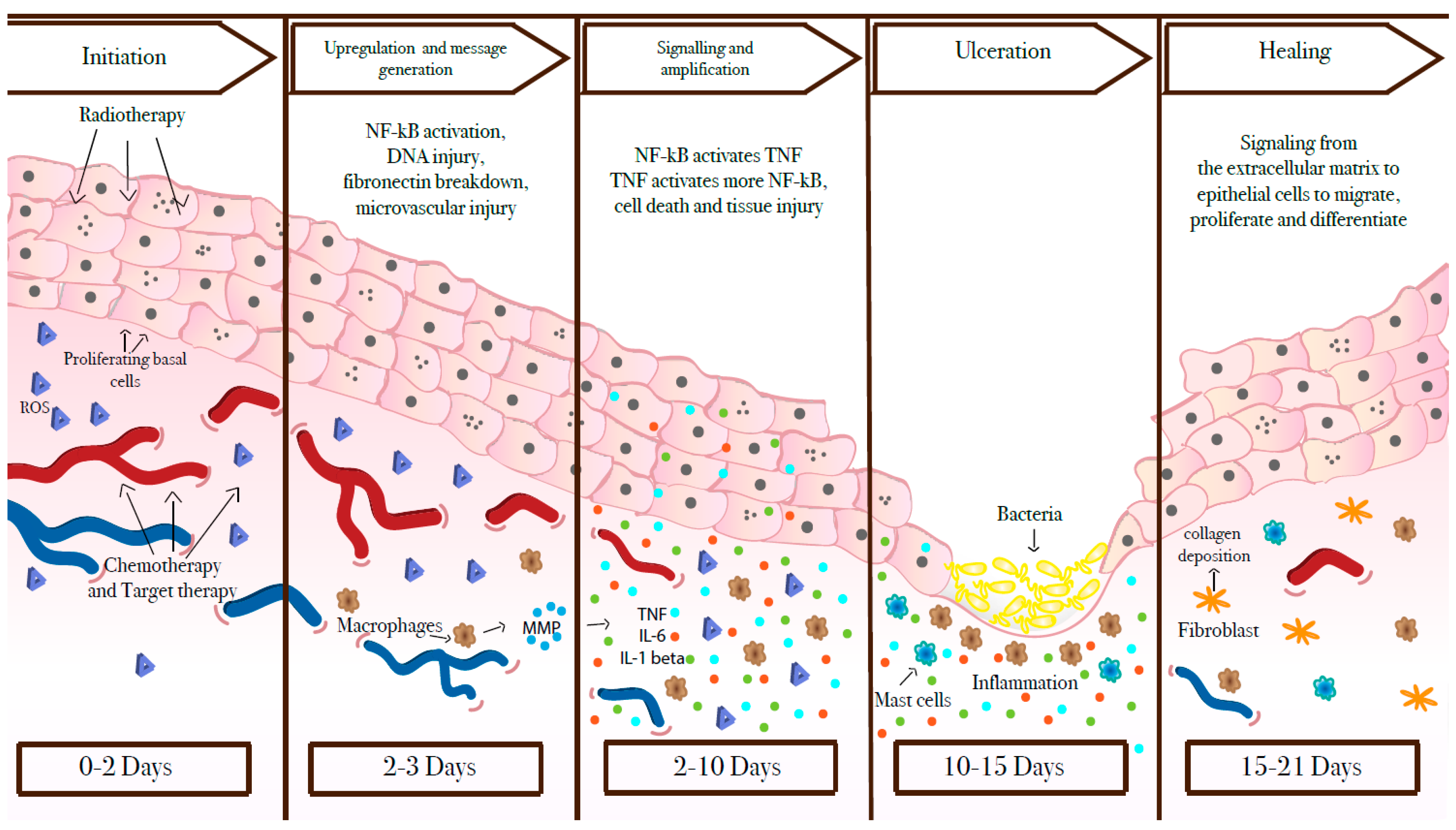
2. What Lies Beneath?

Five stages of pathogenesis have been identified for mucositis: initiation, upregulation/activation, signal amplification, ulceration, and healing (Figure 1) [7,13].
Initiation: This phase is triggered by exposure to anti-cancer procedures, which induces direct damage to the DNA, oxidative stress and, consequently, generates reactive oxygen species (ROS). The injury to epithelial, submucosal and endothelial cells provokes the release of endogenous damage-associated pattern molecules (CRAMPs) [15].
Upregulation/activation: ROS, innate immune response and binding of CRAMPs to receptors propagate further the damage of the cell membranes and activate several transcriptional pathways [15,16]; among these, one of the best known is the pathway of nuclear factor NF-κB [17,18,19]. This latter, once activated, induces an increase in production of pro-inflammatory cytokines, such as tumor necrosis factor-α (TNF-α), interleukin (IL)-6, stress responders such as cyclooxygenase-2 (COX-2), and cytokine modulators by fibroblasts and endothelial cells, which, in turn, can lead to cell apoptosis [20,21,22,23]. In this phase, there is also an increased expression of genes related to adhesion molecules and angiogenesis [18,19].
Amplification: The primary response initiated during the first two phases triggers the expression of several molecules that, in turn, influence local response. NF-κB can be up-regulated by TNF-α, which can initiate mitogen-activated protein kinase (MAPK) signaling, leading to activation of Jun N-terminal kinase signaling. Apoptotic pathways are activated on submucosal and basal epithelial cells causing mucosal ulceration and atrophic transformations. Metalloproteinases (MMPs) too can be dysregulated leading to important pathogenic effects [22,24].
Ulceration: In this phase, clinically symptomatic deep ulcerations appear. In OM, secondary to CT administered by a bolus schedule, the time lapse between initial injury and clinical damage is about 4 days; shortly thereafter, bacterial colonization from both gram-negative and gram-positive bacteria happens; at the same time, CT-induced leukopenia favors progression of the infection. Differently, GIM becomes clinically evident much sooner, within 1–2 days after CT administration.
Healing: In the last stage, there is spontaneous healing of the ulcers, but an increased risk of recurrence due to residual angiogenesis may persist. This final stage is characterized by epithelial proliferation, migration, and differentiation stimulated by the extracellular matrix, and the simultaneous restoration of the local microbial flora [15,25].

3. Risk Factors

Like other adverse (treatment-related) events (AEs), the onset of mucositis is influenced by the features of the anti-cancer regimen, as well as by the patient’s characteristics. Patient-related risk factors include genetic polymorphisms, systemic comorbidities, and other general host-related risk factors. The impact of gender on the risk of GIM is well established for patients exposed to 5-fluorouracil (5-FU)-based CT, the risk for women is higher than for men [26,27,28]. Patients in extreme age groups appear to be at higher risk of developing OM, as demonstrated by increased incidence in pediatric and older patients [11,29]. In addition, the elderly seem to be at higher risk for suffering more severe mucositis [30]. Body composition can play a role in drug metabolism and patients with a low, lean body mass, women and other patients with a low BMI, can be exposed to higher levels of drugs and, as a consequence, develop more severe toxicities [31].

3.1. Anti-Cancer Treatment

The spectrum of the pathobiology, onset, duration and severity, is particularly wide during anti-cancer treatment, differing according to the adopted regimen, dosage, and delivery schedule. Moreover, risk and grade intensify with the proceeding of treatment for a cumulative effect. More details are provided in the next section.

3.2. Genetic Polymorphisms

Polymorphisms in genes codifying for enzymes engaged in drug metabolism have been linked to an increased risk of developing treatment-related toxicities. A higher blood level of 5-FU in patients with dihydropyrimidine dehydrogenase (DPD) deficiency can result in severe or fatal myelosuppression and mucositis [32]. Even patients with polymorphism of the thymidylate synthase (TYMS) gene tend to have more severe toxicity from 5-FU [33]. Patients who carry the 677TT genotype for methylentetrahydrofolate reductase (MTHFR) experience a more severe mucositis when treated with methotrexate for HCT [34]. In addition, polymorphisms in cytokine genes, like TNF, have been associated with a higher severity of CT-related toxicities, including mucositis, in a Japanese population treated with cisplatin and 5-FU [35]. Patients with systemic disease characterized by an increased (e.g., Addison disease) or a reduced apoptosis’s rate (e.g., psoriasis) seem to be, respectively, at higher and lower risk of developing mucositis [36].
More recently, six pathways were identified as being associated with mucositis: PTK6 signaling and the Wnt signaling pathway, two pathways associated with transforming the growth factor (TGF)-b, ERK signaling and the inflammatory response pathway. Of these, the first two pathways seem more specific to mucositis, while the other four are involved in the development of other ailments such as IBS. PTK6 is normally expressed in intestinal cells and is thought to play a role in epithelial barrier function. Suppression of PTK6 is associated with an increase in apoptosis of proliferating cells. On the other side, the Wnt pathways interact with the NK-kB pathway and could be involved in immune response and antigen presentation. Activation of the TGF-b signaling pathway is associated with treatment-related injury, and its regulation through Smad7 or inhibitions of b1 integrins changes the subsequent development of mucositis. The ERK signaling pathway acts in conjunction with extra- and intracellular signaling events, while the inflammatory response pathway is likely linked to the activation of the innate immune response. It is interesting to note how these pathways are shared with diseases with similar clinical presentation (such as IBS), therefore lending strength to the hypothesis that risk for development of a certain clinical phenotype (i.e., diarrhea) is related to a patient specific and individual genomic profile [37].

3.3. Role of Microbial Flora

There is no conclusive evidence that oral and intestinal microbial flora play an active role in the development of mucositis. However, it is known that microbiota can stimulate the production of pro-inflammatory cytokines [38]. At the same time, probiotic bacteria can promote the production of cytoprotective pathways [39]. Moreover, changes in the composition of luminal microbiota can result in proliferation of pathogenic species, thus providing a chance for bacteremia and sepsis through the disruption of the epithelium following a decrease in the immune response. However, microbiota play a key role in activating immune cells including natural killer (NK) cells, mast cells, macrophages and dendritic cells by recognizing pathogen-associated molecular patterns (PAMPS). Then, Toll-like receptors (TRL) recognize these molecules, which activate NF-κB and stimulate the production of pro-inflammatory cytokines. These may, in turn, recruit leukocytes and stimulate them to further secrete more pro-inflammatory cytokines [40]. During anti-cancer treatment, microbial composition may become imbalanced and, thus, the physiological microbiota may become underrepresented (dysbiosis) [41]. The entity and type of variations in microbiota composition differ along the gastrointestinal tract, according to the CT regimen, schedule and doses. Notably, these changes are potentially involved in initiating and exacerbating mucosal impairment.

3.4. Role of the Innate and Adaptive Response in Mucosal Injury

Immune response is involved in maintaining pathophysiological homeostasis. However, it could also induce regimen-related toxicities during anti-cancer therapy. Both in OM and GIM, a key role is played by the innate immune system through the recruitment of inflammatory cells and the interplay among epithelium, dendritic cells, and macrophages [42,43]. More in detail, the anticancer treatment could activate cell injury through the generation of damage-associated molecular patterns (DAMPs) or PAMPs identified by pattern recognition receptors (PRRs), thus increasing pro-inflammatory cytokine production [44,45,46]. Furthermore, GIM alterations in epithelial intestinal barriers promote a rearrangement of microbial flora and bacterial translocation to the intestinal lamina propria, which can enhance neutrophil recruitment and tissue impairment [45,46]. However, even antibody-mediated and cell-mediated immunotoxicity triggered by adaptive immune response are responsible for tissue damage. Guabiraba et al. showed that neutrophils were highly expressed in the blood three days after irinotecan treatment in mice. Moreover, even macrophages play an important role in CT-induced mucositis [47]. Recent evidence showed that T regulatory cells played a critical role in controlling irinotecan-related GIM [48]. Moreover, inflammasome influenced secretion of IL-33 at the level of the intestinal epithelial cells of mice treated with irinotecan. Treatment of mice with an anti-IL33 resulted in attenuation of intestinal damage [49].

4. Prophylaxis

Several approaches are currently available for OM and/or GIM prophylaxis [7]. The MASCC/ISOO and European Society of Medical Oncology (ESMO) guidelines provide a systematic review of the recommended or suggested preventive interventions during cancer treatments (Table 1) [50,51].

4.1. Basic Oral Care and Topic Therapies for OM and mTOR-Induced-Mucositis Prophylaxis

The first preventive measure checked during cancer treatments is the daily oral hygiene routine, evaluated with a baseline dentist evaluation in order to remove potential sources of infection or trauma. The recommended oral care protocol should include a soft toothbrush twice a day, flossing, and alcohol-free mouthwashes after oral hygiene (with bland 0.9% saline, sodium bicarbonate or plain water). Additionally, dietary and nutritional restrictions should reduce potential irritating stimuli, such as hot and spicy food, dehydration, alcohol and smoking [51,54]. Taking into account specific topical therapies, oral rinses with benzydamine HCl, a non-steroidal anti-inflammatory agent, was shown to be effective in preventing OM in patients with HNC receiving exclusive RT [55]. Similarly, dexamethasone-containing mouthwashes (0.5 mg/5 mL oral solution) are specifically recommended for prophylaxis of mTOR (mammalian target of rapamycin)-induced-mucositis in patients with metastatic breast cancer, even if a proper randomized, placebo-controlled trial is still missing [52].

4.2. Physical Therapies for OM Prophylaxis

Several trials have demonstrated the efficacy of oral cryotherapy (intraoral ice-chip therapy) for the prevention of 5-FU induced stomatitis [56,57,58]. As shown by a recent Cochrane meta-analysis, including five trials and 444 patients treated with short-term 5-FU bolus, oral cryotherapy reduced the incidence of all grades and, specifically, severe OM (RR 0.61, 95% CI 0.52–0.72 and RR 0.40, 95% CI 0.27–0.61, respectively) [59]. Moreover, the same meta-analysis examined the data of an additional 271 patients receiving high-dose melphalan-based CT before HSCT, which showed a reduction in all grades of mucositis (RR 0.59, 95% CI 0.35–1.01) and a significant reduction in severe mucositis (RR 0.38, 95% CI 0.20–0.72) [59]. Since both drugs have a short duration, a possible mechanism of action relies on cold-induced vasoconstriction, which could reduce mucosal delivery of CT and therefore its potential local toxicity. Another physical technique investigated for OM prevention was low level laser therapy. It was mainly evaluated in two clinical settings: HSCT [60,61,62,63] and RT for patients with HNC [50,51]. Numerous trials provided consistent evidence that a mucosal pre-treatment with a helium-neon laser was likely to reduce mucosal injury and to promote the epithelial healing process [50,51]. However, there was no consensus on the optimal wavelength, possible detrimental biological effects or on its cost–benefit value [62,64].

4.3. Systemic Therapies for OM and GIM Prophylaxis

Mucosal injury can be approached from a preventive systemic point of view. Oral zinc supplements have shown activity in preventing OM in patients with oral cancer who are candidates for CT-RT or RT [50,65,66].
Another preventive option is represented by amifostine, an organic phosphorylated aminothiol administered intravenously, able to recruit ROS scavengers, thus protecting the normal epithelium and connective tissue [67]. The FDA indication for amifostine refers to xerostomia prophylaxis in post-operative HNC patients treated with RT, with conflicting data on OM prevention [53,68]. Its preventive use is also recommended for RT-induced proctitis and esophagitis [50]. Unfortunately, significant side-effects (mainly nausea and hypotension) limited its prescription in clinical practice. Additionally, sulfasalazine, a salicylate used in inflammatory bowel disease management, may be indicated as preventive remedy for RT-induced enteropathy (500 mg orally twice a day) in patients receiving pelvic RT [50,51]. On the contrary, similar compounds such as acetylsalicylic acid or mesalazine have shown no role in this setting.
Finally, despite the lack of definitive data on their mechanisms of action [69], probiotic agents containing the lactobacillus species may be of value for diarrhea prophylaxis in patients with pelvic malignancy exposed to CT or RT [50,51], and they are currently being evaluated for prevention of CT-RT-induced OM (NCT01707641) and irinotecan-induced diarrhea (NCT02819960).

5. Across Old and New Anti-Cancer Treatments

5.1. Chemotherapy Induced Mucositis

Approximately 40% of CT treated patients develop mucositis. The incidence is conditioned by the regimen; antimetabolites (5-FU, capecitabine and S-1), anthracyclines, irinotecan and taxanes are drugs leading to a higher rate of mucositis [1]. Among antimetabolites, S-1 and capecitabine carry a lower risk of mucositis than 5-FU [70]. For regimens such as docetaxel, cisplatin and fluorouracil (TPF), and in combination treatments (such as RT-CT for HNC), OM occurs in over 50% of patients. Mucin reduction seems to be one of the mechanisms underlying OM in platinum-based CT.
As for GIM, even though the etiology of the cellular damage induced by different CT drugs differs, all pathways ultimately converge in the shortening of crypt length, dampening and fusion of villi, enterocyte hyperplasia and increased apoptosis (more commonly located in the small bowel). A role of pro-inflammatory cytokines and proteins involved in apoptosis regulation has been suggested by many studies evaluating diverse cytotoxic agents (5-FU, methotrexate an irinotecan) [16,46].
The pathobiological mechanisms of GIM are similar to those that promote the development of OM; such mechanisms include disruption of tight junctions and matrix metalloproteinase-mediated connective tissue impairment [71,72].
One of the chemotherapeutic drugs more extensively studied in this regard is irinotecan. Irinotecan is a topoisomerase inhibitor that seems to induce mucositis by activating caspases and p53, downregulating the PI3K/Akt pathway, and promoting the MAPK and PKC pathways, which in turn induce specific effects, such as the reduction in goblet cell number and mucin hypersecretion, which contribute to amplification of the magnitude of diarrhea [73]. There are two main clinical presentations of GIM during irinotecan treatment. Activation of the parasympathetic system, the subsequent inhibition of acetylcholinesterase and the release of acetylcholine lead to cholinergic syndrome and early-onset diarrhea. Conversely, both changes in intestinal motility and direct damage to the mucosa induced by cytokines and inflammatory-mediated effects contribute to late-onset diarrhea [46]. GIM occurs more frequently with a combination of irinotecan and fluoropyrimidines, mainly with capecitabine [70]. In fact, patients treated with the capecitabine and irinotecn (XELIRI). regimen reported higher gastrointestinal toxicity than with fluorouracil and irinotecan (FOLFIRI). For regimens such as fluorouracil and oxaliplatin (FOLFOX) or FOLFIRI, GIM is reported to be 50% and 89% respectively [74].
Notably, GIM induced by oxaliplatin and carboplatin tends to have a lower grade toxicity compared to cisplatin [75]. Taxane treatment induces mild or moderate mucositis in 29–63% of patients, mainly in those treated with docetaxel.
Clinical presentation: OM appears shortly after the first cycle with gradual recovery 2–3 weeks after the discontinuation of treatment [8]. The clinical course may be protracted when complicated by infection, in particular if associated with severe neutropenia [6]. Nausea, vomiting, dysphagia and dyspepsia, with or without pain, can be caused either by infections such as candidosis or, less commonly, as a direct effect of treatment [76]. GIM is usually acute, with rapid onset of diarrhea (generally within 24–48 h of treatment), abdominal pain, nausea, vomiting, anorexia and, in severe cases, weight loss, dehydration and sepsis [24,77].
Management: Different strategies have been tested and are currently under evaluation for the treatment of CT-induced mucositis. However, few agents have been approved while for most of them the evidence is not sufficient to establish a standard therapy [78].
In standard-dose CT-induced OM, studies have failed to determine a benefit of chlorhexidine. No significant difference in the ratings and duration of pain was observed in a double-blind clinical trial conducted on 23 patients receiving CT and evaluating the effectiveness of a standardized oral care protocol (PRO-SELF) plus mouthwash, salt and soda rinses, and chlorhexidine [79].
The efficacy of sucralfate for established OM needs further evidence. Sucralfate is an aluminum salt that protects mucosa from mechanical damage. It also prevents the release of inflammatory cytokines and stimulates angiogenesis, fibroblast, and epidermal cell proliferation contributing to tissue repair. Its beneficial effect has only been observed as prophylaxis for patients treated with 5-FU [80].
Topical vitamin E could be beneficial in reducing the severity of OM but no therapeutic gain would be achieved by using systemic vitamin E in this setting [81].
The increasing knowledge of mechanisms underlying mucositis allows us to consider the use of antioxidant agents as a potential interventional method.
In regard to GIM, the efficacy of octreotide after loperamide failure in 5-FU-induced diarrhea has been assessed in multiple clinical trials [82,83,84,85,86]. Octreotide is a somatostatin analogue that regulates intestinal water and electrolyte transport, inhibits the release of gastrointestinal hormones and plays a role in the preservation of the epithelial barrier. In patients who develop loperamide-refractory diarrhea after standard or high-dose CT for HSCT, octreotide is recommended at a dose of at least 100 μg, administered subcutaneously twice daily [50].

5.2. Radiotherapy-Induced Mucositis

Mucositis is a common side effect in patients treated with RT and CT-RT. Although CT and RT-induced mucositis share the same pathobiological mechanisms, the timing of cellular damage and the onset of symptoms differ; RT induces damage within a few seconds of first exposure and this damage is reiterated in time due to the fractionated schedule. Clinically, symptoms stemming from atrophic changes, such as erythema and soreness, start at the end of the first week of exposure; this is followed, as the cumulative dose increases, by ulceration, which can last for up to 6 weeks after completion of RT [8]. In patients with HNC undergoing concomitant treatment with RT associated with cetuximab, a p16-negative status seems to be associated with a higher rate of grade 3–4 OM [87]. Also the increase of salivary concentration of cytokines (in particular, IL-6 and IL-1β) seems to be related to the severity of mucositis, although there is no relation to baseline concentration [88].
Clinical presentation: Nearly all HNC patients undergoing RT develop some degree of OM. Asymptomatic redness progresses to elevated desquamative patches, which evolve into large, painful contiguous pseudo-membranous lesions associated with dysphagia and, subsequently, a decrease in oral intake [89]. Oral lesions usually appear during the first weeks of treatment and do not disappear until 4–5 weeks after the last administration. The rate and severity of these toxicities increase with concomitant CT-RT [90].
Usually, proctitis occurs after RT (either on its own or in combination with CT) in rectal or other pelvic cancers. The onset can be acute (during or within six weeks after RT)—with diarrhea, tenesmus, urgency and mucus discharge—or chronic—with bleeding, symptoms of obstructed defecation and rectal pain [76].
Management: Lacking or conflicting evidence does not allow us to provide guidelines for mouth rinses to treat OM in HNC patients receiving RT [91].
Amifostine has been proven to reduce the incidence of acute and late xerostomia [92,93]. However, no randomized trial has proven its role and a certain toxicity caused by its intravenous administration has limited its clinical use [94].
A deficiency in glutamine levels can negatively affect mucosal resistance. A double-blind, randomized, placebo-controlled trial was conducted in 40 patients receiving CT-RT for HNC, to evaluate the efficacy of oral glutamine in attenuating the seriousness of mucositis. This agent seemed to decrease the incidence of grade 4 mucositis, thus reducing treatment delays, duration of supplemental nutrition and opioid use [95]. However, its clinical use is still not supported by convincing scientific evidence.
In patients undergoing CT-RT for oral cancer, the use of zinc sulphate might be considered as it is crucial to tissue repair processes. A study showed that zinc sulphate, administered daily throughout the duration of CT, reduced xerostomia and pain [66].
Among anti-inflammatory agents, benzydamine HCl oral rinses have demonstrated analgesic, anesthetic and antimicrobial characteristics [96]. Its use has been associated with a decreased severity of ulcerative oral lesions and pain in RT-induced OM, delaying the need for opioids [55].
MASCC/ISOO clinical guidelines for mucositis have recommended against the use of systemic sucralfate in patients with solid tumors receiving RT who develop GIM.
Conversely, sucralfate enemas represent a useful interventional strategy for chronic RT-induced proctitis in patients with rectal bleeding [50]. Interestingly, 37 patients with RT-induced proctitis were randomized to receive a 4-week course of oral sulfasalazine and prednisolone enemas or rectal sucralfate enemas and an oral placebo. Both regimens determined a significant clinical improvement, assessed at endoscopic evaluation [97]. Other therapies for chronic RT proctitis with bleeding include argon beam coagulation, electrocoagulation, formalin treatment, and hyperbaric oxygen treatment [50].

5.3. Targeted Therapy-Induced Mucosal Injury

Targeted therapies have reshaped anti-cancer treatment, introducing a new peculiar toxicity profile. Regardless of the specific drug category, patient education on the prophylactic measures and early identification of symptoms and signs are crucial to the management of mucositis.

5.3.1. mTOR Inhibitors

Among targeted drugs, the mTOR inhibitors are capable of causing the worst mucosal damage [98].
mTOR inhibitors avoid the activation of mTOR complex 1 (mTORC1) by binding to FKBP-12 and forming a ternary complex, which regulates cellular metabolism [21,99]. Epithelial injury leads to the release of pro-inflammatory cytokines, such as IL-1, IL-6, IL-8, and the activation of innate and adaptive immune responses. Namely, it causes limited infiltration of T regulatory and cytotoxic cells and upregulation of heat shock protein 27 [99]. Recent studies showed absence of microorganisms in mucosal changes. In phase II and III studies, the overall incidence of everolimus-related stomatitis was about 50%, mostly moderate, although 10% was grade 3/4 [100]. Management of stomatitis depends on the severity of the clinical manifestations. Recent meta-analyses showed that incidence of any grade stomatitis ranges between 33.5–52.9%, and between 4.1–5.4% for grade 3. In the BOLERO-2 trial, treatment with exemestane and everolimus in metastatic breast cancer patients was limited by stomatitis, representing the most common side effect with an all-grade incidence of 67% (33% grade ≥ 2, 8% grade 3), leading to dose reduction or interruption [101,102]. Moreover, it was the second cause of treatment discontinuation [102].
Clinical Presentation: mTOR inhibitor-associated OM (mIAOM) almost entirely concerns the non-keratinized, movable oral surface (lateral tongue, buccal and labial mucosa, soft palate and floor of mouth) with isolated or multiple ulcerations, which are usually smaller (0.5 cm) than the ones induced by CT [21]. Typically, they have a rapid onset (usually within 5 days), mainly during the first cycle of treatment, and can spontaneously improve or resolve despite continuation of mTOR inhibitor treatment [103]. In some patients, it may persist over an extended period of time. Moreover, although usually limited, lesions can be very painful [102].
Management: Aggressive treatments, such as alcohol, hydrogen peroxide iodine or thyme derivative solutions, should be avoided [102]. The use of non-alcoholic or salt-water (0.9%) mouthwashes is recommended [104]. If high-grade stomatitis occurs, a wide range of treatments could be used, including anesthetic mouthwashes (e.g., “magic or miracle” solution with viscous lidocaine 2%, diphenhydramine and/or aluminum hydroxide or magnesium hydroxide) [105], topical analgesics (e.g., benzocaine, butyl aminobenzoate, menthol, phenol or tetracaine hydrochloride), coating agents, topical steroidal anti-inflammatories (e.g., clobetasol gel 0.05% or dexamethasone 0.1 mg/mL in patients with multiple or not easily reachable lesions), non-steroidal anti-inflammatories (e.g., amlexanon 5% oral paste), or systemic analgesics (1, 98).The SWISH trial showed that the administration of a topical dexamethasone solution was effective in the treatment of mIAOM in breast cancer patients receiving everolimus in combination with exemestane [52]. After 8 weeks, the incidence of grade ≥ 2 stomatitis was only 2%, compared with the historical data of 33% of the BOLERO-2 study (p = 0.0001), with no grade 3 toxicity [52]. Moreover, a temporary interruption of treatment associated with the administration of a topical dexamethasone solution, followed by topical miconazole gel, have proved effective in the treatment of mIAOM in breast cancer patients receiving everolimus in combination with exemestane (Figure 2) [106].
If severe pain occurs, amlexanox 5% oral paste and an early use of fast acting opioids could be considered [51]. A novel and potentially useful approach in this case could be the use of low-level laser therapy [102,107].

5.3.2. EGFR/HER-1 Inhibitors

Epidermal growth factor receptors (EGFR) play a key role in homeostasis, hence their inhibition causes mucosal toxicity in 15% of treated patients. Among EGFR inhibitors, afatinib causes the higher rate of all-grade mucositis (25–72.1%). Cetuximab and panitumumab have an all-grade OM incidence of 7% and 5%, respectively; only 1% of patients experience ≥ grade 3 toxicity [108,109]. This significantly rises when combined with CT [102].
Clinical presentation: Lesions appear as limited, well-defined superficial ulcers with a modest erythema involving non-keratinized oral mucosa, with a pattern not dissimilar to lesions induced by mTOR inhibitors. Lip erythema, erosions, cracks and angular cheilitis are quite common. Deeper mucosal lesions are not very frequent; they only occur during combination treatment. EGFR-induced mucositis is associated with mucosal hypersensitivity/dysesthesia and dysgeusia, and is sometimes painful. They appear shortly after starting treatment, and rapid onset and gradual disappearance has been reported [102].
Management: Management is based on MASCC guidelines for CT and RT. As recommended by the ESMO guidelines, steroids (topical, intralesional or systemic) are considered the first line of therapy; 0.2% morphine mouth-wash or doxepin rinses may be useful in patients with ≥G3 OM [51]; in the latter case temporary discontinuation of drugs and dosage adjustment should be considered.

5.3.3. Anti-HER2 Agents

Recent studies reported, during treatment with the antibody drug conjugate ado-trastuzumab emtansine (TDM-1), the development of mucosal and cutaneous telangiectasias similar to Osler–Weber–Rendu syndrome [102,110,111]. Approximately 30% of patients treated with TDM-1 developed epistaxis, GI or gynecological bleeding despite the absence of thrombocytopenia. These events may, at least in part, be justified by mucosal vascular alterations. Hence, an early screening for mucosal telangiectasia should be performed.
Clinical presentation: Lips, palate, tongue and jugal mucosa are the sites most affected. During diascopy they become blanch, with a domed form and expanded vessels [102].

5.3.4. VEGF/VEGFR Inhibitors

This class of drugs includes angiogenesis inhibitors and tyrosine kinase inhibitors (TKIs). The incidence of mucositis with these agents is very low. Nevertheless, dysgeusia, xerostomia and aphtha may occur, mainly with TKIs. Geographic tongue has been reported during treatment with bevacizumab or TKIs. Sunitinib and cabozantinib induce all-grade OM (34.21–40.08%) [112]; however, dose adjustment or interruption is rarely needed.
Clinical presentation: Xerostomia, discomfort and taste changes, in absence of morphological changes, are the most common clinical presentation. Symptoms appear in the first two months of treatment and then progressively disappear. Some studies showed a correlation between OM and hand–foot syndrome [113,114].
Management: Rinses with benzydamine HCl have shown efficacy for pain relief [115]. Treatments such as antifungal agents must not be prescribed because they are ineffective [116].

5.3.5. BRAF Inhibitors

Mucositis induced by BRAF inhibitors has only been reported recently; the incidence is unknown but it seems an uncommon side effect. Mucosal damage is secondary to hyperkeratotic stimulus of keratinized and non-keratinized mucosa and often occurs within the first weeks of treatment [102].
Clinical presentation: All lesions exhibit similar features—asymptomatic, hyperkeratotic, multifocal and with rapid onset—and sometimes have a verrucous or papillomatous appearance at the level of the linea alba, tongue and labial mucosa [102].
Management: By blocking the downstream MAPK pathway, MEK inhibitors significantly restrict the development of secondary hyperkeratotic lesions. There is, otherwise, no specific management for these lesions, apart from a topical one.

5.3.6. CDK4/6 Inhibitors

CDK4/6 inhibitors act by preventing the release of the Retinoblastoma (Rb) protein, thus arresting the cell cycle in a quiescent phase. Gastrointestinal epithelium is the most affected tissue. Preclinical studies showed that the binding of cyclin D3 to CDK4/6 was essential for the proliferation of GI epithelial cells. The mucosal damage was either a direct effect of these drugs or a functional defect appearing in some enzymes, or as disturbances in cellular response to injury and interplay with Wnt/β -catenin, MAPK and NF-κB physiologically expressed in GI cells [117,118].
In patient receiving CDK4/6 inhibitors the incidence of all-grade OM was low, 1% ≥ G3 OM [119]. No high-grade stomatitis required dose reductions or treatment discontinuation.
Studies evaluating ribociclib and palbociclib showed a low occurrence of diarrhea. Conversely, in MONARCH trials, abemaciclib induced early-onset GIM in approximately 70–90% of patients, usually in the first treatment cycles [120].
Clinical presentation: Stomatitis is characterized by aphthous ulcers, usually of mild–moderate severity [121]. The median duration of GIM for abemaciclib was 7.5 days (G2) and 4–5 days (G3) [119]. A significant decrease in severe diarrhea after cycle 4 and 5 has been observed [119].
Management: Clinical experience with these drugs is still limited and data on their proper management are not yet available. Blood tests may identify alterations in electrolyte levels. [122]. Nausea and vomiting could be treated with the usual antiemetics (i.e., metoclopramide, serotonin 5-HT3 antagonists) if necessary [122]. However, clinicians should be aware of potential interactions between ribociclib and co-medications, due to the risk of QTc prolongation. Upfront management of diarrhea includes hydration and dietary modification. If needed, the treatment could be extended to antidiarrheal agents (loperamide, diphenoxylate/atropine, octreotide, etc.) (Figure 3).

5.4. Immunotherapy

Immunotherapy-related adverse events (irAEs) are caused by the stimulation and recruitment of the immune system. IrAEs include autoimmune enteropathy and/or autoimmune colitis and inflammatory bowel disease-like colitis. Most clinical trials have reported GI tract toxicities as the most common serious irAE, often leading to discontinuation of treatment. Regimens containing CTLA-4 agents are more likely to cause GI toxicities (27–54%), potentially determining drug discontinuation [123,124].
Very few data are available on the GI irAES caused by anti-programmed death 1 (PD1) and anti- anti-programmed death ligand 1 (PDL1). Usually, grade 3 and 4 occur in 1–2% of cases [125,126]. However, a spectrum of associated adverse oral events has recently emerged.
Use of both PD-L1 and PD-1 inhibitors has been associated with nonspecific stomatitis or oral mucosal inflammation in sporadic cases; no grade ≥ 3 side effects have been observed. Recently, oral lesions more characteristic of PD-1 or PD-L1 inhibitors have been reported.
Histopathological analysis of the mucosal lesions associated with anti-PD1 and anti-PDL1 therapy revealed an abundant histiocytic and T cell infiltration.
Clinical Presentation: Xerostomia, dysgeusia and lichenoid reactions, possibly associated with pain, generally occur several months after initiation of treatment [102,127]. The lesions may appear as whitish papules in reticular or linear streaks.
GI mucosa could be congested, erythematous and granular in endoscopy, sometimes accompanied by nausea, vomiting, fatigue, and weight loss [102].
The onset of diarrhea varies from 1 to 19 weeks, with a median interval of 3 months after starting treatment, more specifically from 1 to 10 infusions for anti-CTLA4 and approximately 3 months for anti-PD1 and anti-PDL1 [128,129]. Diarrhea is the most common symptom; it can be severe and/or hemorrhagic, with abdominal pain, constipation, anorexia and dehydration [130,131]. Endoscopically, colic mucosa could be ulcerated and friable, either diffusely or unevenly, mainly during anti-CTLA4 treatment [102]. Conversely, anti-PD1/anti-PDL1 endoscopic findings comprise either normal mucosa or inflammatory changes ranging from mild erythema to severe inflammation. Common laboratory findings are anemia, increased serum C-reactive protein and low serum albumin level [132].
Management: Two randomized trials exploring prevention of anti-CTLA4 induced enterocolitis failed to demonstrate any benefit from oral budesonide [133,134]. Patients with diarrhea should undergo a comprehensive clinical and laboratory assessment, including complete blood count, serum electrolytes, stool analyses for enteropathogens and Clostridium difficile. If non-severe diarrhea occurs, an antidiarrheal, fluids and electrolyte supplementation should be administered [123]. Conversely, in the case of severe diarrhea, immunotherapy should be discontinued and systemic corticosteroids administered (1–2 mg/kg per day, intravenous i.v.). After 3–5 days of i.v. corticosteroids, the treatment should be switched to infliximab (5mg/kg in a single dose). A second dose of infliximab after 2 weeks may be needed in non-responsive patients (Figure 4) [123,124,128,129].

6. A Comprehensive Management

6.1. Pain Management

Management of oral pain is essential to improving the quality of life of patients experiencing CT-induced mucositis. Considering the lack of studies in this setting, clinical guidelines for the management of pain should be considered, adopting individualized approaches. Opioids, non-opioids and adjuvant medications might be considered, preferably with transdermal or intravenous administration.
Morphine seems to require relatively lower doses and might be better tolerated than other opioids [50]. Notably, 0.2% morphine mouthwash may be effective in patients undergoing CT-RT for the treatment of pain due to OM [50].
Topical therapy with doxepin rinse, a tricyclic antidepressant with anesthetic and analgesic potential, has been evaluated in patients receiving RT for HN malignancies, and HSCT and its use has been observed to significantly reduce the intensity of pain [135]. A randomized, double-blind, placebo-controlled trial testing the efficacy of doxepin in terms of the reduction of RT-induced pain revealed that doxepin determined a greater reduction in mean mouth and throat pain compared to the placebo (p < 0.001) [136]. Doxepin hydrochloride mouthwash may therefore be effective to treat pain related to OM in all types of cancer and treatment modalities [50,65].
Fentanyl may be effective to treat pain due to OM in patients treated with CT, with or without RT [50]. A single-center study showed that transdermal fentanyl reduces the mean pain scores when administered at 25 μg/h during CT-RT [137].
Lastly, different topical preparations have been evaluated. Most frequently, these include lidocaine, benzocaine, milk of magnesia, kaolin, pectin, chlorhexidine, and diphenhydramine. However, there is no significant evidence to support their effectiveness or tolerability and the absorption of amine anesthetics (e.g., lidocaine) through damaged mucosa may in fact exacerbate the toxicity [65].

6.2. Treatment of Infections

When mucosal damage is established, oral ulcers may easily become a portal for bacterial and fungal invasion, potentially leading to systemic infections in neutropenic patients. The use of chlorexidine has been studied as a prophylactic strategy inferior to cryotherapy in reducing the frequency and duration of severe mucositis, while no data are available for the “on-treatment” settings [58].
Other antimicrobial agents, including miconazole and fluconazole, have recently been considered for the treatment of mucositis [138,139]. Nevertheless, there is currently no conclusive evidence to support the use of miconazole or fluconazole in treating CT-induced mucositis and future randomized, double-blind, placebo-controlled clinical trials are needed to evaluate their potential effectiveness in the treatment of this toxicity.

6.3. Nutritional Assessment and Implementation

Nutritional screening and regular follow-up should be offered to patients during anti-cancer treatment [140]. Available clinically validated tools include: the malnutrition universal screening tool (MUST), the malnutrition screening tool (MST), the patient-generated subjective global assessment (PG-SGA), nutritional risk screening (NRS 2002), the mini nutritional assessment (MNA) and the NUTRISCORE tool.
In the event of OM or GIM, early recognition and nutritional intervention are required. Patients with OM usually benefit from a soft diet, irritating food must be avoided. [140]. Oral nutritional supplements (ONS) should be considered when dietary measures are not sufficient [141].
In the event of severe undernourishment, supplemental artificial nutrition may be offered. This should be considered particularly when food intake is predicted to be less than 60% of the estimated expenditure for more than 10 days [141].

7. Conclusions

Mucositis is the most common debilitating side effect related to chemotherapy (CT), radiation therapy (RT), targeted agents, and immunotherapy. An adequate classification of lesions is crucial in choosing the most suitable management and, eventually, the appropriate dose modifications of cancer therapy. Understanding the pathophysiology and management of mucosal injury as a secondary effect of anti-cancer treatment is an important area of clinical research; henceforth, prophylaxis, early diagnosis, and adequate management of complications are essential to increase therapeutic success and, thus, improve survival outcomes of cancer patients.

Funding

This research received no external funding.

Conflicts of Interest

The authors declare no conflict of interest.

References

  1. Pico, J.-L.; Avila-Garavito, A.; Naccache, P. Mucositis: Its occurrence, consequences, and treatment in the oncology setting. Oncologist 1998, 3, 446–451. [Google Scholar]
  2. Bensinger, W.; Schubert, M.; Ang, K.K.; Brizel, D.; Brown, E.; Eilers, J.G.; Elting, L.; Mittal, B.B.; Schattner, M.A.; Speilberg, R.; et al. NCCN Task Force Report—Prevention and management of mucositis in cancer care. J. Natl. Compr. Cancer Netw. 2008, 6, S1–S21. [Google Scholar]
  3. Sonis, S.T.; Elting, L.S.; Keefe, D.; Peterson, D.E.; Schubert, M.; Hauer-Jensen, M.; Bekele, B.N.; Raber-Durlacher, J.; Donnelly, J.P.; Rubenstein, E.B.; et al. Perspectives on cancer therapy-induced mucosal injury: Pathogenesis, measurement, epidemiology, and consequences for patients. Cancer 2004, 100, 1995–2025. [Google Scholar] [CrossRef] [PubMed]
  4. Mortensen, H.R.; Overgaard, J.; Specht, L.; Overgaard, M.; Johansen, J.; Evensen, J.F.; Andersen, L.J.; Andersen, E.; Grau, C. Prevalence and peak incidence of acute and late normal tissue morbidity in the DAHANCA 6&7 randomised trial with accelerated radiotherapy for head and neck cancer. Radiother. Oncol. J. Eur. Soc. Ther. Radiol. Oncol. 2012, 103, 69–75. [Google Scholar]
  5. Blijlevens, N.; Schwenkglenks, M.; Bacon, P.; D’Addio, A.; Einsele, H.; Maertens, J.; Niederwieser, D.; Rabitsch, W.; Roosaar, A.; Ruutu, T.; et al. Prospective oral mucositis audit: Oral mucositis in patients receiving high-dose melphalan or BEAM conditioning chemotherapy--European Blood and Marrow Transplantation Mucositis Advisory Group. J. Clin. Oncol. 2008, 26, 1519–1525. [Google Scholar] [CrossRef] [PubMed]
  6. Chaveli-López, B. Oral toxicity produced by chemotherapy: A systematic review. J. Clin. Exp. Dent. 2014, 6, 81–90. [Google Scholar] [CrossRef] [PubMed]
  7. Sonis, S.T. The pathobiology of mucositis. Nat. Rev. Cancer 2004, 4, 277–284. [Google Scholar] [CrossRef]
  8. Villa, A.; Sonis, S.T. Mucositis: Pathobiology and management. Curr. Opin. Oncol. 2015, 27, 159–164. [Google Scholar] [CrossRef] [PubMed]
  9. Vitale, M.C.; Modaffari, C.; Decembrino, N.; Zhou, F.X.; Zecca, M.; Defabianis, P. Preliminary study in a new protocol for the treatment of oral mucositis in pediatric patients undergoing hematopoietic stem cell transplantation (HSCT) and chemotherapy (CT). Lasers Med. Sci. 2017, 32, 1423–1428. [Google Scholar] [CrossRef]
  10. Lino, M.D.M.da.C.; de Carvalho, F.B.; de Oliveira, L.R.; Magalhães, E.B.; Pinheiro, A.L.B.; Ramalho, L.M.P. Laser phototherapy as a treatment for radiotherapy-induced oral mucositis. Braz. Dent. J. 2011, 22, 162–165. [Google Scholar] [CrossRef] [Green Version]
  11. McCarthy, G.M.; Awde, J.D.; Ghandi, H.; Vincent, M.; Kocha, W.I. Risk factors associated with mucositis in cancer patients receiving 5-fluorouracil. Oral Oncol. 1998, 34, 484–490. [Google Scholar] [CrossRef]
  12. Vokurka, S.; Bystrická, E.; Koza, V.; Scudlová, J.; Pavlicová, V.; Valentová, D.; Visokaiová, M.; Misaniová, L. Higher incidence of chemotherapy induced oral mucositis in females: A supplement of multivariate analysis to a randomized multicentre study. Support. Care Cancer 2006, 14, 974–976. [Google Scholar] [CrossRef] [PubMed]
  13. Wong, H.M. Oral complications and management strategies for patients undergoing cancer therapy. ScientificWorldJournal 2014, 2014, 581795. [Google Scholar] [CrossRef] [PubMed]
  14. Elting, L.S.; Cooksley, C.D.; Chambers, M.S.; Garden, A.S. Risk, outcomes, and costs of radiation-induced oral mucositis among patients with head-and-neck malignancies. Int. J. Radiat. Oncol. Biol. Phys. 2007, 68, 1110–1120. [Google Scholar] [CrossRef] [PubMed]
  15. Blijlevens, N.; Sonis, S. Palifermin (recombinant keratinocyte growth factor-1): A pleiotropic growth factor with multiple biological activities in preventing chemotherapy- and radiotherapy-induced mucositis. Ann. Oncol. Off. J. Eur. Soc. Med. Oncol. 2007, 18, 817–826. [Google Scholar] [CrossRef] [PubMed]
  16. Logan, R.M.; Gibson, R.J.; Bowen, J.M.; Stringer, A.M.; Sonis, S.T.; Keefe, D.M.K. Characterisation of mucosal changes in the alimentary tract following administration of irinotecan: Implications for the pathobiology of mucositis. Cancer Chemother. Pharmacol. 2008, 62, 33–41. [Google Scholar] [CrossRef] [PubMed]
  17. Boers-Doets, C.B.; Epstein, J.B.; Raber-Durlacher, J.E.; Ouwerkerk, J.; Logan, R.M.; Brakenhoff, J.A.; Lacouture, M.E.; Gelderblom, H. Oral adverse events associated with tyrosine kinase and mammalian target of rapamycin inhibitors in renal cell carcinoma: A structured literature review. Oncologist 2012, 17, 135–144. [Google Scholar] [CrossRef]
  18. Sonis, S.T. The biologic role for nuclear factor-kappaB in disease and its potential involvement in mucosal injury associated with anti-neoplastic therapy. Crit. Rev. Oral Biol. Med. Off. Publ. Am. Assoc. Oral Biol. 2002, 13, 380–389. [Google Scholar] [CrossRef]
  19. Logan, R.M.; Gibson, R.J.; Sonis, S.T.; Keefe, D.M.K. Nuclear factor-kappaB (NF-κB) and cyclooxygenase-2 (COX-2) expression in the oral mucosa following cancer chemotherapy. Oral Oncol. 2007, 43, 395–401. [Google Scholar] [CrossRef]
  20. Logan, R.M.; Stringer, A.M.; Bowen, J.M.; Gibson, R.J.; Sonis, S.T.; Keefe, D.M.K. Serum levels of NFκB and pro-inflammatory cytokines following administration of mucotoxic drugs. Cancer Biol. Ther. 2008, 7, 1139–1145. [Google Scholar] [CrossRef]
  21. Boers-Doets, C.B.; Raber-Durlacher, J.E.; Treister, N.S.; Epstein, J.B.; Arends, A.B.P.; Wiersma, D.R.; Lalla, R.V.; Logan, R.M.; van Erp, N.P.; Gelderblom, H. Mammalian target of rapamycin inhibitor-associated stomatitis. Future Oncol. Lond. Engl. 2013, 9, 1883–1892. [Google Scholar] [CrossRef] [PubMed] [Green Version]
  22. Castellani, P.; Balza, E.; Rubartelli, A. Inflammation, DAMPs, tumor development, and progression: A vicious circle orchestrated by redox signaling. Antioxid. Redox Signal. 2014, 20, 1086–1097. [Google Scholar] [CrossRef] [PubMed]
  23. Logan, R.M.; Stringer, A.M.; Bowen, J.M.; Yeoh, A.S.-J.; Gibson, R.J.; Sonis, S.T.; Keefe, D.M.K. The role of pro-inflammatory cytokines in cancer treatment-induced alimentary tract mucositis: Pathobiology, animal models and cytotoxic drugs. Cancer Treat. Rev. 2007, 33, 448–460. [Google Scholar] [CrossRef] [PubMed]
  24. Al-Dasooqi, N.; Gibson, R.J.; Bowen, J.M.; Logan, R.M.; Stringer, A.M.; Keefe, D.M. Matrix metalloproteinases are possible mediators for the development of alimentary tract mucositis in the dark agouti rat. Exp. Biol. Med. Maywood NJ 2010, 235, 1244–1256. [Google Scholar] [CrossRef] [PubMed]
  25. Al-Ansari, S.; Zecha, J.A.E.M.; Barasch, A.; de Lange, J.; Rozema, F.R.; Raber-Durlacher, J.E. Oral Mucositis Induced by Anticancer Therapies. Curr. Oral Health Rep. 2015, 2, 202–211. [Google Scholar] [CrossRef] [PubMed]
  26. Sloan, J.A.; Goldberg, R.M.; Sargent, D.J.; Vargas-Chanes, D.; Nair, S.; Cha, S.S.; Novotny, P.J.; Poon, M.A.; O’Connell, M.J.; Loprinzi, C.L. Women experience greater toxicity with fluorouracil-based chemotherapy for colorectal cancer. J. Clin. Oncol. 2002, 20, 1491–1498. [Google Scholar] [CrossRef]
  27. Chansky, K.; Benedetti, J.; Macdonald, J.S. Differences in toxicity between men and women treated with 5-fluorouracil therapy for colorectal carcinoma. Cancer 2005, 103, 1165–1171. [Google Scholar] [CrossRef] [PubMed]
  28. Schwab, M.; Zanger, U.M.; Marx, C.; Schaeffeler, E.; Klein, K.; Dippon, J.; Kerb, R.; Blievernicht, J.; Fischer, J.; Hofmann, U.; et al. Role of genetic and nongenetic factors for fluorouracil treatment-related severe toxicity: A prospective clinical trial by the German 5-FU Toxicity Study Group. J. Clin. Oncol. 2008, 26, 2131–2138. [Google Scholar] [CrossRef]
  29. Sonis, S.T.; Sonis, A.L.; Lieberman, A. Oral complications in patients receiving treatment for malignancies other than of the head and neck. J. Am. Dent. Assoc. 1939 1978, 97, 468–472. [Google Scholar] [CrossRef]
  30. Zalcberg, J.; Kerr, D.; Seymour, L.; Palmer, M. Haematological and non-haematological toxicity after 5-fluorouracil and leucovorin in patients with advanced colorectal cancer is significantly associated with gender, increasing age and cycle number. Tomudex International Study Group. Eur. J. Cancer Oxf. Engl. 1998, 34, 1871–1875. [Google Scholar] [CrossRef]
  31. Prado, C.M.M.; Baracos, V.E.; McCargar, L.J.; Mourtzakis, M.; Mulder, K.E.; Reiman, T.; Butts, C.A.; Scarfe, A.G.; Sawyer, M.B. Body composition as an independent determinant of 5-fluorouracil-based chemotherapy toxicity. Clin. Cancer Res. 2007, 13, 3264–3268. [Google Scholar] [CrossRef] [PubMed]
  32. Amstutz, U.; Henricks, L.M.; Offer, S.M.; Barbarino, J.; Schellens, J.H.M.; Swen, J.J.; Klein, T.E.; McLeod, H.L.; Caudle, K.E.; Diasio, R.B.; et al. Clinical Pharmacogenetics Implementation Consortium (CPIC) Guideline for Dihydropyrimidine Dehydrogenase Genotype and Fluoropyrimidine Dosing: 2017 Update. Clin. Pharmacol. Ther. 2018, 103, 210–216. [Google Scholar] [CrossRef] [PubMed]
  33. Cho, H.-J.; Park, Y.S.; Kang, W.K.; Kim, J.-W.; Lee, S.-Y. Thymidylate synthase (TYMS) and dihydropyrimidine dehydrogenase (DPYD) polymorphisms in the Korean population for prediction of 5-fluorouracil-associated toxicity. Ther. Drug Monit. 2007, 29, 190–196. [Google Scholar] [CrossRef] [PubMed]
  34. Robien, K.; Schubert, M.M.; Bruemmer, B.; Lloid, M.E.; Potter, J.D.; Ulrich, C.M. Predictors of oral mucositis in patients receiving hematopoietic cell transplants for chronic myelogenous leukemia. J. Clin. Oncol. 2004, 22, 1268–1275. [Google Scholar] [CrossRef] [PubMed]
  35. Sakamoto, K.; Oka, M.; Yoshino, S.; Hazama, S.; Abe, T.; Okayama, N.; Hinoda, Y. Relation between cytokine promoter gene polymorphism and toxicity of 5-fluorouracil plus cisplatin chemotherapy. Oncol. Rep. 2006, 16, 381–387. [Google Scholar] [CrossRef] [PubMed] [Green Version]
  36. Sonis, S.T. Regimen-related gastrointestinal toxicities in cancer patients. Curr. Opin. Support. Palliat. Care 2010, 4, 26–30. [Google Scholar] [CrossRef] [PubMed]
  37. Bachour, P.C.; Sonis, S.T. Predicting mucositis risk associated with cytotoxic cancer treatment regimens: Rationale, complexity, and challenges. Curr. Opin. Support. Palliat. Care 2018, 12, 198–210. [Google Scholar] [CrossRef] [PubMed]
  38. Stringer, A.M.; Logan, R.M. The role of oral flora in the development of chemotherapy-induced oral mucositis. J. Oral Pathol. Med. 2015, 44, 81–87. [Google Scholar] [CrossRef] [PubMed]
  39. Ciorba, M.A. A gastroenterologist’s guide to probiotics. Clin. Gastroenterol. Hepatol. 2012, 10, 960–968. [Google Scholar] [CrossRef]
  40. Zelia, M.G.; Raquel, D.; Caio, T.F.; Danielle, G.S. Mechanisms Underlying Chemotherapy-Associated Mucositis: The Role of Inflammatory Mediators and Potential Therapeutic Targets. Eur. Med. J. 2018, 7, 82–91. [Google Scholar]
  41. Touchefeu, Y.; Montassier, E.; Nieman, K.; Gastinne, T.; Potel, G.; Bruley des Varannes, S.; Le Vacon, F.; de La Cochetière, M.F. Systematic review: The role of the gut microbiota in chemotherapy- or radiation-induced gastrointestinal mucositis—current evidence and potential clinical applications. Aliment. Pharmacol. Ther. 2014, 40, 409–421. [Google Scholar] [CrossRef] [PubMed]
  42. Srikrishna, G.; Freeze, H.H. Endogenous damage-associated molecular pattern molecules at the crossroads of inflammation and cancer. Neoplasia 2009, 11, 615–628. [Google Scholar] [CrossRef]
  43. Sonis, S.T. Oral mucositis. Anticancer Drugs 2011, 22, 607–612. [Google Scholar] [CrossRef] [PubMed]
  44. Newton, K.; Dixit, V.M. Signaling in innate immunity and inflammation. Cold Spring Harb. Perspect. Biol. 2012, 4, a006049. [Google Scholar] [CrossRef]
  45. Wong, D.V.T.; Lima-Júnior, R.C.P.; Carvalho, C.B.M.; Borges, V.F.; Wanderley, C.W.S.; Bem, A.X.C.; Leite, C.A.V.G.; Teixeira, M.A.; Batista, G.L.P.; Silva, R.L.; et al. The Adaptor Protein Myd88 Is a Key Signaling Molecule in the Pathogenesis of Irinotecan-Induced Intestinal Mucositis. PLoS ONE 2015, 10, e0139985. [Google Scholar] [CrossRef] [PubMed]
  46. Ribeiro, R.A.; Wanderley, C.W.S.; Wong, D.V.T.; Mota, J.M.S.C.; Leite, C.A.V.G.; Souza, M.H.L.P.; Cunha, F.Q.; Lima-Júnior, R.C.P. Irinotecan- and 5-fluorouracil-induced intestinal mucositis: Insights into pathogenesis and therapeutic perspectives. Cancer Chemother. Pharmacol. 2016, 78, 881–893. [Google Scholar] [CrossRef]
  47. Lima-Júnior, R.C.P.; Freitas, H.C.; Wong, D.V.T.; Wanderley, C.W.S.; Nunes, L.G.; Leite, L.L.; Miranda, S.P.; Souza, M.H.L.P.; Brito, G.A.C.; Magalhães, P.J.C.; et al. Targeted inhibition of IL-18 attenuates irinotecan-induced intestinal mucositis in mice. Br. J. Pharmacol. 2014, 171, 2335–2350. [Google Scholar] [CrossRef] [PubMed]
  48. Fernandes, C.; Wanderley, C.W.S.; Silva, C.M.S.; Muniz, H.A.; Teixeira, M.A.; Souza, N.R.P.; Cândido, A.G.F.; Falcão, R.B.; Souza, M.H.L.P.; Almeida, P.R.C.; et al. Role of regulatory T cells in irinotecan-induced intestinal mucositis. Eur. J. Pharm. Sci. Eur. Fed. Pharm. Sci. 2018, 115, 158–166. [Google Scholar] [CrossRef] [PubMed]
  49. Guabiraba, R.; Besnard, A.G.; Menezes, G.B.; Secher, T.; Jabir, M.S.; Amaral, S.S.; Braun, H.; Lima-Junior, R.C.P.; Ribeiro, R.A.; Cunha, F.Q.; et al. IL-33 targeting attenuates intestinal mucositis and enhances effective tumor chemotherapy in mice. Mucosal Immunol. 2014, 7, 1079–1093. [Google Scholar] [CrossRef] [Green Version]
  50. Lalla, R.V.; Bowen, J.; Barasch, A.; Elting, L.; Epstein, J.; Keefe, D.M.; McGuire, D.B.; Migliorati, C.; Nicolatou-Galitis, O.; Peterson, D.E.; et al. MASCC/ISOO clinical practice guidelines for the management of mucositis secondary to cancer therapy. Cancer 2014, 120, 1453–1461. [Google Scholar] [CrossRef] [PubMed] [Green Version]
  51. Peterson, D.E.; Boers-Doets, C.B.; Bensadoun, R.J.; Herrstedt, J. ESMO Guidelines Committee Management of oral and gastrointestinal mucosal injury: ESMO Clinical Practice Guidelines for diagnosis, treatment, and follow-up. Ann. Oncol. 2015, 26, 139–151. [Google Scholar] [CrossRef]
  52. Rugo, H.S.; Seneviratne, L.; Beck, J.T.; Glaspy, J.A.; Peguero, J.A.; Pluard, T.J.; Dhillon, N.; Hwang, L.C.; Nangia, C.; Mayer, I.A.; et al. Prevention of everolimus-related stomatitis in women with hormone receptor-positive, HER2-negative metastatic breast cancer using dexamethasone mouthwash (SWISH): A single-arm, phase 2 trial. Lancet Oncol. 2017, 18, 654–662. [Google Scholar] [CrossRef]
  53. Culy, C.R.; Spencer, C.M. Amifostine: An update on its clinical status as a cytoprotectant in patients with cancer receiving chemotherapy or radiotherapy and its potential therapeutic application in myelodysplastic syndrome. Drugs 2001, 61, 641–684. [Google Scholar] [CrossRef] [PubMed]
  54. Mallick, S.; Benson, R.; Rath, G.K. Radiation induced oral mucositis: A review of current literature on prevention and management. Eur. Arch. Oto-Rhino-Laryngol 2016, 273, 2285–2293. [Google Scholar] [CrossRef] [PubMed]
  55. Epstein, J.B.; Silverman, S.; Paggiarino, D.A.; Crockett, S.; Schubert, M.M.; Senzer, N.N.; Lockhart, P.B.; Gallagher, M.J.; Peterson, D.E.; Leveque, F.G. Benzydamine HCl for prophylaxis of radiation-induced oral mucositis: Results from a multicenter, randomized, double-blind, placebo-controlled clinical trial. Cancer 2001, 92, 875–885. [Google Scholar] [CrossRef]
  56. Mahood, D.J.; Dose, A.M.; Loprinzi, C.L.; Veeder, M.H.; Athmann, L.M.; Therneau, T.M.; Sorensen, J.M.; Gainey, D.K.; Mailliard, J.A.; Gusa, N.L. Inhibition of fluorouracil-induced stomatitis by oral cryotherapy. J. Clin. Oncol. 1991, 9, 449–452. [Google Scholar] [CrossRef] [PubMed]
  57. Cascinu, S.; Fedeli, A.; Fedeli, S.L.; Catalano, G. Oral cooling (cryotherapy), an effective treatment for the prevention of 5-fluorouracil-induced stomatitis. Eur. J. Cancer. B. Oral Oncol. 1994, 30, 234–236. [Google Scholar] [CrossRef]
  58. Sorensen, J.B.; Skovsgaard, T.; Bork, E.; Damstrup, L.; Ingeberg, S. Double-blind, placebo-controlled, randomized study of chlorhexidine prophylaxis for 5-fluorouracil-based chemotherapy-induced oral mucositis with nonblinded randomized comparison to oral cooling (cryotherapy) in gastrointestinal malignancies. Cancer 2008, 112, 1600–1606. [Google Scholar] [CrossRef]
  59. Riley, P.; Glenny, A.-M.; Worthington, H.V.; Littlewood, A.; Clarkson, J.E.; McCabe, M.G. Interventions for preventing oral mucositis in patients with cancer receiving treatment: Oral cryotherapy. Cochrane Database Syst. Rev. 2015, 23, CD011552. [Google Scholar]
  60. Barasch, A.; Peterson, D.E.; Tanzer, J.M.; D’Ambrosio, J.A.; Nuki, K.; Schubert, M.M.; Franquin, J.C.; Clive, J.; Tutschka, P. Helium-neon laser effects on conditioning-induced oral mucositis in bone marrow transplantation patients. Cancer 1995, 76, 2550–2556. [Google Scholar] [CrossRef]
  61. Cowen, D.; Tardieu, C.; Schubert, M.; Peterson, D.; Resbeut, M.; Faucher, C.; Franquin, J.C. Low energy Helium-Neon laser in the prevention of oral mucositis in patients undergoing bone marrow transplant: Results of a double blind randomized trial. Int. J. Radiat. Oncol. Biol. Phys. 1997, 38, 697–703. [Google Scholar] [CrossRef]
  62. Schubert, M.M.; Eduardo, F.P.; Guthrie, K.A.; Franquin, J.-C.; Bensadoun, R.-J.J.; Migliorati, C.A.; Lloid, C.M.E.; Eduardo, C.P.; Walter, N.-F.; Marques, M.M.; et al. A phase III randomized double-blind placebo-controlled clinical trial to determine the efficacy of low level laser therapy for the prevention of oral mucositis in patients undergoing hematopoietic cell transplantation. Support. Care Cancer 2007, 15, 1145–1154. [Google Scholar] [CrossRef] [PubMed]
  63. Ferreira, B.; da Motta Silveira, F.M.; de Orange, F.A. Low-level laser therapy prevents severe oral mucositis in patients submitted to hematopoietic stem cell transplantation: A randomized clinical trial. Support. Care Cancer 2016, 24, 1035–1042. [Google Scholar] [CrossRef] [PubMed]
  64. Sonis, S.T.; Hashemi, S.; Epstein, J.B.; Nair, R.G.; Raber-Durlacher, J.E. Could the biological robustness of low level laser therapy (Photobiomodulation) impact its use in the management of mucositis in head and neck cancer patients. Oral Oncol. 2016, 54, 7–14. [Google Scholar] [CrossRef] [PubMed]
  65. Van Sebille, Y.Z.A.; Stansborough, R.; Wardill, H.R.; Bateman, E.; Gibson, R.J.; Keefe, D.M. Management of Mucositis During Chemotherapy: From Pathophysiology to Pragmatic Therapeutics. Curr. Oncol. Rep. 2015, 17, 50. [Google Scholar] [CrossRef]
  66. Arbabi-kalati, F.; Arbabi-kalati, F.; Deghatipour, M.; Ansari Moghadam, A. Evaluation of the efficacy of zinc sulfate in the prevention of chemotherapy-induced mucositis: A double-blind randomized clinical trial. Arch. Iran. Med. 2012, 15, 413–417. [Google Scholar]
  67. Koukourakis, M.I.; Maltezos, E. Amifostine administration during radiotherapy for cancer patients with genetic, autoimmune, metabolic and other diseases. Anticancer. Drugs 2006, 17, 133–138. [Google Scholar] [CrossRef]
  68. Nicolatou-Galitis, O.; Sarri, T.; Bowen, J.; Di Palma, M.; Kouloulias, V.E.; Niscola, P.; Riesenbeck, D.; Stokman, M.; Tissing, W.; Yeoh, E.; et al. Systematic review of amifostine for the management of oral mucositis in cancer patients. Support. Care Cancer 2013, 21, 357–364. [Google Scholar] [CrossRef]
  69. Prisciandaro, L.D.; Geier, M.S.; Butler, R.N.; Cummins, A.G.; Howarth, G.S. Probiotic factors partially improve parameters of 5-fluorouracil-induced intestinal mucositis in rats. Cancer Biol. Ther. 2011, 11, 671–677. [Google Scholar] [CrossRef] [PubMed] [Green Version]
  70. Abdel-Rahman, O.; ElHalawani, H.; Essam-Eldin, S. S-1-based regimens and the risk of oral and gastrointestinal mucosal injury: A meta-analysis with comparison to other fluoropyrimidines. Expert Opin. Drug Saf. 2016, 15, 5–20. [Google Scholar] [CrossRef]
  71. Wardill, H.R.; Bowen, J.M.; Al-Dasooqi, N.; Sultani, M.; Bateman, E.; Stansborough, R.; Shirren, J.; Gibson, R.J. Irinotecan disrupts tight junction proteins within the gut: Implications for chemotherapy-induced gut toxicity. Cancer Biol. Ther. 2014, 15, 236–244. [Google Scholar] [CrossRef] [PubMed]
  72. Chen, P.; Mancini, M.; Sonis, S.T.; Fernandez-Martinez, J.; Liu, J.; Cohen, E.E.W.; Toback, F.G. A Novel Peptide for Simultaneously Enhanced Treatment of Head and Neck Cancer and Mitigation of Oral Mucositis. PLoS ONE 2016, 11, e0152995. [Google Scholar] [CrossRef] [PubMed]
  73. Mayo, B.J.; Stringer, A.M.; Bowen, J.M.; Bateman, E.H.; Keefe, D.M. Irinotecan-induced mucositis: The interactions and potential role of GLP-2 analogues. Cancer Chemother. Pharmacol. 2017, 79, 233–249. [Google Scholar] [CrossRef]
  74. Keefe, D.M.; Elting, L.S.; Nguyen, H.T.; Grunberg, S.M.; Aprile, G.; Bonaventura, A.; Selva-Nayagam, S.; Barsevick, A.; Koczwara, B.; Sonis, S.T. Risk and outcomes of chemotherapy-induced diarrhea (CID) among patients with colorectal cancer receiving multi-cycle chemotherapy. Cancer Chemother. Pharmacol. 2014, 74, 675–680. [Google Scholar] [CrossRef]
  75. Hartmann, J.T.; Lipp, H.-P. Toxicity of platinum compounds. Expert Opin. Pharmacother. 2003, 4, 889–901. [Google Scholar] [CrossRef]
  76. Aprile, G.; Rihawi, K.; De Carlo, E.; Sonis, S.T. Treatment-related gastrointestinal toxicities and advanced colorectal or pancreatic cancer: A critical update. World J. Gastroenterol. 2015, 21, 11793–11803. [Google Scholar] [CrossRef] [PubMed]
  77. Cinausero, M.; Aprile, G.; Ermacora, P.; Basile, D.; Vitale, M.G.; Fanotto, V.; Parisi, G.; Calvetti, L.; Sonis, S.T. New Frontiers in the Pathobiology and Treatment of Cancer Regimen-Related Mucosal Injury. Front. Pharmacol. 2017, 8, 354. [Google Scholar] [CrossRef] [PubMed]
  78. Worthington, H.V.; Clarkson, J.E.; Bryan, G.; Furness, S.; Glenny, A.-M.; Littlewood, A.; McCabe, M.G.; Meyer, S.; Khalid, T. Interventions for preventing oral mucositis for patients with cancer receiving treatment. Cochrane Database Syst. Rev. 2011, 13, CD000978. [Google Scholar] [CrossRef]
  79. Dodd, M.J.; Dibble, S.L.; Miaskowski, C.; MacPhail, L.; Greenspan, D.; Paul, S.M.; Shiba, G.; Larson, P. Randomized clinical trial of the effectiveness of 3 commonly used mouthwashes to treat chemotherapy-induced mucositis. Oral Surg. Oral Med. Oral Pathol. Oral Radiol. Endod. 2000, 90, 39–47. [Google Scholar] [CrossRef]
  80. Ala, S.; Saeedi, M.; Janbabai, G.; Ganji, R.; Azhdari, E.; Shiva, A. Efficacy of Sucralfate Mouth Wash in Prevention of 5-fluorouracil Induced Oral Mucositis: A Prospective, Randomized, Double-Blind, Controlled Trial. Nutr. Cancer 2016, 68, 456–463. [Google Scholar] [CrossRef] [PubMed]
  81. Arash, A.; Somayeh, A.; Pardis, P.; Ahmad, R.M. Efficacy of Topical and Systemic Vitamin E in Preventing Chemotherapy-Induced Oral Mucositis. Rep. Radiother. Oncol. 2015, 2, e796. [Google Scholar]
  82. Cascinu, S.; Fedeli, A.; Fedeli, S.L.; Catalano, G. Octreotide versus loperamide in the treatment of fluorouracil-induced diarrhea: A randomized trial. J. Clin. Oncol. 1993, 11, 148–151. [Google Scholar] [CrossRef] [PubMed]
  83. Goumas, P.; Naxakis, S.; Christopoulou, A.; Chrysanthopoulos, C.; Nikolopoulou, V.; Kalofonos, H.P. Octreotide acetate in the treatment of fluorouracil-induced diarrhea. Oncologist 1998, 3, 50–53. [Google Scholar]
  84. Cascinu, S.; Fedeli, A.; Fedeli, S.L.; Catalano, G. Control of chemotherapy-induced diarrhea with octreotide. A randomized trial with placebo in patients receiving cisplatin. Oncology 1994, 51, 70–73. [Google Scholar] [CrossRef]
  85. Zidan, J.; Haim, N.; Beny, A.; Stein, M.; Gez, E.; Kuten, A. Octreotide in the treatment of severe chemotherapy-induced diarrhea. Ann. Oncol. 2001, 12, 227–229. [Google Scholar] [CrossRef]
  86. Barbounis, V.; Koumakis, G.; Vassilomanolakis, M.; Demiri, M.; Efremidis, A.P. Control of irinotecan-induced diarrhea by octreotide after loperamide failure. Support. Care Cancer 2001, 9, 258–260. [Google Scholar] [CrossRef] [PubMed]
  87. Bonner, J.A.; Giralt, J.; Harari, P.M.; Baselga, J.; Spencer, S.; Bell, D.; Raben, D.; Liu, J.; Schulten, J.; Ang, K.K.; et al. Association of human papillomavirus and p16 status with mucositis and dysphagia for head and neck cancer patients treated with radiotherapy with or without cetuximab: Assessment from a phase 3 registration trial. Eur. J. Cancer Oxf. Engl. 1990 2016, 64, 1–11. [Google Scholar] [CrossRef] [PubMed] [Green Version]
  88. Bossi, P.; Bergamini, C.; Miceli, R.; Cova, A.; Orlandi, E.; Resteghini, C.; Locati, L.; Alfieri, S.; Imbimbo, M.; Granata, R.; et al. Salivary Cytokine Levels and Oral Mucositis in Head and Neck Cancer Patients Treated with Chemotherapy and Radiation Therapy. Int. J. Radiat. Oncol. Biol. Phys. 2016, 96, 959–966. [Google Scholar] [CrossRef]
  89. Vera-Llonch, M.; Oster, G.; Hagiwara, M.; Sonis, S. Oral mucositis in patients undergoing radiation treatment for head and neck carcinoma. Cancer 2006, 106, 329–336. [Google Scholar] [CrossRef]
  90. Trotti, A.; Bellm, L.A.; Epstein, J.B.; Frame, D.; Fuchs, H.J.; Gwede, C.K.; Komaroff, E.; Nalysnyk, L.; Zilberberg, M.D. Mucositis incidence, severity and associated outcomes in patients with head and neck cancer receiving radiotherapy with or without chemotherapy: A systematic literature review. Radiother. Oncol. 2003, 66, 253–262. [Google Scholar] [CrossRef]
  91. McGuire, D.B.; Fulton, J.S.; Park, J.; Brown, C.G.; Correa, M.E.P.; Eilers, J.; Elad, S.; Gibson, F.; Oberle-Edwards, L.K.; Bowen, J.; et al. Systematic review of basic oral care for the management of oral mucositis in cancer patients. Support. Care Cancer 2013, 21, 3165–3177. [Google Scholar] [CrossRef] [PubMed] [Green Version]
  92. Brizel, D.M.; Wasserman, T.H.; Henke, M.; Strnad, V.; Rudat, V.; Monnier, A.; Eschwege, F.; Zhang, J.; Russell, L.; Oster, W.; et al. Phase III randomized trial of amifostine as a radioprotector in head and neck cancer. J. Clin. Oncol. 2000, 18, 3339–3345. [Google Scholar] [CrossRef] [PubMed]
  93. Büntzel, J.; Küttner, K.; Fröhlich, D.; Glatzel, M. Selective cytoprotection with amifostine in concurrent radiochemotherapy for head and neck cancer. Ann. Oncol. 1998, 9, 505–509. [Google Scholar] [CrossRef] [PubMed]
  94. Yuan, A.; Sonis, S. Emerging therapies for the prevention and treatment of oral mucositis. Expert Opin. Emerg. Drugs 2014, 19, 343–351. [Google Scholar] [CrossRef] [PubMed]
  95. Tsujimoto, T.; Yamamoto, Y.; Wasa, M.; Takenaka, Y.; Nakahara, S.; Takagi, T.; Tsugane, M.; Hayashi, N.; Maeda, K.; Inohara, H.; et al. L-glutamine decreases the severity of mucositis induced by chemoradiotherapy in patients with locally advanced head and neck cancer: A double-blind, randomized, placebo-controlled trial. Oncol. Rep. 2015, 33, 33–39. [Google Scholar] [CrossRef] [PubMed]
  96. Rubenstein, E.B.; Peterson, D.E.; Schubert, M.; Keefe, D.; McGuire, D.; Epstein, J.; Elting, L.S.; Fox, P.C.; Cooksley, C.; Sonis, S.T.; et al. Clinical practice guidelines for the prevention and treatment of cancer therapy-induced oral and gastrointestinal mucositis. Cancer 2004, 100, 2026–2046. [Google Scholar] [CrossRef] [PubMed]
  97. Kochhar, R.; Patel, F.; Dhar, A.; Sharma, S.C.; Ayyagari, S.; Aggarwal, R.; Goenka, M.K.; Gupta, B.D.; Mehta, S.K. Radiation-induced proctosigmoiditis. Prospective, randomized, double-blind controlled trial of oral sulfasalazine plus rectal steroids versus rectal sucralfate. Dig. Dis. Sci. 1991, 36, 103–107. [Google Scholar] [CrossRef]
  98. Martins, F.; de Oliveira, M.A.; Wang, Q.; Sonis, S.; Gallottini, M.; George, S.; Treister, N. A review of oral toxicity associated with mTOR inhibitor therapy in cancer patients. Oral Oncol. 2013, 49, 293–298. [Google Scholar] [CrossRef]
  99. Tian, T.; Li, X.; Zhang, J. mTOR Signaling in Cancer and mTOR Inhibitors in Solid Tumor Targeting Therapy. Int. J. Mol. Sci. 2019, 20, 755. [Google Scholar] [CrossRef]
  100. Bachelot, T.; Bourgier, C.; Cropet, C.; Ray-Coquard, I.; Ferrero, J.-M.; Freyer, G.; Abadie-Lacourtoisie, S.; Eymard, J.-C.; Debled, M.; Spaëth, D.; et al. Randomized phase II trial of everolimus in combination with tamoxifen in patients with hormone receptor-positive, human epidermal growth factor receptor 2-negative metastatic breast cancer with prior exposure to aromatase inhibitors: A GINECO study. J. Clin. Oncol. 2012, 30, 2718–2724. [Google Scholar] [CrossRef]
  101. Shameem, R.; Lacouture, M.; Wu, S. Incidence and risk of high-grade stomatitis with mTOR inhibitors in cancer patients. Cancer Invest. 2015, 33, 70–77. [Google Scholar] [CrossRef] [PubMed]
  102. Vigarios, E.; Epstein, J.B.; Sibaud, V. Oral mucosal changes induced by anticancer targeted therapies and immune checkpoint inhibitors. Support. Care Cancer 2017, 25, 1713–1739. [Google Scholar] [CrossRef] [PubMed] [Green Version]
  103. Sonis, S.; Treister, N.; Chawla, S.; Demetri, G.; Haluska, F. Preliminary characterization of oral lesions associated with inhibitors of mammalian target of rapamycin in cancer patients. Cancer 2010, 116, 210–215. [Google Scholar] [CrossRef] [PubMed]
  104. Paplomata, E.; Zelnak, A.; O’Regan, R. Everolimus: Side effect profile and management of toxicities in breast cancer. Breast Cancer Res. Treat. 2013, 140, 453–462. [Google Scholar] [CrossRef] [PubMed]
  105. Harris, D.J.; Eilers, J.; Harriman, A.; Cashavelly, B.J.; Maxwell, C. Putting evidence into practice: Evidence-based interventions for the management of oral mucositis. Clin. J. Oncol. Nurs. 2008, 12, 141–152. [Google Scholar] [CrossRef] [PubMed]
  106. Nicolatou-Galitis, O.; Nikolaidi, A.; Athanassiadis, I.; Papadopoulou, E.; Sonis, S. Oral ulcers in patients with advanced breast cancer receiving everolimus: A case series report on clinical presentation and management. Oral Surg. Oral Med. Oral Pathol. Oral Radiol. 2013, 116, 110–116. [Google Scholar] [CrossRef]
  107. Zecha, J.A.E.M.; Raber-Durlacher, J.E.; Nair, R.G.; Epstein, J.B.; Elad, S.; Hamblin, M.R.; Barasch, A.; Migliorati, C.A.; Milstein, D.M.J.; Genot, M.-T.; et al. Low-level laser therapy/photobiomodulation in the management of side effects of chemoradiation therapy in head and neck cancer: Part 2: Proposed applications and treatment protocols. Support. Care Cancer 2016, 24, 2793–2805. [Google Scholar] [CrossRef]
  108. Price, T.J.; Peeters, M.; Kim, T.W.; Li, J.; Cascinu, S.; Ruff, P.; Suresh, A.S.; Thomas, A.; Tjulandin, S.; Zhang, K.; et al. Panitumumab versus cetuximab in patients with chemotherapy-refractory wild-type KRAS exon 2 metastatic colorectal cancer (ASPECCT): A randomised, multicentre, open-label, non-inferiority phase 3 study. Lancet Oncol. 2014, 15, 569–579. [Google Scholar] [CrossRef]
  109. Cunningham, D.; Humblet, Y.; Siena, S.; Khayat, D.; Bleiberg, H.; Santoro, A.; Bets, D.; Mueser, M.; Harstrick, A.; Verslype, C.; et al. Cetuximab monotherapy and cetuximab plus irinotecan in irinotecan-refractory metastatic colorectal cancer. N. Engl. J. Med. 2004, 351, 337–345. [Google Scholar] [CrossRef]
  110. Sibaud, V.; Niec, R.E.; Schindler, K.; Busam, K.J.; Roché, H.; Modi, S.; Delord, J.P.; Lacouture, M.E. Ado-trastuzumab emtansine-associated telangiectasias in metastatic breast cancer: A case series. Breast Cancer Res. Treat. 2014, 146, 451–456. [Google Scholar] [CrossRef]
  111. Sibaud, V.; Vigarios, E.; Combemale, P.; Lamant, L.; Lacouture, M.E.; Lacaze, J.L.; Dalenc, F.; Delord, J.P. T-DM1-related telangiectasias: A potential role in secondary bleeding events. Ann. Oncol. Off. J. Eur. Soc. Med. Oncol. 2015, 26, 436–437. [Google Scholar] [CrossRef] [PubMed]
  112. Arena, C.; Troiano, G.; De Lillo, A.; Testa, N.F.; Lo Muzio, L. Stomatitis and VEGFR-Tyrosine Kinase Inhibitors (VR-TKIs): A Review of Current Literature in 4369 Patients. BioMed Res. Int. 2018, 2018, 5035217. [Google Scholar] [CrossRef] [PubMed]
  113. Yuan, A.; Kurtz, S.L.; Barysauskas, C.M.; Pilotte, A.P.; Wagner, A.J.; Treister, N.S. Oral adverse events in cancer patients treated with VEGFR-directed multitargeted tyrosine kinase inhibitors. Oral Oncol. 2015, 51, 1026–1033. [Google Scholar] [CrossRef] [PubMed]
  114. Kollmannsberger, C.; Bjarnason, G.; Burnett, P.; Creel, P.; Davis, M.; Dawson, N.; Feldman, D.; George, S.; Hershman, J.; Lechner, T.; et al. Sunitinib in metastatic renal cell carcinoma: Recommendations for management of noncardiovascular toxicities. Oncologist 2011, 16, 543–553. [Google Scholar] [CrossRef] [PubMed]
  115. Califano, R.; Tariq, N.; Compton, S.; Fitzgerald, D.A.; Harwood, C.A.; Lal, R.; Lester, J.; McPhelim, J.; Mulatero, C.; Subramanian, S.; et al. Expert Consensus on the Management of Adverse Events from EGFR Tyrosine Kinase Inhibitors in the UK. Drugs 2015, 75, 1335–1348. [Google Scholar] [CrossRef] [PubMed] [Green Version]
  116. Lacouture, M.; Sibaud, V. Toxic Side Effects of Targeted Therapies and Immunotherapies Affecting the Skin, Oral Mucosa, Hair, and Nails. Am. J. Clin. Dermatol. 2018, 19, 31–39. [Google Scholar] [CrossRef] [PubMed] [Green Version]
  117. Krausova, M.; Korinek, V. Wnt signaling in adult intestinal stem cells and cancer. Cell. Signal. 2014, 26, 570–579. [Google Scholar] [CrossRef] [Green Version]
  118. Xu, H.; Yu, S.; Liu, Q.; Yuan, X.; Mani, S.; Pestell, R.G.; Wu, K. Recent advances of highly selective CDK4/6 inhibitors in breast cancer. J. Hematol. Oncol. 2017, 10, 97. [Google Scholar] [CrossRef] [Green Version]
  119. Sledge, G.W.; Toi, M.; Neven, P.; Sohn, J.; Inoue, K.; Pivot, X.; Burdaeva, O.; Okera, M.; Masuda, N.; Kaufman, P.A.; et al. MONARCH 2: Abemaciclib in Combination With Fulvestrant in Women With HR+/HER2- Advanced Breast Cancer Who Had Progressed While Receiving Endocrine Therapy. J. Clin. Oncol. 2017, 35, 2875–2884. [Google Scholar] [CrossRef]
  120. Dickler, M.N.; Tolaney, S.M.; Rugo, H.S.; Cortés, J.; Diéras, V.; Patt, D.; Wildiers, H.; Hudis, C.A.; O’Shaughnessy, J.; Zamora, E.; et al. MONARCH 1, A Phase II Study of Abemaciclib, a CDK4 and CDK6 Inhibitor, as a Single Agent, in Patients with Refractory HR+/HER2- Metastatic Breast Cancer. Clin. Cancer Res. 2017, 23, 5218–5224. [Google Scholar] [CrossRef]
  121. Lasheen, S.; Shohdy, K.S.; Kassem, L.; Abdel-Rahman, O. Fatigue, alopecia and stomatitis among patients with breast cancer receiving cyclin-dependent kinase 4 and 6 inhibitors: A systematic review and meta-analysis. Expert Rev. Anticancer Ther. 2017, 17, 851–856. [Google Scholar] [CrossRef] [PubMed]
  122. Thill, M.; Schmidt, M. Management of adverse events during cyclin-dependent kinase 4/6 (CDK4/6) inhibitor-based treatment in breast cancer. Ther. Adv. Med. Oncol. 2018, 10, 1758835918793326. [Google Scholar] [CrossRef] [PubMed]
  123. Gupta, A.; Felice, K.M.D.; Loftus, E.V.; Khanna, S. Systematic review: Colitis associated with anti-CTLA-4 therapy. Aliment. Pharmacol. Ther. 2015, 42, 406–417. [Google Scholar] [CrossRef] [PubMed]
  124. Horvat, T.Z.; Adel, N.G.; Dang, T.-O.; Momtaz, P.; Postow, M.A.; Callahan, M.K.; Carvajal, R.D.; Dickson, M.A.; D’Angelo, S.P.; Woo, K.M.; et al. Immune-Related Adverse Events, Need for Systemic Immunosuppression, and Effects on Survival and Time to Treatment Failure in Patients With Melanoma Treated With Ipilimumab at Memorial Sloan Kettering Cancer Center. J. Clin. Oncol. 2015, 33, 3193–3198. [Google Scholar] [CrossRef] [PubMed] [Green Version]
  125. Robert, C.; Ribas, A.; Wolchok, J.D.; Hodi, F.S.; Hamid, O.; Kefford, R.; Weber, J.S.; Joshua, A.M.; Hwu, W.-J.; Gangadhar, T.C.; et al. Anti-programmed-death-receptor-1 treatment with pembrolizumab in ipilimumab-refractory advanced melanoma: A randomised dose-comparison cohort of a phase 1 trial. Lancet 2014, 384, 1109–1117. [Google Scholar] [CrossRef]
  126. Larkin, J.; Hodi, F.S.; Wolchok, J.D. Combined Nivolumab and Ipilimumab or Monotherapy in Untreated Melanoma. N. Engl. J. Med. 2015, 373, 1270–1271. [Google Scholar] [CrossRef] [PubMed]
  127. Sibaud, V.; Meyer, N.; Lamant, L.; Vigarios, E.; Mazieres, J.; Delord, J.P. Dermatologic complications of anti-PD-1/PD-L1 immune checkpoint antibodies. Curr. Opin. Oncol. 2016, 28, 254–263. [Google Scholar] [CrossRef]
  128. Beck, K.E.; Blansfield, J.A.; Tran, K.Q.; Feldman, A.L.; Hughes, M.S.; Royal, R.E.; Kammula, U.S.; Topalian, S.L.; Sherry, R.M.; Kleiner, D.; et al. Enterocolitis in patients with cancer after antibody blockade of cytotoxic T-lymphocyte-associated antigen 4. J. Clin. Oncol. 2006, 24, 2283–2289. [Google Scholar] [CrossRef]
  129. Haanen, J.B.A.G.; Carbonnel, F.; Robert, C.; Kerr, K.M.; Peters, S.; Larkin, J.; Jordan, K. On behalf of the ESMO Guidelines Committee Management of toxicities from immunotherapy: ESMO Clinical Practice Guidelines for diagnosis, treatment and follow-up. Ann. Oncol. 2017, 28, 119–142. [Google Scholar] [CrossRef]
  130. Hofmann, L.; Forschner, A.; Loquai, C.; Goldinger, S.M.; Zimmer, L.; Ugurel, S.; Schmidgen, M.I.; Gutzmer, R.; Utikal, J.S.; Göppner, D.; et al. Cutaneous, gastrointestinal, hepatic, endocrine, and renal side-effects of anti-PD-1 therapy. Eur. J. Cancer Oxf. Engl. 1990 2016, 60, 190–209. [Google Scholar] [CrossRef]
  131. Collins, L.K.; Chapman, M.S.; Carter, J.B.; Samie, F.H. Cutaneous adverse effects of the immune checkpoint inhibitors. Curr. Probl. Cancer 2017, 41, 125–128. [Google Scholar] [CrossRef] [PubMed]
  132. Marthey, L.; Mateus, C.; Mussini, C.; Nachury, M.; Nancey, S.; Grange, F.; Zallot, C.; Peyrin-Biroulet, L.; Rahier, J.F.; Bourdier de Beauregard, M.; et al. Cancer Immunotherapy with Anti-CTLA-4 Monoclonal Antibodies Induces an Inflammatory Bowel Disease. J. Crohns Colitis 2016, 10, 395–401. [Google Scholar] [CrossRef] [PubMed]
  133. Berman, D.; Parker, S.M.; Siegel, J.; Chasalow, S.D.; Weber, J.; Galbraith, S.; Targan, S.R.; Wang, H.L. Blockade of cytotoxic T-lymphocyte antigen-4 by ipilimumab results in dysregulation of gastrointestinal immunity in patients with advanced melanoma. Cancer Immun. 2010, 10, 11. [Google Scholar] [PubMed]
  134. Weber, J.; Thompson, J.A.; Hamid, O.; Minor, D.; Amin, A.; Ron, I.; Ridolfi, R.; Assi, H.; Maraveyas, A.; Berman, D.; et al. A randomized, double-blind, placebo-controlled, phase II study comparing the tolerability and efficacy of ipilimumab administered with or without prophylactic budesonide in patients with unresectable stage III or IV melanoma. Clin. Cancer Res. 2009, 15, 5591–5598. [Google Scholar] [CrossRef] [PubMed]
  135. Epstein, J.B.; Gorsky, M.; Cabay, R.J.; Day, T.; Gonsalves, W. Screening for and diagnosis of oral premalignant lesions and oropharyngeal squamous cell carcinoma: Role of primary care physicians. Can. Fam. Physician Med. Fam. Can. 2008, 54, 870–875. [Google Scholar]
  136. Leenstra, J.L.; Miller, R.C.; Qin, R.; Martenson, J.A.; Dornfeld, K.J.; Bearden, J.D.; Puri, D.R.; Stella, P.J.; Mazurczak, M.A.; Klish, M.D.; et al. Doxepin rinse versus placebo in the treatment of acute oral mucositis pain in patients receiving head and neck radiotherapy with or without chemotherapy: A phase III, randomized, double-blind trial (NCCTG-N09C6 [Alliance]). J. Clin. Oncol. 2014, 32, 1571–1577. [Google Scholar] [CrossRef] [PubMed]
  137. Guo, S.-P.; Wu, S.-G.; Zhou, J.; Feng, H.-X.; Li, F.-Y.; Wu, Y.-J.; Sun, J.-Y.; He, Z.-Y. Transdermal fentanyl for pain due to chemoradiotherapy-induced oral mucositis in nasopharyngeal cancer patients: Evaluating efficacy, safety, and improvement in quality of life. Drug Des. Devel. Ther. 2014, 8, 497–503. [Google Scholar] [PubMed]
  138. Orvain, C.; Moles-Moreau, M.P.; François, S.; Mercier, M.; Moal, F.; Hamel, J.F.; Parot-Schinkel, E.; Ifrah, N.; Hunault-Berger, M.; Tanguy-Schmidt, A. Miconazole mucoadhesive buccal tablet in high-dose therapy with autologous stem cell transplantation (HDT/ASCT)-induced mucositis. Support. Care Cancer 2015, 23, 359–364. [Google Scholar] [CrossRef]
  139. Rao, N.G.; Han, G.; Greene, J.N.; Tanvetyanon, T.; Kish, J.A.; De Conti, R.C.; Chuong, M.D.; Shridhar, R.; Biagioli, M.C.; Caudell, J.J.; et al. Effect of prophylactic fluconazole on oral mucositis and candidiasis during radiation therapy for head-and-neck cancer. Pract. Radiat. Oncol. 2013, 3, 229–233. [Google Scholar] [CrossRef]
  140. Arends, J.; Bachmann, P.; Baracos, V.; Barthelemy, N.; Bertz, H.; Bozzetti, F.; Fearon, K.; Hütterer, E.; Isenring, E.; Kaasa, S.; et al. ESPEN guidelines on nutrition in cancer patients. Clin. Nutr. Edinb. Scotl. 2017, 36, 11–48. [Google Scholar] [CrossRef]
  141. Caccialanza, R.; Pedrazzoli, P.; Cereda, E.; Gavazzi, C.; Pinto, C.; Paccagnella, A.; Beretta, G.D.; Nardi, M.; Laviano, A.; Zagonel, V. Nutritional Support in Cancer Patients: A Position Paper from the Italian Society of Medical Oncology (AIOM) and the Italian Society of Artificial Nutrition and Metabolism (SINPE). J. Cancer 2016, 7, 131–135. [Google Scholar] [CrossRef] [PubMed]
Figure 1. Mucositis pathobiology. This model has proven relevant for oral mucositis and gastrointestinal mucositis induced by chemotherapy and radiotherapy [7,13]. Conversely, there is still little information about the pathogenesis of mucositis associated with the newer anticancer treatment, but they likely differ from “classical mucositis”.
Cancers 11 00857 g001
Figure 2. Dose modifications and management of mTOR inhibitors.
Cancers 11 00857 g002
Figure 3. Dose modifications and management of CDK4/6 inhibitors.
Cancers 11 00857 g003
Figure 4. Dose modifications and management of immunotherapy.
Cancers 11 00857 g004
Table 1. Prophylaxis strategies for oral and gastrointestinal mucositis.
InterventionAimTreatment SettingRef.
Prevention of OM
Basic oral care protocolPrevention of OMAll cancer treatments[50,51]
Oral cryotherapyPrevention of OMBolus 5-FU chemotherapy HD melphalan ± TB-RT for HSCT[50,51] [50,51]
Benzydamine mouthwashPrevention of OMRT for HN cancer patients[50,51]
Photobiomodulation (PBM)Prevention of OMHDCT ± TB-RT for HSCT RT for HN cancer patients[50,51] [50,51]
PaliferminPrevention of OMHDCT and TB-RT for HSCT[50,51]
Zinc supplementsPrevention of OMRT or CT[50,51]
Prevention of mIAS
Dexamethasone-containing mouthwashesPrevention of mIAS BC patients treated with everolimus[52]
Prevention of GIM
AmifostinePrevention of xerostomia Post-operative RT for HN cancer patients [53]
Prevention of proctitisRT for pelvic malignancy[50,51]
Prevention of esophagitisConcomitant CT-RT in NSCLC[50,51]
SulfasalazinePrevention of enteropathyPelvis RT[50,51]
ProbioticsPrevention of diarrheaCT and/or RT for pelvic malignancy[50,51]
Legend: OM—oral mucositis; mIAS—mTOR inhibitor-associated stomatitis; GIM—gastrointestinal mucositis; FU—florouracil; HD—high-dose; TB—total body; RT—radio therapy; HSCT—hematopoietic stem cell transplantation; HN—head and neck; BC—breast cancer; HDCT—high-dose chemotherapy; NSCLC—non-small cell lung cancer; CT—chemotherapy.

