Absence of Intraocular Lymphatic Vessels in Uveal Melanomas with Extrascleral Growth

 ,
,
Abstract
1. Introduction
2. Results
2.1. Patients
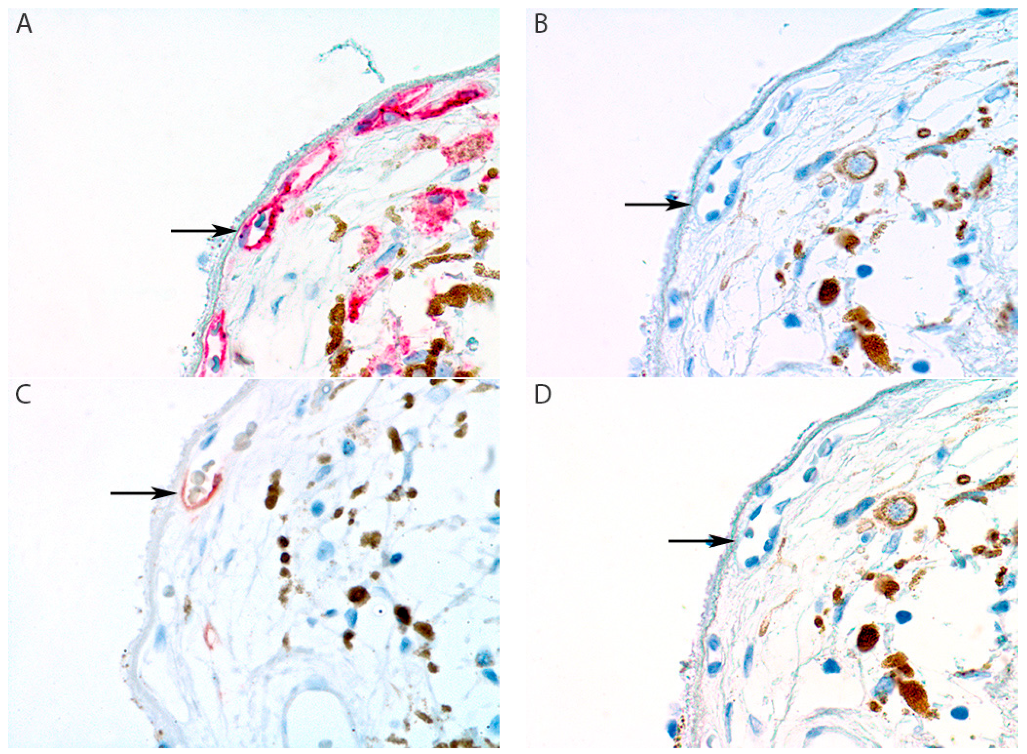
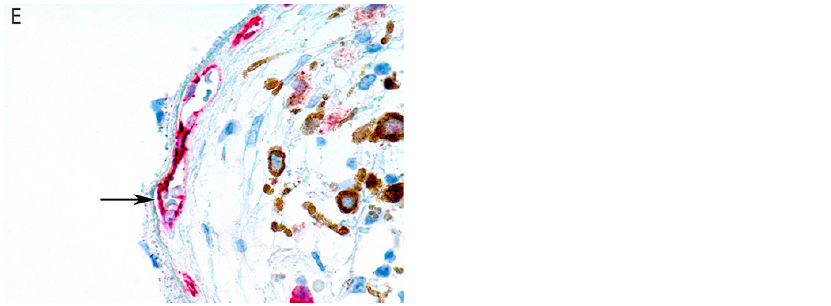
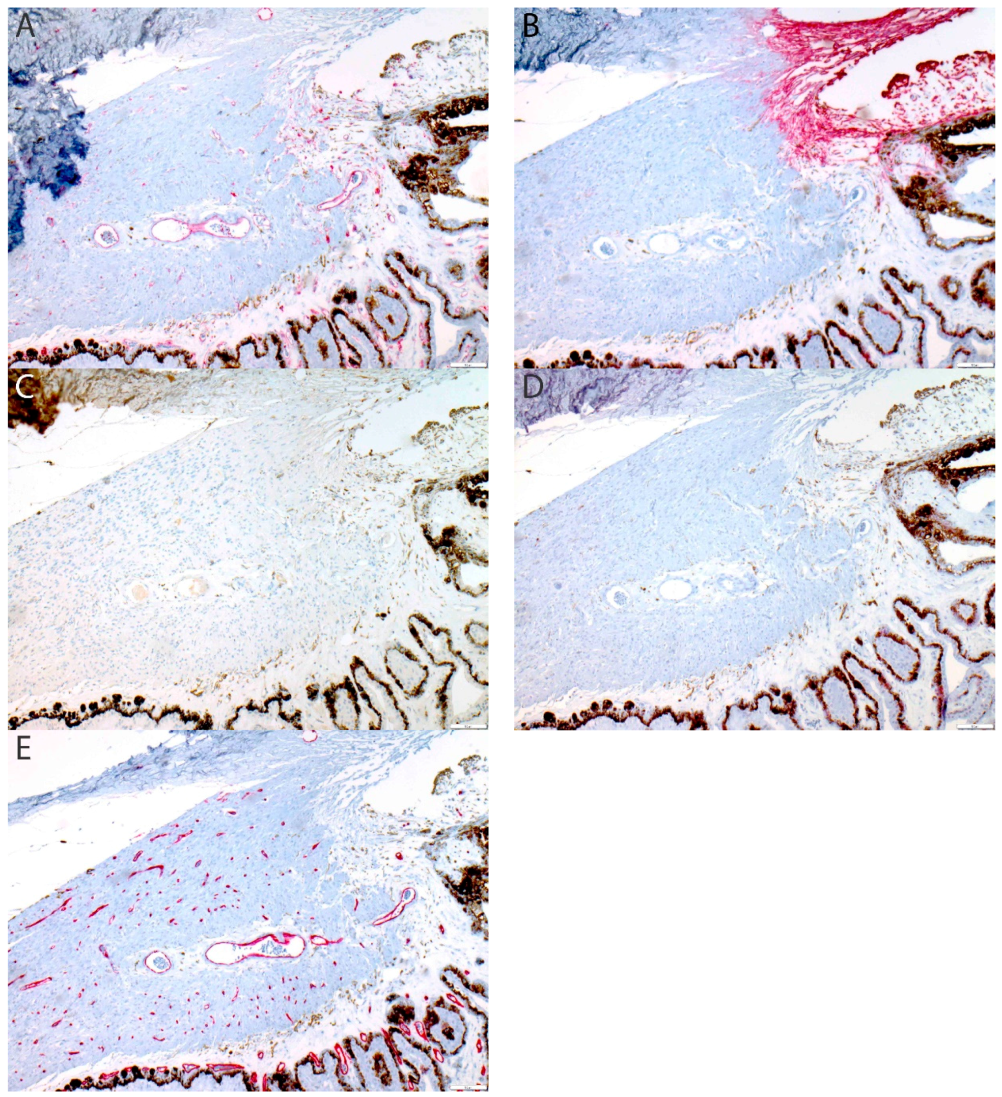
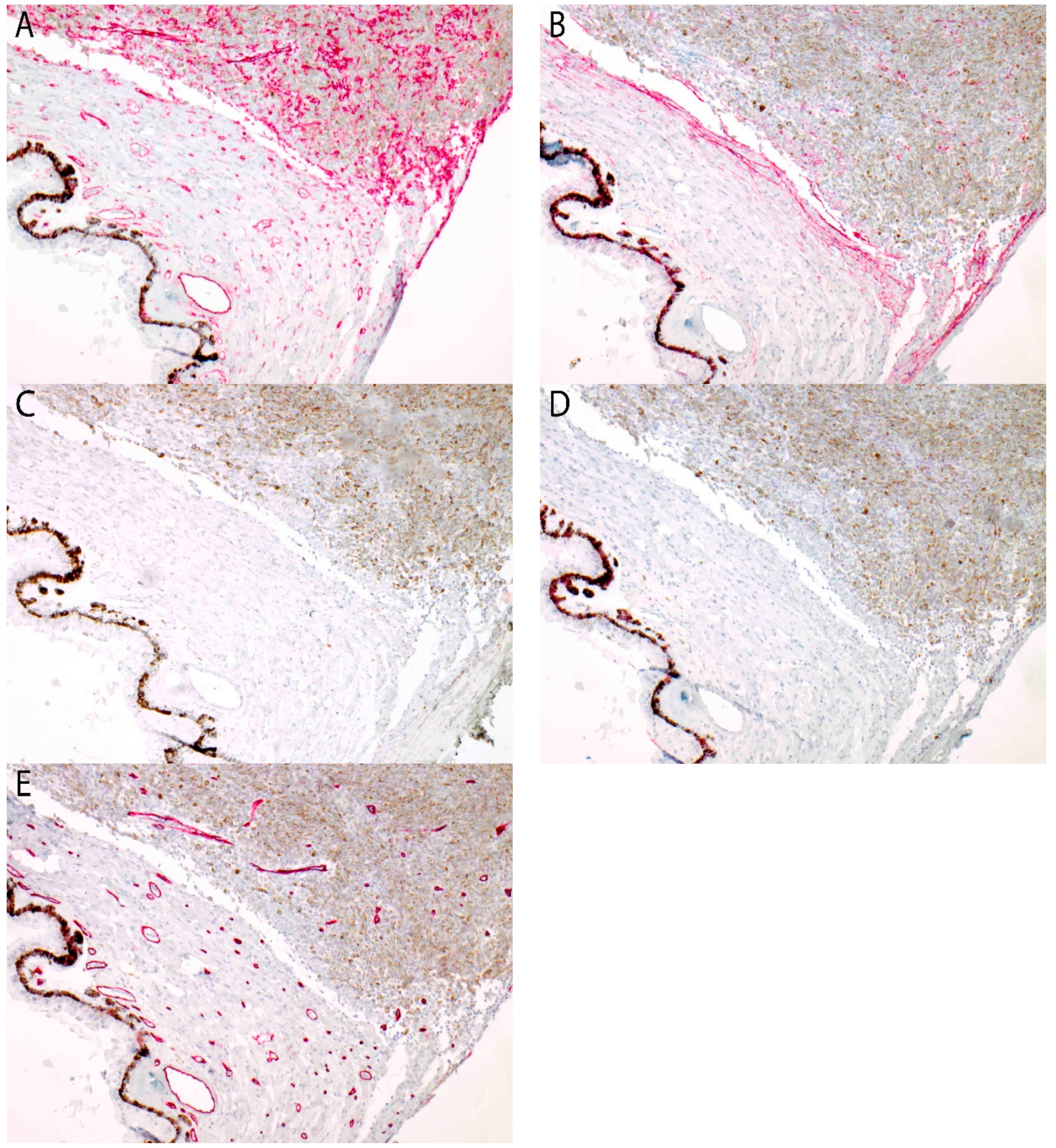
2.2. Immunohistochemistry
3. Discussion
4. Materials and Methods
4.1. Sample Selection
4.2. Tissue Processing
4.3. Immunohistochemistry
4.4. Scoring of Immunohistochemistry
5. Conclusions
Author Contributions
Funding
Acknowledgments
Conflicts of Interest
Appendix A


References
- Bishop, K.D.; Olszewski, A.J. Epidemiology and survival outcomes of ocular and mucosal melanomas: A population-based analysis. Int. J. Cancer 2014, 134, 2961–2971. [Google Scholar] [CrossRef] [PubMed]
- Tojo, D.; Wenig, B.L.; Resnick, K.I. Incidence of cervical metastasis from uveal melanoma: Implications for treatment. Head Neck 1995, 17, 137–139. [Google Scholar] [CrossRef] [PubMed]
- Dithmar, S.; Diaz, C.E.; Grossniklaus, H.E. Intraocular melanoma spread to regional lymph nodes: Report of two cases. Retina 2000, 20, 76–79. [Google Scholar] [CrossRef] [PubMed]
- Ardjomand, N.; Komericki, P.; Langmann, G.; Mattes, D.; Moray, M.; Scarpatetti, M.; El-Shabrawi, Y. Lymph node metastases arising from uveal melanoma. Wien. Klin. Wochenschr. 2005, 117, 433–435. [Google Scholar] [CrossRef] [PubMed]
- Heindl, L.M.; Hofmann, T.N.; Adler, W.; Knorr, H.L.; Holbach, L.M.; Naumann, G.O.; Kruse, F.E.; Cursiefen, C. Intraocular tumor-associated lymphangiogenesis a novel prognostic factor for ciliary body melanomas with extraocular extension? Ophthalmology 2010, 117, 334–342. [Google Scholar] [CrossRef] [PubMed]
- van Beek, J.G.; Koopmans, A.E.; Vaarwater, J.; de Rooi, J.J.; Paridaens, D.; Naus, N.C.; de Klein, A.; Verdijk, R.M.; Kilic, E. The prognostic value of extraocular extension in relation to monosomy 3 and gain of chromosome 8q in uveal melanoma. Investig. Ophthalmol. Vis. Sci. 2014, 55, 1284–1291. [Google Scholar] [CrossRef] [PubMed]
- Coupland, S.E.; Campbell, I.; Damato, B. Routes of extraocular extension of uveal melanoma: Risk factors and influence on survival probability. Ophthalmology 2008, 115, 1778–1785. [Google Scholar] [CrossRef]
- Birke, K.; Lutjen-Drecoll, E.; Kerjaschki, D.; Birke, M.T. Expression of podoplanin and other lymphatic markers in the human anterior eye segment. Investig. Ophthalmol. Vis. Sci. 2010, 51, 344–354. [Google Scholar] [CrossRef]
- Schlereth, S.L.; Neuser, B.; Herwig, M.C.; Muller, A.M.; Koch, K.R.; Reitsamer, H.A.; Schrodl, F.; Cursiefen, C.; Heindl, L.M. Absence of lymphatic vessels in the developing human sclera. Exp. Eye Res. 2014, 125, 203–209. [Google Scholar] [CrossRef]
- Schroedl, F.; Brehmer, A.; Neuhuber, W.L.; Kruse, F.E.; May, C.A.; Cursiefen, C. The normal human choroid is endowed with a significant number of lymphatic vessel endothelial hyaluronate receptor 1 (LYVE-1)-positive macrophages. Investig. Ophthalmol. Vis. Sci. 2008, 49, 5222–5229. [Google Scholar] [CrossRef]
- Zimmermann, P.; Dietrich, T.; Bock, F.; Horn, F.K.; Hofmann-Rummelt, C.; Kruse, F.E.; Cursiefen, C. Tumour-associated lymphangiogenesis in conjunctival malignant melanoma. Br. J. Ophthalmol. 2009, 93, 1529–1534. [Google Scholar] [CrossRef] [PubMed]
- Heindl, L.M.; Hofmann-Rummelt, C.; Adler, W.; Holbach, L.M.; Naumann, G.O.; Kruse, F.E.; Cursiefen, C. Tumor-associated lymphangiogenesis in the development of conjunctival squamous cell carcinoma. Ophthalmology 2010, 117, 649–658. [Google Scholar] [CrossRef] [PubMed]
- Heindl, L.M.; Hofmann-Rummelt, C.; Adler, W.; Bosch, J.J.; Holbach, L.M.; Naumann, G.O.; Kruse, F.E.; Cursiefen, C. Tumor-associated lymphangiogenesis in the development of conjunctival melanoma. Investig. Ophthalmol. Vis. Sci. 2011, 52, 7074–7083. [Google Scholar] [CrossRef] [PubMed]
- Khan, A.M.; Kagan, D.B.; Gupta, N.; Navajas, E.V.; Jin, Y.P.; Yucel, Y.H. Ciliary body lymphangiogenesis in uveal melanoma with and without extraocular extension. Ophthalmology 2013, 120, 306–310. [Google Scholar] [CrossRef]
- Heindl, L.M.; Hofmann, T.N.; Knorr, H.L.; Rummelt, C.; Schrodl, F.; Schlotzer-Schrehardt, U.; Holbach, L.M.; Naumann, G.O.; Kruse, F.E.; Cursiefen, C. Intraocular lymphangiogenesis in malignant melanomas of the ciliary body with extraocular extension. Investig. Ophthalmol. Vis. Sci. 2009, 50, 1988–1995. [Google Scholar] [CrossRef] [PubMed]
- Van der Auwera, I.; Cao, Y.; Tille, J.C.; Pepper, M.S.; Jackson, D.G.; Fox, S.B.; Harris, A.L.; Dirix, L.Y.; Vermeulen, P.B. First international consensus on the methodology of lymphangiogenesis quantification in solid human tumours. Br. J. Cancer 2006, 95, 1611–1625. [Google Scholar] [CrossRef] [PubMed]
- Ordonez, N.G. Immunohistochemical endothelial markers: A review. Adv. Anat. Pathol. 2012, 19, 281–295. [Google Scholar] [CrossRef] [PubMed]
- Schroedl, F.; Kaser-Eichberger, A.; Schlereth, S.L.; Bock, F.; Regenfuss, B.; Reitsamer, H.A.; Lutty, G.A.; Maruyama, K.; Chen, L.; Lutjen-Drecoll, E.; et al. Consensus statement on the immunohistochemical detection of ocular lymphatic vessels. Investig. Ophthalmol. Vis. Sci. 2014, 55, 6440–6442. [Google Scholar] [CrossRef] [PubMed]
- Adamczyk, L.A.; Gordon, K.; Kholova, I.; Meijer-Jorna, L.B.; Telinius, N.; Gallagher, P.J.; van der Wal, A.C.; Baandrup, U. Lymph vessels: The forgotten second circulation in health and disease. Virchows Arch. 2016, 469, 3–17. [Google Scholar] [CrossRef] [PubMed]
- Kerrigan, A.M.; Navarro-Nunez, L.; Pyz, E.; Finney, B.A.; Willment, J.A.; Watson, S.P.; Brown, G.D. Podoplanin-expressing inflammatory macrophages activate murine platelets via CLEC-2. J. Thromb. Haemost. 2012, 10, 484–486. [Google Scholar] [CrossRef] [PubMed]
- Sosa-Pineda, B.; Wigle, J.T.; Oliver, G. Hepatocyte migration during liver development requires Prox1. Nat. Genet. 2000, 25, 254–255. [Google Scholar] [CrossRef] [PubMed]
- Rodriguez-Niedenfuhr, M.; Papoutsi, M.; Christ, B.; Nicolaides, K.H.; von Kaisenberg, C.S.; Tomarev, S.I.; Wilting, J. Prox1 is a marker of ectodermal placodes, endodermal compartments, lymphatic endothelium and lymphangioblasts. Anat. Embryol. (Berl.) 2001, 204, 399–406. [Google Scholar] [CrossRef] [PubMed]
- Wilting, J.; Papoutsi, M.; Christ, B.; Nicolaides, K.H.; von Kaisenberg, C.S.; Borges, J.; Stark, G.B.; Alitalo, K.; Tomarev, S.I.; Niemeyer, C.; et al. The transcription factor Prox1 is a marker for lymphatic endothelial cells in normal and diseased human tissues. FASEB J. 2002, 16, 1271–1273. [Google Scholar] [CrossRef] [PubMed]
- Yang, J.F.; Walia, A.; Huang, Y.H.; Han, K.Y.; Rosenblatt, M.I.; Azar, D.T.; Chang, J.H. Understanding lymphangiogenesis in knockout models, the cornea, and ocular diseases for the development of therapeutic interventions. Surv. Ophthalmol. 2016, 61, 272–296. [Google Scholar] [CrossRef] [PubMed]
- Mouta Carreira, C.; Nasser, S.M.; di Tomaso, E.; Padera, T.P.; Boucher, Y.; Tomarev, S.I.; Jain, R.K. LYVE-1 is not restricted to the lymph vessels: Expression in normal liver blood sinusoids and down-regulation in human liver cancer and cirrhosis. Cancer Res. 2001, 61, 8079–8084. [Google Scholar] [PubMed]
- McKenney, J.K.; Weiss, S.W.; Folpe, A.L. CD31 expression in intratumoral macrophages: A potential diagnostic pitfall. Am. J. Surg. Pathol. 2001, 25, 1167–1173. [Google Scholar] [CrossRef] [PubMed]
- Clarijs, R.; Schalkwijk, L.; Ruiter, D.J.; de Waal, R.M. Lack of lymphangiogenesis despite coexpression of VEGF-C and its receptor Flt-4 in uveal melanoma. Investig. Ophthalmol. Vis. Sci. 2001, 42, 1422–1428. [Google Scholar]
- Heindl, L.M.; Schrodl, F.; Lutjen-Drecoll, E.; Cursiefen, C. Ciliary body lymphangiogenesis. Ophthalmology 2013, 120, e41–e42. [Google Scholar] [CrossRef]
- Truong, T.N.; Li, H.; Hong, Y.K.; Chen, L. Novel characterization and live imaging of Schlemm’s canal expressing Prox-1. PLoS ONE 2014, 9, e98245. [Google Scholar] [CrossRef]
- Aspelund, A.; Tammela, T.; Antila, S.; Nurmi, H.; Leppanen, V.M.; Zarkada, G.; Stanczuk, L.; Francois, M.; Makinen, T.; Saharinen, P.; et al. The Schlemm’s canal is a VEGF-C/VEGFR-3-responsive lymphatic-like vessel. J. Clin. Investig. 2014, 124, 3975–3986. [Google Scholar] [CrossRef]
- Brierley, J. Malignant Melanoma of the Uvea. In TNM Classification of Malignant Tumours, 8th ed.; Brierley, J., Gospodarowicz, M., Wittekind, C., Eds.; John Wiley & Sons, Ltd.: Oxford, UK; Hoboken, NJ, USA, 2017; pp. 221–225. [Google Scholar]
- Maruyama, K.; Ii, M.; Cursiefen, C.; Jackson, D.G.; Keino, H.; Tomita, M.; Van Rooijen, N.; Takenaka, H.; D’Amore, P.A.; Stein-Streilein, J.; et al. Inflammation-induced lymphangiogenesis in the cornea arises from CD11b-positive macrophages. J. Clin. Investig. 2005, 115, 2363–2372. [Google Scholar] [CrossRef] [PubMed]
- de Waard-Siebinga, I.; Hilders, C.G.; Hansen, B.E.; van Delft, J.L.; Jager, M.J. HLA expression and tumor-infiltrating immune cells in uveal melanoma. Graefes Arch. Clin. Exp. Ophthalmol. 1996, 234, 34–42. [Google Scholar] [CrossRef] [PubMed]
- World Medical Association. World Medical Association Declaration of Helsinki: Ethical principles for medical research involving human subjects. JAMA 2013, 310, 2191–2194. [Google Scholar] [CrossRef] [PubMed]
- McLean, I.W.; Foster, W.D.; Zimmerman, L.E.; Gamel, J.W. Modifications of Callender’s classification of uveal melanoma at the Armed Forces Institute of Pathology. Am. J. Ophthalmol. 1983, 96, 502–509. [Google Scholar] [CrossRef]


| Patient Characteristics | Patients, n = 16 |
|---|---|
| Gender, No. (%) | |
| Men | 7 (44) |
| Women | 9 (56) |
| Age in years, mean (SD) 1 | 66 (14) |
| Tumor size classification | |
| T1 | 3 |
| T2 | 3 |
| T3 | 7 |
| T4 | 3 |
| Largest diameter of the extension of the tumor in mm, mean (SD) 1 | 2.6 (2.5) |
| Tumor location | |
| Choroid | 9 |
| Ciliary body | 7 |
| Cell type | |
| Epithelioid | 4 |
| Mixed | 7 |
| Spindle | 5 |
| Disease-free survival in months, mean (SD) 1 | 77 (64) |
| Alive, n | 9 |
| Metastases, n | 4 |
| Death, because of uveal melanoma, n | 3 |
| Death other cause, n | 3 |
| Lost to follow-up, n | 1 |
| Markers | Expression of CD31 | Expression of D2-40 | Expression of LYVE-1 | Expression of Prox-1 | Expression of CD34 |
|---|---|---|---|---|---|
| Lymphatic vessel | + | + | + | + | − |
| Sample 1 | + | − | − | − | + |
| Sample 2 | + | − | − | − | + |
| Sample 3 | + | − | − | − | + |
| Sample 4 | + | − | − | − | + |
| Sample 5 | + | − | − | − | + |
| Sample 6 | + | − | − | − | + |
| Sample 7 | + | − | − | − | + |
| Sample 8 | + | − | + | − | + |
| Sample 9 | + | − | − | − | + |
| Sample 10 | + | − | − | − | + |
| Sample 11 | + | − | − | − | + |
| Sample 12 | + | − | − | − | + |
| Sample 13 | + | − | − | − | + |
| Sample 14 | + | − | − | − | + |
| Sample 15 | + | − | + | − | + |
| Sample 16 | + | − | − | − | + |
© 2019 by the authors. Licensee MDPI, Basel, Switzerland. This article is an open access article distributed under the terms and conditions of the Creative Commons Attribution (CC BY) license (http://creativecommons.org/licenses/by/4.0/).
Share and Cite
van Beek, J.G.M.; van den Bosch, Q.C.C.; Naus, N.; Paridaens, D.; de Klein, A.; Kiliç, E.; Verdijk, R.M. Absence of Intraocular Lymphatic Vessels in Uveal Melanomas with Extrascleral Growth. Cancers 2019, 11, 228. https://doi.org/10.3390/cancers11020228
van Beek JGM, van den Bosch QCC, Naus N, Paridaens D, de Klein A, Kiliç E, Verdijk RM. Absence of Intraocular Lymphatic Vessels in Uveal Melanomas with Extrascleral Growth. Cancers. 2019; 11(2):228. https://doi.org/10.3390/cancers11020228
Chicago/Turabian Stylevan Beek, Jackelien G. M., Quincy C. C. van den Bosch, Nicole Naus, Dion Paridaens, Annelies de Klein, Emine Kiliç, and Robert M. Verdijk. 2019. "Absence of Intraocular Lymphatic Vessels in Uveal Melanomas with Extrascleral Growth" Cancers 11, no. 2: 228. https://doi.org/10.3390/cancers11020228
APA Stylevan Beek, J. G. M., van den Bosch, Q. C. C., Naus, N., Paridaens, D., de Klein, A., Kiliç, E., & Verdijk, R. M. (2019). Absence of Intraocular Lymphatic Vessels in Uveal Melanomas with Extrascleral Growth. Cancers, 11(2), 228. https://doi.org/10.3390/cancers11020228