Review

Signal-Targeted Therapies and Resistance Mechanisms in Pancreatic Cancer: Future Developments Reside in Proteomics

by
Célia Cintas
1,2,
Thibaut Douché
1,2,
Nicole Therville
1,2,
Silvia Arcucci
1,2,
Fernanda Ramos-Delgado
1,2,
Céline Basset
1,2,3,
Benoît Thibault
1,2 and
Julie Guillermet-Guibert
1,2,*
1
INSERM U1037, CRCT, Université Paul Sabatier, 31037 Toulouse, France
2
Laboratoire d’Excellence TouCAN, 31037 Toulouse, France
3
Pathology Department, IUCT-O, Hôpitaux de Toulouse, 31037 Toulouse, France
*
Author to whom correspondence should be addressed.
Cancers 2018, 10(6), 174; https://doi.org/10.3390/cancers10060174
Submission received: 9 May 2018 / Revised: 29 May 2018 / Accepted: 30 May 2018 / Published: 1 June 2018

Abstract

:
For patients with metastatic pancreatic cancer that are not eligible for surgery, signal-targeted therapies have so far failed to significantly improve survival. These therapeutic options have been tested in phase II/III clinical trials mostly in combination with the reference treatment gemcitabine. Innovative therapies aim to annihilate oncogenic dependency, or to normalize the tumoural stroma to allow immune cells to function and/or re-vascularisation to occur. Large scale transcriptomic and genomic analysis revealed that pancreatic cancers display great heterogeneity but failed to clearly delineate specific oncogene dependency, besides oncogenic Kras. Beyond these approaches, proteomics appears to be an appropriate approach to classify signal dependency and to identify specific alterations at the targetable level. However, due to difficulties in sampling, proteomic data for this pathology are scarce. In this review, we will discuss the current state of clinical trials for targeted therapies against pancreatic cancer. We will then highlight the most recent proteomic data for pancreatic tumours and their metastasis, which could help to identify major oncogenic signalling dependencies, as well as provide future leads to explain why pancreatic tumours are intrinsically resistant to signal-targeted therapies. We will finally discuss how studies on phosphatidylinositol-3-kinase (PI3K) signalling, as the paradigmatic pro-tumoural signal downstream of oncogenic Kras in pancreatic cancer, would benefit from exploratory proteomics to increase the efficiency of targeted therapies.

Graphical Abstract

1. Introduction

To date, surgical resection provides the best chance of a cure for patients with pancreatic cancer. However, this curative approach is only proposed in 15 to 20% of cases. Moreover, there is a high rate of relapse following tumour resection, with a greater than 50% chance of local recurrence after surgery. The 5-year overall survival rate in resected patients is 28% and the median survival time is 18 months [1]. This high rate of recurrence is associated with the presence of micrometastasis at the time of surgery. Hence, adjuvant chemotherapy is often prescribed and can significantly increase patient survival. A very recent meta-analysis of 14 articles showed that adjuvant treatment (chemotherapy or chemoradiotherapy) improves the survival of patients with pancreatic ductal adenocarcinoma (PDAC) at one year and three year [2]. In this context, the use of targeted therapies, including those targeting the phosphatidylinositol-3-kinase (PI3K)/Akt pathway, needs to be considered [3].
Indeed, in PDAC there are many altered signalling pathways downstream oncogenic Kras such as mitogen-activated protein kinase (MAPK) and PI3K pathways, but also Braf signalling, transforming growth factor (TGFβ), Notch and DNA repair pathways [4] and among them, the PI3K/Akt is one of the most critically affected. The lipid kinase PI3Kα was shown to drive initiation of pancreatic cancer downstream of the main driving oncogene of this cancer [5,6,7,8], oncogenic Kras, which is found to be mutated in more than 80% of pancreatic cancer patients.
For 80–85% of patients with non-operable pancreatic cancer with locoregional or distant metastases (mainly liver and lung) chemotherapy can be used to improve quality of life and survival by relieving symptoms of disease. Since 1997, gemcitabine monotherapy remains the standard palliative chemotherapy for patients with metastatic PDAC [9,10,11,12], given the many clinical failures to combine it with other agents [13]. Recently, the combination of gemcitabine and nab-paclitaxel was found to be superior to treatment with gemcitabine alone (8.5 months versus 6.7 months survival), but presented a higher toxicity [14,15]. nab-paclitaxel is an albumin-bound paclitaxel, with the addition of albumin possibly leading to improved pharmacokinetics, greater specificity of distribution within the tumour, higher intra-tumoural concentration and better efficacy. The first major advance in the palliative treatment of patients with pancreatic cancer occurred in 2011 using FOLFIRINOX, a combination chemotherapy combining 5-fluorouracil, folinic acid, irinotecan and oxaliplatin. It has become the standard treatment for metastatic PDAC in patients in good general condition. Its efficacy is superior to that of gemcitabine (mean overall survival of 11.1 months versus 6.7 months), but it produces more toxic side effects [16,17]. If the determination of the most efficient chemotherapy regimen is improving, how to select the most efficient targeted therapy for these patients remains an open question in the field.
In 2016 the American Society of Clinical Oncology (ASCO) published its recommendations for the management of potentially curable PDACs (grade I, II), locally advanced PDACs (grade III), and metastatic PDACs (grade IV) (www.asco.org/guidelineswiki). Their recommendations for therapeutic interventions, including FOLFORINOX, irradiation and/or gemcitabine in combination with nab-paclitaxel, are based on relevant articles published between 2004 and 2015. However, it remains necessary and urgent to discover new targets and associated therapeutics, as well as improve irradiation protocols and current therapeutic strategies, by determining the mechanisms of intrinsic pancreatic resistance to anti-tumour agents including targeted therapies. Targeted anti-cancer therapies are strategies that aim to block or annihilate cancer progression by specifically addressing some of their abnormalities, including at the protein level. Here, we will first analyse the state-of-the art clinical trials involving targeted therapies in pancreatic cancer and, in light of the latest studies on PDAC proteomes, discuss how analysis of protein levels or post-translational modifications by proteomics can aid in the design of efficient personalized therapies for pancreatic cancer patients.
Proteomics (see Figure 1) is a powerful and promising tool for:
  • the identification of early diagnostic and prognostic protein biomarkers;
  • the identification of deregulated proteins and signalling pathways;
  • the identification of protein biomarkers predictive of the response to treatment;
  • the identification of early resistance mechanisms leading to the development of adapted combinatorial treatments.
It has now become clear that state-of-the art proteomics can be applied to PDAC research to help address these and other questions related to the clinical management of PDAC (Table 1). However, with the exception of studies into biomarkers, the proteomics data obtained for this pathology remains poor compared to data for other liquid or solid tumours.

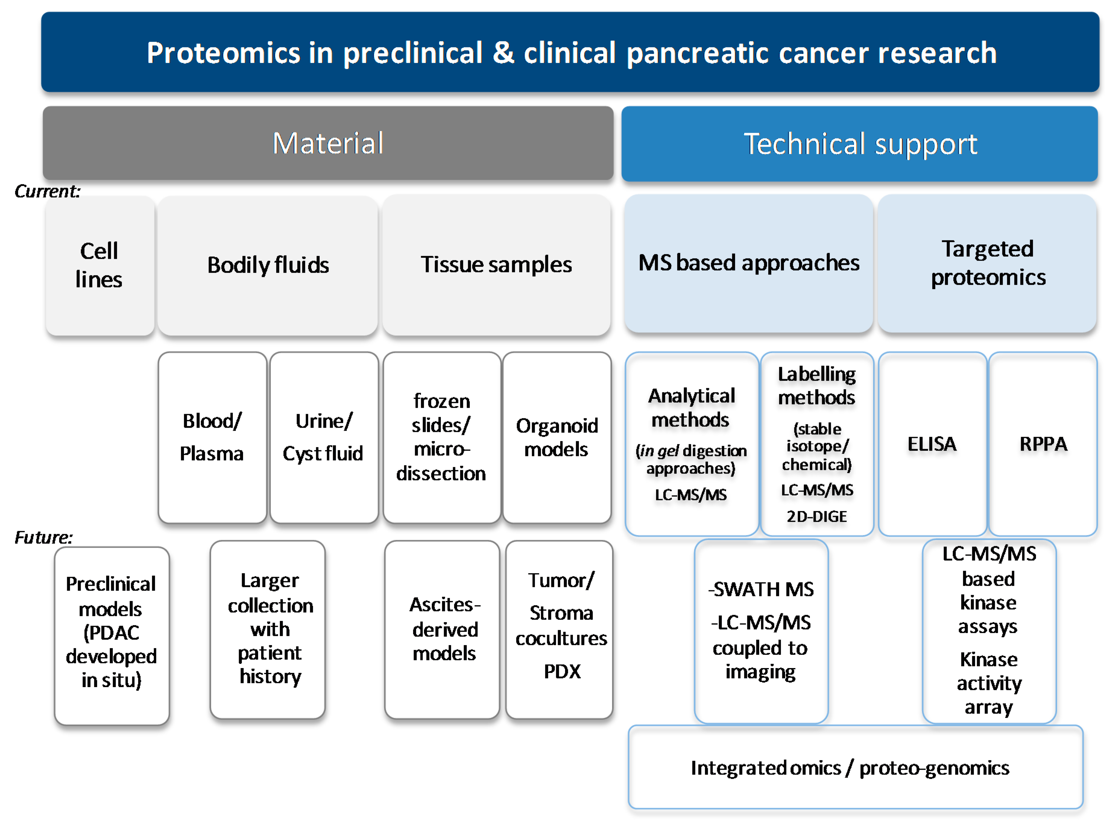
1.1. Proteomics Is a Technological Tool Which Can Help to Improve the Clinical Management of Pancreatic Cancer

Proteomics is the study of all the proteins of an organism, a biological fluid, an organ, a cell or even a cellular compartment. This set of proteins is called the “proteome”. The latter is a dynamic and complex entity. The proteome contains a much larger number of proteins than the genome contains genes. In human cells, an estimated 22,000 genes can yield up to one million proteins [18]. Despite these approximations, it is considered that proteins represent about 60% of a cell. The large scale study of proteins grew dramatically during the 1990s, with the advent of mass spectrometers (in 2002 the Nobel Prize in Chemistry was awarded to John Fenn and Koichi Tanaka for developing a novel method for the mass spectrometric analysis of biological macromolecules) [19]. Mass spectrometry (MS) is an analytical method that ionizes chemical species and sorts the ions based on their mass-to-charge ratio; it aims to identify and separate molecules (proteins but also small molecules and drugs) with very good resolution and sensitivity. New methodologies now allow the qualitative and quantitative analysis of complex biological samples, which may contain thousands of proteins, some of which are present in small quantities [20,21]. Post-translational modifications such as phosphorylation and ubiquitination can be studied by these techniques with prior enrichment of the starting material (mostly with beads coated with anti-pTyr antibodies or anti-ubiquitin antibodies for studies of tyrosine phosphorylations or ubiquitinations, and immobilized metal affinity chromatography (IMAC) or titanium dioxide (TiO2) beads for serine, threonine, or tyrosine phosphorylations) [22,23]. Such studies are called phosphoproteomics and ubiquitin-omics, respectively. Post-translational modification can regulate the biological activity of a protein. Therefore, analyses of the post-translational modifications of proteins in a tumour could identify the Achille’s heal of each tumour, and lead to the identification of novel therapeutic targets [24].
Proteomics provides insights into proteome-related changes in disease, such as observed changes in protein abundance, subcellular localization, post-translational modifications and cell signalling. Published proteomic results for pancreatic cancer are derived from the analysis of fluid (blood [25], plasma [26,27,28,29,30], serum [27,28,31,32,33,34,35,36,37,38,39,40,41], pancreatic juice [42] and cystic fluid [43]) and/or solid samples mostly microdissected beforehand [30,44,45], not from formalin-fixed-paraffin embedded (FFPE) tissue sections but from frozen sections [31,33,44,45,46,47], from healthy versus sick patients or patients at risk of developing pancreatic cancer (e.g., chronic pancreatitis (CP) [27,29,31,32,34,35,37,38,39,42]), from murine models of PDAC [30,33,48,49,50], as well as from conditioned medium [37,41,50,51,52] and established or experimental tumor-derived or patient-derived cell lines [25,27,30,37,48,50,51,52,53,54,55,56] (Figure 2).
Some articles report the quantification of total or phosphorylated protein amounts using reverse phase protein array (RPPA) or “reversed phase protein chips” [27,37,47] and enzyme-linked immunosorbent assay (ELISA), mostly for validation purposes [26,28,34] (Figure 2). Nevertheless, pancreatic cancer samples are mainly studied using higher throughput methods such as mass spectrometry or two-dimensional differential gel electrophoresis (2D-DIGE) [30,41,42,44,45,52].
Bottom-up (or shotgun) approaches, that is, the identification of proteins from the analysis of their peptide components, have led to increased proteome coverage (i.e., the identification and quantification of a maximal number of proteins from a mixture). Tryptic digestion followed by peptide fractionation, peptide separation through liquid chromatography, and on-line electrospray ionization coupled to tandem MS/MS orbitrap-based analyzers is the most common choice for in-depth proteomic analysis. Improvements in sample preparation, the sensitivity of mass analyzers and computational developments now allow researchers to quantify what is considered to be close to a full proteome (over 10,000 proteins expressed per cell) [57]. This level of protein identification and quantification is rarely described in studies involving PDAC samples [50]. In PDAC, due to the low amount of samples, key studies involve the utilization of more discriminatory techniques such as 2D-DIGE [45], that consists of labelling proteins with fluorescent probes prior to a 2D-electrophoresis separation according to their isoelectric point and molecular weight. Subsequently, spots can be excised from the gel, proteins digested with trypsin and peptides identified by matrix-assisted laser desorption/ionization time-of-flight (MALDI-TOF). Laser capture microdissection (LCM) has proven to be a beneficial preanalytical tissue-processing technique for isolating and selectively enriching discrete cellular populations from frozen tissue sections [30,44,45]. In PDAC, desmoplasia (tumour microenvironment) can pollute samples. Laser dissection can be used to discriminate epithelial cells from stroma and hence increase the chance of discovering biomarkers of early disease [30,44,45].
For quantitative proteomic approaches (as reviewed in [58]), differences in sample preparation, sample injection, and between each MS run can have profound effects on MS results. This is particularly true when attempting to quantify differential protein abundance between samples. This results in large numbers of replicate runs being required before any quantitative difference can be confidently identified. This is critical in all label-free MS experiments [57]. To overcome this, mass tags, such as stable isotope labelling with amino acids in cell culture (SILAC), isobaric tags for relative and absolute quantitation (iTRAQ), and chemical dimethyl labelling, can be incorporated into samples, typically labelling proteins or peptides, during tissue culture (SILAC) or sample lysis (superSILAC, iTRAQ and dimethyl labeling). The addition of these mass tags to one of the comparative groups allows multiple samples to be mixed and prepared in an identical manner while facilitating their separation within the mass spectrometer. Such labelling techniques allow the simultaneous investigation of multiple samples, facilitating accurate relative quantification, reducing bias, and increasing reproducibility [57] and are critical to reproducibly study post-translational modifications. These techniques are starting to be explored in PDAC [35,46,48]. Other strategies involve LC-MS/MS based [59] or protein array [60] technologies to measure and infer selected kinase activity (Figure 2). Although of easier use, targeted proteomics on protein expression and phosphorylation alterations most commonly found in cancer by techniques such as RPPA do not always give satisfactory answers due to their possible lower sensitivity and their narrowed analysis [48]. Mass spectrometry-based proteomic profiling of well-characterized cohorts of PDAC patients with detailed clinical information has the potential to provide effective stratification of PDAC patients and identify potential targets for novel treatments.

1.2. Genomic Characterization Has Failed to Identify Molecular Subtypes of Pancreatic Cancer to Guide the Choice of Targeted Therapies; Proteomics Appears to Be A Better Approach

Anti-cancer drugs fall into four broad categories based on their pharmacological action: conventional chemotherapy, radiopharmaceuticals, immunotherapies and inhibitors of (mostly) oncogenic mechanisms (but also of angiogenesis or immunomodulation) that include targeted therapies and hormone therapies (Figure 3A). Targeted anti-cancer therapies are strategies that aim to block the growth and/or spread of tumour cells by specifically addressing some of their abnormalities. Their main mode of action is an inhibition of the mechanisms of oncogenesis with a higher specificity towards cancer cells or their microenvironment. They are intracellular inhibitors (small chemical molecules such as protein or lipid kinase inhibitors) or extracellular inhibitors (biological drugs such as monoclonal antibodies to receptor tyrosine kinases (RTKs) or their ligands) (Table 2).
Targeted therapies are part of what is called “precision medicine”. This term refers to a medicine that is based on an in-depth knowledge of the biological mechanisms leading to the appearance and development of tumours. The use of these treatments is therefore guided, as far as possible, by the molecular characteristics of each patient’s tumour (for example: tumour differentiation, genetic alterations such as mutations/overexpression of oncogenes, loss of function of tumour suppressor genes, proteome). Most targeted therapies are currently used as a monotherapy (62% in France). The first targeted therapy was approved for use in France in 2000. This was trastuzumab, an antibody targeting the extracellular domain of the HER2 receptor, used in the treatment of human epidermal growth factor receptor 2 (HER2)-positive metastatic breast cancer as a monotherapy in patients that had already been treated with at least two chemotherapies for their metastatic disease. By the end of 2015, the French Cancer Health Institute INCa had identified 47 targeted therapies which had marketing authorization (MA) in France for the treatment of cancer.
Only one molecule, an inhibitor of the RTK epidermal growth factor receptor Epithelial Growth Factor Receptor (EGFR), has an indication in PDAC, used in combination with gemcitabine without any patient stratification (Figure 3B,C). So far, the efficiency of these molecules in PDAC remains modest, if compared to the spectacular action of targeted therapies in the first year of treatment in other aggressive solid tumours. In the successful cases, the choice of therapy is guided by oncogenic alterations, such as BRAF inhibitors in melanomas or EGFR inhibitors in lung cancers. Although overall survival (OS) and progression-free survival (PFS) improved slightly in PDAC patients treated with an EGFR inhibitor in combination with gemcitabine, there was no significant difference in the objective response (measurable response) as compared to placebo plus gemcitabine [61,62]. At present, in France, 38 clinical trials testing one or more anti-cancer molecules are in progress for pancreatic ductal cancer (data excluding neuroendocrine tumours), including 20 novel targeted therapies mostly used in combination with chemotherapy (Table 3). These studies were designed without the inclusion of any predictive markers. Targeted therapies against pancreatic microenvironment, which is thought to contribute to tumour aggressiveness, are not yet in phase I clinical trials [63].
In PDAC, the efficacy of 15 targeted therapies (mostly in combination with the chemotherapy gemcitabine) has been evaluated [64], the details of which are presented in Table 4. Despite patients displaying an initial anti-tumour response after treatment with these targeted therapies, as demonstrated by an ameliorated general state of the patients, resistance is induced very quickly. Besides, the poor general state of patients before starting their treatment prevents an increase in dosage required to reach a better target coverage and is an issue when testing a combination of several targeted therapies due to their increased toxicity. In the case of mTOR inhibitors, clinical trials led to decreased survival, possibly due to an acceleration of resistance mechanisms as explained below (paragraph 1.6). In general, it has been found that improvements to patient survival time are limited to a few weeks-months. Therefore, more effective drug combinations which prevent resistance while sparing general toxicity need to be developed.
Pancreatic cancer cells are mostly initially resistant (corresponding to a process called innate resistance). Moreover, adaptive resistance can also occur in pancreatic cancer. Whereby one or more tumour subpopulations with different characteristics compared to the sensitive cells could emerge allowing them to survive and continue to proliferate in the presence of pharmacological inhibition, leading to therapeutic failure [65,66]. Heterogeneity within a tumour is thus a critical component of resistance mechanisms [67]. Technological progress and large volumes of transcriptional, epigenetic and genetic data have led to a characterisation of the molecular identity of pancreatic cancer (mostly from resected tumours) [68,69,70,71]. However, in contrast to other cancers such as in lung cancer with the search of EGFR mutations at the genome level, these global approaches failed to identify simple therapeutic strategies based on a stratification of PDAC patients. Recent integrated omics approaches for all cancers highlight that the search for genetic or genomic alterations is not sufficient to predict which patients will benefit from targeted therapies [69,72]. In contrast, proteomic approaches appear better than transcriptomics or genomics at predicting sensitivity to a targeted therapy (for example PI3K inhibitors) because they reflect the real activity of the targets in tumors or in metastases [72,73]. It is all the more important in PDAC where the genomic and transcriptomic heterogeneity does not lead to obvious molecular classifications. This suggest that proteomic methods should be used to investigate resistance to treatment in pancreatic cancer patients, and moreover predict which therapy will be most efficient (Figure 1) [73]. Recent pioneering data, which need to be complemented by wider studies, also argue for the development of such unbiased exploratory strategies in pancreatic cancer.

1.3. Proteomic Profiling of PDAC Tissues

Proteomic studies give insights into metastatic PDAC biology, leading to the discovery of novel targets. Oncogenic Kras is altered in more than 80% of all PDAC patients. In PDAC, preneoplastic lesions (pancreatic intraepithelial neoplasia (PanIN), mucinous cystic neoplasm (MCM) and intra-ductal papillary mucinous neoplasm (IPMN)) progressively evolve towards at first in situ and then infiltrating metastatic adenocarcinoma, histologically defined as differentiated, moderately differentiated or dedifferentiated. Molecular classifications based on transcriptomics and genomics data have now been developed, including so called squamous, pancreatic progenitor, immunogenic and aberrantly differentiated subtypes [68,69,70,71]. The majority of proteomic studies performed to identify biomarkers of pancreatic cancer do not discriminate the different grades of the tumours studied or refer to the new molecular classification. This is due to the relatively low number of patients used in these studies (maximum 36 cell lines [54], 46 tumour samples [44], 173 plasma samples [25], serum samples [37] or 24 cystic fluid samples [43]), with sub-classification rendering a statistical approach irrelevant. However, 2D-DIGE technology performed on microdissections of human pancreatic tumours identified that the calcium-binding protein S100A6 was upregulated in moderately or poorly differentiated tumours [44], and was found to regulate epithelial-mesenchymal transition via an activation of β-catenin [74]. In 2009, Sitek and collaborators identified 86 differentially regulated proteins involved in pancreatic tumour progression using only microdissections of 37 single lesions of different grades of PanIN (PanIN is a precursor lesion of PDAC) from 9 patients, compared against PDAC cells and normal ductal cells [45]. These results are complemented by proteomic analysis performed by Dufresne et al., involving 2D-DIGE of microdissected murine PanIN cells and plasma samples from genetically engineered mouse models of the different precancerous stages [30]. The identification of peptide signatures specific to each type of precancerous lesion would thus make it possible to discriminate them from normal pancreatic tissue and could potentially lead to the discovery of both biomarkers and targets.
Independently of PDAC molecular classification, one study proposes the classification of pancreatic cancer cell lines based on the level and site of tyrosine phosphorylation (pTyr) of RTKs, potentially identifying three groups of cell lines classified as “low pTyr”, “RTK enriched pTyr” and “mixed pTyr” (on BAIAP2 Y337, PKP3 Y84, PKP2 Y166, CTNND1 Y174, CTNND1 Y 904) [54]. All sub-types presented a high level of tyrosine phosphorylation of RON, EPHA2, MET and ERBB2. “RTK enriched pTyr” cell lines presented high levels of tyrosine-phosphorylated EGFR, MET, RON, EPHA4, EPHB2/3/4, DDR2 as compared to the two other subtypes. These data indicate that a combination of RTKs are usually activated in pancreatic cancer, suggesting that single agent strategies towards RTKs are likely to be inefficient. If this classification could be performed on patient biopsies, it could guide the choice of treatment (including combinative strategies). A diagnostic tool based on proteomic profiling of easy-to-access liquid biopsies could be a clinically-feasible approach, as recent data show that the surface proteome of circulating exosomes provides unique opportunities to analyse the heterogeneity of metastatic pancreatic cancer [25].
A large scale proteomic analysis of a cell line with stemness properties (a subclone of Panc-1 cells characterized by increased expression of ceruplasmin, galectin-3 and myristoylated alanine-rich C-kinase substrate MARCKS) and its secretome revealed the importance of fatty acid synthesis and mevalonate pathways, as well as glycolysis, through the regulation of fatty acid synthase FASN and acetyl-CoA acetyltransferase2 ACAT2 protein expression, in this subtype of cells. Results obtained from this study could accelerate the development of novel therapeutic strategies targeting pancreatic cancer stem cells [51,53]. Using lectin microarray and nano LC-MS/MS, several glycoproteins were found to be enriched (e.g., cytokeratin 8 or ICAM1) or down regulated (e.g., aminopeptidase N) in this population of cells [75]. Stem cells may be the origin of tumour relapse in patients undergoing chemotherapy, therefore the discovery of specific targets for this pool of cells from patient-derived organoids could improve the current treatments. Indeed, large scale proteomics data on patient-derived organoids or xenografts (PDX) are lacking (Table 1); these data would provide a proof-of-concept for the use of proteomics in precision medicine (Figure 1).

1.4. Proteomic Approaches to Search for New Biomarkers of Early Disease

Early detection of pancreatic cancer is key for improving patient survival. Recent review articles discuss the use of several proteomic techniques for the early diagnosis of PDAC [76,77,78]. Currently, clinically used tumour markers lack sensitivity and specificity. For example, the carbohydrate antigen marker CA-19-9 (secreted by exocrine cells and tumours) present in the serum makes it possible to estimate pancreatic tumour progression during treatment [79]. However, the presence of this marker is not sufficient to diagnose pancreatic cancer, due to its lack of specificity (a high concentration is also found in the sera of patients with acute and chronic pancreatitis, hepatitis and biliary obstruction).
After the identification of potential protein biomarkers by large scale approaches, in the long term, the development of targeted proteomic technologies, such as selected reaction monitoring (SRM) also called multiple reaction monitoring (MRM) (using other MS apparatus such as triple quadrupole mass spectrometers), which can provide accurate and reproducible measurements of biomarkers of interest may be critical for the clinical use of the identified protein biomarkers [57]. To confirm the identity of a protein a match to at least two proteotypic peptides (peptides that are most likely to be identified by current MS-based methods, unique to this protein, without missing cleavage sites and not susceptible to post-translational modifications) is required. In the cancer proteome, aberrant proteins are present, which are absent from protein databases. This can also explain why the identification of cancer specific peptides is difficult, especially for this very heterogeneous type of cancer.
Numerous studies report new specific protein biomarkers for the diagnosis of pancreatic cancer identified from pancreas tissue lysate or liquid biopsies (such as secretome, but mostly plasma/serum) by proteomics [31,32,36,37,38,39,40,41,46,52,80]. Some biomarkers have been characterized and evaluated alone or in combination with CA19-9 for their ability to prediagnose pancreatic cancer [34]. For example, Park et al. established recently, from a large-cohort study of 500 patient sera, a panel of two proteins, apolipoprotein A4 (APOA4) and tissue inhibitor of metalloproteinase-1 (TIMP1), that in combination with CA 19-9 detection, were able to discriminate pancreatitis from early PDAC [38]. Preneoplastic samples are difficult to obtain due to the late diagnosis of this disease, nevertheless a recent analysis of 25 human PanIN organoids has led to a major step forward. Thrombospondin 2 detected in plasma either alone or in combination with CA19-9 was validated (98% combination specificity, 87% sensitivity) to distinguish all stages of the disease (resectable I and II, locally advanced III and metastatic IV), and in particular was able to discriminate resectable stage I cancer as readily as stage III/IV PDAC tumors [81]. Studies of other preneoplastic lesion such as IPMN and other cystic precursor lesions of the pancreas identify the proteins TIMP1 in plasma, mucin-5AC, mucin-2, cyst-fluid carcinoembryonic antigen and prostate stem-cell antigen in cyst fluids as potential protein biomarkers [29,43]. These studies should lead to further insights regarding the early diagnosis of pancreatic cancer. Identification using iTRAQ and validation using MRM of thrombospondin-1 (TSP-1) as a protein biomarker in patients at risk of developing pancreatic cancer, such as those with neo-onset diabetes, show that this condition impacts positively the diagnostic performance of CA19-9 [35]. These advances in early diagnostics are expected to lead to an increase in the efficiency of targeted therapies.
Despite the successful identification of biomarkers in preclinical or early phase clinical proteomic studies, thus far no proteomics-based discovery is used in clinical practice to diagnose PDAC. Honda et al., recently developed an antibody-based assay (ELISA) to measure circulating apolipoprotein A2 (APOA2) isoforms APOA2-ATQ/AT (C-terminal truncations of the apoAII homo-dimer) for the early detection of pancreatic cancer. However, clinical validation on 151 cases of stage I/II pancreatic cancer did not allow the early stages of pancreatic cancer to be statistically distinguished from healthy controls. Its clinical use should be reassessed in patients at high risk of pancreatic malignancy [28,34,35]. The definition of high risk patients remains difficult in PDAC. No single protein biomarker has a high enough discriminatory power to diagnose PDAC, therefore the development of a panel of multiple protein or glycoproteins biomarkers [82], which are more sensitive and specific, is essential for the expansion of routine clinical tools.
As is the case for most of these studies, replication of the results in larger cohorts of patients is necessary to form conclusions regarding the use of individual markers. Alternative approaches, such as the sequential window acquisition of all theoretical mass spectra (SWATH-MS) methodology which was developed recently [83], based on data-independent acquisition, can provide large coverage with a high degree of reproducibility (Table 1). Indeed, SWATH-MS allows a complete and permanent recording of all fragment ions of the detectable peptide precursors present in a biological sample. It thus combines the advantages of shotgun (high throughput) with those of SRM (high reproducibility and consistency). However, this technology has not yet been largely applied to PDAC patient samples.

1.5. Proteomic Approaches to Search for New Biomarkers of Predictive Response

Global bottom-up approaches to analyse the proteome and post-translationally modified proteomes (phosphorylation, glycosylation, ubiquitination) are scarcely used in clinical PDAC. Indeed, proteomics can be used as an adapted tool to predict the response to targeted therapies. Studying inter-patient tumour heterogeneity with quantitative MS-based high throughput proteomics and phosphoproteomics (as performed for 14 resected head PDAC patients) allows the identification of commonly activated signals such as Akt activation [46]. Proteomics can also analyse the changes in signalling pathways during treatment course of patients [67]. Using a quantitative proteomic approach (using first SILAC for protein labelling, then an enrichment in tyrosine phosphorylated peptides), Kim and collaborators revealed the heterogeneity of three metastatic sites of pancreatic cancer (liver, lung and peritoneum) by creating three cell lines from each organ of the same patient [55]. This heterogeneity is characterized by changes in the expression of the entire proteome as well as changes in the activity of tyrosine kinases, such as Axl tyrosine kinase. These data further demonstrate that neoplastic cells developing in different organs in the same PDAC patient display differential sensitivity to targeted therapies. So far, these properties have not been taken into account in clinical trials, partly explaining their relative inefficiency (Table 3 and Table 4). In contrast, in a large scale genomic and genetic analysis, genetic alterations in metastatic sites were found to be maintained as compared to the primary site of tumourigenesis [67]. Thus, the latest proteo-genomics data highlight the importance of a personalized combination of therapeutics targeting all the subclonal features of metastases, using proteomics to guide the therapeutic choice.
Proteomics has been found to be a powerful tool in the study of tumour cell/microenvironment cell signalling interactions and modifications. For example, Jorgensen’s team has recently demonstrated using murine Kras-mutated pancreatic cancer cells that oncogenic Kras signalling in tumour cells not only activates a cell-autonomous signalling network, but that it also activates the non-autonomous oncogenic signalling of stromal cells through a selective modification of their proteomes and their post-translationnal proteomes. Conversely, stromal cells can in turn modify and amplify oncogenic signalling increasing the number of protein signalling nodes and activated kinases involved in the survival of the same or other tumour cells. The total proteome or the phosphorylated proteome is different when cells are co-cultured, and lead to the identification of another oncogene dependency (here, insulin growth factor receptor 1 IGFR1/Axl). Thus, oncogenic signalling is no longer limited only to tumour cells but to the entire tumour compartment [50]. The temporal unbiaised exploration of extracellular communication signal would also help to design efficient signal targeted therapies. Indeed, the extracellular matrix found in abundance in desmoplasia is an important area to explore, using, for example, SWATH-MS technologies as developed for breast cancer [84].
Hence, besides genetic and genomic alterations, prediction of sensitivity to targeted therapies is influenced by heterotypic tumour-stroma signalling. The only way to assess this sensitivity appears to be by proteomic analysis of patient-derived samples, including all the cellular partners at stake in the primary tumour and the metastatic niche (Figure 1). This will require a proper analysis of post-translationally modified proteins in each cell compartment when cultured alone of in combination with their environment. Cross-comparison with heterotypic signalling in tumors from a different organ could pave the way to explain the intrinsic resistance to targeted therpies of pancreatic cancer.

1.6. Proteomic Approaches to Identify Resistance Mechanisms-Towards an Evolution in Precision Medicine

Global studies to understand the adaptive responses to targeted therapies in pancreatic cancer are starting to be published. Adaptive and reversible resistance to inhibition of cancer-driving mutated Kras in pancreatic cancer cells has been found to involve the tyrosine phosphorylation of focal adhesion pathway components, while strikingly no significant mutational or transcriptional changes were observed [48]. Alteration of other post-translational modifications other than tyrosine phosphorylation was not analysed. Large scale proteomics analysing the temporal effects of paclitaxel on pancreatic cancer cells, highlighted the role of proteins involved in mitochondrial function, survival (PI3K pathway) and cell cycle arrest in a key resistance mechanism [56]. Proteomics has identified that several negative feedback mechanisms are relieved upon mTORC1 inhibition, thus explaining the disappointing results from clinical trials on this target [38,39,57,59]. In other cancer settings, Hsu and colleagues have shown, for example, through global phosphoproteome analysis after SILAC labelling of cells (a method of labelling cells which allows a very precise differential quantitative approach) and enrichment in serine-, tyrosine- and threonine- phosphorylated peptides that the mTORC1 complex is able to inhibit and degrade insulin and IGF-1 receptors through phosphorylation of the adapter protein growth factor bound protein 10 (Grb10) [85,86]. It is now generally agreed that to target the PI3K/Akt/mTOR pathway, which is hyperactivated in 50% of all PDAC patients and associated with poor prognosis [6], hitting upstream PI3K is the best strategy to prevent positive feedback due to mTOR (mTORC1) inhibition. Whilst some mechanisms of resistance to PI3K inhibitors have been identified such as mitogen-activated protein kinase kinase activation also known as MEK [87,88], other mechanisms of resistance remain elusive in pancreatic cancer. Integrating different types of omics approaches to identify such mechanisms could increase the efficiency of these innovative targeted therapies towards driving signalling pathways [3,72].

2. Conclusions/Discussion/Perspectives

It is challenging to perform in-depth and large-scale clinical studies on PDAC. The sparsity of samples and the difficulty in defining the population at risk renders this task even more difficult. However, recent technological developments have led to advances both at fundamental and clinical research stages. Besides a better understanding of the oncogenic dependency of PDAC [89], the current development of MS-based methods, coupled to imaging [33,49], which need less material and are more quantitative [90] and of non-MS based robust targeted proteomic approaches [91], together with improvements to patient-derived ex vivo cultures to better mimic each patient’s situation will be instrumental in improving the management of patients with pancreatic cancer. Proteomic profiling of metastatic tumour cells will also be necessary (Table 1). Finally, integration of MS-based proteomic profiling with other state-of-the art technologies (such as next generation sequencing) will help to define driver proteins and to identify therapeutic targets. Integration of these data in a so called proteo-genomics approach is grabbing tremendous attention and has already been applied to other cancers [92]. Mostly used to provide protein-level evidence of gene expression and to help refine gene models, proteogenomics also includes computational strategies for building and using customized protein sequence databases. The later information are used to help identify novel peptides (not present in reference protein sequence databases) from mass spectrometry–based proteomic data [92]. These novel computational methods will be critical to achieve full personalized management of PDAC. Again, defining the best culture conditions, using conditions which mimic patient heterogeneity while maintaining the signalling pathways highly specific to the organ niche [50] is key to this success (Figure 1).
Cancers, such as pancreatic cancer, with complex molecular pathways are more refractory than those with less complex pathways [93]. Any improvement in the knowledge of this complexity will increase the efficiency of targeted-therapies in PDAC patients. This will only be possible first if we analyse in better detail cell-cell communication in simpler systems, while maintaining at best the conditions found in tumours or metastatic sites.
From reviewing the literature, it is clear that key questions still need to be answered (Table 1). Increased access to material from PDAC patients (via organoid culture), technologies adapted to the discovery of biomarkers such as SWATH technology and newly developed spatially resolved proteomic techniques will strive towards these aims. A few key questions are:
  • Can we use proteomics to detect PDAC earlier? Are earlier detected tumours more sensitive to targeted therapies towards PDAC oncogenic dependency (e.g., PI3K)? Will this knowledge increase life expectancy of PDAC patients?
  • Can we use proteomics to refine the current (epi)-genetic and genomic characterization of PDAC to better stratify patients? Sampling patients with PDAC is difficult. Can we develop and adapt methodological work flows to aid patient sampling? These workflows should also incorporate a better understanding of the metastatic disease.
  • Can we use proteomics to identify new targets (extracellular, membrane or intracellular) which take into account tumour-stroma heterotypic signalling? Is this signal different in each tumoral niche?
  • Can we use proteomics to understand, at a targetable protein level/modification (phosphorylation, ubiquitination), the specific resistance of PDAC patients to targeted therapies?
Early (or at least earlier) diagnosis using proteomics is a growing field with promising leads, but to be clinically validated larger cohorts of at-risk patients are needed. New leads in terms of oncogenic dependency have been discovered, such as the importance of PI3Kα in driving Kras-induced pancreatic cancerogenesis [5,8]. Clinical trials targeting such enzymes are on-going (Table 3 and Table 5). However, there is a lack of preclinical data to identify patients that are more sensitive to PI3K inhibitors due to an absence of commonly found alterations of the pathway (such as the loss of PTEN expression and the mutation of PIK3CA gene encoding PI3Kα) [3]. Similarly, adaptive resistance to such therapies cannot be anticipated. This increase in knowledge should be accompanied with efforts to adapt proteomic technologies to the limited amount of cells which can be accessed in PDAC patients [94].
In conclusion, the next challenge for PDAC proteomics will be to increase our knowledge so that patients can be stratified according to each innovative therapeutic approach (Figure 1). A more comprehensive knowledge of the resistance mechanisms induced by targeted therapies, observed at the level of the target protein, will allow researchers and clinicians to develop effective therapeutic strategies adapted to each target/oncogenic pathway in each patient-specific environment. Thus abolishing, preventing or delaying the appearance of resistance, which may ameliorate the survival curve of these patients.

Funding

J.G.-G.’s laboratory belongs to Toucan, Laboratoire d’Excellence, A.N.R., an integrated research program on Signal-targeted Drug Resistance. J.G.-G.’s laboratory for this topic was/is funded by French Charity Ligue Nationale Contre le Cancer (GB/MA/VSP-10443- salary to C.C.), Europe EU-ERG FP7 (270696 PaCa/PI3K), ARC (PJA20171206596), Toucan ANR Laboratory of Excellence, MSCA-ITN/ETN PhD-PI3K (Project ID: 675392- salary to F.R.D. and S.A.), Fondation de France (salary to B.T.), GSO, Fondation Toulouse Cancer Santé.

Acknowledgments

J.G.-G. is a member of COST action EU-Pancreas BM1204.

Conflicts of Interest

The authors declare no conflict of interests.

Glossary

Targeted therapy: Also known as molecularly targeted therapy. In cancer, these therapies differ from conventional therapies as they act on specific molecular targets that aim to interfere with crucial cell processes. Precision medicine: Emerging medical field that compiles information coming from a patient’s genes and proteins to prevent, diagnose and/or treat disease. Heterotypic signalling: Signalling between different cell types. intrinsic resistance: Innate ability to show resistance or to tolerate a treatment. Adaptative resistance: Acquired resistance towards a treatment/molecule due to frequent exposure.

Abbreviations

ALKAnaplastic lymphoma kinase;
APOA2Apolipoprotein A2;
APOA4Apolipoprotein A4;
ASCOAmerican Society of Clinical Oncology;
BTKBruton’s tyrosine kinase;
CA 19-9Cancer antigen 19-9;
CDK4/6Cyclin-dependent kinase 4/6;
CSF-1RColony-stimulating factor 1 receptor;
CPChronic pancreatitis;
CTCCirculating tumoral cell;
DDR1/2Discoidin domain receptor tyrosine kinase 1/2;
EGFREpidermal growth factor receptor;
ELISAEnzyme-linked immunosorbent assay;
FFPEFormalin-fixed paraffin-embedded;
FGFRFibroblast growth factor receptor;
FLT3Fms-related tyrosine kinase 3;
GAS6Growth arrest-specific 6;
Grb10Growth factor bound protein 10;
HER2human epidermal growth factor receptor 2;
Hsp27Heat shock protein 27;
IGF-1(R)Insulin-like growth factor 1 (Receptor);
IMACimmobilized metal affinity chromatography;
IPMNIntraductal papillary mucinous neoplasm;
iTRAQisobaric tags for relative and absolute quantitation;
JAKJanus kinase;
LckLymphocyte-specific protein tyrosine kinase;
LCMLaser capture microdissection;
MAMarketing authorization;
MALDI-TOFMatrix assisted laser desorption ionization-time of flight mass spectrometry;
MEKMitogen-activated protein kinase kinase;
METHepatocyte growth factor receptor;
MRMMultiple reaction monitoring;
MSMass spectrometry;
mTORMammalian target of rapamycin;
NNRTIInhibitor of non-nucleoside reverse transcriptase;
PanINPancreatic intraepithelial neoplasia;
PARPPoly ADP ribose polymerase;
PDACPancreatic ductal adenocarcinoma;
PDGFRPlatelet-derived growth factor receptor;
PDXPatient-derived xenograft;
PI3KPhosphatidylinositol-3-kinase;
PSCAProstate stem cell antigen;
pTyrphosphorylated tyrosine;
OSOverall survival;
PFSProgression-free survival;
RANKLReceptor activator of nuclear factor kappa-Β ligand;
ROS1C-ros oncogene 1;
RPPAReverse phase protein array;
RTKReceptor tyrosine kinase;
Tie2Tyrosine kinase with immunoglobulin and EGF homology domains;
TIMP Tissue inhibitor of metalloproteinase-1;
TiO2Titanium dioxide;
TGFβRTransforming growth factor β receptor;
TKTyrosine kinase;
SILACStable isotope labelling with amino acids in cell culture;
SMOSmoothened kinase;
SRMSelected reaction monitoring
VEGF(R)Vascular endothelial growth factor (receptor);

References

  1. Neoptolemos, J.P.; Kleeff, J.; Michl, P.; Costello, E.; Greenhalf, W.; Palmer, D.H. Therapeutic developments in pancreatic cancer: Current and future perspectives. Nat. Rev. Gastroenterol. Hepatol. 2018, 15, 333–348. [Google Scholar] [CrossRef] [PubMed]
  2. Xu, J.; Jiang, B.; Chen, Y.; Qi, F.Z.; Zhang, J.H.; Yuan, H. Optimal adjuvant chemotherapy for resected pancreatic adenocarcinoma: A systematic review and network meta-analysis. Oncotarget 2017, 8, 81419–81429. [Google Scholar] [CrossRef] [PubMed]
  3. Pons-Tostivint, E.; Thibault, B.; Guillermet-Guibert, J. Targeting PI3K Signaling in Combination Cancer Therapy. Trends Cancer 2017, 3, 454–469. [Google Scholar] [CrossRef] [PubMed]
  4. Witkiewicz, A.K.; McMillan, E.A.; Balaji, U.; Baek, G.; Lin, W.C.; Mansour, J.; Mollaee, M.; Wagner, K.U.; Koduru, P.; Yopp, A.; et al. Whole-exome sequencing of pancreatic cancer defines genetic diversity and therapeutic targets. Nat. Commun. 2015, 6, 6744. [Google Scholar] [CrossRef] [PubMed]
  5. Baer, R.; Cintas, C.; Dufresne, M.; Cassant-Sourdy, S.; Schonhuber, N.; Planque, L.; Lulka, H.; Couderc, B.; Bousquet, C.; Garmy-Susini, B.; et al. Pancreatic cell plasticity and cancer initiation induced by oncogenic Kras is completely dependent on wild-type PI 3-kinase p110alpha. Genes Dev. 2014, 28, 2621–2635. [Google Scholar] [CrossRef] [PubMed]
  6. Baer, R.; Cintas, C.; Therville, N.; Guillermet-Guibert, J. Implication of PI3K/Akt pathway in pancreatic cancer: When PI3K isoforms matter? Adv. Biol. Regul. 2015, 59, 19–35. [Google Scholar] [CrossRef] [PubMed]
  7. Eser, S.; Reiff, N.; Messer, M.; Seidler, B.; Gottschalk, K.; Dobler, M.; Hieber, M.; Arbeiter, A.; Klein, S.; Kong, B.; et al. Selective requirement of PI3K/PDK1 signaling for Kras oncogene-driven pancreatic cell plasticity and cancer. Cancer Cell 2013, 23, 406–420. [Google Scholar] [CrossRef] [PubMed]
  8. Wu, C.Y.; Carpenter, E.S.; Takeuchi, K.K.; Halbrook, C.J.; Peverley, L.V.; Bien, H.; Hall, J.C.; DelGiorno, K.E.; Pal, D.; Song, Y.; et al. PI3K regulation of RAC1 is required for KRAS-induced pancreatic tumorigenesis in mice. Gastroenterology 2014, 147. [Google Scholar] [CrossRef] [PubMed]
  9. Burris, H.A., III; Moore, M.J.; Andersen, J.; Green, M.R.; Rothenberg, M.L.; Modiano, M.R.; Cripps, M.C.; Portenoy, R.K.; Storniolo, A.M.; Tarassoff, P.; Nelson, R.; et al. Improvements in survival and clinical benefit with gemcitabine as first-line therapy for patients with advanced pancreas cancer: A randomized trial. J. Clin. Oncol. 1997, 15, 2403–2413. [Google Scholar] [CrossRef] [PubMed]
  10. Adamska, A.; Domenichini, A.; Falasca, M. Pancreatic Ductal Adenocarcinoma: Current and Evolving Therapies. Int. J. Mol. Sci. 2017, 18, 1338. [Google Scholar] [CrossRef] [PubMed]
  11. Danovi, S.A.; Wong, H.H.; Lemoine, N.R. Targeted therapies for pancreatic cancer. Br. Med. Bull. 2008, 87, 97–130. [Google Scholar] [CrossRef] [PubMed] [Green Version]
  12. Barati Bagherabad, M.; Afzaljavan, F.; ShahidSales, S.; Hassanian, S.M.; Avan, A. Targeted Therapies in Pancreatic Cancer: Promises and Failures. J. Cell. Biochem. 2017. [Google Scholar] [CrossRef] [PubMed]
  13. Paulson, A.S.; Cao, H.S.T.; Tempero, M.A.; Lowy, A.M. Therapeutic advances in pancreatic cancer. Gastroenterology 2013, 144, 1316–1326. [Google Scholar] [CrossRef] [PubMed]
  14. Von Hoff, D.D.; Ramanathan, R.K.; Borad, M.J.; Laheru, D.A.; Smith, L.S.; Wood, T.E.; Korn, R.L.; Desai, N.; Trieu, V.; Iglesias, J.L.; et al. Gemcitabine plus nab-paclitaxel is an active regimen in patients with advanced pancreatic cancer: A phase I/II trial. J. Clin. Oncol. 2011, 29, 4548–4554. [Google Scholar] [CrossRef] [PubMed]
  15. De Vita, F.; Ventriglia, J.; Febbraro, A.; Laterza, M.M.; Fabozzi, A.; Savastano, B.; Petrillo, A.; Diana, A.; Giordano, G.; Troiani, T.; et al. NAB-paclitaxel and gemcitabine in metastatic pancreatic ductal adenocarcinoma (PDAC): From clinical trials to clinical practice. BMC Cancer 2016, 16, 709. [Google Scholar] [CrossRef] [PubMed]
  16. Conroy, T.; Desseigne, F.; Ychou, M.; Bouche, O.; Guimbaud, R.; Becouarn, Y.; Adenis, A.; Raoul, J.L.; Gourgou-Bourgade, S.; de la Fouchardiere, C.; et al. FOLFIRINOX versus gemcitabine for metastatic pancreatic cancer. N. Engl. J. Med. 2011, 364, 1817–1825. [Google Scholar] [CrossRef] [PubMed]
  17. Gunturu, K.S.; Yao, X.; Cong, X.; Thumar, J.R.; Hochster, H.S.; Stein, S.M.; Lacy, J. FOLFIRINOX for locally advanced and metastatic pancreatic cancer: Single institution retrospective review of efficacy and toxicity. Med. Oncol. 2013, 30, 361. [Google Scholar] [CrossRef] [PubMed]
  18. Humphery-Smith, I. A human proteome project with a beginning and an end. Proteomics 2004, 4, 2519–2521. [Google Scholar] [CrossRef] [PubMed]
  19. Fenn, J.B.; Mann, M.; Meng, C.K.; Wong, S.F.; Whitehouse, C.M. Electrospray ionization for mass spectrometry of large biomolecules. Science 1989, 246, 64–71. [Google Scholar] [CrossRef] [PubMed] [Green Version]
  20. Bantscheff, M.; Lemeer, S.; Savitski, M.M.; Kuster, B. Quantitative mass spectrometry in proteomics: Critical review update from 2007 to the present. Anal. Bioanal. Chem. 2012, 404, 939–965. [Google Scholar] [CrossRef] [PubMed]
  21. Yang, Y.; Franc, V.; Heck, A.J.R. Glycoproteomics: A Balance between High-Throughput and In-Depth Analysis. Trends Biotechnol. 2017, 35, 598–609. [Google Scholar] [CrossRef] [PubMed]
  22. Heap, R.E.; Gant, M.S.; Lamoliatte, F.; Peltier, J.; Trost, M. Mass spectrometry techniques for studying the ubiquitin system. Biochem. Soc. Trans. 2017, 45, 1137–1148. [Google Scholar] [CrossRef] [PubMed]
  23. Hogrebe, A.; von Stechow, L.; Bekker-Jensen, D.B.; Weinert, B.T.; Kelstrup, C.D.; Olsen, J.V. Benchmarking common quantification strategies for large-scale phosphoproteomics. Nat. Commun. 2018, 9, 1045. [Google Scholar] [CrossRef] [PubMed]
  24. Mueller, M.; Martens, L.; Apweiler, R. Annotating the human proteome: Beyond establishing a parts list. Biochim. Biophys. Acta 2007, 1774, 175–191. [Google Scholar] [CrossRef] [PubMed]
  25. Castillo, J.; Bernard, V.; Lucas, F.A.S.; Allenson, K.; Capello, M.; Kim, D.U.; Gascoyne, P.; Mulu, F.C.; Stephens, B.M.; Huang, J.; et al. Surfaceome profiling enables isolation of cancer-specific exosomal cargo in liquid biopsies from pancreatic cancer patients. Ann. Oncol. 2018, 29, 223–229. [Google Scholar] [CrossRef] [PubMed]
  26. Capello, M.; Bantis, L.E.; Scelo, G.; Zhao, Y.; Li, P.; Dhillon, D.S.; Patel, N.J.; Kundnani, D.L.; Wang, H.; Abbruzzese, J.L.; et al. Sequential Validation of Blood-Based Protein Biomarker Candidates for Early-Stage Pancreatic Cancer. J. Natl. Cancer Inst. 2017, 109. [Google Scholar] [CrossRef] [PubMed]
  27. Grote, T.; Siwak, D.R.; Fritsche, H.A.; Joy, C.; Mills, G.B.; Simeone, D.; Whitcomb, D.C.; Logsdon, C.D. Validation of reverse phase protein array for practical screening of potential biomarkers in serum and plasma: Accurate detection of CA19-9 levels in pancreatic cancer. Proteomics 2008, 8, 3051–3060. [Google Scholar] [CrossRef] [PubMed] [Green Version]
  28. Honda, K.; Kobayashi, M.; Okusaka, T.; Rinaudo, J.A.; Huang, Y.; Marsh, T.; Sanada, M.; Sasajima, Y.; Nakamori, S.; Shimahara, M.; et al. Plasma biomarker for detection of early stage pancreatic cancer and risk factors for pancreatic malignancy using antibodies for apolipoprotein-AII isoforms. Sci. Rep. 2015, 5, 15921. [Google Scholar] [CrossRef] [PubMed] [Green Version]
  29. Ilies, M.; Sappa, P.K.; Iuga, C.A.; Loghin, F.; Salazar, M.G.; Weiss, F.U.; Beyer, G.; Lerch, M.M.; Volker, U.; Mayerle, J.; et al. Plasma protein profiling of patients with intraductal papillary mucinous neoplasm of the pancreas as potential precursor lesions of pancreatic cancer. Clin. Chim. Acta 2018, 477, 127–134. [Google Scholar] [CrossRef] [PubMed]
  30. Ligat, L.; Saint-Laurent, N.; El-Mrani, A.; Gigoux, V.; al Saati, T.; Tomasini, R.; Nigri, J.; Dejean, S.; Pont, F.; Baer, R.; et al. Pancreatic preneoplastic lesions plasma signatures and biomarkers based on proteome profiling of mouse models. Br. J. Cancer 2015, 113, 1590–1598. [Google Scholar] [CrossRef] [PubMed] [Green Version]
  31. Crnogorac-Jurcevic, T.; Gangeswaran, R.; Bhakta, V.; Capurso, G.; Lattimore, S.; Akada, M.; Sunamura, M.; Prime, W.; Campbell, F.; Brentnall, T.A.; et al. Proteomic analysis of chronic pancreatitis and pancreatic adenocarcinoma. Gastroenterology 2005, 129, 1454–1463. [Google Scholar] [CrossRef] [PubMed]
  32. Fakelman, F.; Felix, K.; Buchler, M.W.; Werner, J. New pre-analytical approach for the deep proteome analysis of sera from pancreatitis and pancreas cancer patients. Arch. Physiol. Biochem. 2010, 116, 208–217. [Google Scholar] [CrossRef] [PubMed]
  33. Gruner, B.M.; Hahne, H.; Mazur, P.K.; Trajkovic-Arsic, M.; Maier, S.; Esposito, I.; Kalideris, E.; Michalski, C.W.; Kleeff, J.; Rauser, S.; et al. MALDI imaging mass spectrometry for in situ proteomic analysis of preneoplastic lesions in pancreatic cancer. PLoS ONE 2012, 7, e39424. [Google Scholar] [CrossRef] [PubMed]
  34. Jenkinson, C.; Elliott, V.; Menon, U.; Apostolidou, S.; Fourkala, O.E.; Gentry-Maharaj, A.; Pereira, S.P.; Jacobs, I.; Cox, T.F.; Greenhalf, W.; et al. Evaluation in pre-diagnosis samples discounts ICAM-1 and TIMP-1 as biomarkers for earlier diagnosis of pancreatic cancer. J. Proteom. 2015, 113, 400–402. [Google Scholar] [CrossRef] [PubMed] [Green Version]
  35. Jenkinson, C.; Elliott, V.L.; Evans, A.; Oldfield, L.; Jenkins, R.E.; O’Brien, D.P.; Apostolidou, S.; Gentry-Maharaj, A.; Fourkala, E.O.; Jacobs, I.J.; et al. Decreased Serum Thrombospondin-1 Levels in Pancreatic Cancer Patients Up to 24 Months Prior to Clinical Diagnosis: Association with Diabetes Mellitus. Clin. Cancer Res. 2016, 22, 1734–1743. [Google Scholar] [CrossRef] [PubMed]
  36. Lin, C.; Wu, W.C.; Zhao, G.C.; Wang, D.S.; Lou, W.H.; Jin, D.Y. ITRAQ-based quantitative proteomics reveals apolipoprotein A-I and transferrin as potential serum markers in CA19-9 negative pancreatic ductal adenocarcinoma. Medicine (Baltimore) 2016, 95, e4527. [Google Scholar] [CrossRef] [PubMed] [Green Version]
  37. Mustafa, S.; Pan, L.; Marzoq, A.; Fawaz, M.; Sander, L.; Ruckert, F.; Schrenk, A.; Hartl, C.; Uhler, R.; Yildirim, A.; et al. Comparison of the tumor cell secretome and patient sera for an accurate serum-based diagnosis of pancreatic ductal adenocarcinoma. Oncotarget 2017, 8, 11963–11976. [Google Scholar] [CrossRef] [PubMed]
  38. Park, J.; Lee, E.; Park, K.J.; Park, H.D.; Kim, J.W.; Woo, H.I.; Lee, K.H.; Lee, K.T.; Lee, J.K.; Park, J.O.; et al. Large-scale clinical validation of biomarkers for pancreatic cancer using a mass spectrometry-based proteomics approach. Oncotarget 2017, 8, 42761–42771. [Google Scholar] [CrossRef] [PubMed]
  39. Saraswat, M.; Joenvaara, S.; Seppanen, H.; Mustonen, H.; Haglund, C.; Renkonen, R. Comparative proteomic profiling of the serum differentiates pancreatic cancer from chronic pancreatitis. Cancer Med. 2017, 6, 1738–1751. [Google Scholar] [CrossRef] [PubMed]
  40. Takano, S.; Sogawa, K.; Yoshitomi, H.; Shida, T.; Mogushi, K.; Kimura, F.; Shimizu, H.; Yoshidome, H.; Ohtsuka, M.; Kato, A.; et al. Increased circulating cell signalling phosphoproteins in sera are useful for the detection of pancreatic cancer. Br. J. Cancer 2010, 103, 223–231. [Google Scholar] [CrossRef] [PubMed] [Green Version]
  41. Wehr, A.Y.; Hwang, W.T.; Blair, I.A.; Yu, K.H. Relative quantification of serum proteins from pancreatic ductal adenocarcinoma patients by stable isotope dilution liquid chromatography-mass spectrometry. J. Proteome Res. 2012, 11, 1749–1758. [Google Scholar] [CrossRef] [PubMed]
  42. Chen, R.; Pan, S.; Cooke, K.; Moyes, K.W.; Bronner, M.P.; Goodlett, D.R.; Aebersold, R.; Brentnall, T.A. Comparison of pancreas juice proteins from cancer versus pancreatitis using quantitative proteomic analysis. Pancreas 2007, 34, 70–79. [Google Scholar] [CrossRef] [PubMed]
  43. Jabbar, K.S.; Arike, L.; Verbeke, C.S.; Sadik, R.; Hansson, G.C. Highly Accurate Identification of Cystic Precursor Lesions of Pancreatic Cancer Through Targeted Mass Spectrometry: A Phase IIc Diagnostic Study. J. Clin. Oncol. 2018, 36, 367–375. [Google Scholar] [CrossRef] [PubMed] [Green Version]
  44. Shekouh, A.R.; Thompson, C.C.; Prime, W.; Campbell, F.; Hamlett, J.; Herrington, C.S.; Lemoine, N.R.; Crnogorac-Jurcevic, T.; Buechler, M.W.; Friess, H.; et al. Application of laser capture microdissection combined with two-dimensional electrophoresis for the discovery of differentially regulated proteins in pancreatic ductal adenocarcinoma. Proteomics 2003, 3, 1988–2001. [Google Scholar] [CrossRef] [PubMed]
  45. Sitek, B.; Sipos, B.; Alkatout, I.; Poschmann, G.; Stephan, C.; Schulenborg, T.; Marcus, K.; Luttges, J.; Dittert, D.D.; Baretton, G.; et al. Analysis of the pancreatic tumor progression by a quantitative proteomic approach and immunhistochemical validation. J. Proteome Res. 2009, 8, 1647–1656. [Google Scholar] [CrossRef] [PubMed]
  46. Britton, D.; Zen, Y.; Quaglia, A.; Selzer, S.; Mitra, V.; Lobetaner, C.; Jung, S.; Bohm, G.; Schmid, P.; Prefot, P.; et al. Quantification of pancreatic cancer proteome and phosphorylome: Indicates molecular events likely contributing to cancer and activity of drug targets. PLoS ONE 2014, 9, e90948. [Google Scholar] [CrossRef] [PubMed]
  47. Huang, Y.J.; Frazier, M.L.; Zhang, N.; Liu, Q.; Wei, C. Reverse-phase protein array analysis to identify biomarker proteins in human pancreatic cancer. Dig. Dis. Sci. 2014, 59, 968–975. [Google Scholar] [CrossRef] [PubMed]
  48. Chen, P.Y.; Muzumdar, M.D.; Dorans, K.J.; Robbins, R.A.; Bhutkar, A.; del Rosario, A.M.; Mertins, P.; Qiao, J.; Schafer, C.; Gertler, F.B.; et al. Adaptive and reversible resistance to Kras inhibition in pancreatic cancer cells. Cancer Res. 2018, 78, 985–1002. [Google Scholar] [CrossRef] [PubMed]
  49. Gruner, B.M.; Winkelmann, I.; Feuchtinger, A.; Sun, N.; Balluff, B.; Teichmann, N.; Herner, A.; Kalideris, E.; Steiger, K.; Braren, R.; et al. Modeling Therapy Response and Spatial Tissue Distribution of Erlotinib in Pancreatic Cancer. Mol. Cancer Ther. 2016, 15, 1145–1152. [Google Scholar] [CrossRef] [PubMed]
  50. Tape, C.J.; Ling, S.; Dimitriadi, M.; McMahon, K.M.; Worboys, J.D.; Leong, H.S.; Norrie, I.C.; Miller, C.J.; Poulogiannis, G.; Lauffenburger, D.A.; et al. Oncogenic KRAS Regulates Tumor Cell Signaling via Stromal Reciprocation. Cell 2016, 165, 1818. [Google Scholar] [CrossRef] [PubMed]
  51. Brandi, J.; Pozza, E.D.; Dando, I.; Biondani, G.; Robotti, E.; Jenkins, R.; Elliott, V.; Park, K.; Marengo, E.; Costello, E.; et al. Secretome protein signature of human pancreatic cancer stem-like cells. J. Proteom. 2016, 136, 1–12. [Google Scholar] [CrossRef] [PubMed]
  52. Gronborg, M.; Kristiansen, T.Z.; Iwahori, A.; Chang, R.; Reddy, R.; Sato, N.; Molina, H.; Jensen, O.N.; Hruban, R.H.; Goggins, M.G.; et al. Biomarker discovery from pancreatic cancer secretome using a differential proteomic approach. Mol. Cell. Proteom. 2006, 5, 157–171. [Google Scholar] [CrossRef] [PubMed]
  53. Brandi, J.; Dando, I.; Pozza, E.D.; Biondani, G.; Jenkins, R.; Elliott, V.; Park, K.; Fanelli, G.; Zolla, L.; Costello, E.; et al. Proteomic analysis of pancreatic cancer stem cells: Functional role of fatty acid synthesis and mevalonate pathways. J. Proteom. 2017, 150, 310–322. [Google Scholar] [CrossRef] [PubMed]
  54. Humphrey, E.S.; Su, S.P.; Nagrial, A.M.; Hochgrafe, F.; Pajic, M.; Lehrbach, G.M.; Parton, R.G.; Yap, A.S.; Horvath, L.G.; Chang, D.K.; et al. Resolution of Novel Pancreatic Ductal Adenocarcinoma Subtypes by Global Phosphotyrosine Profiling. Mol. Cell. Proteom. 2016, 15, 2671–2685. [Google Scholar] [CrossRef] [PubMed]
  55. Kim, M.S.; Zhong, Y.; Yachida, S.; Rajeshkumar, N.V.; Abel, M.L.; Marimuthu, A.; Mudgal, K.; Hruban, R.H.; Poling, J.S.; Tyner, J.W.; et al. Heterogeneity of pancreatic cancer metastases in a single patient revealed by quantitative proteomics. Mol. Cell. Proteom. 2014, 13, 2803–2811. [Google Scholar] [CrossRef] [PubMed]
  56. Wang, X.; Niu, J.; Li, J.; Shen, X.; Shen, S.; Straubinger, R.M.; Qu, J. Temporal Effects of Combined Birinapant and Paclitaxel on Pancreatic Cancer Cells Investigated via Large-scale, Ion-Current-Based Quantitative Proteomics (IonStar). Mol. Cell. Proteom. 2018, 17, 655–671. [Google Scholar] [CrossRef] [PubMed]
  57. Domon, B.; Aebersold, R. Options and considerations when selecting a quantitative proteomics strategy. Nat. Biotechnol. 2010, 28, 710–721. [Google Scholar] [CrossRef] [PubMed]
  58. Kosako, H.; Nagano, K. Quantitative phosphoproteomics strategies for understanding protein kinase-mediated signal transduction pathways. Expert Rev. Proteom. 2011, 8, 81–94. [Google Scholar] [CrossRef] [PubMed] [Green Version]
  59. Cutillas, P.R.; Jorgensen, C. Biological signalling activity measurements using mass spectrometry. Biochem. J. 2011, 434, 189–199. [Google Scholar] [CrossRef] [PubMed] [Green Version]
  60. Sikkema, A.H.; den Dunnen, W.F.; Diks, S.H.; Peppelenbosch, M.P.; de Bont, E.S. Optimizing targeted cancer therapy: Towards clinical application of systems biology approaches. Crit. Rev. Oncol. Hematol. 2012, 82, 171–186. [Google Scholar] [CrossRef] [PubMed]
  61. Moore, M.J.; Goldstein, D.; Hamm, J.; Figer, A.; Hecht, J.R.; Gallinger, S.; Au, H.J.; Murawa, P.; Walde, D.; Wolff, R.A.; et al. Erlotinib plus gemcitabine compared with gemcitabine alone in patients with advanced pancreatic cancer: A phase III trial of the National Cancer Institute of Canada Clinical Trials Group. J. Clin. Oncol. 2007, 25, 1960–1966. [Google Scholar] [CrossRef] [PubMed]
  62. Rougier, P.; Riess, H.; Manges, R.; Karasek, P.; Humblet, Y.; Barone, C.; Santoro, A.; Assadourian, S.; Hatteville, L.; Philip, P.A. Randomised, placebo-controlled, double-blind, parallel-group phase III study evaluating aflibercept in patients receiving first-line treatment with gemcitabine for metastatic pancreatic cancer. Eur. J. Cancer 2013, 49, 2633–2642. [Google Scholar] [CrossRef] [PubMed]
  63. Amanam, I.; Chung, V. Targeted Therapies for Pancreatic Cancer. Cancers 2018, 10, 36. [Google Scholar] [CrossRef] [PubMed]
  64. Di Marco, M.; Grassi, E.; Durante, S.; Vecchiarelli, S.; Palloni, A.; Macchini, M.; Casadei, R.; Ricci, C.; Panzacchi, R.; Santini, D.; et al. State of the art biological therapies in pancreatic cancer. World J. Gastrointest. Oncol. 2016, 8, 55–66. [Google Scholar] [CrossRef] [PubMed]
  65. Burrell, R.A.; Swanton, C. Tumour heterogeneity and the evolution of polyclonal drug resistance. Mol. Oncol. 2014, 8, 1095–1111. [Google Scholar] [CrossRef] [PubMed]
  66. Almendro, V.; Marusyk, A.; Polyak, K. Cellular heterogeneity and molecular evolution in cancer. Annu. Rev. Pathol. 2013, 8, 277–302. [Google Scholar] [CrossRef] [PubMed]
  67. Makohon-Moore, A.P.; Zhang, M.; Reiter, J.G.; Bozic, I.; Allen, B.; Kundu, D.; Chatterjee, K.; Wong, F.; Jiao, Y.; Kohutek, Z.A.; et al. Limited heterogeneity of known driver gene mutations among the metastases of individual patients with pancreatic cancer. Nat. Genet. 2017, 49, 358–366. [Google Scholar] [CrossRef] [PubMed] [Green Version]
  68. Waddell, N.; Pajic, M.; Patch, A.M.; Chang, D.K.; Kassahn, K.S.; Bailey, P.; Johns, A.L.; Miller, D.; Nones, K.; Quek, K.; et al. Whole genomes redefine the mutational landscape of pancreatic cancer. Nature 2015, 518, 495–501. [Google Scholar] [CrossRef] [PubMed] [Green Version]
  69. Bailey, P.; Chang, D.K.; Nones, K.; Johns, A.L.; Patch, A.M.; Gingras, M.C.; Miller, D.K.; Christ, A.N.; Bruxner, T.J.; Quinn, M.C.; et al. Genomic analyses identify molecular subtypes of pancreatic cancer. Nature 2016, 531, 47–52. [Google Scholar] [CrossRef] [PubMed]
  70. Sivakumar, S.; de Santiago, I.; Chlon, L.; Markowetz, F. Master Regulators of Oncogenic KRAS Response in Pancreatic Cancer: An Integrative Network Biology Analysis. PLoS Med. 2017, 14, e1002223. [Google Scholar] [CrossRef] [PubMed]
  71. Collisson, E.A.; Sadanandam, A.; Olson, P.; Gibb, W.J.; Truitt, M.; Gu, S.; Cooc, J.; Weinkle, J.; Kim, G.E.; Jakkula, L.; et al. Subtypes of pancreatic ductal adenocarcinoma and their differing responses to therapy. Nat. Med. 2011, 17, 500–503. [Google Scholar] [CrossRef] [PubMed] [Green Version]
  72. Zhang, Y.; Kwok-Shing Ng, P.; Kucherlapati, M.; Chen, F.; Liu, Y.; Tsang, Y.H.; de Velasco, G.; Jeong, K.J.; Akbani, R.; Hadjipanayis, A.; et al. A Pan-Cancer Proteogenomic Atlas of PI3K/AKT/mTOR Pathway Alterations. Cancer Cell 2017, 31. [Google Scholar] [CrossRef] [PubMed]
  73. Cintas, C.; Guillermet-Guibert, J. Heterogeneity of Phosphatidylinositol-3-Kinase (PI3K)/AKT/Mammalian Target of Rapamycin Activation in Cancer: Is PI3K Isoform Specificity Important? Front. Oncol. 2017, 7, 330. [Google Scholar] [CrossRef] [PubMed]
  74. Chen, X.; Liu, X.; Lang, H.; Zhang, S.; Luo, Y.; Zhang, J. S100 calcium-binding protein A6 promotes epithelial-mesenchymal transition through beta-catenin in pancreatic cancer cell line. PLoS ONE 2015, 10, e0121319. [Google Scholar]
  75. Zhu, J.; He, J.; Liu, Y.; Simeone, D.M.; Lubman, D.M. Identification of glycoprotein markers for pancreatic cancer CD24+CD44+ stem-like cells using nano-LC-MS/MS and tissue microarray. J. Proteome Res. 2012, 11, 2272–2281. [Google Scholar] [CrossRef] [PubMed]
  76. Huang, R.; Chen, Z.; He, L.; He, N.; Xi, Z.; Li, Z.; Deng, Y.; Zeng, X. Mass spectrometry-assisted gel-based proteomics in cancer biomarker discovery: Approaches and application. Theranostics 2017, 7, 3559–3572. [Google Scholar] [CrossRef] [PubMed]
  77. Kim, H.; Park, J.; Wang, J.I.; Kim, Y. Recent advances in proteomic profiling of pancreatic ductal adenocarcinoma and the road ahead. Expert Rev. Proteom. 2017, 14, 963–971. [Google Scholar] [CrossRef] [PubMed]
  78. Pan, S.; Brentnall, T.A.; Chen, R. Proteomics analysis of bodily fluids in pancreatic cancer. Proteomics 2015, 15, 2705–2715. [Google Scholar] [CrossRef] [PubMed] [Green Version]
  79. Wakabayashi, T.; Sawabu, N.; Takemori, Y.; Satomura, Y.; Kidani, H.; Ohta, H.; Watanabe, H.; Yamakawa, O.; Takahashi, H.; Watanabe, K.; et al. Diagnostic significance of cancer-associated carbohydrate antigen (CA19-9) concentrations in pancreatic juice: Analysis in pure pancreatic juice collected by endoscopic aspiration and immunohistochemical study in chronic pancreatitis. Pancreas 1993, 8, 151–159. [Google Scholar] [CrossRef] [PubMed]
  80. Sun, C.; Rosendahl, A.H.; Ansari, D.; Andersson, R. Proteome-based biomarkers in pancreatic cancer. World J. Gastroenterol. 2011, 17, 4845–4852. [Google Scholar] [CrossRef] [PubMed]
  81. Kim, J.; Bamlet, W.R.; Oberg, A.L.; Chaffee, K.G.; Donahue, G.; Cao, X.J.; Chari, S.; Garcia, B.A.; Petersen, G.M.; Zaret, K.S. Detection of early pancreatic ductal adenocarcinoma with thrombospondin-2 and CA19-9 blood markers. Sci. Transl. Med. 2017, 9. [Google Scholar] [CrossRef] [PubMed]
  82. Nie, S.; Lo, A.; Wu, J.; Zhu, J.; Tan, Z.; Simeone, D.M.; Anderson, M.A.; Shedden, K.A.; Ruffin, M.T.; Lubman, D.M. Glycoprotein biomarker panel for pancreatic cancer discovered by quantitative proteomics analysis. J. Proteome Res. 2014, 13, 1873–1884. [Google Scholar] [CrossRef] [PubMed]
  83. Gillet, L.C.; Navarro, P.; Tate, S.; Rost, H.; Selevsek, N.; Reiter, L.; Bonner, R.; Aebersold, R. Targeted data extraction of the MS/MS spectra generated by data-independent acquisition: A new concept for consistent and accurate proteome analysis. Mol. Cell. Proteom. 2012, 11. [Google Scholar] [CrossRef] [PubMed]
  84. Krasny, L.; Bland, P.; Kogata, N.; Wai, P.; Howard, B.A.; Natrajan, R.C.; Huang, P.H. SWATH mass spectrometry as a tool for quantitative profiling of the matrisome. J. Proteom. 2018. [Google Scholar] [CrossRef] [PubMed]
  85. Hsu, P.P.; Kang, S.A.; Rameseder, J.; Zhang, Y.; Ottina, K.A.; Lim, D.; Peterson, T.R.; Choi, Y.; Gray, N.S.; Yaffe, M.B.; et al. The mTOR-regulated phosphoproteome reveals a mechanism of mTORC1-mediated inhibition of growth factor signaling. Science 2011, 332, 1317–1322. [Google Scholar] [CrossRef] [PubMed] [Green Version]
  86. Yu, Y.; Yoon, S.O.; Poulogiannis, G.; Yang, Q.; Ma, X.M.; Villen, J.; Kubica, N.; Hoffman, G.R.; Cantley, L.C.; Gygi, S.P.; et al. Phosphoproteomic analysis identifies Grb10 as an mTORC1 substrate that negatively regulates insulin signaling. Science 2011, 332, 1322–1326. [Google Scholar] [CrossRef] [PubMed]
  87. Alagesan, B.; Contino, G.; Guimaraes, A.R.; Corcoran, R.B.; Deshpande, V.; Wojtkiewicz, G.R.; Hezel, A.F.; Wong, K.K.; Loda, M.; Weissleder, R.; et al. Combined MEK and PI3K inhibition in a mouse model of pancreatic cancer. Clin. Cancer Res. 2015, 21, 396–404. [Google Scholar] [CrossRef] [PubMed]
  88. Junttila, M.R.; Devasthali, V.; Cheng, J.H.; Castillo, J.; Metcalfe, C.; Clermont, A.C.; Otter, D.D.; Chan, E.; Bou-Reslan, H.; Cao, T.; et al. Modeling targeted inhibition of MEK and PI3 kinase in human pancreatic cancer. Mol. Cancer Ther. 2015, 14, 40–47. [Google Scholar] [CrossRef] [PubMed]
  89. Ying, H.; Dey, P.; Yao, W.; Kimmelman, A.C.; Draetta, G.F.; Maitra, A.; DePinho, R.A. Genetics and biology of pancreatic ductal adenocarcinoma. Genes Dev. 2016, 30, 355–385. [Google Scholar] [CrossRef] [PubMed] [Green Version]
  90. Dalla Pozza, E.; Manfredi, M.; Brandi, J.; Buzzi, A.; Conte, E.; Pacchiana, R.; Cecconi, D.; Marengo, E.; Donadelli, M. Trichostatin A alters cytoskeleton and energy metabolism of pancreatic adenocarcinoma cells: An in depth proteomic study. J. Cell. Biochem. 2018, 119, 2696–2707. [Google Scholar] [CrossRef] [PubMed]
  91. Hilhorst, R.; Houkes, L.; van den Berg, A.; Ruijtenbeek, R. Peptide microarrays for detailed, high-throughput substrate identification, kinetic characterization, and inhibition studies on protein kinase A. Anal. Biochem. 2009, 387, 150–161. [Google Scholar] [CrossRef] [PubMed]
  92. Shukla, H.D. Comprehensive Analysis of Cancer-Proteogenome to Identify Biomarkers for the Early Diagnosis and Prognosis of Cancer. Proteomes 2017, 5, 28. [Google Scholar] [CrossRef] [PubMed]
  93. Breitkreutz, D.; Hlatky, L.; Rietman, E.; Tuszynski, J.A. Molecular signaling network complexity is correlated with cancer patient survivability. Proc. Natl. Acad. Sci. USA 2012, 109, 9209–9212. [Google Scholar] [CrossRef] [PubMed] [Green Version]
  94. Bournet, B.; Vignolle-Vidoni, A.; Grand, D.; Roques, C.; Breibach, F.; Cros, J.; Muscari, F.; Carrere, N.; Selves, J.; Cordelier, P.; et al. Endoscopic ultrasound-guided fine-needle aspiration plus KRAS and GNAS mutation in malignant intraductal papillary mucinous neoplasm of the pancreas. Endosc. Int. Open 2016, 4, E1228–E1235. [Google Scholar] [CrossRef] [PubMed]
Figure 1. Using proteomics to improve the clinical care of patients with pancreatic cancer.
Cancers 10 00174 g001
Figure 2. Major materials and technical tools used in preclinical pancreatic cancer research. RPPA: reverse phase protein array; ELISA: Enzyme-linked immunosorbent assay; LC-MS/MS: Liquid chromatography–tandem mass spectrometry; 2D-DIGE: two-dimensional differential gel electrophoresis; PDX: patient derived xenograft.
Cancers 10 00174 g002
Figure 3. Evolution of the use of targeted therapies in cancers using France as an example. (A) Contribution in percentage of targeted therapies as compared to the therapeutic arsenal authorized for use in cancers (excluding radiotherapy). Distribution (B) and number (C) of marketing authorizations for targeted therapies. Adapted from INCa 2015 and completed with clinicaltrials.gouv.
Cancers 10 00174 g003
Table 1. Open questions in PDAC research that could be addressed by applying proteomics to a large range of biological samples. CTC: circulating tumoral cell; PDX: patient-derived xenografts.
SourceDetails of SourcePDAC Clinical ApplicationPDAC Tumoural BiologyActual Limitations/Developments
Biopsies (fine needle under echo-endoscopy)Diagnostic
Active surveillance in patients at risk (chronic pancreatitis, mucinous lesions, hereditary)
  • Diagnostic markers
  • Prognostic markers
  • Therapeutic options
  • Markers of therapeutic response
  • Heterogeneity
  • Molecular subtyping
  • Stroma
  • New targets
  • Resistance to drugs
Poor cellularity
Development of organoids or PDX
MetastasisLoco-regional metastasis (peritoneum, ascites)
Distant metastasis (lung, liver)
  • Diagnostic markers
  • Prognostic markers
  • Therapeutic options
  • Markers of therapeutic response
  • Metastatic niche
  • Molecular subtyping [55]
  • Stroma
  • New targets
  • Resistance to drugs
Limited sampling
ResectionNormal adjacent tissue
Tumour
Desmoplastic reaction
  • Diagnostic markers [47]
  • Prognostic markers
  • Therapeutic options
  • Markers of therapeutic response
  • Heterogeneity [31,44,45,46,47,52]
  • Molecular subtyping
  • Stroma
  • New targets [46]
  • Resistance to drugs
Limited to 15–20% of all PDAC patients-do not represent the most aggressive patients
Development of MS coupled to imaging
Body fluidsBlood, blood fractions (serum, plasma, exosomes, CTCs, etc...)
Urine
Ascites
  • New targets
  • Resistance to drugs
Selection of patients based on circulating DNA
Development of ascites-based PDX as an easy access to metastatic cells in their environment (e.g., immune cells)
Cell lines/in situ experimental PDAC Not applicable
  • Heterogeneity [30,33]
  • Metastatic niche
  • Molecular subtyping [54]
  • Stroma [50]
  • New targets
  • Resistance to drugs [48,56]
  • Secretions [50]
Do not fully represent the heterogeneity of PDAC
Study of heterotypic communication between stromal and cancer cells
Conditioned medium Not applicable
  • Secretions [25,37,41,51,52]
  • Metastatic niche
  • Molecular subtyping
  • Stroma
  • New targets
  • Resistance to drugs
Study of heterotypic communication between stromal and cancer cells
Table 2. List of the 47 anti-cancer targeted therapies authorized in France. Adapted from INCa data and completed with Vidal.fr, updated in January 2018.
Intracellular InhibitorsExtracellular Inhibitors
Inhibitors of Protein Kinase(s)Ab Directed against RTK(s)
NameTarget(s)NameTarget(s)
AfatinibEGFRCetuximabAb anti-EGFR
AxitinibVEGFRPanitumumabAb anti-EGFR
OsimertinibEGFRPertuzumabAb anti-HER2
BosutinibBcr-Abl, SrcRamucirumabAb anti-VEGF
CabozantinibMET, AXL, VEGFR, GAS6, RET, ROS1, FLT3, Tie2TrastuzumabAb anti-HER2
CeritinibALKTrastuzumab emtansineAb anti-HER2
CobimetinibMEKAb directed against ligand(s)
CrizotinibALK and METAfliberceptAb anti-VEGF
DabrafenibRAFBevabizumabAb anti-VEGF
DasatinibBcr-Abl, SrcDenosumabAb anti-RANKL
ErlotinibEGFR
EverolimusmTOR
GefitinibEGFR
IbrutinibBTK
Idelalisibp110δ (PI3K)
ImatinibBcr-Abl, c-Kit, DDR1/2, CSF-1R, PDGFR
LapatinibEGFR, ErbB2
LenvatinibVEGFR, FGFR, PDGFR
NilotinibBcr-Abl
NintedanibPDGFR, FGFR, VEGFR, FLT3, Lck, Lyn, Src
OlaparibPARP
OsimertinibEGFR
PalbociclibCDK4/6
PazopanibVEGFR, c-Kit, PDGFR
PonatinibBcr-Abl
RegorafenibVEGFR, c-Kit, PDGFR
RibociclibCyclin D1/CDK4, CDK6
RuxolitinibJAK1/2
SonidegibSMO
SorafenibRAF, VEGFR, FGFR, c-Kit, PDGFR
SunitinibVEGFR, c-Kit, c-Kit, CSF-1R, RET, PDGFR
TemsirolimusmTOR
TivozanibVEGF
TrametinibMEK1/2
VandetanibVEGFR, EGFR, RET
VenetoclaxBcl2
VemurafenibERK, BRAF
VismodegibSMO
Table 3. Ongoing clinical trials for pancreatic cancer in France. In grey: clinical trials combining a targeted therapy with a chemotherapy. In blue: clinical trials using a targeted therapy only. Adapted from clinical.gouv.fr, updated in August 2017.
Name of the StudyMolecule TestedType of TherapyType of DrugPhasePathologies
D081FC00001-POLOOlaparib vs. placeboTargeted therapyInhibitor of PARPIIIMetastatic PDAC with BRCA mutation
SIRINOXOxaliplatin + IrinotecanChemotherapyPlatinum salts, DNA topoisomerase I inhibitorIDigestive adenocarcinoma (pancreas, oesophagus, stomach, small intestine and biliary tract)
PRODIGE 29FOLFIRINOX vs. GemcitabineChemotherapyAnti-metabolite, DNA topoisomerase I inhibitor, Platinum saltsIIILocally advanced PDAC
PAMELA-70FOLFIRINOXChemotherapyAnti-metabolite, DNA topoisomerase I inhibitor, Platinum saltsIIMetastatic PDAC
RC48Adoptive transfer of allogeneic lymphocyte cells with natural cytotoxic activity + CetuximabCellular therapyAntibody anti-EGFRI/IIHepatic metastasis of PDAC, colorectal or small intestine cancer
PRODIGE 24 − ACCORD 24Gemcitabine vs. FOLFIRINOXChemotherapyAnti-metabolite, DNA topoisomerase I inhibitor, Platinum saltsIIIPDAC
FIRGEMAXNab-paclitaxel + Gemcitabine vs. Nab-paclitaxel + Gemcitabine plus FOLFIRI3ChemotherapyAnti-metabolite, DNA topoisomerase I inhibitor, Platinum saltsIIMetastatic PDAC
PANOPTIMOXFOLFIRINOX +/− LV5FU2 vs FOLFIRINOX +/− FIRGEMChemotherapyAnti-metabolite, DNA topoisomerase I inhibitor, Platinum saltsIIMetastatic PDAC
MOAnab1Gemcitabine + Nab-paclitaxelChemotherapyAnti-metaboliteIMetastatic PDAC
GABRINOXGemcitabine + Nab-paclitaxel followed by FOLFIRINOXChemotherapyAnti-metaboliteIMetastatic PDAC
JANUS-2Ruxolitinib + CapecitabinTargeted therapy + ChemotherapyInhibitor of Janus kinase (JAK) + Anti-metaboliteIIILocally advanced or metastatic PDAC
CMEK162X2111MEK162 + GanitumabTargeted therapiesMEK inhibitor + antibody anti-IGF1RI/IIMetastatic PDAC, colorectal adenocarcinoma and melanoma
AFUGEMABI-007 + Gemcitabine vs. ABI-007 + LV5FU2ChemotherapyAnti-metaboliteIIMetastatic PDAC
H9H-MC-JBAJGemcitabine + LY2157299Targeted therapy + ChemotherapyTGFβR inhibitor + Anti-metaboliteI/IILocally advanced or metastatic PDAC
2009-011992-61Gemcitabine + AS703026Targeted therapy + ChemotherapyMEK inhibitor + Anti-metaboliteIIMetastatic PDAC
NEOPAC/IPC 2011-002Neoadjuvant Gemcitabine + Oxaliplatin and adjuvant Gemcitabine vs. adjuvant Gemcitabine ChemotherapyAnti-metabolite +/− Platinum saltsIIIPDAC (head of the pancreas)
CAOU6Gemcitabine +/− ABI-007 ChemotherapyAnti-metaboliteIIIMetastatic PDAC
TherGAPAnti-tumoural complex CYL-02Gene therapyEnzymatic, MetabolicIPDAC
ESPAC-4Gemcitabine +/− CapecitabinChemotherapyAnti-metaboliteIIIResectable PDAC
CO-101-001Gemcitabine + CO-1.01ChemotherapyAnti-metaboliteIIMetastatic PDAC
GATE 1Gemcitabine + Trastuzumab + ErlotinibTargeted therapies + ChemotherapyHER2 inhibitor, mTOR inhibitor,
Anti-metabolite
IIMetastatic PDAC
ASTELLAS 200800Gemcitabine + AGS-1C4D4Targeted therapy + ChemotherapyAntibody anti-PSCA + Anti-metaboliteIIMetastatic PDAC
AB SCIENCE AB07012Gemcitabine +/− MasitinibTargeted therapy + ChemotherapyTyrosine kinase inhibitor + Anti-metaboliteIIILocally advanced or metastatic PDAC
THERAPYCetuximab + TrastuzumabTargeted therapies + ChemotherapyHER2 and EGFR inhibitorsI/IIMetastatic PDAC
PANTEREfavirenzTargeted therapyInhibitor of non-nucleoside reverse transcriptase (NNRTI)IIPDAC
GERCOR LAP 07 D07-1Gemcitabine +/− Erlotinib followed by Gemcitabine or chemoradiotherapy with CapecitabinTargeted therapy + Chemotherapy +/− ChemoradiotherapyEGFR inhibitor + Anti-metabolite IIILocally advanced PDAC
SciClone SCI-RP-Pan-P2-001Gemcitabine +/−RP101Targeted therapy + ChemotherapyHsp27 inhibitor + Anti-metaboliteIIUnresectable, locally advanced or metastatic PDAC
Hoffmann-La Roche BO21129ErlotinibTargeted therapyEGFR inhibitorIILocally advanced PDAC
Pharmexa PRIMOVAXGemcitabine + GV001 vs. Gemcitabine Targeted therapy + ChemotherapyStimulator of LT CD8 + Anti-metaboliteIIIPDAC
Hoffmann-La Roche BO21128Gemcitabine + ErlotinibTargeted therapy + ChemotherapyEGFR inhibitor + Anti-metaboliteIIMetastatic PDAC
Sanofi-Aventis EFC10203tegafur (a prodrug of 5FU) + gimeracil (5-chloro-2,4 dihydropyridine, CDHP + oteracil (potassium oxonate, Oxo) vs. 5-FUChemotherapyAnti-metaboliteIIIMetastatic PDAC
Pfizer A4061028Gemcitabine +/−AG-013736 (Axitinib)Targeted therapy + ChemotherapyVEGFR inhibitor + Anti-metaboliteIIILocally advanced or metastatic unresectable PDAC
Sanofi-Aventis EFC10547Gemcitabine + AfliberceptTargeted therapy + ChemotherapyAntibody anti-VEGF1/2 + GemcitabineIIIMetastatic PDAC
CAPERGEMGemcitabine + Capecitabin + ErlotinibTargeted therapy + ChemotherapyEGFR inhibitor + Anti-metabolitesIAdvanced PDAC
ACCORD 11 PRODIGE 4Gemcitabine vs. FOLFIRINOX ChemotherapyAnti-metabolite, DNA topoisomerase I inhibitor, Platinum saltsIIIMetastatic PDAC
ACCORD 09Radiotherapy (RT) + Docetaxel + 5-FU or RT + Docetaxel and CisplatinChemoradiotherapyRadiotherapy, Alkylating agent, Anti-metabolite, Platinum saltsIIPDAC
Phase 1-2 (RECF0016)Radiotherapy + IrinotecanChemoradiotherapyDNA topoisomerase I inhibitorI/IILocally advanced PDAC
BAYPANGemcitabine +/− SorafenibTargeted therapy + ChemotherapyC-Raf and B-Raf inhibitor + Anti-metaboliteIIILocally advanced or metastatic PDAC
Table 4. Outcome of initial clinical trials for targeted therapies in pancreatic cancer. Some of the targeted therapies listed here did not have a high selectivity towards their targets [10,11,12].
TargetTherapyNumber of PatientsMean Survival (Months)
(Treatment Versus Chemo Only)
TelomeraseGemcitabine + GV100110628.4 vs. 6.9
VEGFGemcitabine + Bevacizumab6025.7 vs. 6.0
KrasGemcitabine + Tipifarnib6886.3 vs. 6.0
EGFRGemcitabine + Cetuximab7666.5 vs. 6.0
Gemcitabine + Erlotinib5696.24 vs. 5.91
ErbB2Trastuzumab444.6 vs. 5.4
GastrinGastrazol + 5-FU983.6 vs. 4.2
mTORGemcitabine + Everolimus 294.5 vs. 6.5
PI3K/PLKGemcitabine + Rigosertib1066.1 vs. 6.4
Sonic HedgehogGemcitabine + Vismodegib 1066.9 vs. 6.1
Notch3Gemcitabine + IPI-929122Not tolerated
IGF1-RGemcitabine + Ganitumab8007.0 vs. 7.2
MMPGemcitabine + Matrimastat2395.4 vs. 5.4
JAK/STATRuxolitinib + Capecitabin1274.5 vs. 4.2
α-secretaseRO4929097 (no Gemcitabine arm)184.1
MEK1/ERK1/2Selumetinib versus Capecitabin385.3 vs. 4.9
Table 5. Ongoing active clinical trials in pancreatic cancer targeting PI3K. Adapted from clinical.gouv.fr, updated in April 2018. Most phase I clinical trials using PI3K inhibitors in monotherapy or in combination therapy are completed; they include advanced pancreatic cancers (ref pons-Tostivint).
Name of the StudyMolecule TestedType of TherapyType of DrugType of PatientsPhase
NCT03065062Palbociclib + GedatolisibTargeted therapyCDK4/6 inhibitor + PI3K/mTOR inhibitorSolid tumorsI
NCT02646748Pembrolizumab + Itacitinib (INCB039110) and/or Pembrolizumab + INCB050465Targeted therapyPD-1 antibody + JAK1 inhibitor and/or PD-1 antibody + PI3Kδ inhibitorSolid tumorsI
MATCH Screening trialMultiple (including GSK2636771)Targeted therapyMultiple (including PI3Kβ inhibitor)Advanced refractory solid cancersII
NCT02077933Alpelisib + Everolimus or Alpelisib + Everolimus + ExemestaneTargeted therapyPI3Kα inhibitor + mTOR inhibitor or PI3Kα inhibitor + mTOR inhibitor + aromatase inhibitorAdvanced breast, renal and pancreatic cancerI
NCT02155088BYL719 + Gemcitabine + Nab-PaclitaxelTargeted therapy + chemotherapyPI3Kα + Anti-metabolite + Microtubule poisonLocally advanced and metastatic pancreatic cancerI

