Article

Transcript Abundance of Photorhabdus Insect-Related (Pir) Toxin in Manduca sexta and Galleria mellonella Infections

by
Anaïs Castagnola
1,
Geraldine Mulley
2,
Nathaniel Davis
3,
Nicholas Waterfield
4 and
S. Patricia Stock
1,5,*
1
Center for Insect Science, University of Arizona, Tucson, AZ 85721, USA
2
School of Biological Sciences, University of Reading, Whiteknights, Reading RG6 6AJ, UK
3
Pima Community College, Tucson, AZ 85721, USA
4
Division of Biomedical Sciences, Warwick Medical School, Warwick University, Coventry CV4 7AL, UK
5
Department of Entomology, University of Arizona, Tucson, AZ 85721, USA
*
Author to whom correspondence should be addressed.
Toxins 2016, 8(10), 287; https://doi.org/10.3390/toxins8100287
Submission received: 6 June 2016 / Revised: 21 September 2016 / Accepted: 22 September 2016 / Published: 29 September 2016
(This article belongs to the Section Bacterial Toxins)

Abstract

:
In this study, we assessed pirAB toxin transcription in Photorhabdus luminescens laumondii (strain TT01) (Enterobacteriaceae) by comparing mRNA abundance under in vivo and in vitro conditions. In vivo assays considered both natural and forced infections with two lepidopteran hosts: Galleria mellonella and Manduca sexta. Three portals of entry were utilized for the forced infection assays: (a) integument; (b) the digestive route (via mouth and anus); and (c) the tracheal route (via spiracles). We also assessed plu4093-2 transcription during the course of a natural infection; this is when the bacteria are delivered by Heterorhabditis bacteriophora nematodes. Transcript abundance in G. mellonella was higher than in M. sexta at two of the observed time points: 15 and 18 h. Expression of pirAB plu4093-2 reached above endogenous control levels at 22 h in G. mellonella but not in M. sexta. Overall, pirAB plu4093-2 transcripts were not as highly expressed in M. sexta as in G. mellonella, from 15 to 22 h. This is the first study to directly compare pirAB plu4093-2 toxin transcript production considering different portals of entry.

1. Introduction

The entomopathogenic Gram-negative bacterium Photorhabdus luminescens laumondii (strain TT01) (Enterobacteriaceae) and its nematode host, Heterorhabditis bacteriophora (Nematoda: Heterorhabditidae), form an insecticidal partnership that has been shown to successfully kill a wide range of insect pests [1,2,3]. Third-stage infective juvenile (IJ) nematodes are the only free-living stage in the nematode’s life cycle and are responsible for vectoring Photorhabdus from one host to another [4,5]. Once a suitable insect host is found, IJs may penetrate it through three different routes: (a) integument; (b) the digestive route (i.e., IJs penetrate into the insect through either the mouth or anus and are readily exposed to gut tissue); and (c) the tracheal route (i.e., IJs enter through the insect’s spiracles) [6,7,8]. Although it has not yet been definitively demonstrated all the locations at which the nematodes can release bacteria during these different routes of infection, it has been shown to predominantly occur in the hemolymph. Nevertheless, anytime Photorhabdus cells are released by the IJs, they subsequently colonize insect tissue, and the anterior portion of the midgut is one of the initial targets. More specifically, the Photorhabdus cells align to the intestinal longitudinal muscles prior to invading the fat body tissue [9].
An examination of the genome of P. luminescens laumondii (here after P. l. laumondii), strain TT01, revealed the presence of a number of pathogenicity islands that contain a multitude of toxins and virulence factors that can contribute to insect mortality [10]. For example, the insect toxin complex proteins, Tc, have been shown to attack the anterior grooves of the intestine in M. sexta (Lepidoptera: Sphingidae) [11]. Another toxin, the Mcf (makes caterpillars floppy) protein has been shown to disrupt gut tissue and induce apoptosis in hemocytes, ultimately making caterpillars floppy due to a loss of turgor pressure and thus giving the toxin its unique name [12,13]. Additionally, the Photorhabdus virulence cassettes (PVCs), which have a phage-like appearance, have been found to kill hemocytes and cause cytoskeletal condensation [14]. Furthermore, the Photorhabdus insect-related (PirAB) toxins, which are encoded in two independent loci, plu4437-plu4436 and plu4093-plu4092, each elaborate a distinct binary toxin [5]. The two PirABs have been shown to have variable toxicity depending on the delivery method and insect host. For example, protein products of plu4093-2 and plu4437-6 have oral toxicity against Plutella xyllostella and mosquito larvae [10,15]. In addition, cloned PirAB (plu4093-2) has injectable toxicity in G. mellonella but no oral or injectable toxicity in M. sexta, nor oral activity in H. virescens or L. dispar [16]. PirAB (plu4437-6) showed no oral or hemocoel-based insecticidal activity to G. mellonella or S. litura [17]. Interestingly, the PirAB toxin from P. asymbiotica has been shown to exhibit oral toxicity against the larvae of the Dengue vector mosquitos Aedes aegypti and Aedes albopictus but did not affect the Mesocyclops thermocyclopoides predator [18]. Although purified PirA and PirB are toxic when applied together, the linking of the two proteins, using artificial translation fusion, has been shown to have a cytotoxic effect in vitro against cultured CF-203 midgut cells of the spruce budworm, Choristoneura fumiferana. These artificial translational fusions did not show toxicity when orally delivered in S. exigua larvae but had injectable toxicity in S. exigua [19]. Recently, a plasmid containing a gene homologous of Pir toxins was found in the shrimp pathogen Vibrio parahaemolyticus [20,21]. The affected shrimp show pathological responses suggesting the involvement of Pir toxin-like proteins in the etiology of acute hepato-pancreatic necrosis disease [20].
Pir proteins have amino acid similarity with the pore-forming domain of Cry2A toxin of Bacillus thuringiensis (Bt) [22]. This sequence correspondence has prompted the development of toxin chimeras with the goal of enhancing their individual toxicity [23]. For example, when PirB was fused with Cry2A, the recombinant toxin displayed oral toxicity in S. exigua larvae, but no oral toxicity was observed in a different host (Helicoverpa armigera), suggesting this chimera is host specific [23].
Although the toxicity and relationship to virulence of PirAB have both been investigated, the expression of this toxin during the natural course of infection has yet not been assessed. Therefore, in this study, we evaluated the abundance of pirAB toxin transcripts of the Photorhabdus l. laumondii TT01 strain when delivered by its vector nematode, H. bacteriophora. Natural and forced infection assays were also investigated.

2. Results

2.1. PirAB plu4093-2 Transcript Expression in Natural Infections

The relative abundance of the pirAB transcript was assessed for both lepidopteran hosts at four time points: 12, 15, 18, and 22 h, considering natural infections (i.e., larvae were exposed to infective juvenile nematodes (IJs)). Our results show that transcript expression in both G. mellonella and M. sexta remained below the endogenous control between 12 and 18 h post-IJ exposure. Transcript abundance in G. mellonella was slightly higher (Student’s t-test α = 0.05) than in M. sexta at three time points: 12, 15, and 18 h (Figure 1). However, a three-fold increase (Students t-test α = 0.05) of pirAB transcripts was detected at 22 h in G. mellonella when compared with that observed in M. sexta.

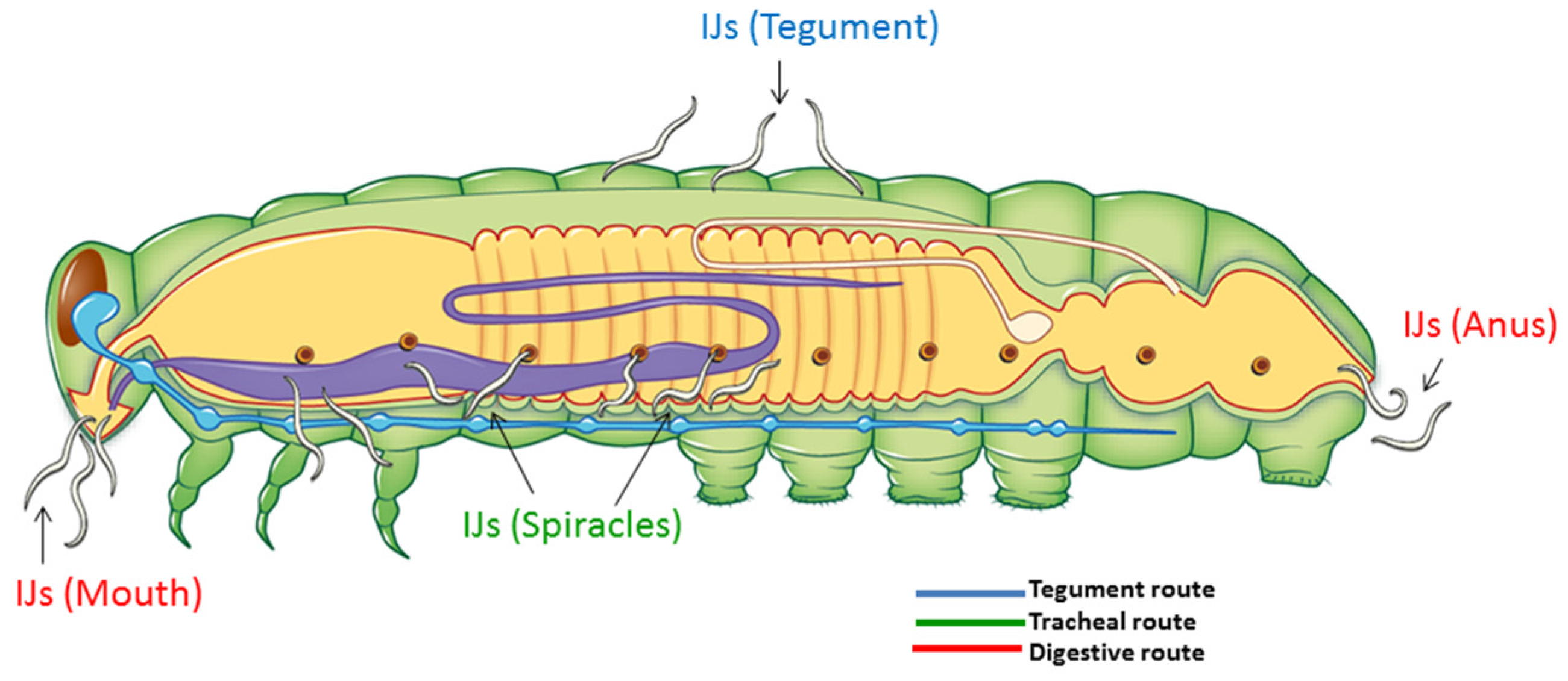
2.2. Portal of Entry and Time Until Death in Forced Infections

Time until death in G. mellonella ranged between 21 and 24 h across different portals of entry (POEs) (Table 1). This timing can be correlated with the increase in pirAB toxin transcript production that was observed in the natural iinfection assays (Figure 1). Our data also suggest that, except for the anus, all other portals of entry to a host had no significant effect on time to death.
Time until death in M. sexta, compared with G. mellonella, was delayed, varying between 25 to 33 h (Table 1). The longest time to death was observed when IJs were delivered via the mouth (Table 1) and is statistically longer when compared with both spiracle and tegument IJ injections. Time until death by both the digestive tract entry routes (mouth and anus) were not significantly different from each other.

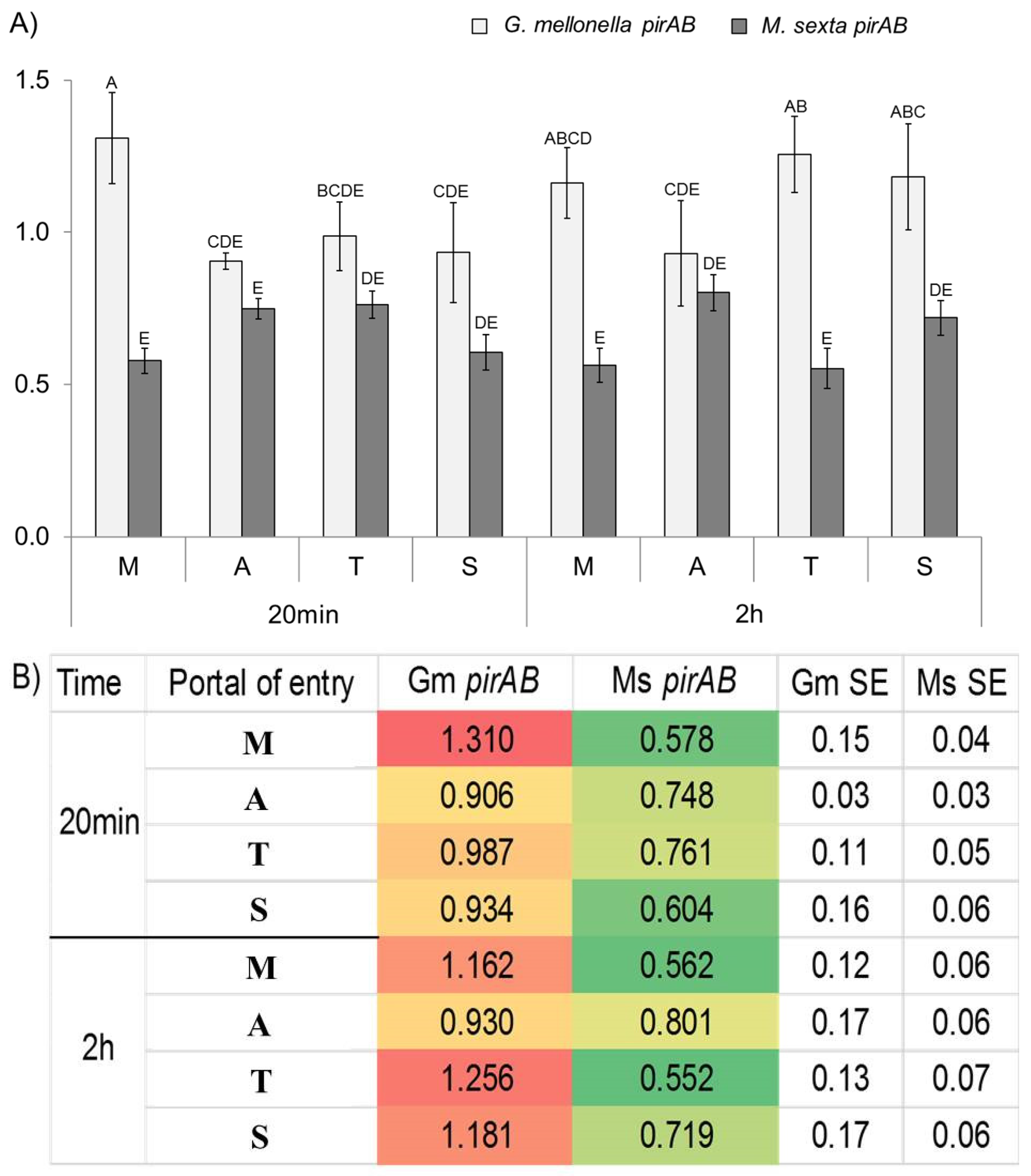
2.3. Transcript Abundance in Forced Infection Assays

PirAB plu4093-2 transcript abundance was measured considering different POEs at two observation time points: 20 min and 2 h post-injection for the two insect hosts tested.
For G. mellonella, the highest expression of pirAB transcripts was observed in the mouth at 20 min post-injection. Expression of this toxin in all other POEs was significantly lower (Figure 2). At 2 h post-infection, toxin expression was moderately expressed in mouth, tegument, and spiracles and remained low for the anus.
Contrastingly, in M. sexta, pirAB transcripts were not as highly expressed as in G. mellonella. PirAB transcript expression at all portals of entry was about the same. Anus and tegument routes were minimally higher at 20 min post-infection; however, differences were not significant (p < 0.05). Transcript expression for the mouth and spiracles entries showed a low expression profile at this time interval. At 2 h post-injection, transcription profiles remained similar to those recorded at 20 min (Figure 2).

3. Discussion

Photorhabdus luminescens PirAB toxins have been reported to possess both injectable and oral activities against a range of insects [10,15,16,17]. It has also been demonstrated that their toxicity is binary and host-specific [15,16,17,22]. For example, histological examination of P. xylostella larvae fed with E. coli expressing PirA/PirB proteins revealed midgut epithelium abnormalities, suggesting gut toxicity [15]. Furthermore, PirAB toxins have shown oral activity against mosquito larvae [10,18]. There is also evidence that, in G. mellonella, both PirA and PirB components, from both plu4093-2 and 4437-6, are necessary for injectable activity because injection of either PirA or PirB alone does not cause mortality [16,17]. Blackburn et al. also showed that, when the plu4092 gene was truncated, mortality of P. xylostella decreased from 100% to <15% [15]. In all these studies, the approach for assessing activity of Pir toxins has considered the injection of bacteria cells or toxin preparations delivered into hemocoel via tegument or orally via the ingestion of PirAB toxin. Additionally, Pir-Cry toxin chimera delivery was through oral delivery by the incorporation of toxin combinations into the insect diet [23]. Until now, the expression of Pir toxins when the Photorhabdus are delivered by their vectoring nematodes had not been investigated.
In this study, we evaluated abundance of pirAB toxin transcripts (plu4093-2 and plu4437) of the P. l. laumondii TT01 strain. Furthermore, until now, expression of Pir toxins when the bacteria are naturally delivered by their vector nematodes had not been investigated. In this respect, we investigated the transcription of independent pirAB loci, plu4093-2 and plu4437-6, in insect homogenates and under in vitro conditions (Suppl. Materials). Our observations showed that the addition of M. sexta hemolymph to LB broth slightly reduced the transcription of all the pir genes. Furthermore, there was a higher decrease of plu4093-2 than plu4437-6. However, the overall mean base mapping value for plu4093-2 was higher than plu4437-6 (Tables S1 and S2, Figure S1). We also observed that all pir genes were more highly expressed during the exponential growth phase other than the lone pirA. There was a big increase in expression of the pirA orphan gene at the stationary phase, while the other pir genes showed only minor differences. Based on these results, we only focused on expression of plu4093-2 for the subsequent experiments.
Our results showed that plu4093-2 pirAB transcripts were transcribed as early as 22 h during the course of a natural infection. However, transcript levels remained below threshold levels between 12 h and 20 h post-infection. Interestingly, a three-fold increase of transcripts was detected at 22 h post-infection in G. mellonella but not in M. sexta. In this respect, we speculate that this may be due to the fact that M. sexta is a more resilient host to Photorhabdus–Heterorhabditis infection. Therefore, the observed low expression of pir transcripts in M. sexta may be due to the fact that more time was needed to show their activity.
With respect to forced infection assays, our data showed that, for G. mellonella, pir toxin transcripts are highly expressed in the mouth at both 20 min and 2 h post-injection in H. bacteriophora IJs. We speculate that IJ nematodes may be able to release Photorhabdus cells when they enter a host through the mouth. Alternatively, it is possible that IJs may be crushed by the insect’s mouth parts, thereby allowing the ectopic release of bacterial cells and thus activation of gene expression.
Other studies have also demonstrated that activation of toxin gene expression of bacteria responding to insect tissue signals [24,25,26]. Specifically, it has been shown that exposure of P. luminescens cells to G. mellonella tissue homogenates activates production Tc and other putatively identified toxins [24]. In contrast, toxin expression in M. sexta was low for all portals of entry at both time points tested (Figure 2). In this respect, we speculate that M. sexta may have a more efficient immune system than G. mellonella; therefore, additional time may be required to propel toxin production and cause insect death. In relation to this, studies have shown that M. sexta can mount a defense against P. luminescens [25]. Low toxin transcript measurements reported in this study are consistent with observations by Daborn et al., who demonstrated that Photorhabdus tc toxin gene expression could not be detected above threshold levels at early infection time points [26].
Overall, this study demonstrated for the first time the transcription of pirAB in natural infections when P. l. laumondii cells are delivered by Heterorhabditis IJs. We also showed a three-fold increase of PirAB toxin transcription at 22 h in G. mellonella, suggesting the deployment of toxin transcripts in early stages of the infection process. Further work is needed to discern the timing of plu4093-2 and plu4437-6 translation and potential for component PirA and PirB complex formation. Furthermore, results from G. mellonella forced injection assays correlated time until death with pirAB toxin transcript production. Additionally, we showed that pirAB transcripts are produced in G. mellonella larvae when the bacteria are delivered by H. bacteriophora IJs via the mouth.

4. Experimental Section

4.1. Insects, Nematodes, and Bacterial Cultures

Fifth-instar larvae of the greater wax moth, Galleria mellonella (Lepidoptera: Pyrallidae), and early fourth instar M. sexta (Lepidoptera: Sphingidae) larvae were considered for the natural and forced infection assays. Selection of hosts was based on their known susceptibility to HeterorhabditisPhotorhabdus infection [27]. G. mellonella larvae were obtained from Timberline Fisheries (Timberline, Marion, IL, USA) and kept in the lab at 15 °C until their use in the experiments. M. sexta larvae were obtained from J. Hildebrandt’s laboratory at the University of Arizona. Prior to the experiments, larvae were surface sterilized with 70% ethanol.
Heterorhabditis bacteriophora strain TT01 was obtained from I. eleftherianos laboratory (G. Washington University, Washington, DC, USA). Nematodes were propagated in G. mellonella larvae following procedures described by Stock and Goodrich-Blair (2012) [28]. IJs were collected from modified White traps and stored in 250-mL tissue culture flasks (at a concentration of 15,000 IJs/mL) at 15 °C until their use in the experiments following procedures described by Stock and Goodrich-Blair (2012) [28].

4.2. Natural and Forced Infection Assays

Two different experimental arenas and inocula were considered for the natural infection and forced injection assays depending on the insect host tested. For the natural infection assays, a 1.7-mL micro-centrifuge tube lined with a small triangle of filter paper (Whatman #1) was used for G. mellonella infections. An inoculum of 110 IJs/larva was delivered on the filter paper, and one larva was added to each tube. A 15-mL conical centrifuge tube was considered for M. sexta infections. An inoculum of 500 IJs was delivered on a small triangle of filter paper (Whatman #1) placed inside the tube.
Forced injections were administered by delivering the same concentration of H. bacteriophora IJs into G. mellonella or M. sexta considering four portals of entry (POE): mouth, anus, spiracles, and cuticle (Figure 3). For this purpose, a single larva was injected with a syringe with a 25-gauge needle considering one portal of entry at a time and controls consisted of larvae injected with M9 buffer solution only. In both cases, one larva was added into each tube. To allow air exchange, the lid of each tube was perforated. Tubes were capped and placed in an incubator in the dark and maintained at 28 °C. There were six tubes per observation time points: 12 h, 15 h, 18 h, and 22 h for natural infections or 20 min and 2 h for forced injections. RNA from H. bacteriophora IJs was collected at these time points. Three replicates were collected per time point, and experiments were repeated twice.
A remote recording video system was built using iSpy Connect Open Source Camera Security Software (iSPY Group, Stockholm, Sweden) and a mounted webcam to record the course of infection for the forced infection assays. A 24-well plate was used as the experimental arena, and one larva per well was added. Three replicates were collected per time point and experiments were repeated twice. A time-lapse video was created with Windows MovieMaker software 2012 (Microsoft Corp., Redmond, WA, USA) to measure time stamp information to record time until death to establish mean time until death.

4.3. Detection of pirAB Transcripts through qPCR

Total RNA was extracted using TriReagent (Molecular Research Center, Cincinnati, OH, USA) according to the manufacturer’s instructions. Contaminating genomic DNA was eliminated by digestion with DNAse I (GE Healthcare, Piscataway, NJ, USA) for one hour at 37 °C. Samples were further purified using the RNA clean-up and concentration kit (Thermo Fisher Scientific, Hampton, NH, USA). Purified total RNA was checked for quality and quantified by 2% agarose gel electrophoresis and Nanodrop spectrophotometer (Nanodrop Technologies, Wilmington, DE, USA).
One microgram of total RNA, which did not amplify from PCR with endogenous control (primer pair 5 and 6 with DreamTaq K1071, according to Thermo’s recommendations) (Table 2), was considered gDNA-free and then used to synthesize cDNA. Reactions were designed according to the RevertAid First Strand cDNA Synthesis Kit (Thermo Scientific, Waltham, MA, USA) according to the manufacturer’s instructions. Relative quantification of pirAB gene expression was measured in a Mastercycler® ep gradient S (Eppendorf, San Diego, CA, USA) for forty cycles using the Realplex4 version 2.2 software (Eppendorf, San Diego, CA, USA) to collect cycle threshold (Ct) values. All primers used in qRT-PCR are presented in Table 2.
Primers targeting pirAB transcripts were designed using Geneious software version 8.0.5 (Biomatters, Auckland, New Zealand) and NCBI primer-blast. For relative quantification, an endogenous control, Gyrase subunit B (gyrB) (P. l. laumondii TTO1 plu004), was considered. The relative standard curve method was used for gene expression analysis comparing transcript levels between target and endogenous control genes between treatments. Primers (Table 2) were validated by a clean melting curve and Sanger sequencing analysis.

4.4. Statistical Analysis

Statistical analysis was performed using JMP® software version 12.2.0 Copyright© SAS Institute Inc. where a comparison of means was performed using Student’s t-test at an alpha of 0.05. For all figures and tables, unique letters denote statistical significance.

Supplementary Materials

The following are available online at www.mdpi.com/2072-6651/8/10/287/s1., Figure S1: Pir-loci expression in P. luminescens TT01 strain grown under two different media, Table S1: DESeq analysis of RNA-seq data showing the mean mapped base counts (across the ORFs) and log2 fold change for each of the pir gene homologues in P. luminescens TT01 grown in different media, LB alone or with 20% v/v M. sexta hemolymph supplement (LB+MSH). Different pir-operons are colour coded. E and S represent mid-exponential and stationary growth phases respectively, Table S2: DESeq comparison of exponential and stationary phase RNA-seq data showing mean mapped base counts (across the ORFs) and log2 fold change for each of the pir gene homologues in P. l. laumondii TT01 grown in the different media, LB alone or with 20% v/v M. sexta hemolymph supplement (LB+MSH). The three different pir-operons are colour coded.

Acknowledgments

The authors would like to acknowledge Belen Molina, an undergraduate research assistant, and Alexandra Roder, a graduate student, in S. Patricia Stock’s laboratory, for their help with experiments. We recognize support from the Postdoctoral Excellence in Research and Teaching (PERT) Program at the Center for Insect Science at The University of Arizona, which funded A. Castagnola through a National Institutes of Health training grant (1K12 GM 00708, IRACDA Program). We are also grateful to the NEMASYM (Nematode-Bacterium Symbioses) Research Coordination Network (NSF-IOS 0840932 to S. Patricia Stock). The opinions shared in this manuscript are not necessarily a reflection of those of the National Institute of Health or the National Science Foundation.

Author Contributions

A.C. and S.P.S. conceived and designed in vivo and qPCR experiments and wrote and edited the manuscript. N.D., performed in vivo injection assays, collected data and did the live video recording. A.C. analyzed the data. G.M. and N.W. designed in vitro and RNA seq experiments and edited the manuscript.

Conflicts of Interest

The authors declare no conflict of interest.

References

  1. Gaugler, R.; Kaya, H.K. Entomopathogenic Nematodes in Biological Control; CRC Press, Inc.: Boca Raton, FL, USA, 1990. [Google Scholar]
  2. Gaugler, R.; Gaugler, R. Entomopathogenic Nematology; CABI Pub.: Cambridge, MA, USA, 2002. [Google Scholar]
  3. Koppenhoefer, A.M.; Lacey, L.A.; Kaya, H.K. Nematodes; Kluwer Academic Publishers: Berlin, Germany, 2000. [Google Scholar]
  4. Thomas, G.M.; Poinar, G.O. Xenorhabdus Gen-Nov, A Genus of entomopathogenic, nematophilic bacteria of the family Enterobacteriaceae. Int. J. Syst. Bacteriol. 1979, 29, 352–360. [Google Scholar] [CrossRef]
  5. Boemare, N.E.; Akhurst, R.J.; Mourant, R.G. DNA relatedness between Xenorhabdus spp. (Enterobacteriaceae), symbiotic bacteria of entomopathogenic nematodes, and a proposal to transfer Xenorhabdus luminescens to a new Genus, Photorhabdus gen. nov. Int. J. Syst. Bacteriol. 1993, 43, 249–255. [Google Scholar] [CrossRef]
  6. Bedding, R.A.; Molyneux, A.S. Penetration of insect cuticle by infective juveniles of Heterorhabditis spp. (Heterorhabditidae, Nematoda). Nematologica 1982, 28, 354–359. [Google Scholar] [CrossRef]
  7. Grewal, P.S.; Gaugler, R.; Lewis, E.E. Host recognition behavior by entomopathogenic nematodes during contact with insect gut contents. J. Parasitol. 1993, 79, 495–503. [Google Scholar] [CrossRef]
  8. Wang, Y.; Gaugler, R. Host and penetration site location by entomopathogenic nematodes against Japanese beetle larvae. J. Invertebr. Pathol. 1998, 72, 313–318. [Google Scholar] [CrossRef] [PubMed]
  9. Silva, C.P.; Waterfield, N.R.; Daborn, P.J.; Dean, P.; Chilver, T.; Au, C.P.Y.; Sharma, S.; Potter, U.; Reynolds, S.E.; ffrench-Constant, R.H. Bacterial infection of a model insect: Photorhabdus luminescens and Manduca sexta. Cell. Microbiol. 2002, 4, 329–339. [Google Scholar] [CrossRef] [PubMed]
  10. Duchaud, E.; Rusniok, C.; Frangeul, L.; Buchrieser, C.; Givaudan, A.; Taourit, S.; Bocs, S.; Boursaux-Eude, C.; Chandler, M.; Charles, J.F.; et al. The genome sequence of the entomopathogenic bacterium Photorhabdus luminescens. Nat. Biotechnol. 2003, 21, 1307–1313. [Google Scholar] [CrossRef] [PubMed]
  11. Waterfield, N.; Hares, M.; Yang, G.; Dowling, A.; ffrench-Constant, R. Potentiation and cellular phenotypes of the insecticidal Toxin complexes of Photorhabdus bacteria. Cell. Microbiol. 2005, 7, 373–382. [Google Scholar] [CrossRef] [PubMed]
  12. Daborn, P.J.; Waterfield, N.; Silva, C.P.; Au, C.P.Y.; Sharma, S.; Ffrench-Constant, R.H. A single Photorhabdus gene, makes caterpillars floppy (MCF), allows Escherichia coli to persist within and kill insects. Proc. Natl. Acad. Sci. USA 2002, 99, 10742–10747. [Google Scholar] [CrossRef] [PubMed]
  13. Dowling, A.J.; Daborn, P.J.; Waterfield, N.R.; Wang, P.; Streuli, C.H.; ffrench-Constant, R.H. The insecticidal toxin Makes caterpillars floppy (MCF) promotes apoptosis in mammalian cells. Cell. Microbiol. 2004, 6, 345–353. [Google Scholar] [CrossRef] [PubMed]
  14. Yang, G.; Dowling, A.J.; Gerike, U.; ffrench-Constant, R.H.; Waterfield, N.R. Photorhabdus virulence cassettes confer injectable insecticidal activity against the wax moth. J. Bacteriol. 2006, 188, 2254–2261. [Google Scholar] [CrossRef] [PubMed]
  15. Blackburn, M.B.; Farrar, R.R.; Novak, N.G.; Lawrence, S.D. Remarkable susceptibility of the diamondback moth (Plutella xylostella) to ingestion of Pir toxins from Photorhabdus luminescens. Entomol. Exp. Appl. 2006, 121, 31–37. [Google Scholar] [CrossRef]
  16. Waterfield, N.; Kamita, S.G.; Hammock, B.D.; Ffrench-Constant, R. The Photorhabdus Pir toxins are similar to a developmentally regulated insect protein but show no juvenile hormone esterase activity. FEMS Microbiol. Lett. 2005, 245, 47–52. [Google Scholar] [CrossRef] [PubMed]
  17. Sun, J.; Liu, C.; Qiu, L. Cloning, expression and insecticidal activity of the pirA2B2 gene from Photorhabdus luminescens TT01. Weishengwu Xuebao 2012, 52, 532–537. [Google Scholar]
  18. Ahantarig, A.; Chantawat, N.; Waterfield, N.R.; Ffrench-Constant, R.; Kittayapong, P. PirAB toxin from Photorhabdus asymbiotica as a larvicide against dengue vectors. Appl. Environ. Microbiol. 2009, 75, 4627–4629. [Google Scholar] [CrossRef] [PubMed]
  19. Li, Y.; Hu, X.; Zhang, X.; Liu, Z.; Ding, X.; Xia, L.; Hu, S. Photorhabdus luminescens PirAB-fusion protein exhibits both cytotoxicity and insecticidal activity. FEMS Microbiol. Lett. 2014, 356, 23–31. [Google Scholar] [CrossRef] [PubMed]
  20. Lee, C.-T.; Chen, I.T.; Yang, Y.-T.; Ko, T.-P.; Huang, Y.-T.; Huang, J.-Y.; Huang, M.-F.; Lin, S.-J.; Chen, C.-Y.; Lin, S.-S.; et al. The opportunistic marine pathogen Vibrio parahaemolyticus becomes virulent by acquiring a plasmid that expresses a deadly toxin. Proc. Natl. Acad. Sci. USA 2015, 112, 10798–10803. [Google Scholar] [CrossRef] [PubMed]
  21. Han, J.E.; Tang, K.F.J.; Tran, L.H.; Lightner, D.V. Photorhabdus insect-related (Pir) toxin-like genes in a plasmid of Vibrio parahaemolyticus, the causative agent of acute hepatopancreatic necrosis disease (AHPND) of shrimp. Dis. Aquat. Org. 2015, 113, 33–40. [Google Scholar] [CrossRef] [PubMed]
  22. Ffrench-Constant, R.H.; Dowling, A.; Waterfield, N.R. Insecticidal toxins from Photorhabdus bacteria and their potential use in agriculture. Toxicon 2007, 49, 436–451. [Google Scholar] [CrossRef] [PubMed]
  23. Hu, X.; Liu, Z.; Li, Y.; Ding, X.; Xia, L.; Hu, S. PirB-Cry2Aa hybrid protein exhibits enhanced insecticidal activity against Spodoptera exigua larvae. J. Invertebr. Pathol. 2014, 120, 40–42. [Google Scholar] [CrossRef] [PubMed]
  24. Muench, A.; Stingl, L.; Jung, K.; Heermann, R. Photorhabdus luminescens genes induced upon insect infection. MCB Genom. 2008, 9. [Google Scholar] [CrossRef] [PubMed]
  25. Eleftherianos, I.; Marokhazi, J.; Millichap, P.J.; Hodgkinson, A.J.; Sriboonlert, A.; ffrench-Constant, R.H.; Reynolds, S.E. Prior infection of Manduca sexta with non-pathogenic Escherichia coli elicits immunity to pathogenic Photorhabdus luminescens: Roles of immune-related proteins shown by RNA interference. Insect Biochem. Mol. Biol. 2006, 36, 517–525. [Google Scholar] [CrossRef] [PubMed]
  26. Daborn, P.J.; Waterfield, N.; Blight, M.A.; Ffrench-Constant, R.H. Measuring virulence factor expression by the pathogenic bacterium Photorhabdus luminescens in culture and during insect infection. J. Bacteriol. 2001, 183, 5834–5839. [Google Scholar] [CrossRef] [PubMed]
  27. Miranda, V.A.; Navarro, P.D.; Davidowitz, G.; Bronstein, J.; Stock, S.P. Effect of insect host age and diet on the fitness of the entomopathogenic nematode-bacteria mutualism. Symbiosis 2013, 61, 145–153. [Google Scholar] [CrossRef]
  28. Stock, S.P.; Goodrich-Blair, H.E. Nematode parasites, pathogens and associates of insects and invertebrates of economic importance. In Manual of Techniques in Invertebrate Pathology, 2nd ed.; Elsevier Ltd.: San Diego, CA, USA, 2012; pp. 373–426. [Google Scholar]
Figure 1. PirAB transcript abundance detected by qPCR during the course of natural infection in G. mellonella and M. sexta. Different letters denote statistically significant means.
Toxins 08 00287 g001
Figure 2. (A) Bar graph of pirAB transcripts in forced injection assays considering different portals of entry (M: Mouth, A: Anus, T: Tegument, S: Spiracle). Different letters denote statistically significant means. (B) Heat map of pirAB transcript abundance detected by qPCR using relative standard curve method. Gm = G. mellonella, Ms = M. sexta.
Toxins 08 00287 g002
Figure 3. Schematic representation showing portals of entry considered and tissue targets for the forced infection assays.
Toxins 08 00287 g003
Table 1. Time until death based on portal of entry in G. mellonella and M. sexta.
Portal of EntryTime Until Death (h ± SE)
G. mellonella *M. sexta *
Mouth21 a ± 0.7233 a ± 2.47
Anus24 b ± 0.8528 a,b ± 2.31
Spiracle23 a,b ± 1.2126 b ± 1.42
Tegument22 a,b ± 0.7425 b ± 1.65
* Unique letters for each species column denote statistical significance based on means comparison using Student’s t-test (p < 0.05). Different letters indicate statistically significant differences.
Table 2. Primers used in this study.
Toxin Transcript DetectedGene IDAmplicon Size (bp)SequenceTm (°C)GC (%)
pirABplu4093-2231GGCACGTTAACACCTTCTCT5848
TGCAGTTGGTCCTTTGAGTGA6248
gyrBplu004105CTCGTGAAGCAGCCCGTAAA6455
CCGGATCACGTTCCTGACAA6455

