Article

Alkaline pH, Low Iron Availability, Poor Nitrogen Sources and CWI MAPK Signaling Are Associated with Increased Fusaric Acid Production in Fusarium oxysporum

1
Department of Agricultural, Environmental and Food Sciences, University of Molise, 86100 Campobasso, Italy
2
Departamento de Genética, Campus de Excelencia Internacional Agroalimentario ceiA3, Universidad de Córdoba, 14014 Córdoba, Spain
3
Department of Agricultural Sciences, Università di Napoli Federico II, 80055 Portici, Italy
4
Center for Studies on Bioinspired Agro-Enviromental Technology, Università di Napoli Federico II, 80055 Portici, Italy
*
Authors to whom correspondence should be addressed.
Current address: Instituto Maimonides de Investigación Biomédica de Cordoba (IMIBIC), Universitdad de Córdoba, 14014 Córdoba, Spain.
Toxins 2023, 15(1), 50; https://doi.org/10.3390/toxins15010050
Submission received: 22 November 2022 / Revised: 18 December 2022 / Accepted: 29 December 2022 / Published: 6 January 2023
(This article belongs to the Special Issue Biological Activities and Potential Applications of Phytotoxins)

Abstract

:
Fusaric acid (FA) is one of the first secondary metabolites isolated from phytopathogenic fungi belonging to the genus Fusarium. This molecule exerts a toxic effect on plants, rhizobacteria, fungi and animals, and it plays a crucial role in both plant and animal pathogenesis. In plants, metal chelation by FA is considered one of the possible mechanisms of action. Here, we evaluated the effect of different nitrogen sources, iron content, extracellular pH and cellular signalling pathways on the production of FA siderophores by the pathogen Fusarium oxysporum (Fol). Our results show that the nitrogen source affects iron chelating activity and FA production. Moreover, alkaline pH and iron limitation boost FA production, while acidic pH and iron sufficiency repress it independent of the nitrogen source. FA production is also positively regulated by the cell wall integrity (CWI) mitogen-activated protein kinase (MAPK) pathway and inhibited by the iron homeostasis transcriptional regulator HapX. Collectively, this study demonstrates that factors promoting virulence (i.e., alkaline pH, low iron availability, poor nitrogen sources and CWI MAPK signalling) are also associated with increased FA production in Fol. The obtained new insights on FA biosynthesis regulation can be used to prevent both Fol infection potential and toxin contamination.
Key Contribution: Production of fusaric acid in Fusarium oxysporum is associated with virulence-promoting conditions: alkaline pH, low iron availability, poor nitrogen sources and CWI MAPK signalling.

1. Introduction

Fusarium oxysporum comprises a cosmopolitan complex of fungal species [1] including both non-pathogenic and pathogenic forms [2], which can infect plants, animals and humans [3]. Plant pathogenic strains cause tracheomycosis or foot and root rots (Fusarium wilt) in a large number of plant species and are grouped into over 150 pathogenic forms (formae speciales) [4]. F. oxysporum f. sp. lycopersici (Fol) is the pathogenic form that causes wilting of tomato plants. Fusarium species are known to synthesize several biologically active compounds with different roles in plant pathogenesis and microbial competition [5]. Fusaric acid (5-butylpyridine-2-carboxylic acid) (FA), the first fungal metabolite discovered to be implicated in tomato pathogenesis [6], is one of the most widely distributed mycotoxins in the genus Fusarium and has been used as an efficient indicator of Fusarium contamination in food and feed grains [7]. Plant treatment with FA causes the rapid development of disease symptoms such as internerval necrosis and foliar desiccation even in the absence of the pathogen [8]. FA toxicity in plants has been attributed to different mechanisms of action, including direct membrane damage, electrolyte loss, decrease in cellular ATP levels, metallo-enzyme inhibition, oxidative burst and metal chelation [6,9,10,11]. In addition to its phytotoxic effect, FA also shows varying degrees of inhibitory activity on rhizobacterial populations. For instance, species of the genera Bacillus and Paenibacillus are susceptible, while those belonging to Pseudomonas are highly resistant [12]. Intriguingly, FA resistance in these species has been shown to rely on the expression of two major siderophores, pyoverdine and enantio-pyochelin, suggesting that the inhibitory effect of FA toxicity on bacteria, similar to plants, depends on its chelating activity [13].
In Fusarium fujikuroi the expression of FA biosynthesis genes has been shown to be regulated by the nitrogen-responsive GATA transcription factor AreB and the pH-responsive transcription factor PacC [14]. Similarly, FA levels in Fol are controlled via PacC-mediated modulation of chromatin condensation at the fub1 locus, which encodes a major FA biosynthetic gene [15]. Additionally, in a banana pathogenic isolate of F. oxysporum f. sp. cubense, several components of the CWI MAPK cascade have been shown to act as positive regulators of FA biosynthetic genes and FA production [16].
In Fol, three different MAPKs (Fmk1, Mpk1 and Hog1) regulating distinct virulence functions have been described. While Fmk1 is essential for invasive growth and plant infection, the other two MAPKs, Mpk1 and Hog1, contribute to plant infection both via Fmk1-shared and -independent functions, albeit to a lesser extent [17,18,19]. This work aimed at evaluating the role of different environmental factors such as nitrogen source, extracellular pH and iron content in the regulation of FA production in Fol. Furthermore, we tested the contribution of the three MAPK pathways as well as the iron and pH response regulators HapX and PacC, respectively, on FA biosynthesis.

2. Results

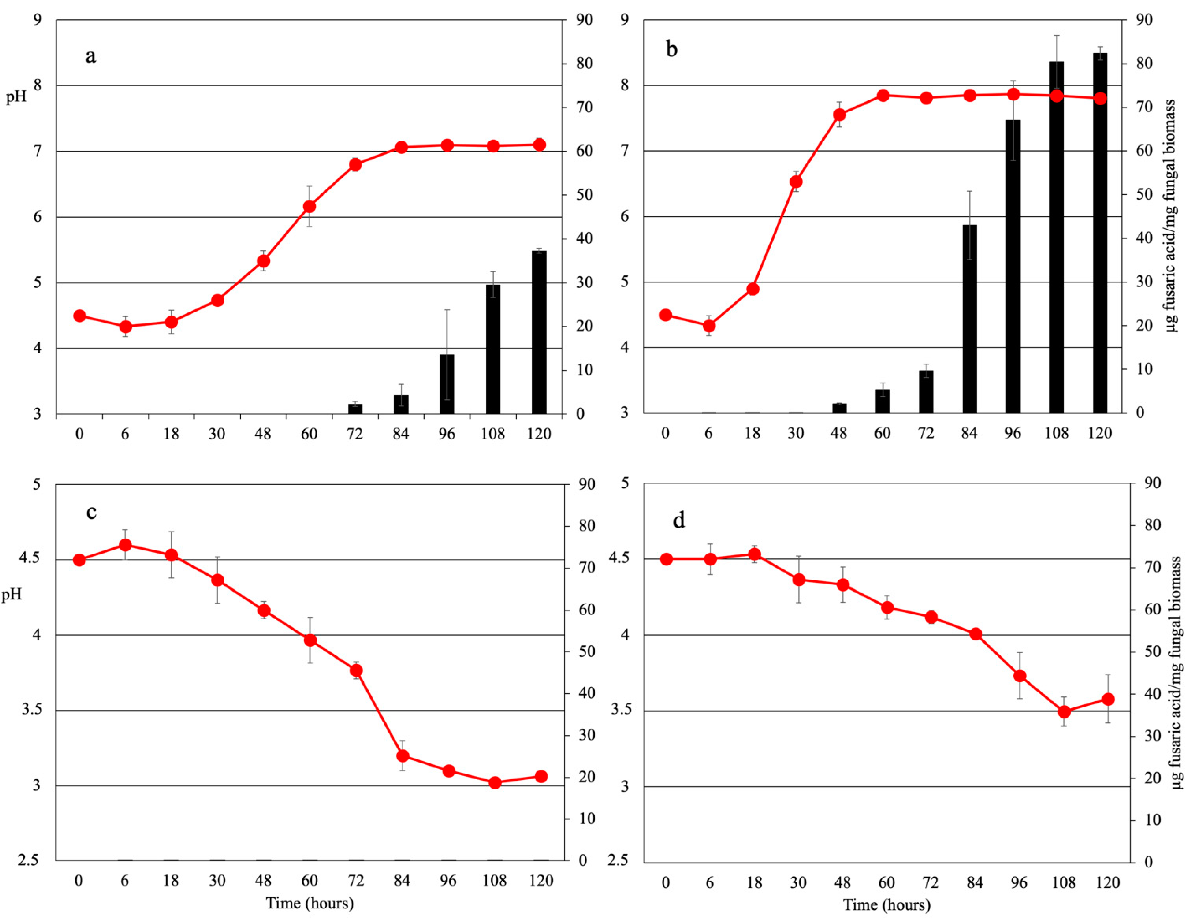
2.1. Nitrogen Source Affects Extracellular pH in F. oxysporum f. sp. lycopersici

We tested the effect of different nitrogen sources on extracellular pH modification, iron chelating activity and FA production by Fol. Microconidia were inoculated in minimal medium (pH 4.5) supplemented with either urea, sodium nitrate, ammonium sulphate or ammonium nitrate as the sole nitrogen source. While urea and sodium nitrate elicited an increase in extracellular pH, ammonium sulphate and ammonium nitrate induced extracellular acidification (Table 1; Figure 1). To investigate the role of MAPK pathways in pH modulation, we measured extracellular pH changes in fmk1Δ, hog1Δ and mpk1Δ knock-out mutants. Interestingly, deletion of the cell wall integrity (CWI) MAPK Mpk1 resulted in increased extracellular alkalinisation on urea and sodium nitrate but did not affect ammonium-dependent acidification. Similarly, mutations in the seven-transmembrane α-pheromone receptor Ste2 or the conserved components of the CWI pathway Bck1 and Mkk2 also led to an increase in pH (Table 1), suggesting for a role of the sex pheromone perception machinery and the CWI pathway in the regulation of this process.

2.2. Siderophore Production Is pH- and Iron-Sensing Dependent

To detect siderophore production by Fol, a CAS assay was performed after 5 days of incubation in the different test conditions. We found that the chelating activity in fungal supernatants was more than 40% higher in media containing urea or sodium nitrate showing high pH, compared to those supplemented with ammonium sulphate or ammonium nitrate which had low pH (Table 1). Furthermore, chelating activity was significantly higher (p ≤ 0.05) in isogenic mutants lacking Ste2 or conserved components of the CWI MAPK pathway, but not in fmk1Δ and hog1Δ mutants, compared with the wild-type strain (Table 1). The correlation between pH signalling and siderophore production was further supported by the finding that deletion the of alkaline pH-responsive factor PacC (pacCΔ) resulted in decreased chelating activity in urea and nitrate media, whereas expression of a dominant activating PacC allele (pacCc), which represses acidic-regulated functions [20,21], resulted in increased chelating activity in ammonium-containing media (Table 1).
Previous studies revealed that deletion of the iron homeostasis regulator HapX induces a slight increase in extracellular chelating activity under iron-limiting conditions when using glutamine (Gln) as the nitrogen source [22]. Here, we detected a significant increase in extracellular chelating activity in hapXΔ as compared to the wild type, which was even more dramatic when ammonium was used as a nitrogen source.

2.3. Fusaric Acid Production Is Regulated by Environmental pH and Iron Availability

Fusaric acid (FA) has been suggested to function in metal cation chelation [13,15]. Here, we found that FA concentrations increased steadily in supernatants of the wild-type strain grown on urea or sodium nitrate, whereas no FA was detected in cultures supplemented with ammonium even after prolonged incubation (Figure 1 and Figure 2; Table 1). These findings suggest that either the nitrogen source or environmental pH regulates FA production. In line with the second hypothesis, buffering the pH to 4.5 completely abolished FA production on urea- or sodium nitrate-containing media, while increasing the pH to 7.0 activated FA production on ammonium-supplemented media (Figure 3). Further corroborating the finding of a pH-dependent FA regulation mechanism in Fol, inappropriate pH sensing in the pacCΔ and pacCc mutants resulted in altered FA levels at alkaline or acidic pH, respectively.
In general, FA production appeared to correlate with chelating activity in fungal supernatants, particularly in sodium nitrate-supplemented cultures, which contained high levels of chelating activity and FA (Table 1). Interestingly, the addition of the general ion chelator EDTA or the specific iron-chelating bacterial siderophore pyoverdine resulted in increased FA concentrations regardless of the nitrogen source used, even though pyoverdine stimulation was most effective on sodium nitrate (Figure 3). By contrast, iron addition dramatically decreased FA production in all tested conditions (Figure 3), suggesting that iron sensing plays an important role in the regulation of FA biosynthesis. Indeed, the deletion of the iron-response transcriptional regulator HapX led to increased FA production in all tested media except for that containing sodium nitrate (Table 1).
Unexpectedly, the ste2Δ, bck1Δ, mkk2Δ and mpk1Δ mutants showed lower levels of FA production in both urea- and sodium nitrate-supplemented media, despite the higher chelating activity detected in these conditions (Table 1).

3. Discussion

Since its discovery more than 70 years ago, fusaric acid (FA) has been among the most studied fungal secondary metabolites produced by Fusarium phytopathogenic species [7,23]. Its wide spectrum toxicity towards plants, animals, bacteria and fungi has attracted the attention of many scientists with the aim of identifying its biosynthetic gene cluster, the environmental conditions eliciting its production/secretion and its mode of action. Recently, different genes (fub1, fub2, fub3, fub4 and fub5) have been described as being responsible for FA biosynthesis in plant pathogenic Fusarium species, and their transcription has been found to be regulated by several environmental factors, including ambient pH, nitrogen source, nutrient availability and presence of a plant host [24]. Although the toxicity mechanism of FA is not fully understood, an increasing body of evidence suggests that metal chelation represents a major mechanism for the toxic effect of FA on plants, mammals and competing rhizobacteria [13,15]. In this work, we investigated the effect of environmental conditions on FA production by Fol. Previous work by Lopez-Berges and co-workers [25] showed that Fol is able to use a large variety of nitrogen sources. While a readily metabolized source ammonium inhibited virulence-related functions, a non-preferred source nitrate promoted such functions [25]. Moreover, it was shown that the ability of fungal pathogens to invade and kill plants depends upon cellular iron homeostasis, environmental iron availability and rhizosphere pH [22,26,27,28,29]. High iron availability and acidic pH inhibit virulence, while low iron availability and alkaline pH promote infection [29].
Here, we found that Fol cultures grown in the presence of ammonium show acidic pH values, while those supplemented with nitrate or urea show high pH values. It is important to note that iron availability in soils depends largely on redox potential and pH, where iron solubility decreases as soil pH increases [30]. To overcome reduced iron availability, soil-inhabiting microbes have evolved a battery of high-affinity, low-molecular-weight iron chelators known as siderophores, which are secreted into the environment for efficient acquisition of limited iron pools [31]. In line with this, Fol cultures grown on nitrogen sources leading to alkaline pH values showed higher chelating activity than those grown in acidic conditions. Interestingly, a similar pattern was observed for FA accumulation in culture supernatants, suggesting a possible role in FA production under iron-limiting conditions. In support of this idea, external addition of the chelating agent EDTA or the bacterial siderophore pyoverdine, as well as buffering the medium to pH 7.0, induced an increase in FA levels, while exogenous addition of iron or buffering to pH 4.5 resulted in reduced FA production. Thus, FA biosynthesis in Fol appears to be regulated by environmental pH and, consequently, iron availability. In line with this, Fol mutants in PacC, which are affected in external pH sensing, were altered in FA production and chelating activity. This is in agreement with previous reports indicating the requirement of PacC for efficient expression of fub genes in F. fujikuroi and siderophore production in Aspergillus nidulans at alkaline pH [14,32]. It is noteworthy that, similarly to siderophore biosynthesis, FA production also appears to depend on iron homeostasis in Fol because deletion of the iron homeostasis regulator HapX [22], a repressor of siderophore biosynthesis [33], led to an overall increase in FA levels and chelating activity. Thus, FA production in Fol might be regulated via two independent mechanisms based on pH and iron sensing.
In the banana pathogen F. oxysporum f. sp. cubense, Bck1, Mkk2 and Mpk1, three conserved elements of the CWI MAPK signalling cascade, were shown to positively regulate the expression of FA biosynthetic genes and FA production [16]. Here, we found that Fol mutants lacking Mpk1, which is required for host sensing and virulence [18], produced less FA, suggesting that FA production in F. oxysporum is regulated by the CWI MAPK pathway. Surprisingly, the α-pheromone specific receptor Ste2, which was recently shown to signal via the CWI MAPK pathway to regulate chemotropism and conidial germination in Fol [18,34,35], also showed lower FA accumulation, suggesting a new role of pheromone autocrine signalling in FA production. Importantly, this effect was independent of extracellular pH and chelating activity, indicating that pheromone signalling-mediated FA production functions downstream of pH and Fe sensing in Fol.
Collectively, our results show that virulence-promoting conditions such as alkaline pH, low iron availability, poor nitrogen sources and CWI MAPK signalling are associated with an increase in FA production, suggesting that Fol has evolved both independent and overlapping strategies to fine-tune the production of this important mycotoxin.

4. Materials and Methods

4.1. Fungal Isolates and Culture Condition

Fungal strains derived from Fusarium oxysporum f. sp. lycopersici (Fol) isolate 4287 (FGSC 9935) and used in this study are reported in Table 2. For microconidia production, fungal strains were grown in Potato Dextrose Broth (PDB; Difco; Fisher Scientific; Rodano, MI, Italy) at 28 °C with shaking at 180 rpm for 5 days. For in vitro fusaric acid (FA) detection, Puhalla minimal medium (MgSO4·7 H2O 2 mM, KH2PO4 7 mM, KCl 6 mM, Sucrose 90 mM) adjusted to pH 5.0 and supplemented with 25 mM of different nitrogen sources (NaNO3; (NH4)2SO4; NH4NO3; CH4N2O) was used [36]. Where indicated, the medium was pH buffered to 7.0 or 5.0 with 100 mM 4-morpholineethanesulfonic acid monohydrate (MES) or supplemented with 100 μM FeCl3, 0.2 mM EDTA or 0.2 mM pyoverdine. Fungal strains were inoculated in the growth medium at a final concentration of 5 × 10⁵ conidia mL−1 and incubated for 7 days at 28 °C on a rotary shaker regulated at 180 rpm. The dry weight of the fungal biomass and pH of the culture broth were evaluated periodically.

4.2. Chrome Azurol S Assay

Siderophore quantification in fungal supernatants was carried out by using the liquid chrome azurol S assay (CAS) (as previously described [37]. The percentage of chelating activity (CA%) was indirectly quantified by measuring the OD655 of the culture supernatant (ODs) and the uninoculated medium (ODc), used as a control, 60 min after the start of the reaction, with the following formula: CA% = [(ODc − ODs)/ODc] × 100.

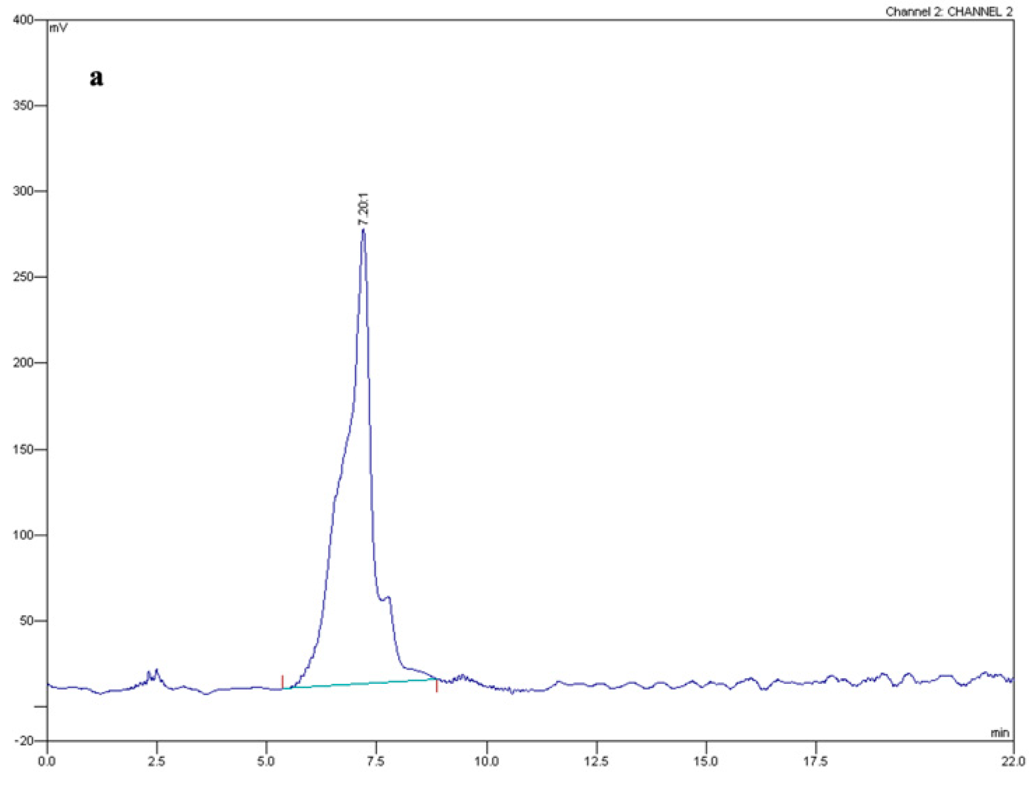
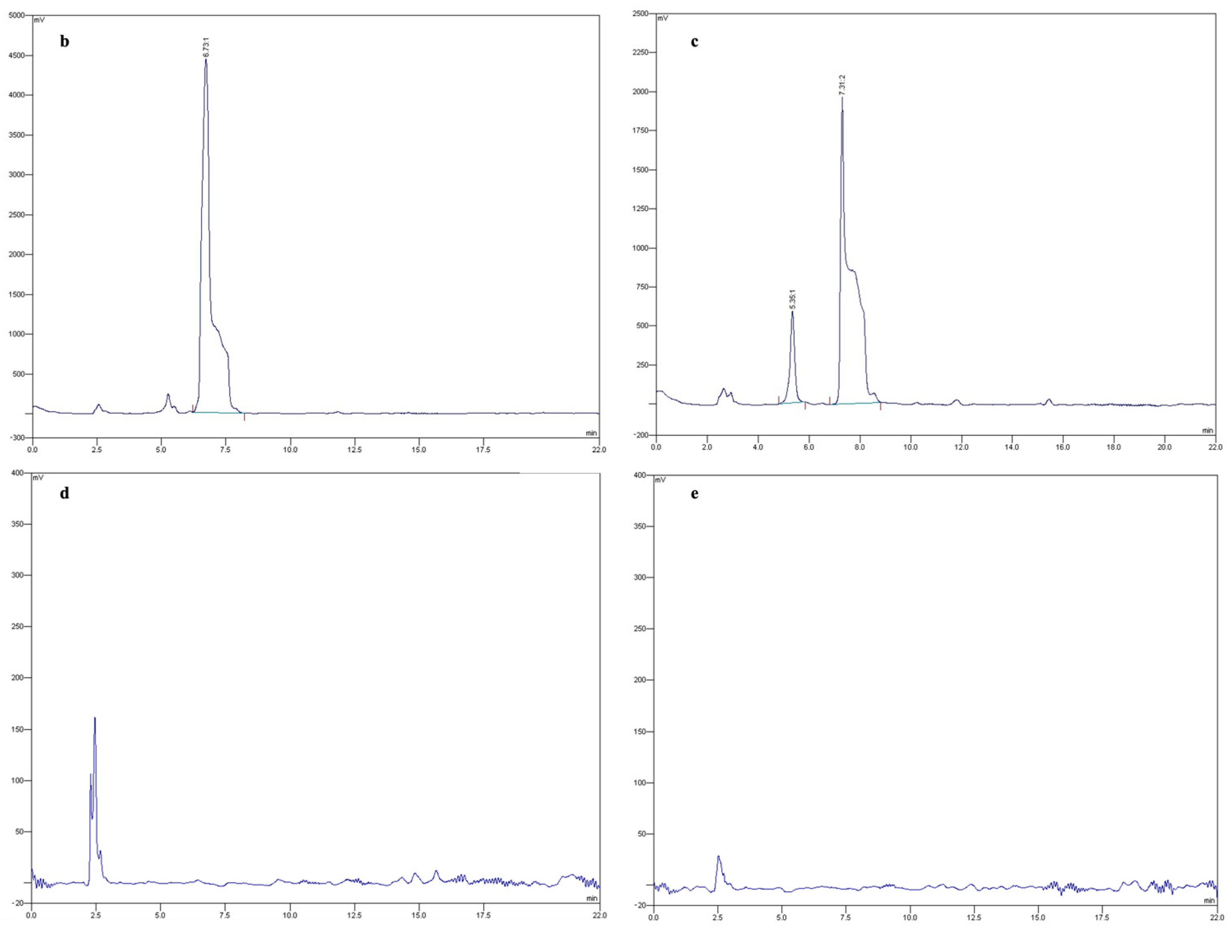
4.3. Fusaric Acid Extraction and Quantification

For FA extraction from fungal cultures, supernatants were collected and filtered through Whatman no. 4 filter paper (Whatman Ltd., Maidstone, UK), adjusted to pH 2.0 with HCl (37% v/v) and extracted with ethyl acetate (1:1 v/v). Ethyl acetate phases were separately collected, dried by rotary evaporation under reduced pressure and resuspended in methanol. FA content was quantified through high-performance liquid chromatography (HPLC-UV; Varian Analytical Instruments; Model 9010; Palo Alto, CA, USA) by using previously reported methods and experimental conditions [38,39] and expressed in µg of FA per mg of dry fungal biomass. To obtain an FA calibration curve, methanol-dissolved FA standard solution (Merck Life Science; Milano, MI, Italy) was injected at concentrations ranging from 0.02 to 2.0 mg mL−1. A linear relationship between peak areas and the investigated FA concentrations was obtained (Y = 0.001x + 0.0105; R2 value of 0.997). Method validation was performed by spiking MM blanks with known concentrations of FA. The average recovery rate was 96% and always exceeded 95%.

4.4. Statistical Analyses

Data were submitted to variance analysis (ANOVA) using the SPSS software v. 16.0 (SPSS Inc., Chicago, IL, USA) and means compared with Tukey’s test. Before analyses, percentages of chelating activity were converted into Bliss angular values (arcsine square root of the percentage value). All experiments were repeated at least three times, with similar results. Homogeneity of variance for independent repetitions of each experiment was tested, and data from separate experiments having homogeneous variances were pooled.

Author Contributions

Conceptualization, D.P. and D.T.; methodology, D.S.; validation, D.P., F.D.C., G.L., A.D.P. and D.T.; resources, M.S.L.-B.; writing—original draft preparation, D.P.; writing—review and editing, A.D.P. and D.T.; supervision, D.T., F.D.C. and G.L.; funding acquisition, D.T., A.D.P. and G.L. All authors have read and agreed to the published version of the manuscript.

Funding

This work was supported by grants from the Spanish Ministry of Science and Innovation (MICINN, grant PID2019-108045RB-I00) and Junta de Andalucía (P20_00179) to A.D.P.; grants by the Italian Ministry of Education, University and Research (PRIN-BiPP grant 2020T58TA3) and the Università degli Studi di Napoli Federico II (FRA-Line B-2020-TOPOPATH grant PG/2021/0034842) to D.T.; and grants by the Italian Ministry of Education (PRIN 20089LSZ2A_003) to G.L. This study was carried out within the Agritech National Research Center and received funding from the European Union Next-GenerationEU (PIANO NAZIONALE DI RIPRESA E RESILIENZA (PNRR)—MISSIONE 4 COMPONENTE 2, INVESTIMENTO 1.4–D.D. 1032 17/06/2022, CN00000022). This manuscript reflects only the authors’ views and opinions, neither the European Union nor the European Commission can be considered responsible for them.

Institutional Review Board Statement

Not applicable.

Informed Consent Statement

Not applicable.

Data Availability Statement

Not applicable.

Conflicts of Interest

The authors declare no conflict of interest.

References

  1. Gordon, T.R.; Martyn, R.D. The evolutionary biology of Fusarium oxysporum. Annu. Rev. Phytopathol. 1997, 35, 111–128. [Google Scholar] [CrossRef] [PubMed] [Green Version]
  2. Fuchs, J.G.; Moënne-Loccoz, Y.; Défago, G. Nonpathogenic Fusarium oxysporum strain fo47 induces resistance to Fusarium wilt in tomato. Plant Dis. 1997, 81, 492–496. [Google Scholar] [CrossRef] [PubMed] [Green Version]
  3. Kang, S.; Demers, J.; del Mar Jimenez-Gasco, M.; Rep, M. Fusarium oxysporum. In Genomics of Plant-Associated Fungi and Oomycetes: Dicot Pathogens; Dean, R.A., Lichens-Park, A., Kole, C., Eds.; Springer: Berlin/Heidelberg, Germany, 2014; pp. 99–119. [Google Scholar]
  4. Michielse, C.B.; Rep, M. Pathogen profile update: Fusarium oxysporum. Mol. Plant Pathol. 2009, 10, 311–324. [Google Scholar] [CrossRef] [PubMed]
  5. Keller, N.P.; Turner, G.; Bennett, J.W. Fungal secondary metabolism [mdash] from biochemistry to genomics. Nat. Rev. Microbiol. 2005, 3, 937–947. [Google Scholar] [CrossRef]
  6. Gaumann, E. Fusaric acid as a wilt toxin. Phytopathology 1957, 47, 342–357. [Google Scholar]
  7. Bacon, C.W.; Porter, J.K.; Norred, W.P.; Leslie, J.F. Production of fusaric acid by Fusarium species. Appl. Environ. Microbiol. 1996, 62, 4039–4043. [Google Scholar] [CrossRef] [Green Version]
  8. Dong, X.; Ling, N.; Wang, M.; Shen, Q.; Guo, S. Fusaric acid is a crucial factor in the disturbance of leaf water imbalance in fusarium-infected banana plants. Plant. Physiol. Biochem. 2012, 60, 171–179. [Google Scholar] [CrossRef]
  9. Bouizgarne, B.; El-Maarouf-Bouteau, H.; Frankart, C.; Reboutier, D.; Madiona, K.; Pennarun, A.M.; Monestiez, M.; Trouverie, J.; Amiar, Z.; Briand, J.; et al. Early physiological responses of Arabidopsis thaliana cells to fusaric acid: Toxic and signalling effects. New Phytol. 2006, 169, 209–218. [Google Scholar] [CrossRef]
  10. Stipanovic, R.D.; Puckhaber, L.S.; Liu, J.; Bell, A.A. Phytotoxicity of fusaric acid and analogs to cotton. Toxicon 2011, 57, 176–178. [Google Scholar] [CrossRef]
  11. Singh, V.K.; Upadhyay, R.S. Fusaric acid induced cell death and changes in oxidative metabolism of Solanum lycopersicum. Bot. Stud. 2014, 55, 66. [Google Scholar] [CrossRef] [Green Version]
  12. Landa, B.B.; Cachinero-Díaz, J.M.; Lemanceau, P.; Jiménez-Díaz, R.M.; Alabouvette, C. Effect of fusaric acid and phytoanticipins on growth of rhizobacteria and Fusarium oxysporum. Can. J. Microbiol. 2002, 48, 971–985. [Google Scholar] [CrossRef] [PubMed]
  13. Ruiz, J.A.; Bernar, E.M.; Jung, K. Production of siderophores increases resistance to fusaric acid in Pseudomonas protegens pf-5. PLoS ONE 2015, 10, e0117040. [Google Scholar] [CrossRef] [PubMed] [Green Version]
  14. Niehaus, E.M.; von Bargen, K.W.; Espino, J.J.; Pfannmuller, A.; Humpf, H.U.; Tudzynski, B. Characterization of the fusaric acid gene cluster in Fusarium fujikuroi. Appl. Microbiol. Biotechnol. 2014, 98, 1749–1762. [Google Scholar] [CrossRef] [PubMed]
  15. López-Díaz, C.; Rahjoo, V.; Sulyok, M.; Ghionna, V.; Martín-Vicente, A.; Capilla, J.; Di Pietro, A.; López-Berges, M.S. Fusaric acid contributes to virulence of Fusarium oxysporum on plant and mammalian hosts. Mol. Plant Pathol. 2018, 19, 440–453. [Google Scholar] [CrossRef] [Green Version]
  16. Ding, Z.; Li, M.; Sun, F.; Xi, P.; Sun, L.; Zhang, L.; Jiang, Z. Mitogen-activated protein kinases are associated with the regulation of physiological traits and virulence in Fusarium oxysporum f. sp. cubense. PLoS ONE 2015, 10, e0122634. [Google Scholar] [CrossRef] [Green Version]
  17. Segorbe, D.; Di Pietro, A.; Pérez-Nadales, E.; Turrà, D. Three Fusarium oxysporum mitogen-activated protein kinases (mapks) have distinct and complementary roles in stress adaptation and cross-kingdom pathogenicity. Mol. Plant Pathol. 2016, 18, 912–924. [Google Scholar] [CrossRef]
  18. Turrà, D.; El Ghalid, M.; Rossi, F.; Di Pietro, A. Fungal pathogen uses sex pheromone receptor for chemotropic sensing of host plant signals. Nature 2015, 527, 521–524. [Google Scholar] [CrossRef]
  19. Di Pietro, A.; García-Maceira, F.I.; Méglecz, E.; Roncero, M.I.G. A map kinase of the vascular wilt fungus Fusarium oxysporum is essential for root penetration and pathogenesis. Mol. Microbiol. 2001, 39, 1140–1152. [Google Scholar] [CrossRef]
  20. Prusky, D.; McEvoy, J.L.; Saftner, R.; Conway, W.S.; Jones, R. Relationship between host acidification and virulence of Penicillium spp. on apple and citrus fruit. Phytopathology 2004, 94, 44–51. [Google Scholar] [CrossRef] [Green Version]
  21. Caracuel, Z.; Roncero, M.I.; Espeso, E.A.; Gonzalez-Verdejo, C.I.; Garcia-Maceira, F.I.; Di Pietro, A. The ph signalling transcription factor pacc controls virulence in the plant pathogen Fusarium oxysporum. Mol. Microbiol. 2003, 48, 765–779. [Google Scholar] [CrossRef]
  22. López-Berges, M.S.; Capilla, J.; Turrà, D.; Schafferer, L.; Matthijs, S.; Jöchl, C.; Cornelis, P.; Guarro, J.; Haas, H.; Di Pietro, A. Hapx-mediated iron homeostasis is essential for rhizosphere competence and virulence of the soilborne pathogen Fusarium oxysporum. Plant Cell 2012, 24, 3805–3822. [Google Scholar] [CrossRef] [PubMed]
  23. Yabuta, T.; Kambe, K.; Hayashi, T. Biochemistry of the bakanae-fungus. I. Fusarinic acid, a new product of the bakanae fungus. Agric. Chem. Soc. Jpn 1937, 10, 1059–1068. [Google Scholar]
  24. Brown, D.W.; Butchko, R.A.; Busman, M.; Proctor, R.H. Identification of gene clusters associated with fusaric acid, fusarin, and perithecial pigment production in Fusarium verticillioides. Fungal Genet. Biol. 2012, 49, 521–532. [Google Scholar] [CrossRef] [Green Version]
  25. López-Berges, M.S.; Rispail, N.; Prados-Rosales, R.C.; Di Pietro, A. A nitrogen response pathway regulates virulence functions in Fusarium oxysporum via the protein kinase tor and the bzip protein meab. Plant Cell 2010, 22, 2459–2475. [Google Scholar] [CrossRef] [PubMed] [Green Version]
  26. Masachis, S.; Segorbe, D.; Turra, D.; Leon-Ruiz, M.; Furst, U.; El Ghalid, M.; Leonard, G.; Lopez-Berges, M.S.; Richards, T.A.; Felix, G. A fungal pathogen secretes plant alkalinizing peptides to increase infection. Nat. Microbiol. 2016, 1, 16043. [Google Scholar] [CrossRef]
  27. Lopez-Berges, M.S.; Turra, D.; Capilla, J.; Schafferer, L.; Matthijs, S.; Jochl, C.; Cornelis, P.; Guarro, J.; Haas, H.; Di Pietro, A. Iron competition in fungus-plant interactions: The battle takes place in the rhizosphere. Plant Signal. Behav. 2013, 8, e23012. [Google Scholar] [CrossRef] [Green Version]
  28. Fernandes, T.R.; Segorbe, D. How alkalinization drives fungal pathogenicity. PLoS Pathog. 2017, 13, e1006621. [Google Scholar] [CrossRef]
  29. Dong, X.; Wang, M.; Ling, N.; Shen, Q.; Guo, S. Effects of iron and boron combinations on the suppression of Fusarium wilt in banana. Sci. Rep. 2016, 6, 38944. [Google Scholar] [CrossRef]
  30. Colombo, C.; Palumbo, G.; He, J.Z.; Pinton, R.; Cesco, S. Review on iron availability in soil: Interaction of fe minerals, plants, and microbes. J. Soils Sedim. 2014, 14, 538–548. [Google Scholar] [CrossRef]
  31. Lemanceau, P.; Bauer, P.; Kraemer, S.; Briat, J.-F. Iron dynamics in the rhizosphere as a case study for analyzing interactions between soils, plants and microbes. Plant Soil 2009, 321, 513–535. [Google Scholar] [CrossRef]
  32. Eisendle, M.; Oberegger, H.; Buttinger, R.; Illmer, P.; Haas, H. Biosynthesis and uptake of siderophores is controlled by the pacc-mediated ambient-ph regulatory system in Aspergillus nidulans. Eukaryot. Cell 2004, 3, 561–563. [Google Scholar] [CrossRef] [PubMed] [Green Version]
  33. Hsu, P.-C.; Yang, C.-Y.; Lan, C.-Y. Candida albicans hap43 is a repressor induced under low-iron conditions and is essential for iron-responsive transcriptional regulation and virulence. Eukaryot. Cell 2011, 10, 207–225. [Google Scholar] [CrossRef]
  34. Vitale, S.; Partida-Hanon, A.; Serrano, S.; Martinez-Del-Pozo, A.; Di Pietro, A.; Turra, D.; Bruix, M. Structure-activity relationship of alpha mating pheromone from the fungal pathogen Fusarium oxysporum. J. Biol. Chem. 2017, 292, 3591–3602. [Google Scholar] [CrossRef] [Green Version]
  35. Vitale, S.; Di Pietro, A.; Turra, D. Autocrine pheromone signalling regulates community behaviour in the fungal pathogen Fusarium oxysporum. Nat. Microbiol. 2019, 4, 1443–1449. [Google Scholar] [CrossRef] [PubMed]
  36. Puhalla, J.E. Compatibility reactions on solid medium and interstrain inhibition in Ustilago maydis. Genetics 1968, 60, 461–474. [Google Scholar] [CrossRef] [PubMed]
  37. Pérez-Miranda, S.; Cabirol, N.; George-Téllez, R.; Zamudio-Rivera, L.S.; Fernández, F.J. O-cas, a fast and universal method for siderophore detection. J. Microbiol. Meth. 2007, 70, 127–131. [Google Scholar] [CrossRef] [PubMed]
  38. Amalfitano, C.; Pengue, R.; Andolfi, A.; Vurro, M.; Zonno, M.C.; Evidente, A. Hplc analysis of fusaric acid, 9,10-dehydrofusaric acid and their methyl esters, toxic metabolites from weed pathogenic Fusarium species. Phytochem. Anal. 2002, 13, 277–282. [Google Scholar] [CrossRef]
  39. Marzano, M.; Gallo, A.; Altomare, C. Improvement of biocontrol efficacy of Trichoderma harzianum vs. Fusarium oxysporum f. sp. lycopersici through uv-induced tolerance to fusaric acid. Biol. Control. 2013, 67, 397–408. [Google Scholar] [CrossRef]
Figure 1. Trend of pH (red line) and fusaric acid concentration (black bars) in the culture supernatants of Fusarium oxysporum f. sp. lycopersici grown in minimal medium supplemented with urea (a), sodium nitrate (b), ammonium sulphate (c) or ammonium nitrate (d) as sole nitrogen sources. Bars represent standard deviations from three independent replicates. Experiments were performed three times, with similar results.
Toxins 15 00050 g001
Figure 2. HPLC chromatograms obtained by injecting 20 µg mL−1 of a fusaric acid standard (a) or an ethyl acetate fraction obtained from Fusarium oxysporum f. sp. lycopersici cultures grown in the presence of urea (b), sodium nitrate (c), ammonium sulphate (d) or ammonium nitrate (e) as sole nitrogen sources. The red vertical lines in the graph mark the start and end of each peak.
Toxins 15 00050 g002aToxins 15 00050 g002b
Figure 3. Fusaric acid content in the culture filtrates of F. oxysporum f. sp. lycopersici grown in minimal medium containing urea (a), sodium nitrate (b), ammonium sulphate (c) or ammonium nitrate (d) as sole nitrogen sources and supplemented with 0.2 mM EDTA, 100 µM FeCl3 or 0.2 mM pyoverdine, or pH buffered with 100 mM MES to pH 4.5 or 7. Bars represent standard deviations from three independent replicates. Experiments were performed three times, with similar results.
Toxins 15 00050 g003
Table 1. Effect of different nitrogen sources on extracellular pH modification, chelating ability and fusaric acid content in the culture supernatants of the indicated F. oxysporum f. sp. lycopersici (Fol) strains. For each fungal strain, values marked by common letters are not different according to Tukey’s test (p ≤ 0.05). For each nitrogen source values marked with the * symbol are statistically different (p ≤ 0.05; according to Tukey’s test) from wild-type values. The values in the table are averages of three independent experiments with three replicates each.
Fol StrainNitrogen SourcepHChelating ActivityFusaric Acid 1
Wild typeUrea7.02a 78.00a 37a
Sodium nitrate7.81b 68.40b 82b
Ammonium sulphate3.07c 27.55c n.d.c
Ammonium nitrate3.64d 25.55c n.d.c
fmk1ΔUrea6.94a 75.78a 41a
Sodium nitrate7.85b 69.46b 75b
Ammonium sulphate2.88c 22.98c n.d.c
Ammonium nitrate4.16d 23.90c n.d.c
hog1ΔUrea6.81a 75.75a 36a
Sodium nitrate7.12b 65.37b 85b
Ammonium sulphate2.93c 25.55c n.d.c
Ammonium nitrate3.60d 25.16c n.d.c
mpk1ΔUrea8.95a*89.05a*12a*
Sodium nitrate8.22b*95.16a*42b*
Ammonium sulphate3.01c 21.09b n.d.c
Ammonium nitrate4.16d 23.51b n.d.c
ste2ΔUrea8.40a*92.05a*9a*
Sodium nitrate8.33a*91.16a*39b*
Ammonium sulphate3.07b 28.09b n.d.c
Ammonium nitrate3.92c 27.51b n.d.c
bck1ΔUrea8.80a*90.59a*11a*
Sodium nitrate8.52a*92.13a*45b*
Ammonium sulphate2.94b 21.09b n.d.c
Ammonium nitrate3.91c 24.76b n.d.c
mkk2ΔUrea8.71a*93.93a*8a*
Sodium nitrate8.35a*93.02a*62b*
Ammonium sulphate3.12b 28.43b n.d.c
Ammonium nitrate4.00c 23.46b n.d.c
hapXΔUrea7.05a 84.80a*63a*
Sodium nitrate7.89b 86.41a*70a
Ammonium sulphate3.02c 60.56b*27b*
Ammonium nitrate3.98d 66.75b*0.22b*
pacCcUrea7.18a 77.89a 37a
Sodium nitrate7.77b 67.77b 50b*
Ammonium sulphate3.12c 58.28c*22c*
Ammonium nitrate4.06d 56.28c*12d*
pacCΔUrea7.08a 35.55a*n.d.a
Sodium nitrate7.58b 32.25a*n.d.a
Ammonium sulphate3.05c 28.35b n.d.a*
Ammonium nitrate4.18d 29.25b n.d.a*
1 Fusaric acid concentration is expressed in µg of the compound per mg of dry fungal biomass. n.d.= not detectable.
Table 2. Fusarium oxysporum f. sp. lycopersici (Fol) wild-type and mutant strains used in the experiments.
Fol StrainGenotypeGene FunctionReference
FGSC 4287Wild type [19]
fmk1Δfmk1::PHLEOMAPK[19]
mpk1Δmpk1::HYGMAPK[18]
hog1Δhog1::HYGMAPK[17]
ste2Δste2::HYGGPCR[18]
mkk2Δmkk2::HYGMAPKK[18]
bck1Δbck1::HYGMAPKKK[18]
hapXΔhapX::HYGTranscription factor[22]
pacCΔpacC::HYGTranscription factor[21]
pacCCpacCC::HYGTranscription factor[21]
Disclaimer/Publisher’s Note: The statements, opinions and data contained in all publications are solely those of the individual author(s) and contributor(s) and not of MDPI and/or the editor(s). MDPI and/or the editor(s) disclaim responsibility for any injury to people or property resulting from any ideas, methods, instructions or products referred to in the content.

