Article

Validation of Predicted Virulence Factors in Listeria monocytogenes Identified Using Comparative Genomics

by
Hossam Abdelhamed
*,
Mark L. Lawrence
,
Reshma Ramachandran
and
Attila Karsi
*
Department of Basic Sciences, College of Veterinary Medicine, Mississippi State University, Mississippi State, MS 39762, USA
*
Authors to whom correspondence should be addressed.
Toxins 2019, 11(9), 508; https://doi.org/10.3390/toxins11090508
Submission received: 30 July 2019 / Revised: 14 August 2019 / Accepted: 24 August 2019 / Published: 30 August 2019
(This article belongs to the Special Issue Toxins and Virulence Factors of Listeria monocytogenes)

Abstract

:
Listeria monocytogenes is an intracellular facultative pathogen that causes listeriosis, a foodborne zoonotic infection. There are differences in the pathogenic potential of L. monocytogenes subtypes and strains. Comparison of the genome sequences among L. monocytogenes pathogenic strains EGD-e and F2365 with nonpathogenic L. innocua CLIP1182 and L. monocytogenes strain HCC23 revealed a set of proteins that were present in pathogenic strains and had no orthologs among the nonpathogenic strains. Among the candidate virulence factors are five proteins: putrescine carbamoyltransferase; InlH/InlC2 family class 1 internalin; phosphotransferase system (PTS) fructose transporter subunit EIIC; putative transketolase; and transcription antiterminator BglG family. To determine if these proteins have a role in adherence and invasion of intestinal epithelial Caco-2 cells and/or contribute to virulence, five mutant strains were constructed. F2365ΔinlC2, F2365Δeiic, and F2365Δtkt exhibited a significant (p < 0.05) reduction in adhesion to Caco-2 cells compared to parent F2365 strain. The invasion of F2365ΔaguB, F2365ΔinlC2, and F2365ΔbglG decreased significantly (p < 0.05) compared with the parent strain. Bacterial loads in mouse liver and spleen infected by F2365 was significantly (p < 0.05) higher than it was for F2365ΔaguB, F2365ΔinlC2, F2365Δeiic, F2365Δtkt, and F2365ΔbglG strains. This study demonstrates that aguB, inlC2, eiic, tkt, and bglG play a role in L. monocytogenes pathogenicity.
Key Contribution: This study identified new virulence factors important for L. monocytogenes pathogenicity. This study also confirms the use of comparative genomics to predict virulence genes in L. monocytogenes.

1. Introduction

Listeria monocytogenes is a Gram-negative intracellular foodborne pathogen that can cause invasive infection with high mortality rates. Compared to many other foodborne pathogens, L. monocytogenes causes a relatively low number of human disease cases, but it is estimated to cause nearly one-fourth of all foodborne-disease-related deaths in the United States each year [1,2]. Pregnant women, newborns, the elderly, and immunocompromised individuals are predominantly affected by an invasive form of illness. Gastrointestinal illness can develop in healthy adults and children as a possible manifestation following ingestion of Listeria.
During the infection process, Listeria can cross the intestinal, placental, and blood-brain barriers and cause meningoencephalitis, mastitis, abortion, metritis, and septicemia [3,4]. L. monocytogenes can grow in a wide range of conditions such as refrigeration (2–4 °C), low pH, and high sodium concentrations [5]. As a result, L. monocytogenes has been reported in a wide range of both raw and processed foods, including dairy products, meat products, poultry products, vegetables, and fish products.
The genus Listeria currently includes seventeen species [6]. Only the three hemolytic species (L. monocytogenes, L. seeligeri, and L. ivanovii) are considered pathogens [7]. Of these, L. monocytogenes is consistently pathogenic and is involved in food-borne outbreaks of listeriosis [8]. Few rare cases of human infections by L. ivanovii and L. seeligeri were reported [9,10,11]. To search for novel virulence factors of Listeria, our group previously performed orthology analysis to identify proteins present in pathogenic strains (EGD-e and F2365) but absent in nonpathogenic strains of the same or closely related species (L. monocytogenes strain HCC23 and L. innocua strain CLIP1182) [12]. The result revealed a list of 58 secreted proteins, enzymes, transporters, and transcriptional regulators that are uniquely encoded by EGD-e and F2365 compared to HCC23 and CLIP1182. The 58 proteins were classified into different categories according to their functional roles: carbon metabolism, biosynthesis of amino acids, microbial metabolism in diverse environments, cell adhesion, regulation of transcription, biosynthesis of secondary metabolites, capsule synthesis, protein metabolism, signal transduction, and transport. Some of the 58 proteins were previously published and identified to contribute to Listeria virulence [12,13,14].
Among the candidate virulence factors, five proteins were selected that were not previously characterized in F2365: putrescine carbamoyltransferase (WP_003724872); InlH/InlC2 family class 1 internalin (WP_003728063); PTS fructose transporter subunit EIIC (WP_003722829); putative transketolase (WP_003725545); and transcription antiterminator BglG family (AAT05528). These five proteins were selected out of 58 proteins to represent different functional groups and classes. Putrescine carbamoyltransferase is one of the four enzymes that constitute agmatine deiminase (AgDI) pathway, which is implicated in bacterial tolerance to acidic environments [15,16]. InlC2 belongs to the internalin protein family, which mediates adhesion and invasion [17,18]. EIIC is one of the phosphotransferase system (PTS) components that contributes to selective transport of sugars across the inner bacterial membrane [19]. Transketolase (TKT) is an enzyme in the non-oxidative branch of pentose phosphate (PP) pathway that connects the PP pathway to glycolysis [20]. BglG represents a group of transcriptional antiterminators that control expression of sugar utilization genes [21,22].
Comparative genomics is an emerging field, and it has the capability of predicting putative genes associated with particular phenotypes, including virulence. Comparative genomic analyses of L. monocytogenes strain EGD-e with nonpathogenic L. innocua strain CLIP1182 revealed potential genetic differences responsible for pathogenicity [23]. Since then, several virulence factors were identified in L. monocytogenes by comparative genomics [12,24]. In the current study, we assessed the contribution to virulence of five L. monocytogenes genes identified solely by comparative genomics. To assess virulence, we compared the ability of aguB, incl2, eiic, tkt, and bglG mutants to invade and adhere to intestinal cells, which is an important step to initiate infection and essential for systemic listeriosis. Also, we elucidated the role of the five genes in L. monocytogenes virulence using a mice virulence assay. Our findings validate the use of comparative genomics, particularly orthology analysis, as a predictive method to identify putative virulence genes.

2. Results

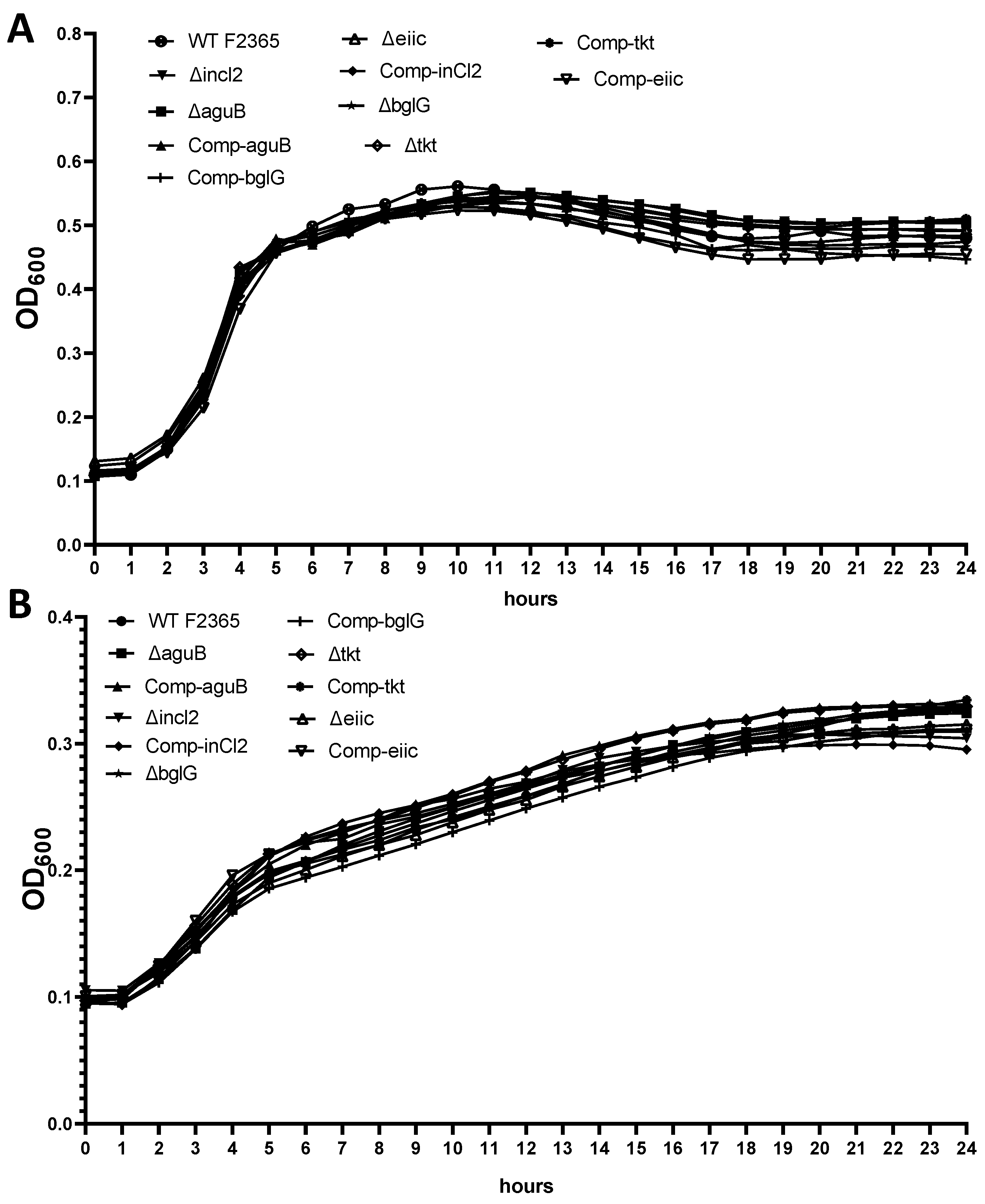
2.1. Growth of Mutants

The mutant strains exhibited a similar growth rate to the wild type F2365 strain when grown in brain heart infusion (BHI) as enriched medium or minimal medium (MM) (Figure 1).

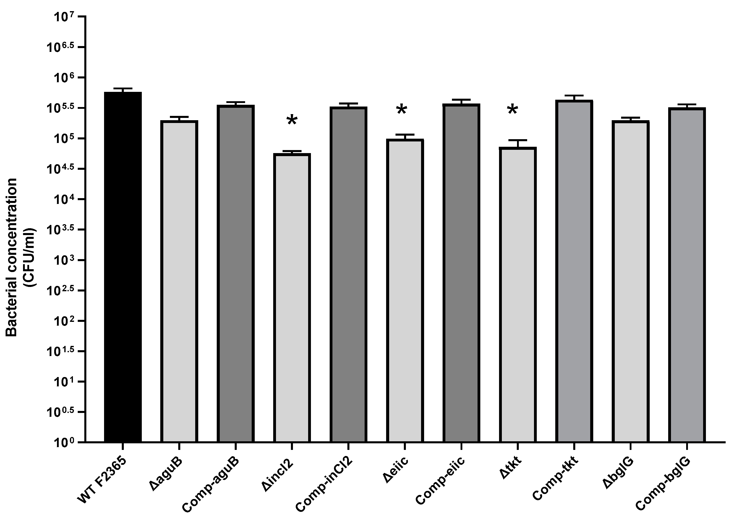
2.2. Role of aguB, inlC2, eiic, tkt, and bglG in L. Monocytogenes Adhesion

The abilities of wild type F2365 strain and mutant strains to adhere to Caco-2 cells were compared. Caco-2 cells were incubated for 1 h at 37 °C with 106 colony-forming unit (CFU) of bacteria. Strain F2365ΔinlC2, F2365Δeiic, and F2365Δtkt showed an approximately 1-log(log10), 0.8-log, and 0.9-log reduction in adhesion to Caco-2 cells compared with parent strain F2365. These differences were statistically (p < 0.05) significant. F2365ΔaguB and F2365ΔbglG showed approximately 0.46-log and 0.5-log reduction in adhesion to Caco-2 cells compared to parent strain F2365, and this was not statistically significant. Complementation of the mutations restored adhesion properties (Figure 2).

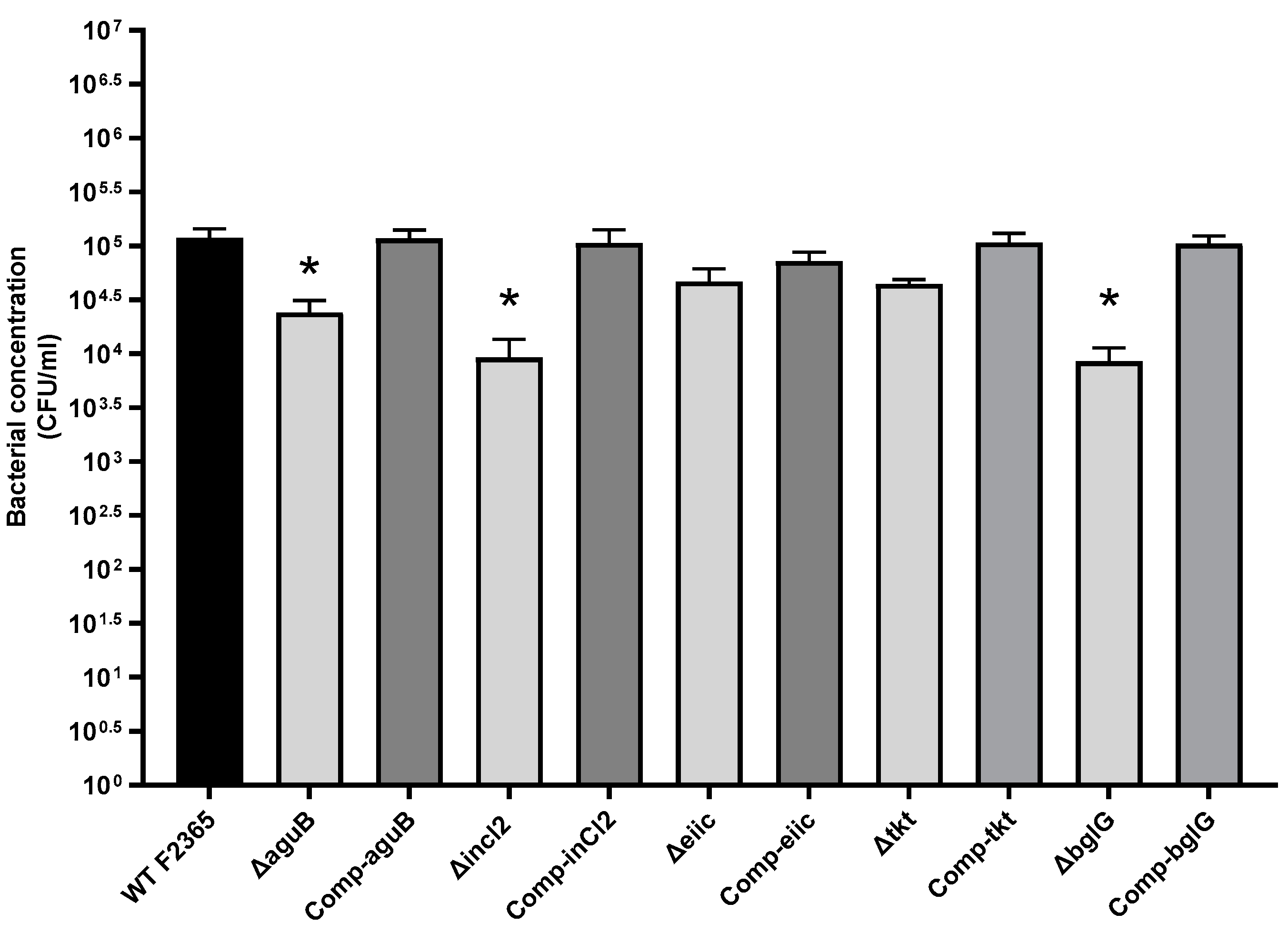
2.3. Role of aguB, inlC2, eiic, tkt, and bglG in L. Monocytogenes Invasion

Invasion of the F2365 strain and mutant strains of L. monocytogenes were compared in Caco-2 cells. Caco-2 cells and bacteria were incubated for 1 h at 37 °C followed by an additional 2.5 h in media with gentamicin to kill extracellular bacteria. There were approximately 0.57-log, 0.9-log, and 0.8-log reductions in the invasion in Caco-2 cells infected with F2365ΔaguB, F2365ΔinlC2, and F2365ΔbglG, compared to parent strain F2365, respectively. These differences were statistically significant (p < 0.05). F2365Δeiic and F2365Δtkt showed an approximate 0.2-log and 0.4-log reduction in invasion compared to parent strain F2365, and this was not statistically significant (Figure 3). Complementation restored the invasion of the mutant strains to wild type levels.

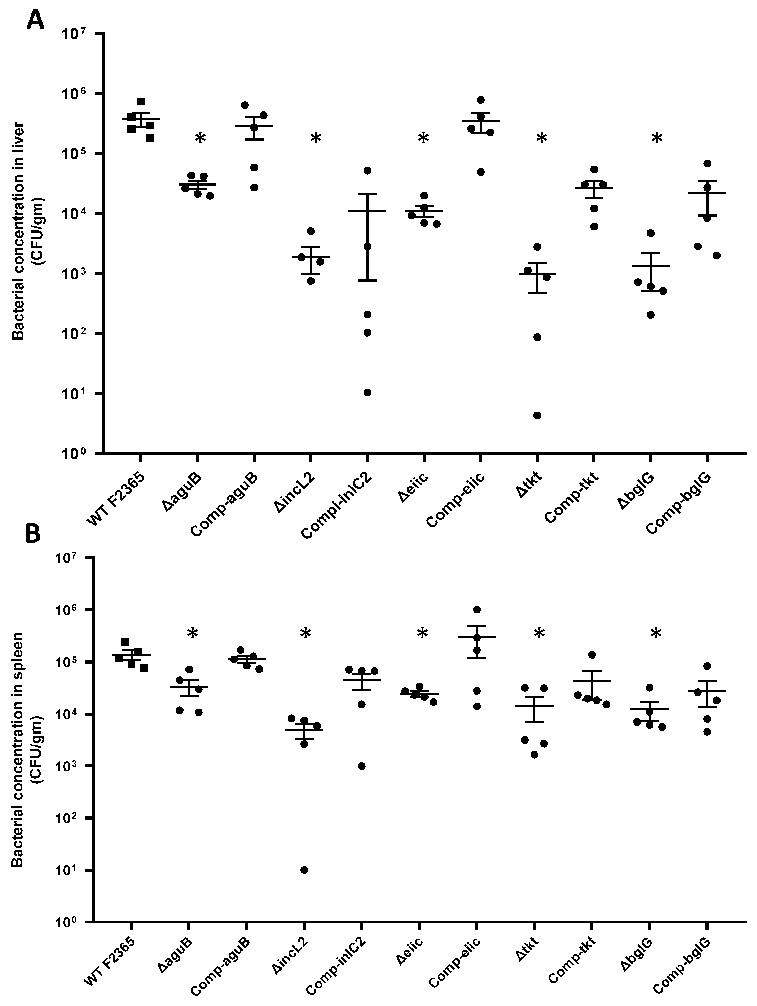
2.4. Virulence and Colonization of Mutant Strains in Mice

Virulence of the five mutant strains was assessed in vivo using BALB/c mice. Mice were infected intraperitoneally with 5 × 10⁴ CFU/mL, and bacterial loads in liver and spleen were enumerated 3 days post-infection. Bacteria concentrations in the liver were significantly (p < 0.05) higher in mice infected with F2365 strain than mice infected with mutant strains. Bacteria concentrations were reduced about 1-log, 2.3-log, 1.5-log, 2.6-log, and 2.4-log in F2365ΔaguB, F2365ΔinlC2, F2365Δeiic, F2365Δtkt, and F2365ΔbglG compared with the wild-type strain, respectively (Figure 4A). Also, bacterial loads were found to be significantly higher in case of aguB, eiic, tkt, and bglG complemented strain as compared to corresponding mutant (p = 0.031, 0.0079, 0.0079, and 0.031, respectively), confirming that it was the deletion of the gene which led to the consequent alterations in the bacterial loads. No significant differences between the wild-type and complement strains were observed in these four strains (p > 0.05). However, bacterial loads in mice infected with inlC2 complemented strain were not significantly (p = 0.39) different compared to deletion strain. This could be due to a defect in the expression of the inlC2 during replication of the complemented strain in liver tissue. Moreover, genetic complementation could differ from the native level of expression [25]. Colonization of strain F2365 and complemented strains in the spleen was significantly (p < 0.05) higher than the tested mutants. Compared to parent strain F2365, the reduction in bacterial concentrations in spleen were 0.6-log, 1.5-log, 0.7-log, 1-log, and 1-log in F2365ΔaguB, F2365ΔinlC2, F2365Δeiic, F2365Δtkt, and F2365ΔbglG, respectively (Figure 4B).

3. Discussion

L. monocytogenes is a facultative intracellular bacterium able to penetrate and replicate within mammalian cells [26]. Serotype 4b strain F2365 was isolated from one of the worst bacterial foodborne outbreaks reported in the United States; it was involved in an outbreak of listeriosis in California in 1985 [27]. L. monocytogenes pathogenesis includes multiple stages such as internalization, vacuolar escape, intracellular replication, movement by actin mobilization, and cell-to-cell spread [28]. Several L. monocytogenes genes whose products are required for these processes have been identified [29]. The aim of the present study was to determine the predictive value of comparative genomics by investigating roles of five previously uncharacterized proteins in L. monocytogenes virulence. L. monocytogenes ability to adhere and invade phagocytic and non-phagocytic cells is an important aspect of disease pathogenesis. Thus, the intestinal epithelium Caco-2 cell line was used as an in vitro model to evaluate the effects of gene deletion on adhesion and invasion of L. monocytogenes. Intraperitoneal inoculation was chosen for this study as it allows Listeria to directly disseminate to the systemic sites via the lymphatic system and blood, bypassing the need for adhesion and invasion of the intestine as in oral infection [30]. To evaluate the level of systematic infection, bacteria loads in the liver and spleen were assayed 72 hours after infection. Liver and spleen play an important role in pathogenesis of listeriosis [31]. Shortly after infection, Listeria rapidly disseminates to the spleen and liver due to quick uptake by dendritic and Kupffer cells [32]. The majority of the invading bacteria accumulate in the liver and spleen and bacterial replication occurs in the liver and spleen during early stages of infection [33]. Peak burden of L. monocytogenes multiplication occurs at 48 to 72 h after infection and mice start to die shortly after this period [34].
In the present study, F2365ΔaguB showed approximately 0.57-log reduction in invasion in Caco-2 cells compared to parent F2365 strain. Also, deletion of aguB resulted in reduced colonization in liver and spleen as compared to wild-type. The aguB gene encoding putrescine carbamoyltransferase mediates phosphorolysis of N-carbamoylputrescine to produce putrescine and carbamoylphosphate [15]. This reaction represents the second step of the agmatine deiminase (AgDI) pathway, which includes four genes: An agmatine–putrescine antiporter (aguD); agmatine deiminase (aguA); putrescine carbamoyltransferase (aguB); and a carbamate kinase (aguC) [35]. The AgDI pathway plays an important role in the control of cytoplasmic pH and generates metabolic energy in the form of ATP [36,37] by converting agmatine into putrescine, ATP, ammonia, and carbon dioxide [38]. In a previous study, L. monocytogenes EGD-e mutants with deletion in ΔarcA and ΔargR (arginine deiminase ADI pathway genes, which closely resembles AgDI pathway) showed a 10-fold reduction in survival in spleens compared with the wild-type strain in mice following intraperitoneal infection [39]. In Salmonella Typhimurium, putrescine plays a critical role in controlling virulence through stimulating the expression of essential virulence loci. Furthermore, a S. Typhimurium putrescine mutant displayed defective invasion and survival/replication in epithelial cells and was attenuated in the mouse model compared to the wild-type [40].
In the present study, a ΔinlC2 mutant was defective in adhesion and invasion to epithelial cells and also showed significant attenuation in the mouse model. These results suggest that inlC2 contributes to L. monocytogenes virulence. Complementation of inlC2 mutant strain restored the adhesion and invasion to epithelial cells. However, full restoration of wild type behavior was not achieved by inlC2 complemented strain in liver tissue. This could be due to defect in the expression of the inlC2 during replication of complement strain in liver. InlC2 is in the internalin family, a diverse group of proteins required for entry of L. monocytogenes into non-professional phagocytic cells, such as epithelial cells, hepatocytes, fibroblasts, or endothelial cells [41]. The internalin family is characterized by tandem arrays of leucine rich repeats (LRR) [18]. inlC2 encodes a secreted protein of 548 aa and is in the same operon with inlD and inlE. The secreted InlC2 protein plays a significant role in L. monocytogenes cell to cell spread [42]. Its amino acid sequence is highly homologous to InlH, with only 13 amino acid differences. Both InlC2 and InlH have the same LRR domain and C-terminal regions, and both are regulated by the same stress-responsive sigma factor σ [43,44]. In some L. monocytogenes strains such as EGD and 10403S, the 5’end of inlC2 is fused with the 3′ end of inlD to result in inlH [45,46]. InlC2 upregulates antigens during infection and thus may be important in L. monocytogenes pathogenesis [47]. Inactivation of inlH (which removed inlD and inlC2) by an in-frame deletion led to attenuated L. monocytogenes EGD virulence, evidenced by significantly reduced numbers of bacteria in both liver and spleen compared to the wild-type strain after oral infection of mice [14]. In contrast, no effect on L. monocytogenes EGD virulence could be established upon inactivation of inlC2 by comparing bacterial concentrations in liver and spleen following intravenous injection [45]. In another study, deletion of inlC2 in L. monocytogenes strain M5 significantly enhanced the adherence and invasion to Hela cells, and virulence in murine mouse model. The authors speculate that increased invasion of ΔinlC2 mutant might be due to elevated production of InlA [48]. This discrepancy between the results may be due to strain differences that lead to difference in protein structure and function or may be due to differences in the cell line type [49,50]. Antibodies to InlC2 are generated in various animal hosts infected with L. monocytogenes, indicating that InlC2 may be induced in vivo [51].
Phosphotransferase component EIIC is an essential component of a phosphotransferase system (PTS) that is responsible for recognition, selective binding, and transport of specific sugar molecules through the cell membrane into the cytoplasm [52]. EIIC components are divided into several families, including subclasses for glucose, sucrose, mannitol, fructose, lactose, mannose, and cellobiose [53]. Understanding the role of EIIC (LMOf2365_0661) is of great importance because growth of L. monocytogenes as an intracellular pathogen depends on efficient use of sugar from the host. In the present study, F2365Δeiic exhibited a significant reduction in adhesion to epithelial cells. Moreover, F2365Δeiic had less bacterial counts in vivo compared to its parent strain. In a previous study, mutation of a PTS EIIC homolog did not impair virulence of Klebsiella pneumoniae in the mouse respiratory infection model [54]. Various PTS systems have been associated with Streptococcus gordonii adhesion and biofilm formation [55]. Also, an in vivo expression technology screen performed with K. pneumoniae strain CG43 indicated that ptfA, encoding a fructose phosphotransferase, was positively expressed during BALB/c mice infection [56].
Our study showed that deletion of tkt, encoding transketolase enzyme, in F2365 significantly decreased adhesion but did not affect invasion of Caco-2 cells compared to the parent strain. The tkt mutation also reduced virulence of L. monocytogenes in mice. Transketolase (tkt) catalyze the reversible transfer of a ketol group between several donor and acceptor substrates [57,58], thus creating a reversible link between glycolysis and the PP pathway. TKT is responsible for the production of essential cell constituents, such as aromatic amino acids, aromatic vitamins, NADPH, several sugar phosphate intermediates, and pyridoxine [59]. In Leishmania mexicana, tkt was found to be essential in establishing mammalian cell infection, and tkt-deleted L. mexicana did not cause any obvious lesions in mice even after 9 weeks [60]. Moreover, tkt1 is induced in vivo in chicken livers and spleens following avian pathogenic Escherichia coli (APEC) infection, demonstrating the importance of tkt in APEC pathogenesis [61]. Genes tktA and tktB are essential for APEC survival in chickens [62,63]. Moreover, tkt-deleted Mycobacterium tuberculosis displayed reduced virulence and intracellular growth in macrophages compared to wild-type strain [64]. In E. coli, deletion of tktA increased antibiotic and oxidative stress susceptibilities through interaction with marRAB operon [65].
In the present study, strains Δeiic and Δtkt adhered to Caco-2 cells less efficiently than wild type strain but the difference in the intracellular multiplication was not significant. This may be due to the fact that the role of these two genes is limited to entry process and contact between L. monocytogenes and host cells. Similar to this finding, the deletion of L. monocytogenes either fliF or fliI abolishes flagella assembly and impairs the bacterial adhesion to nonphagocytic cells. However, intracellular multiplication of null mutants is apparently not affected [66]. In the same study, the authors monitored the adhesion and intracellular multiplication of less motile L. monocytogenes strains. The result demonstrated that all the strains adhered less efficiently (between 2 and 10 times less) than EGD-e to Caco-2 cells but the intracellular survival was not affected in any of the isolates, suggesting that expression of these genes might play a role in the adhesion to epithelial cells but has no impact on intracytosolic multiplication [66].
The present work demonstrated that mutation of bglG impaired bacterial invasion severely but not adhesion. Furthermore, bglG deletion reduced the virulence of L. monocytogenes in mice. BglG is an RNA-binding transcriptional antiterminator that regulates expression of phosphoenolpyruvate phosphate transferase systems (PEP-PTS) by preventing premature termination of the transcription process [67]. In E. coli, bglG control expression of β-glucoside utilization (bgl) operon by binding to bgl mRNA at sites that stabilizing and alleviating the formation of the terminator structure and allowing transcription of downstream genes [68,69]. It is more likely that bglG is involved in sensing and consumption of carbohydrates from the host cell during infection, thus facilitating pathogenesis [70]. A previous study identified 15 putative bglG transcriptional antiterminators in the L. monocytogenes chromosome, all of which appear to be located either up or downstream from genes encoding PTS system [71]. Deletion mutants in lmo0402 and lmo0501 genes, which encode a bglG transcriptional antiterminator, exhibited they are essential for attachment of L. monocytogenes EGD-e strain at lower temperatures [72].
The in vivo study was performed to determine bacterial count in liver and spleen following dissemination of bacteria in the internal organs. However, the health-related parameters such as body temperature and weight loss were not recorded during the animal study. Even though body temperature and weight loss are often considered as benchmarks of wellbeing in animal models of acute and chronic disease, measurement of these parameters require handling of animals, which could cause distress and immune suppression that can confound the data and thereby potentially lead to erroneous conclusions [73]. Moreover, body temperature is influenced by numerous factors, including the time of day, the presence and type of bedding, and the number of cage mates [74,75]. Weight loss is a complex factor and does not always indicate the severity of illness or the likelihood of imminent death [76,77].
This study highlights the significant contributions of five genes in the pathogenic potential of L. monocytogenes by whether influencing the adhesion (inlC2, eiic, and tkt), invasion (aguB, inlC2, and bglG), and virulence (aguB, inlC2, eiic, tkt, and bglG) in mice. These results correlated with our comparative genomics and orthology analysis and support the role of such comprehensive and predictive methods in identifying putative and undiscovered virulence factors. However, the mechanisms leading to attenuated virulence in these mutants are not yet completely clear. Future work will focus on further identifying mechanism of attenuation on these strains.

4. Material and Methods

4.1. Ethics Statement

All animal experiment presented in this study was approved by the Animal Care Committee of the Mississippi State University (protocol # 17-105). Approval date: 11 March 2017.

4.2. Bacterial Strains and Growth Conditions

All of the strains and plasmids used in this study are listed in Table 1. Brain heart infusion (BHI) (Difco Laboratories, Detroit, MI, USA) and Luria–Bertani (LB) (Difco Laboratories) broth and agar were used to grow L. monocytogenes and Escherichia coli strains, respectively. L. monocytogenes and E. coli were incubated at 37 °C. Strains carrying plasmids were grown in the presence of the following antibiotics: ampicillin (100 μg/mL) for E. coli strains, erythromycin (10 μg/mL) and chloramphenicol (10 μg/mL) for L. monocytogenes strains. Anhydrotetracycline (ATc, 1.5 μg/mL) was used for inducing the expression of antisense secY RNA in the pHoss1 plasmid as described below [78]. Human enterocyte-like Caco-2 (TIB37, ATCC) cell lines were grown in Dulbecco’s Modified Eagle’s Medium (DMEM) (ATCC, Gibco, Manassas, VA, USA) supplemented with 20% fetal bovine serum (Atlanta Biologicals, Norcross, GA, USA) and 1% glutamine incubated at 37 °C with 5% CO2 under humidified conditions.

4.3. Construction of L. Monocytogenes Mutants

Five genes (Table 2) were targeted for in-frame deletion using a previously published mutagenesis gene excision method [78]. A generic map and more details for the strategy used for genes deletion are present in our previously published manuscript [78]. The sequence of primers used for construction and validation of the mutants are listed in Table 3. Briefly, approximately 1 kb fragment of the upstream and the downstream region of each gene was amplified by PCR using genomic DNA from L. monocytogenes strain F2365 as a template. Overlap PCR reactions were used to generate in-frame deletion DNA fragments [81]. These fragments were then cloned into the digested pHoss1 vector and transformed into chemically competent E. coli DH5α. After sequencing, the resulting plasmids were electroporated into L. monocytogenes strain F2365 and selected in BHI plus erythromycin at 30 °C for 3 days. A single colony was streaked onto BHI without erythromycin and incubated at the nonpermissive temperature (42 °C) for two days to allow the plasmid to integrate into the chromosome. This process was repeated three times. Single colonies were then passed three times on BHI broth at 30 °C before spreading BHI agar containing ATc. The induction of secY antisense RNA expression in the presence of ATc inhibits the growth of bacteria that retain the integrated plasmid and allow positive selection for chromosomal excision and loss of the plasmid [82,83]. The presence of mutation in the erythromycin-sensitive clones were verified by colony PCR using the primer pair A and D. Final confirmation of the deletion was confirmed by sanger sequencing. The constructed mutants were designated F2365ΔaguB, F2365ΔinlC2, F2365Δeiic, F2365Δtkt, and F2365ΔbglG.

4.4. Complementation of the Mutant Strains

Complementation procedures were performed by inserting a copy of the wild-type gene with its promoter region into the appropriate mutant strains using the pPL2 site-specific shuttle integration vector [80]. The integration site of pPL2 have no observed effect on virulence phenotypes of Listeria [80]. Briefly, coding fragments of aguB, inlC2, eiic, tkt, and bglG were amplified by PCR using primers listed in Table 3. PCR products were digested with SacI and SalI enzymes, ligated into pPL2, and transformed into E. coli DH5α. To verify the correct assembly of the plasmids, colony PCR and sequencing were performed. The resulting plasmids were then electroporated into L. monocytogenes mutant strains and selected on BHI agar plates containing chloramphenicol. Chromosomal insertion of aguB, inlC2, eiic, tkt, and bglG was confirmed by PCR. Complemented strains generated through this approach were designated F2365ΔaguB::aguB, F2365ΔinlC2::inlC2, F2365Δeiic::eiic, F2365Δtkt::tkt, and F2365ΔbglG::bglG.

4.5. Growth Kinetics of Mutants

Growth F2365 wild-type strain, mutants, and complemented strains in BHI broth and Minimal Medium (MM) [84] at 37 °C were compared. Growth assays were achieved in a 96-well plate. Overnight cultures of each strain in BHI were adjusted to an optical density at λ = 600 nm (OD600) of 1.00 and diluted 1:50 into BHI or MM. The plates were incubated in a Multi-Mode Reader (BioTek Cytation 5) at 37 °C for 24 h. OD600 was measured every hour. All growth experiments were performed in three independent experiments, and each experiment was run with six replicates.

4.6. Adherence and Invasion Assay

Adherence of L. monocytogenes F2365 wild-type, mutant, and complemented strains to Caco-2 epithelial cells was evaluated as described [85]. In brief, Caco-2 cells were seeded into 24-well tissue culture plates at 105 cells per well. The cells were infected with bacteria (106 CFU) at a multiplicity of infection (MOI) of 10 bacteria per cell and incubated at 37 °C for 1 h in the presence of 5% CO2, after which the medium was removed. Infected monolayer cells were then washed three times with phosphate-buffered saline (PBS) to eliminate non-adherent bacteria and lysed with 0.5% Triton X-100. Appropriate serial dilutions were prepared in PBS and spread on BHI agar for enumeration of bacterial cells. Adherence assay were performed in three independent experiments with four biological replicates for each infection.
L. monocytogenes F2365, mutant, and complemented strains were tested for their ability to invade Caco-2 cells as described previously [86]. Bacteria were added to the Caco-2 cell monolayer to yield MOI of 10 bacteria per cell and incubated at 37 °C for 1 h. Infected cells were washed three times with PBS and incubated in medium containing gentamicin (100 μg/mL) for 2.5 h to kill extracellular bacteria. Gentamicin (100 μg/mL) was used to rapidly eliminate extracellular bacteria and prevent further infection by bacteria released from dead cells [87]. Aminoglycosides poorly penetrate the eukaryotic cell membrane when used in the incubation medium for short periods (usually less than 4 h) [88,89]. Cells were then washed three times with PBS and lysed using 0.5% Triton X-100 for 10 min. Appropriate dilutions of the lysates were spread on BHI agar. Three independent experiments were performed with four biological replicates for each infection.

4.7. Virulence in Mice

The virulence of L. monocytogenes strain F2365, mutant strains, and complemented strains were compared in a mouse infection model as previously described [86]. Sixty 8-week-old specific pathogen-free female BALB/c mice (Jackson Laboratory, Sacramento, CA, USA) were housed in 12 cages (5 mice/cage) according to treatment groups. Bacteria were harvested at an OD600 of ∼1.0 and washed with PBS. The bacterial density in overnight cultures was estimated from a previously prepared standard curve that correlated OD600 to viable-bacteria numbers. Average CFU was calculated based on serial dilutions and calculating the average colony numbers on agar plates. The OD600 was adjusted so that approximately 5 × 10⁴ CFU of each bacterial strain was inoculated intraperitoneally in each mouse in 100 μL of sterile PBS. Five mice were used for each strain. Mice were visually evaluated every two hours during the day for signs of illness or discomfort throughout the experiment. At 72 h post-infection, mice were euthanized followed by collection of liver and spleen samples. Tissue samples were weighed and homogenized in PBS. Homogenates were serially diluted and spread on BHI agar to determine numbers of viable bacteria.

4.8. Statistical Analysis

The data from adhesion, invasion, and in vivo virulence are presented as mean ± standard error (SE). The dot plots of bacterial numbers in each mouse were generated using GraphPad Prism 8 software, along with the median values. For in vivo experiment, a non-parametric Mann-Whitney test was used to detect the statistical significance in bacterial load in liver and spleen of infected mice between F2365 parent strain, mutants, and complemented strains. p values < 0.05 were considered statistically significant in all analyses. Statistical analysis of adhesion and invasion assays were performed as previously described [86]. Briefly, visual assessment of histograms using PROC UNIVARIATE in SAS for Windows v9.4 (SAS Institute Inc., Cary, NC, USA) indicated that the data was approximately normally distributed following log10 transformation. Pairwise comparison of means was conducted with Tukey’s test. Analysis of variance (ANOVA) was performed using PROC GLM in SAS for Windows v9.4 to assess significant differences.

Author Contributions

H.A. and R.R. performed the experiments. H.A., M.L.L., and A.K. designed the experiment. H.A. wrote the manuscript. All authors read and approved the final manuscript.

Funding

This study was supported by the Center for Biomedical Research Excellence in Pathogen–Host Interactions, National Institute of General Medical Sciences, (P20 GM103646-06), SCA no. 58-6066-7081 titled Mississippi Center for Food Safety and Post-Harvest Technology, MS Agricultural and Forestry Experiment Station, and the College of Veterinary Medicine.

Acknowledgments

We thank Bridget Willeford, Lucy Senter, and the Mississippi State University Laboratory Animal Resources and Care unit for animal and veterinary care. We also thank Breanna Brown, Austin Lawrence, Iman Ibrahim, Basant Gomaa, and Michelle Banes for their help in mice experiment. We also thank John Harkness for review of the manuscript. Growth experiments were performed in COBRE Mini-Research Core.

Conflicts of Interest

The authors declare that there are no conflicts of interest.

References

  1. Mead, P.S.; Slutsker, L.; Dietz, V.; McCaig, L.F.; Bresee, J.S.; Shapiro, C.; Griffin, P.M.; Tauxe, R.V. Food-related illness and death in the United States. Emerg. Infect. Dis. 1999, 5, 607–625. [Google Scholar]
  2. Norton, D.M.; Scarlett, J.M.; Horton, K.; Sue, D.; Thimothe, J.; Boor, K.J.; Wiedmann, M. Characterization and pathogenic potential of Listeria monocytogenes isolates from the smoked fish industry. Appl. Environ. Microbiol. 2001, 67, 646–653. [Google Scholar] [CrossRef]
  3. Liu, D.; Lawrence, M.L.; Wiedmann, M.; Gorski, L.; Mandrell, R.E.; Ainsworth, A.J.; Austin, F.W. Listeria monocytogenes subgroups IIIA, IIIB, and IIIC delineate genetically distinct populations with varied pathogenic potential. J. Clin. Microbiol. 2006, 44, 4229–4233. [Google Scholar] [CrossRef]
  4. Hamon, M.A.; Batsche, E.; Regnault, B.; Tham, T.N.; Seveau, S.; Muchardt, C.; Cossart, P. Histone modifications induced by a family of bacterial toxins. Proc. Natl. Acad. Sci. USA 2007, 104, 13467–13472. [Google Scholar] [Green Version]
  5. Gandhi, M.; Chikindas, M.L. Listeria: A foodborne pathogen that knows how to survive. Int. J. Food Microbiol. 2007, 113, 1–15. [Google Scholar] [CrossRef]
  6. Orsi, R.H.; Wiedmann, M. Characteristics and distribution of Listeria spp., including Listeria species newly described since 2009. Appl. Microbiol. Biotechnol. 2016, 100, 5273–5287. [Google Scholar] [Green Version]
  7. Cocolin, L.; Rantsiou, K.; Iacumin, L.; Cantoni, C.; Comi, G. Direct identification in food samples of Listeria spp. and Listeria monocytogenes by molecular methods. Appl. Environ. Microbiol. 2002, 68, 6273–6282. [Google Scholar] [CrossRef]
  8. Scharff, R.L. Economic Burden from Health Losses Due to Foodborne Illness in the United States. J. Food Prot. 2012, 75, 123–131. [Google Scholar] [CrossRef]
  9. Guillet, C.; Join-Lambert, O.; Le Monnier, A.; Leclercq, A.; Mechai, F.; Mamzer-Bruneel, M.F.; Bielecka, M.K.; Scortti, M.; Disson, O.; Berche, P.; et al. Human listeriosis caused by Listeria ivanovii. Emerg. Infect. Dis. 2010, 16, 136–138. [Google Scholar] [CrossRef]
  10. Snapir, Y.M.; Vaisbein, E.; Nassar, F. Low virulence but potentially fatal outcome-Listeria ivanovii. Eur. J. Intern. Med. 2006, 17, 286–287. [Google Scholar]
  11. Sauders, B.D.; Overdevest, J.; Fortes, E.; Windham, K.; Schukken, Y.; Lembo, A.; Wiedmann, M. Diversity of Listeria species in urban and natural environments. Appl. Environ. Microbiol. 2012, 78, 4420–4433. [Google Scholar]
  12. Paul, D.; Steele, C.; Donaldson, J.R.; Banes, M.M.; Kumar, R.; Bridges, S.M.; Arick, M., 2nd; Lawrence, M.L. Genome comparison of Listeria monocytogenes serotype 4a strain HCC23 with selected lineage I and lineage II L. monocytogenes strains and other Listeria strains. Genom. Data 2014, 2, 219–225. [Google Scholar] [CrossRef] [Green Version]
  13. Begley, M.; Bron, P.A.; Heuston, S.; Casey, P.G.; Englert, N.; Wiesner, J.; Jomaa, H.; Gahan, C.G.; Hill, C. Analysis of the isoprenoid biosynthesis pathways in Listeria monocytogenes reveals a role for the alternative 2-C-methyl-D-erythritol 4-phosphate pathway in murine infection. Infect. Immun. 2008, 76, 5392–5401. [Google Scholar] [CrossRef]
  14. Raffelsbauer, D.; Bubert, A.; Engelbrecht, F.; Scheinpflug, J.; Simm, A.; Hess, J.; Kaufmann, S.H.; Goebel, W. The gene cluster inlC2DE of Listeria monocytogenes contains additional new internalin genes and is important for virulence in mice. Mol. Gen. Genet. 1998, 260, 144–158. [Google Scholar]
  15. Chen, J.; Cheng, C.; Xia, Y.; Zhao, H.; Fang, C.; Shan, Y.; Wu, B.; Fang, W. Lmo0036, an ornithine and putrescine carbamoyltransferase in Listeria monocytogenes, participates in arginine deiminase and agmatine deiminase pathways and mediates acid tolerance. Microbiology 2011, 157 Pt 11, 3150–3161. [Google Scholar]
  16. Cheng, C.; Chen, J.; Fang, C.; Xia, Y.; Shan, Y.; Liu, Y.; Wen, G.; Song, H.; Fang, W. Listeria monocytogenes aguA1, but not aguA2, encodes a functional agmatine deiminase: Biochemical characterization of its catalytic properties and roles in acid tolerance. J. Biol. Chem. 2013, 288, 26606–26615. [Google Scholar]
  17. Bonazzi, M.; Lecuit, M.; Cossart, P. Listeria monocytogenes internalin and E-cadherin: From structure to pathogenesis. Cell. Microbiol. 2009, 11, 693–702. [Google Scholar] [CrossRef]
  18. Bierne, H.; Sabet, C.; Personnic, N.; Cossart, P. Internalins: A complex family of leucine-rich repeat-containing proteins in Listeria monocytogenes. Microbes Infect. 2007, 9, 1156–1166. [Google Scholar]
  19. McCoy, J.G.; Levin, E.J.; Zhou, M. Structural insight into the PTS sugar transporter EIIC. Biochim. Biophys. Acta 2015, 1850, 577–585. [Google Scholar]
  20. Jung, Y.M.; Lee, J.N.; Shin, H.D.; Lee, Y.H. Role of tktA gene in pentose phosphate pathway on odd-ball biosynthesis of poly-beta-hydroxybutyrate in transformant Escherichia coli harboring phbCAB operon. J. Biosci. Bioeng. 2004, 98, 224–227. [Google Scholar] [CrossRef]
  21. Nussbaum-Shochat, A.; Amster-Choder, O. BglG, the transcriptional antiterminator of the bgl system, interacts with the beta’ subunit of the Escherichia coli RNA polymerase. Proc. Natl. Acad. Sci. USA 1999, 96, 4336–4341. [Google Scholar] [CrossRef]
  22. Raveh, H.; Lopian, L.; Nussbaum-Shochat, A.; Wright, A.; Amster-Choder, O. Modulation of transcription antitermination in the bgl operon of Escherichia coli by the PTS. Proc. Natl. Acad. Sci. USA 2009, 106, 13523–13528. [Google Scholar] [CrossRef]
  23. Glaser, P.; Frangeul, L.; Buchrieser, C.; Rusniok, C.; Amend, A.; Baquero, F.; Berche, P.; Bloecker, H.; Brandt, P.; Chakraborty, T.; et al. Comparative genomics of Listeria species. Science 2001, 294, 849–852. [Google Scholar]
  24. Casey, A.; Jordan, K.; Coffey, A.; Fox, E.M.; McAuliffe, O. Comparative Genomic Analysis of Two Serotype 1/2b Listeria monocytogenes Isolates from Analogous Environmental Niches Demonstrates the Influence of Hypervariable Hotspots in Defining Pathogenesis. Front. Nutr. 2016, 3, 54. [Google Scholar] [Green Version]
  25. Husna, A.U.; Wang, N.; Cobbold, S.A.; Newton, H.J.; Hocking, D.M.; Wilksch, J.J.; Scott, T.A.; Davies, M.R.; Hinton, J.C.; Tree, J.J.; et al. Methionine biosynthesis and transport are functionally redundant for the growth and virulence of Salmonella Typhimurium. J. Biol. Chem. 2018, 293, 9506–9519. [Google Scholar] [CrossRef] [Green Version]
  26. Drevets, D.A.; Sawyer, R.T.; Potter, T.A.; Campbell, P.A. Listeria monocytogenes infects human endothelial cells by two distinct mechanisms. Infect. Immun. 1995, 63, 4268–4276. [Google Scholar] [Green Version]
  27. Linnan, M.J.; Mascola, L.; Lou, X.D.; Goulet, V.; May, S.; Salminen, C.; Hird, D.W.; Yonekura, M.L.; Hayes, P.; Weaver, R.; et al. Epidemic listeriosis associated with Mexican-style cheese. N. Engl. J. Med. 1988, 319, 823–828. [Google Scholar] [CrossRef]
  28. Tilney, L.G.; Portnoy, D.A. Actin filaments and the growth, movement, and spread of the intracellular bacterial parasite, Listeria monocytogenes. J. Cell Biol. 1989, 109 Pt 1, 1597–1608. [Google Scholar] [CrossRef]
  29. Portnoy, D.A.; Chakraborty, T.; Goebel, W.; Cossart, P. Molecular determinants of Listeria monocytogenes pathogenesis. Infect. Immun. 1992, 60, 1263–1267. [Google Scholar] [Green Version]
  30. Sobyanin, K.A.; Sysolyatina, E.V.; Chalenko, Y.M.; Kalinin, E.V.; Ermolaeva, S.A. Route of Injection Affects the Impact of InlB Internalin Domain Variants on Severity of Listeria monocytogenes Infection in Mice. BioMed Res. Int. 2017, 2101575. [Google Scholar] [CrossRef]
  31. Cousens, L.P.; Wing, E.J. Innate defenses in the liver during Listeria infection. Immunol. Rev. 2000, 174, 150–159. [Google Scholar] [CrossRef] [PubMed]
  32. Neuenhahn, M.; Busch, D.H. Unique functions of splenic CD8α+ dendritic cells during infection with intracellular pathogens. Immunol. Lett. 2007, 114, 66–72. [Google Scholar] [CrossRef] [PubMed]
  33. Hoelzer, K.; Pouillot, R.; Dennis, S. Animal models of listeriosis: A comparative review of the current state of the art and lessons learned. Vet. Res. 2012, 43, 18. [Google Scholar] [CrossRef] [PubMed]
  34. Kernbauer, E.; Maier, V.; Rauch, I.; Muller, M.; Decker, T. Route of Infection Determines the Impact of Type I Interferons on Innate Immunity to Listeria monocytogenes. PLoS ONE 2013, 8, e65007. [Google Scholar] [CrossRef] [PubMed]
  35. Griswold, A.R.; Jameson-Lee, M.; Burne, R.A. Regulation and physiologic significance of the agmatine deiminase system of Streptococcus mutans UA159. J. Bacteriol. 2006, 188, 834–841. [Google Scholar] [CrossRef] [PubMed]
  36. Llacer, J.L.; Polo, L.M.; Tavarez, S.; Alarcon, B.; Hilario, R.; Rubio, V. The gene cluster for agmatine catabolism of Enterococcus faecalis: Study of recombinant putrescine transcarbamylase and agmatine deiminase and a snapshot of agmatine deiminase catalyzing its reaction. J. Bacteriol. 2007, 189, 1254–1265. [Google Scholar] [CrossRef] [PubMed]
  37. Smith, J.L.; Liu, Y.; Paoli, G.C. How does Listeria monocytogenes combat acid conditions? Can. J. Microb. 2012, 59, 141–152. [Google Scholar] [CrossRef]
  38. Lucas, P.M.; Blancato, V.S.; Claisse, O.; Magni, C.; Lolkema, J.S.; Lonvaud-Funel, A. Agmatine deiminase pathway genes in Lactobacillus brevis are linked to the tyrosine decarboxylation operon in a putative acid resistance locus. Microbiology 2007, 153 Pt 7, 2221–2230. [Google Scholar] [CrossRef]
  39. Ryan, S.; Begley, M.; Gahan, C.G.; Hill, C. Molecular characterization of the arginine deiminase system in Listeria monocytogenes: Regulation and role in acid tolerance. Environ. Microbiol. 2009, 11, 432–445. [Google Scholar] [CrossRef]
  40. Jelsbak, L.; Thomsen, L.E.; Wallrodt, I.; Jensen, P.R.; Olsen, J.E. Polyamines are required for virulence in Salmonella enterica serovar Typhimurium. PLoS ONE 2012, 7, e36149. [Google Scholar] [CrossRef]
  41. Gaillard, J.L.; Berche, P.; Frehel, C.; Gouln, E.; Cossart, P. Entry of L. monocytogenes into cells is mediated by internalin, a repeat protein reminiscent of surface antigens from gram-positive cocci. Cell 1991, 65, 1127–1141. [Google Scholar] [CrossRef]
  42. Rajabian, T.; Gavicherla, B.; Heisig, M.; Muller-Altrock, S.; Goebel, W.; Gray-Owen, S.D.; Ireton, K. The bacterial virulence factor InlC perturbs apical cell junctions and promotes cell-to-cell spread of Listeria. Nat. Cell Biol. 2009, 11, 1212–1218. [Google Scholar] [CrossRef] [PubMed] [Green Version]
  43. Hain, T.; Hossain, H.; Chatterjee, S.S.; Machata, S.; Volk, U.; Wagner, S.; Brors, B.; Haas, S.; Kuenne, C.T.; Billion, A.; et al. Temporal transcriptomic analysis of the Listeria monocytogenes EGD-e sigmaB regulon. BMC Microbiol. 2008, 8, 20. [Google Scholar] [CrossRef] [PubMed]
  44. Kazmierczak, M.J.; Mithoe, S.C.; Boor, K.J.; Wiedmann, M. Listeria monocytogenes sigma B regulates stress response and virulence functions. J. Bacteriol. 2003, 185, 5722–5734. [Google Scholar] [CrossRef] [PubMed]
  45. Dramsi, S.; Dehoux, P.; Lebrun, M.; Goossens, P.L.; Cossart, P. Identification of four new members of the internalin multigene family of Listeria monocytogenes EGD. Infect. Immun. 1997, 65, 1615–1625. [Google Scholar] [PubMed]
  46. Jia, Y.; Nightingale, K.K.; Boor, K.J.; Ho, A.; Wiedmann, M.; McGann, P. Distribution of internalin gene profiles of Listeria monocytogenes isolates from different sources associated with phylogenetic lineages. Foodborne Pathog. Dis. 2007, 4, 222–232. [Google Scholar] [CrossRef] [PubMed]
  47. Yu, W.L.; Dan, H.; Lin, M. Novel protein targets of the humoral immune response to Listeria monocytogenes infection in rabbits. J. Med. Microbiol. 2007, 56 Pt 7, 888–895. [Google Scholar] [CrossRef]
  48. Jiang, J.; Chen, J.; Cheng, C.; Hu, H.; Bai, F.; Chen, N.; Yan, G.; Fang, W. Disruption of InlC2 enhances the internalization of Listeria monocytogenes by epithelial cells. World J. Microbiol. Biotechnol. 2011, 27, 2155–2161. [Google Scholar] [CrossRef]
  49. Gözel, B.; Monney, C.; Aguilar-Bultet, L.; Rupp, S.; Frey, J.; Oevermann, A. Hyperinvasiveness of Listeria monocytogenes sequence type 1 is independent of lineage I-specific genes encoding internalin-like proteins. MicrobiologyOpen 2019, 8, e00790. [Google Scholar] [CrossRef] [PubMed]
  50. Bierne, H.; Cossart, P. Listeria monocytogenes surface proteins: From genome predictions to function. Microbiol. Mol. Biol. Rev. 2007, 71, 377–397. [Google Scholar] [CrossRef] [PubMed]
  51. Yu, W.L.; Dan, H.; Lin, M. InlA and InlC2 of Listeria monocytogenes Serotype 4b Are Two Internalin Proteins Eliciting Humoral Immune Responses Common to Listerial Infection of Various Host Species. Curr. Microbiol. 2008, 56, 505–509. [Google Scholar] [CrossRef] [PubMed]
  52. Grisafi, P.L.; Scholle, A.; Sugiyama, J.; Briggs, C.; Jacobson, G.R.; Lengeler, J.W. Deletion mutants of the Escherichia coli K-12 mannitol permease: Dissection of transport-phosphorylation, phospho-exchange, and mannitol-binding activities. J. Bacteriol. 1989, 171, 2719–2727. [Google Scholar] [CrossRef] [PubMed]
  53. Postma, P.W.; Lengeler, J.W.; Jacobson, G.R. Phosphoenolpyruvate:carbohydrate phosphotransferase systems of bacteria. Microbiol. Rev. 1993, 57, 543–594. [Google Scholar]
  54. Boddicker, J.D.; Anderson, R.A.; Jagnow, J.; Clegg, S. Signature-tagged mutagenesis of Klebsiella pneumoniae to identify genes that influence biofilm formation on extracellular matrix material. Infect. Immun. 2006, 74, 4590–4597. [Google Scholar] [CrossRef] [PubMed]
  55. Kilic, A.O.; Tao, L.; Zhang, Y.; Lei, Y.; Khammanivong, A.; Herzberg, M.C. Involvement of Streptococcus gordonii beta-glucoside metabolism systems in adhesion, biofilm formation, and in vivo gene expression. J. Bacteriol. 2004, 186, 4246–4253. [Google Scholar] [CrossRef] [PubMed]
  56. Lai, Y.C.; Peng, H.L.; Chang, H.Y. Identification of genes induced in vivo during Klebsiella pneumoniae CG43 infection. Infect. Immun. 2001, 69, 7140–7145. [Google Scholar] [CrossRef] [PubMed]
  57. Kochetov, G.A. Transketolase: Structure and mechanism of action. Biokhimiia 1986, 51, 2010–2029. [Google Scholar] [PubMed]
  58. Datta, A.G.; Racker, E. Mechanism of action of transketolase. II. The substrate-enzyme intermediate. J. Biol. Chem. 1961, 236, 624–628. [Google Scholar]
  59. Zhao, G.; Winkler, M.E. An Escherichia coli K-12 tktA tktB mutant deficient in transketolase activity requires pyridoxine (vitamin B6) as well as the aromatic amino acids and vitamins for growth. J. Bacteriol. 1994, 176, 6134–6138. [Google Scholar] [CrossRef] [Green Version]
  60. Kovarova, J.; Pountain, A.W.; Wildridge, D.; Weidt, S.; Bringaud, F.; Burchmore, R.J.S.; Achcar, F.; Barrett, M.P. Deletion of transketolase triggers a stringent metabolic response in promastigotes and loss of virulence in amastigotes of Leishmania mexicana. PLoS Pathog. 2018, 14, e1006953. [Google Scholar] [CrossRef]
  61. Tuntufye, H.N.; Lebeer, S.; Gwakisa, P.S.; Goddeeris, B.M. Identification of Avian Pathogenic Escherichia coli Genes That Are Induced In Vivo during Infection in Chickens. Appl. Environ. Microbiol. 2012, 78, 3343–3351. [Google Scholar] [CrossRef] [PubMed]
  62. Li, G.; Tivendale, K.A.; Liu, P.; Feng, Y.; Wannemuehler, Y.; Cai, W.; Mangiamele, P.; Johnson, T.J.; Constantinidou, C.; Penn, C.W.; et al. Transcriptome analysis of avian pathogenic Escherichia coli O1 in chicken serum reveals adaptive responses to systemic infection. Infect. Immun. 2011, 79, 1951–1960. [Google Scholar] [CrossRef] [PubMed]
  63. Li, G.; Laturnus, C.; Ewers, C.; Wieler, L.H. Identification of genes required for avian Escherichia coli septicemia by signature-tagged mutagenesis. Infect. Immun. 2005, 73, 2818–2827. [Google Scholar] [CrossRef] [PubMed]
  64. Kolly, G.S.; Sala, C.; Vocat, A.; Cole, S.T. Assessing essentiality of transketolase in Mycobacterium tuberculosis using an inducible protein degradation system. FEMS Microbiol. Lett. 2014, 358, 30–35. [Google Scholar] [CrossRef] [PubMed]
  65. Domain, F.; Bina, X.R.; Levy, S.B. Transketolase A, an enzyme in central metabolism, derepresses the marRAB multiple antibiotic resistance operon of Escherichia coli by interaction with MarR. Mol. Microbiol. 2007, 66, 383–394. [Google Scholar] [CrossRef] [PubMed]
  66. Bigot, A.; Pagniez, H.; Botton, E.; Frehel, C.; Dubail, I.; Jacquet, C.; Charbit, A.; Raynaud, C. Role of FliF and FliI of Listeria monocytogenes in flagellar assembly and pathogenicity. Infect. Immun. 2005, 73, 5530–5539. [Google Scholar] [CrossRef] [PubMed]
  67. Schnetz, K.; Rak, B. Regulation of the bgl operon of Escherichia coli by transcriptional antitermination. EMBO J. 1988, 7, 3271–3277. [Google Scholar] [CrossRef]
  68. Mahadevan, S.; Wright, A. A bacterial gene involved in transcription antitermination: Regulation at a rho-independent terminator in the bgl operon of E. coli. Cell 1987, 50, 485–494. [Google Scholar] [CrossRef]
  69. Amster-Choder, O.; Wright, A. Transcriptional regulation of the bgl operon of Escherichia coli involves phosphotransferase system-mediated phosphorylation of a transcriptional antiterminator. J. Cell. Biochem. 1993, 51, 83–90. [Google Scholar] [CrossRef]
  70. Rothe, F.M.; Bahr, T.; Stulke, J.; Rak, B.; Gorke, B. Activation of Escherichia coli antiterminator BglG requires its phosphorylation. Proc. Natl. Acad. Sci. USA 2012, 109, 15906–15911. [Google Scholar] [CrossRef]
  71. Glenn, S.M. Genes Involved in Attachment of Listeria Monocytogenes to Abiotic Surfaces. Ph.D. Thesis, University of Leicester, Leicester, UK, 2012. [Google Scholar]
  72. Gorski, L.; Palumbo, J.D.; Mandrell, R.E. Attachment of Listeria monocytogenes to radish tissue is dependent upon temperature and flagellar motility. Appl. Environ. Microbiol. 2003, 69, 258–266. [Google Scholar] [CrossRef] [PubMed]
  73. Ray, M.A.; Johnston, N.A.; Verhulst, S.; Trammell, R.A.; Toth, L.A. Identification of markers for imminent death in mice used in longevity and aging research. J. Am. Assoc. Lab. Anim. Sci. 2010, 49, 282–288. [Google Scholar] [PubMed]
  74. Gordon, C.J. Effect of cage bedding on temperature regulation and metabolism of group-housed female mice. Comp. Med. 2004, 54, 63–68. [Google Scholar] [PubMed]
  75. Overton, J.M.; Williams, T.D. Behavioral and physiologic responses to caloric restriction in mice. Physiol. Behav. 2004, 81, 749–754. [Google Scholar] [CrossRef] [PubMed]
  76. Nemzek, J.A.; Xiao, H.Y.; Minard, A.E.; Bolgos, G.L.; Remick, D.G. Humane endpoints in shock research. Shock 2004, 21, 17–25. [Google Scholar] [CrossRef] [PubMed]
  77. Krarup, A.; Chattopadhyay, P.; Bhattacharjee, A.K.; Burge, J.R.; Ruble, G.R. Evaluation of surrogate markers of impending death in the galactosamine-sensitized murine model of bacterial endotoxemia. Lab. Anim. Sci. 1999, 49, 545–550. [Google Scholar]
  78. Abdelhamed, H.; Lawrence, M.L.; Karsi, A. A novel suicide plasmid for efficient gene mutation in Listeria monocytogenes. Plasmid 2015, 81, 1–8. [Google Scholar] [CrossRef]
  79. Nelson, K.E.; Fouts, D.E.; Mongodin, E.F.; Ravel, J.; DeBoy, R.T.; Kolonay, J.F.; Rasko, D.A.; Angiuoli, S.V.; Gill, S.R.; Paulsen, I.T.; et al. Whole genome comparisons of serotype 4b and 1/2a strains of the food-borne pathogen Listeria monocytogenes reveal new insights into the core genome components of this species. Nucleic Acids Res. 2004, 32, 2386–2395. [Google Scholar] [CrossRef] [Green Version]
  80. Lauer, P.; Chow, M.Y.; Loessner, M.J.; Portnoy, D.A.; Calendar, R. Construction, characterization, and use of two Listeria monocytogenes site-specific phage integration vectors. J. Bacteriol. 2002, 184, 4177–4186. [Google Scholar] [CrossRef]
  81. Horton, R.M.; Hunt, H.D.; Ho, S.N.; Pullen, J.K.; Pease, L.R. Engineering hybrid genes without the use of restriction enzymes: Gene splicing by overlap extension. Gene 1989, 77, 61–68. [Google Scholar] [CrossRef]
  82. Monk, I.R.; Shah, I.M.; Xu, M.; Tan, M.W.; Foster, T.J. Transforming the Untransformable. Application of Direct Transformation To Manipulate Genetically Staphylococcus aureus and Staphylococcus epidermidis. mBio 2012, 3, e00277-11. [Google Scholar] [CrossRef] [PubMed]
  83. Bae, T.; Schneewind, O. Allelic replacement in Staphylococcus aureus with inducible counter-selection. Plasmid 2006, 55, 58–63. [Google Scholar] [CrossRef] [PubMed]
  84. Premaratne, R.J.; Lin, W.J.; Johnson, E.A. Development of an improved chemically defined minimal medium for Listeria monocytogenes. Appl. Environ. Microbiol. 1991, 57, 3046–3048. [Google Scholar] [PubMed]
  85. Cowart, R.E.; Lashmet, J.; McIntosh, M.E.; Adams, T.J. Adherence of a virulent strain of Listeria monocytogenes to the surface of a hepatocarcinoma cell line via lectin-substrate interaction. Arch. Microbiol. 1990, 153, 282–286. [Google Scholar] [CrossRef] [PubMed]
  86. Reddy, S.; Turaga, G.; Abdelhamed, H.; Banes, M.M.; Wills, R.W.; Lawrence, M.L. Listeria monocytogenes PdeE, a phosphodiesterase that contributes to virulence and has hydrolytic activity against cyclic mononucleotides and cyclic dinucleotides. Microb. Pathog. 2017, 110, 399–408. [Google Scholar] [CrossRef]
  87. Kortebi, M.; Milohanic, E.; Mitchell, G.; Pechoux, C.; Prevost, M.C.; Cossart, P.; Bierne, H. Listeria monocytogenes switches from dissemination to persistence by adopting a vacuolar lifestyle in epithelial cells. PLoS Pathog. 2017, 13, e1006734. [Google Scholar] [CrossRef]
  88. Maurin, M.; Raoult, D. Use of aminoglycosides in treatment of infections due to intracellular bacteria. Antimicrob. Agents Chemother. 2001, 45, 2977–2986. [Google Scholar] [CrossRef]
  89. Vaudaux, P.; Waldvogel, F.A. Gentamicin antibacterial activity in the presence of human polymorphonuclear leukocytes. Antimicrob. Agents Chemother. 1979, 16, 743–749. [Google Scholar] [CrossRef] [Green Version]
Figure 1. Growth curves for wild-type, mutants, and complement strains in brain heart infusion (BHI) broth (A) and minimal medium (B) for 24 h at 37 °C. Data represent the mean of three independent experiments, and each was performed in six replicates.
Toxins 11 00508 g001
Figure 2. Adhesion of F2365, mutant, and complement strains to Caco-2 cells. Numbers on the Y axis represent bacterial concentration (CFU/mL). Data represent the mean from three independent experiments, and each was performed in four replicates. Error bars reflect standard error from each mean. Asterisks indicate statistical significance between F2365 and mutant strains (p < 0.05). Non-significant data were unmarked.
Toxins 11 00508 g002
Figure 3. Invasion of Caco-2 cells by F2365, mutant, and complement strains. Numbers on the Y axis represent bacterial concentrations (CFU/mL). Data represent mean colony forming units (CFU) from three independent experiments performed in four biological replicates. Error bars reflect standard error from each mean. Asterisks indicate statistical significance between F2365 and mutant strains (p < 0.05).
Toxins 11 00508 g003
Figure 4. Bacterial concentrations (CFU/gm) in liver (A) and spleen (B) collected from mice at 3 days post-infection with L. monocytogenes F2365, mutant strains, and complement strains. Each dot represents the bacterial concentration in one mouse. The mean ± SE (n = 5) numbers of CFU for each strain are indicated by horizontal lines. Statistical analysis was performed by a non-parametric Mann-Whitney test. *, p < 0.05.
Toxins 11 00508 g004
Table 1. Bacterial strains and plasmids used in this study.
Bacterial Strains, Plasmid DescriptionSource/Reference
E. coli
DH5α and Top10Competent cells Invitrogen (Carlsbad, CA, USA)
L. monocytogenes
F2365wild-type serotype 4b strain[79]
F2365ΔaguB F2365ΔaguB mutant strainThis study
F2365ΔinlC2 F2365ΔinlC2 mutant strainThis study
F2365ΔeiicF2365Δeiic mutant strainThis study
F2365ΔtktF2365Δtkt mutant strainThis study
F2365ΔbglGF2365ΔbglG mutant strainThis study
F2365ΔaguB::aguBF2365ΔaguB::pPL2-aguB complemented strainThis study
F2365ΔinlC2::inlC2F2365ΔinlC2::pPL2-inlC2 complemented strainThis study
F2365Δeiic::eiicF2365Δeiic::pPL2-eiic complemented strainThis study
F2365Δtkt::tktF2365Δtkt::pPL2-tkt complemented strainThis study
F2365ΔbglG::bglGF2365ΔbglG::pPL2-bglG complemented strainThis study
Plasmids
pHoss18995 bp, pMAD::secY antisense, ΔbgaB[78]
pPL26123 bp, PSA attPP, chlr[80]
pLmΔaguBpHoss1::ΔaguB This study
pLmΔinlC2pHoss1::ΔinlC2This study
pLmΔeiicpHoss1::ΔeiicThis study
pLmΔtktpHoss1::ΔtktThis study
pLmΔbglGpHoss1::ΔbglGThis study
pPl2-aguBpPL2::aguBThis study
pPL2-inlC2pPL2::inlC2This study
pPL2-eiicpPL2::eiicThis study
pPL2-tktpPL2::tktThis study
pPL2-bglGpPL2::bglGThis study
Table 2. Summarizes information on the mutant strains and deleted proteins function.
MutantsLocus TagEncoded Protein Function
F2365ΔaguBLMOf2365_0045Putrescine carbamoyltransferaseCatalyzes the formation of putrescine from carbamoyl-putrescine during agmatine degradation
F2365ΔinlC2LMOf2365_0281Internalin C2Virulence, modulate host inflammation
F2365ΔeiicLMOf2365_0661Fructose-like permease EIIC subunit 2Putative fructose-like permease EIIC subunit 2 phosphotransferase system (PTS) enzyme
F2365ΔtktLMOf2365_1054Transketolase_CTransketolase, C-terminal subunit, putative transketolase, N-terminal subunit
F2365ΔbglGLMOf2365_2763Transcription antiterminator, BglG familyBeta-glucoside operon family transcription antiterminator
Table 3. Primers used to generate and verify in-frame deletion strains.
PrimersDescriptionSequence (5’→3’) bRE a
AguB_F01AAAGTCGACTCCGTTCCAGTAGTCGCTCTASalI
AguB_R938BCGGAATCACCCTGTAACTCGT
AguB_F833CACGAGTTACAGGGTGATTCCGCGTTTTAGTTGTGGAATCTGC
AguB_F01DAACCATGGTTTCGCTGCATACATTGCTACNcoI
AguB_Seq ATTGCGGAGTTGAAAGGCAAT
InlC2_F02AAAGTCGACTTCATGGACCAAGCTACCAATSalI
InlC2_R954BACCCTTCTGTGCGAAAGATGT
InlC2_F933CACATCTTTCGCACAGAAGGGTGGCAATTAGCTTTTGGGTAGG
InlC2_R02DAACCATGGATATTCGGGCTTGCATAAACANcoI
InlC2_seq CGAATCAGAATAAACTGTTGC
Eiic_F01AAAGTCGACGCAAAAGTGACAACCCCACTASalI
Eiic_R967BTGGACAAATTCTTCCTCTTCA
Eiic_F964CTGAAGAGGAAGAATTTGTCCACGAGGAAGCAGATTGTGTCAT
Eiic_F01DAACCATGGCGCCATTATGCTCTTTCAAACNcoI
Eiic_Seq TATGGTTGGTTCGATTGTAGG
Tkt_F01AAAGTCGACAATTGCCGTCTATCTGATCCASalI
Tkt_R900BCCTTTCTCTATTCACCGCGTA
Tkt_F900CTACGCGGTGAATAGAGAAAGGCCTTGGAGACGAAAGAAGATG
Tkt_R01 DATACCATGGCAATGTGTCTCCACAAGAACGNcoI
Tkt_seq GTTACTGGGTAAAGCGAGAGG
BglG_F01AAAGTCGACCCGGTTGCCCTATATTTTAGCSalI
BglG_R975BCGCTGTCAATGGGTTTTGTTA
BglG_F936CTAACAAAACCCATTGACAGCGGCATTACCAGACTACGGTTTCA
BglG_F01DAACCATGGCTGCTTGGCTCATATTGGAAANcoI
BglG_Seq GGGTATTATTGCTTGGATATGA
AguB_Comp_F01 AAAGAGCTCATGGTTGAGGTGATAGAAATGASacI
AguB_Comp_R01 AAAGTCGACATCTTATAAGCCAGCGCCATTSalI
InlC2_Comp-F01 AAAGAGCTCAATGGTAGCTGCTATTCTCGGTASacI
InlC2_Comp-R01 AAAGTCGACCACTTTGATTGTTTTGCGGAGSalI
EiiC_Comp_F01 AAAGAGCTCGGAGGATAACTAAATGAGAACGCTTASacI
Eiic_Comp_R01 AAAGTCGACTCATCCTTTCTAAATGTCTTCAASalI
Tkt_Comp-F01 AAAGAGCTCTACGCGGTGAATAGAGAAAGGSacI
Tkt_Comp-R01 AAAGTCGACCTTCATCTTCTTTCGTCTCCAAGGSalI
BglG_Comp_F01 AAAGAGCTCTGGTGATTTGTTTGAGAATTGAGSacI
BglG_Comp_F01 AAAGTCGACTCGCGGTAACAAGCCTATTAGTSalI
a RE stands for restriction enzyme embedded to the 5’ end of the primer. b The bold sequences represent the restriction enzyme recognition sites added to primer A and D. Underlined sequences in primer C reflect reverse complemented primer B sequence.

