“Appropriate Treatment” and Therapeutic Window in Spasticity Treatment with IncobotulinumtoxinA: From 100 to 1000 Units


Abstract
1. Introduction
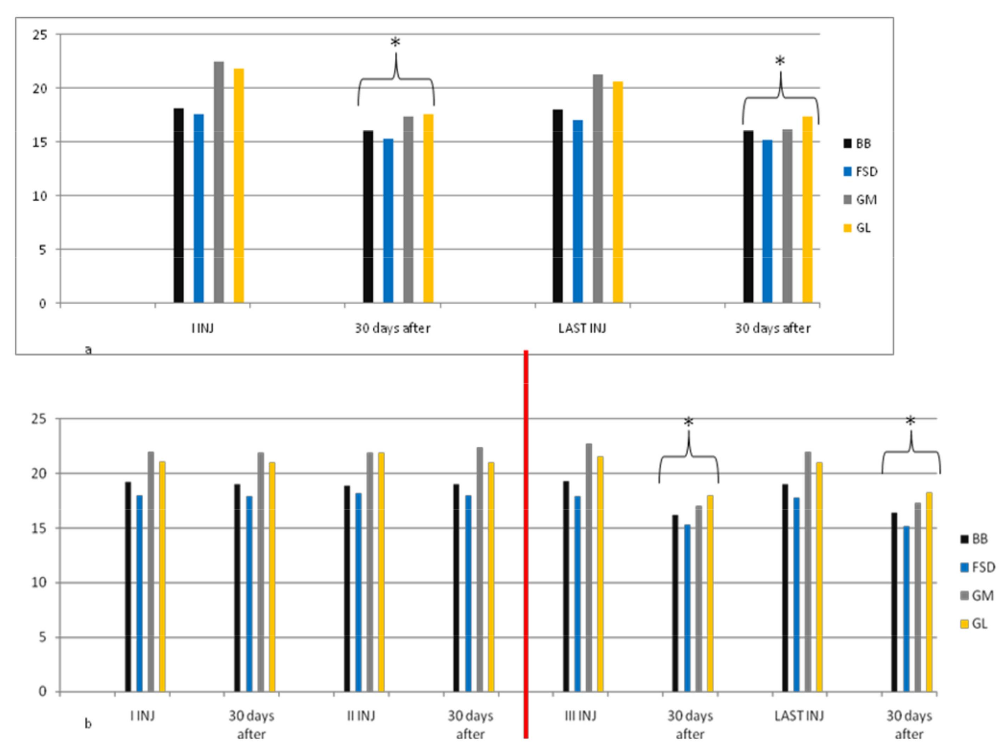
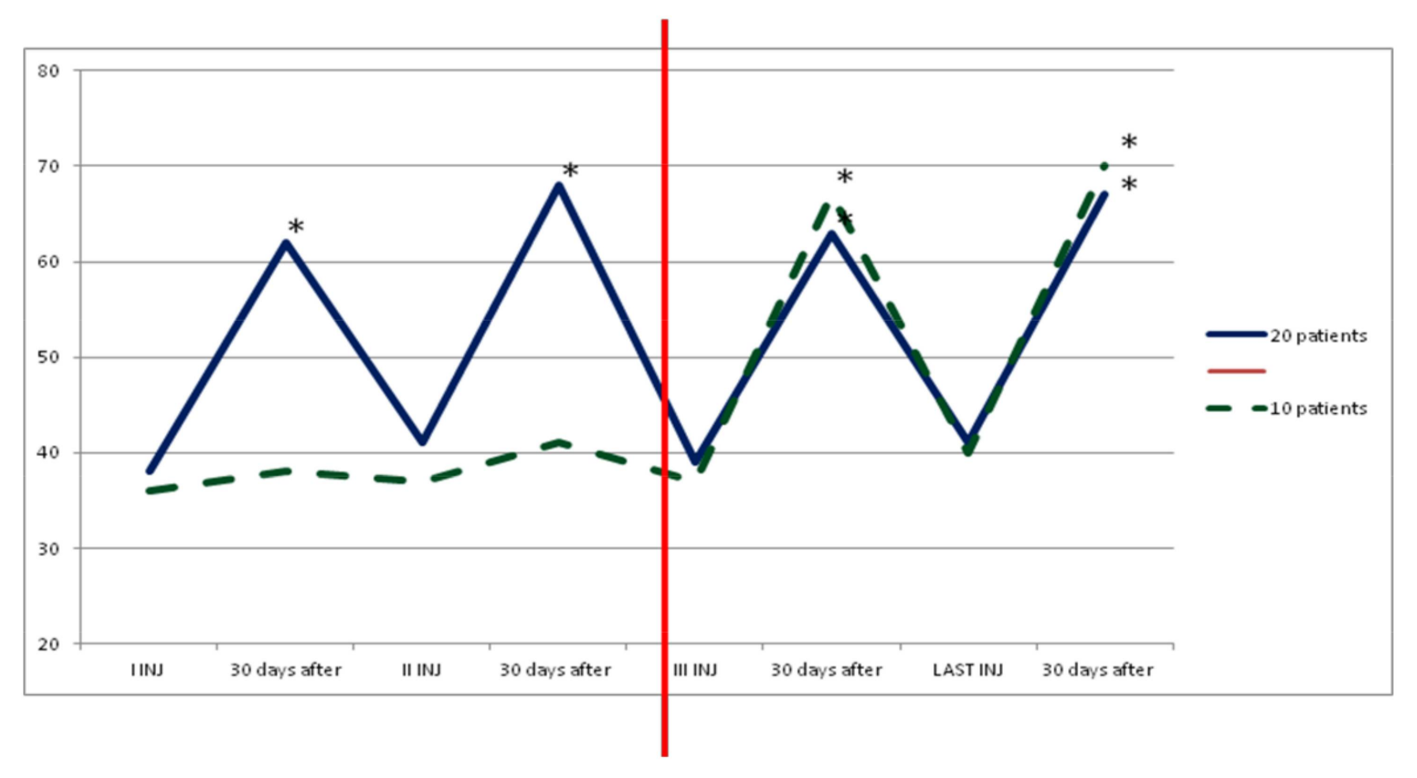
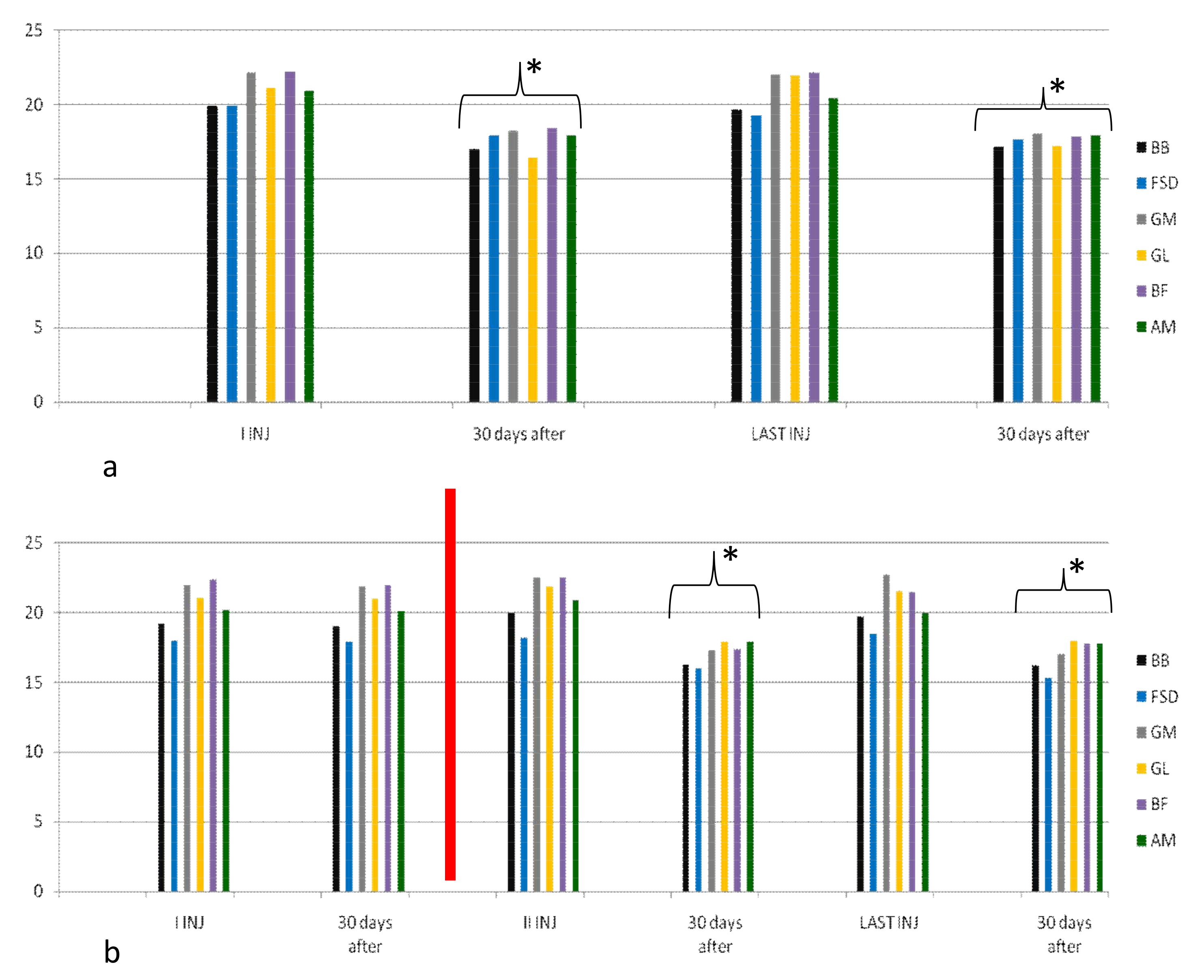
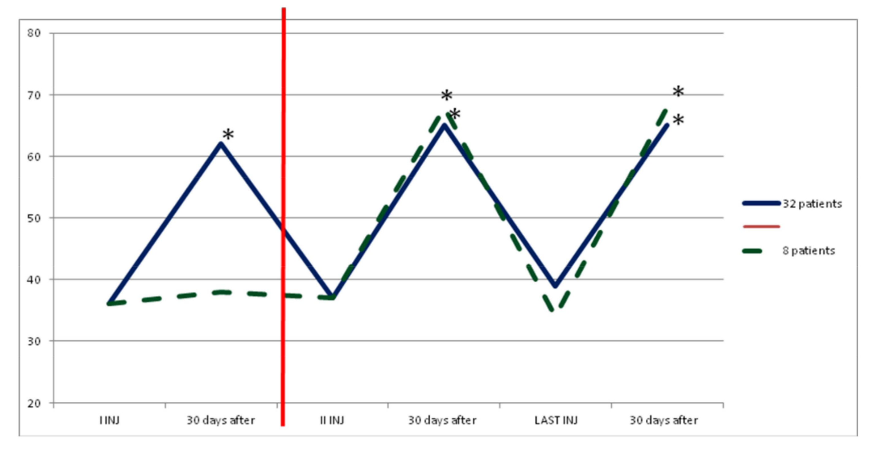
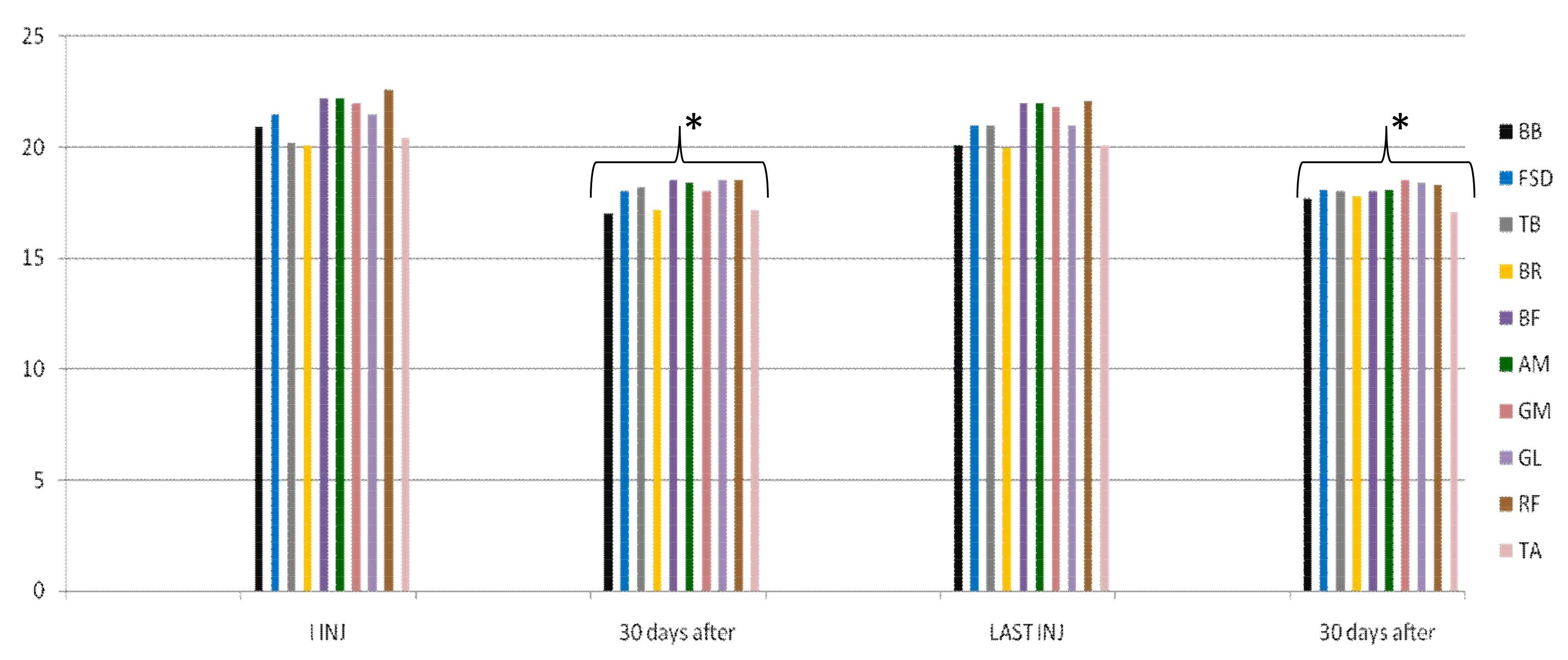
2. Results
Adverse Events
- -
- local muscle weakness: no case in group A, two cases in group B (5%) and two cases in group C (4%);
- -
- transient generalized weakness: no cases in group A and B, two cases in group C (4%);
- -
- mild dysphagia: no case in group A and B, one case in group C (2%; in the first cycle of injection, we also treated in this patient the left sternocleidomastoid muscle with 50 U of IncobotulinumtoxinA 1% saline due to muscle dystonia; this probably caused an adverse event because in the second cycle we treated the same muscle with 35 U of drug with good clinical response of dystonia and no dysphagia).
3. Discussion
4. Conclusions
5. Materials and Methods
5.1. Study Design
- (1)
- Group A (30 patients) up to 400 U
- (2)
- Group B (40 patients) from 400 U to 700 U
- (3)
- Group C (50 patients) from 700 U to 1000 U (maximum dose 600 U per limb).
5.2. Outcome Measures
5.3. Statistical Analysis
Acknowledgments
Author Contributions
Conflicts of Interest
References
- Jost, W.H.; Hefter, H.; Reissig, A.; Kollewe, K.; Wissel, J. Efficacy and safety of botulinum toxin type A (Dysport) for the treatment of post-stroke arm spasticity: Results of the German-Austrian open-label post-marketing surveillance prospective study. J. Neurol. Sci. 2014, 337, 86–90. [Google Scholar] [CrossRef] [PubMed]
- Hefter, H.; Jost, W.H.; Reissig, A.; Zakine, B.; Bakheit, A.M.; Wissel, J. Classification of posture in poststroke upper limb spasticity: A potential decision tool for botulinum toxin A treatment? Int. J. Rehabil. Res. 2012, 35, 227–233. [Google Scholar] [CrossRef] [PubMed]
- Simpson, D.M.; Gracies, J.M.; Graham, H.K.; Miyasaki, J.M.; Naumann, M.; Russman, B.; Simpson, L.L.; So, Y. Therapeutics and Technology Assessment Subcommittee of the American Academy of Neurology. Assessment: Botulinum neurotoxin for the treatment of spasticity (an evidence-based review): Report of the Therapeutics and Technology Assessment Subcommittee of the American Academy of Neurology. Neurology 2008, 70, 1691–1698. [Google Scholar] [CrossRef] [PubMed]
- Simpson, D.M.; Hallett, M.; Ashman, E.J.; Comella, C.L.; Green, M.W.; Gronseth, G.S.; Armstrong, M.J.; Gloss, D.; Potrebic, S.; Jankovic, J.; et al. Practice guideline update summary: Botulinum neurotoxin for the treatment of blepharospasm, cervical dystonia, adult spasticity, and headache: Report of the Guideline Development Subcommittee of the American Academy of Neurology. Neurology 2016, 86, 1818–1826. [Google Scholar] [CrossRef] [PubMed]
- Royal College of Physicians, British Society of Rehabilitation Medicine. Chartered Society of Physiotherapy, Association of Chartered Physiotherapists Interested in Neurology. Spasticity in Adults: Management Using Botulinum Toxin: National Guidelines. Available online: www.rcplondon.ac.uk/sites/default/files/documents/spasticity-in-adults-management-botulinum-toxin.pdf (accessed on 1 January 2016).
- Kocabas, H.; Salli, A.; Demir, A.H.; Ozerbil, O.M. Comparison of phenol and alcohol neurolysis of tibial nerve motor branches to the gastrocnemius muscle for treatment of spastic foot after stroke: A randomized controlled pilot study. Eur. J. Phys. Rehabil. Med. 2010, 46, 5–10. [Google Scholar] [PubMed]
- Bensmail, D.; Hanschmann, A.; Wissel, J. Satisfaction with botulinum toxin treatment in post-stroke spasticity: Results from two cross-sectional surveys (patients and physicians). J. Med. Econ. 2014, 17, 618–625. [Google Scholar] [CrossRef] [PubMed]
- Yang, D.J.; Park, S.K.; Uhm, Y.H.; Park, S.H.; Chun, D.W.; Kim, J.H. The correlation between muscle activity of the quadriceps and balance and gait in stroke patients. J. Phys. Ther. Sci. 2016, 28, 2289–2292. [Google Scholar] [CrossRef] [PubMed]
- Mohammad, H.; Shahram, A.; Seyed, A.H. Validity of Modified Ashworth Scale as a Measure of Wrist Spasticity in Stroke Patients. Iran. Rehabil. J. 2011, 9, 26–30. [Google Scholar]
- Fleuren, J.F.; Voerman, G.E.; Erren-Wolters, C.V.; Snoek, G.J.; Rietman, J.S.; Hermens, H.J.; Nene, A.V. Stop using the Ashworth Scale for the assessment of spasticity. J. Neurol. Neurosurg. Psychiatry 2010, 81, 46–52. [Google Scholar] [CrossRef] [PubMed]
- Von Werder, S.C.; Disselhorst-Klug, C. The role of biceps brachii and brachioradialis for the control of elbow flexion and extension movements. J. Electromyogr. Kinesiol. 2016, 28, 67–75. [Google Scholar] [CrossRef] [PubMed]
- Van Deun, B.; Hobbelen, J.S.; Cagnie, B.; Van Eetvelde, B.; Van Den Noortgate, N.; Cambier, D. Reproducible Measurements of Muscle Characteristics Using the MyotonPRO Device: Comparison Between Individuals with and without Paratonia. J. Geriatr. Phys. Ther. 2016. [Google Scholar] [CrossRef] [PubMed]
- Wissel, J.; Bensmail, D.; Ferreira, J.J.; Molteni, F.; Satkunam, L.; Moraleda, S.; Rekand, T.; McGuire, J.; Scheschonka, A.; Flatau-Baqué, B.; et al. Safety and efficacy of incobotulinumtoxinA doses up to 800 U in limb spasticity: The TOWER study. Neurology 2017, 88, 1321–1328. [Google Scholar] [CrossRef] [PubMed]
- Merz Pharma UK Ltd. XEOMIN® 100 U Summary of Product Characteristics. Available online: www.medicines.org.uk/emc/medicine/20666 (accessed on 6 January 2016).
- Merz Pharmaceuticals, LLC. Xeomin® US Prescribing Information. Available online: www.xeomin.com/wp-content/uploads/xeomin-full-prescribing-information.pdf (accessed on 6 January 2016).
- Tamargo, J.; Le Heuzey, J.Y.; Mabo, P. Narrow therapeutic index drugs: A clinical pharmacological consideration to flecainide. Eur. J. Clin. Pharmacol. 2015, 71, 549–567. [Google Scholar] [CrossRef] [PubMed]
- Reiffel, J.A. Formulation substitution and other pharmacokinetic variability: Underappreciated variables affecting antiarrhythmic efficacy and safety in clinical practice. Am. J. Cardiol. 2000, 85, 46D–52D. [Google Scholar] [CrossRef]
- Reiffel, J.A. Issues in the use of generic antiarrhythmic drugs. Curr. Opin. Cardiol. 2001, 16, 23–29. [Google Scholar] [CrossRef] [PubMed]
- Ianieri, G.; Saggini, R.; Marvulli, R.; Tondi, G.; Aprile, A.; Ranieri, M.; Benedetto, G.; Altini, S.; Lancioni, G.E.; Goffredo, L.; et al. New approach in the assessment of the tone, elasticity and the muscular resistance: Nominal scales vs MYOTON. Int. J. Immunopathol. Pharmacol. 2009, 22 (Suppl. 3), 21–24. [Google Scholar] [CrossRef] [PubMed]
- Marvulli, R.; Megna, M.; Romanelli, E.; Mastromauro, L.; Conte, E.; Lancioni, G.; Dargenio, M.; De Venuto, G.; Gallo, G.A.; Lerario, R.; et al. Effectiveness of the Treatment with Botulinum Toxin Type A (BTX-A) in the Management of the Spasticity in Patients with Amyotrophic Lateral Sclerosis (ALS). Clin. Immunol. Endocr. Metab. Drugs 2016, 3. [Google Scholar] [CrossRef]
- Marvulli, R.; Mastromauro, L.; Romanelli, E.; Lopopolo, A.; Dargenio, M.; Fornarelli, F.; Conte, E.; Fiore, P.; Megna, M.; Ianieri, G. How Botulinum Toxin Type A—Occupational Therapy (OT)-Functional Electrical Stimulation (FES) Modify Spasticity and Functional Recovery in Patients with Upper Limb Spasticity Post Stroke. Clin. Immunol. Endocr. Metab. Drugs 2016, 3, 62–67. [Google Scholar] [CrossRef]
- Munari, D.; Pedrinolla, A.; Smania, N.; Picelli, A.; Gandolfi, M.; Saltuari, L.; Schena, F. High-intensity treadmill training improves gait ability, VO2peak and cost of walking in stroke survivors: Preliminary results of a pilot randomized controlled trial. Eur. J. Phys. Rehabil. Med. 2016. Epub ahead of print. Available online: https://www.minervamedica.it/en/journals/europa-medicophysica/article.php?cod=R33Y9999N00A16083004 (accessed on 27 March 2018).
- Picelli, A.; Bacciga, M.; Melotti, C.; LA Marchina, E.; Verzini, E.; Ferrari, F.; Pontillo, A.; Corradi, J.; Tamburin, S.; Saltuari, L.; et al. Combined effects of robot-assisted gait training and botulinum toxin type A on spastic equinus foot in patients with chronic stroke: A pilot, single blind, randomized controlled trial. Eur. J. Phys. Rehabil. Med. 2016, 52, 759–766. [Google Scholar] [PubMed]
- Demetrios, M.; Khan, F.; Turner-Stokes, L.; Brand, C.; McSweeney, S. Multidisciplinary rehabilitation following botulinum toxin and other focal intramuscular treatment for post-stroke spasticity. Cochrane Database Syst. Rev. 2013, 6, CD009689. [Google Scholar] [CrossRef] [PubMed]
- Picelli, A.; Tamburin, S.; Cavazza, S.; Scampoli, C.; Manca, M.; Cosma, M.; Berto, G.; Vallies, G.; Roncari, L.; Melotti, C.; et al. Relationship between ultrasonographic, electromyographic, and clinical parameters in adult stroke patients with spastic equinus: An observational study. Arch. Phys. Med. Rehabil. 2014, 95, 1564–1570. [Google Scholar] [CrossRef] [PubMed]
- Synnot, A.; Chau, M.; Pitt, V.; O’Connor, D.; Gruen, R.L.; Wasiak, J.; Clavisi, O.; Pattuwage, L.; Phillips, K. Interventions for managing skeletal muscle spasticity following traumatic brain injury. Cochrane Database Syst. Rev. 2017, 11, CD008929. [Google Scholar] [CrossRef] [PubMed]
- Gao, F.; Grant, T.H.; Roth, E.J.; Zhang, L.Q. Changes in passive mechanical properties of the gastrocnemius muscle at the muscle fascicle and joint levels in stroke survivors. Arch. Phys. Med. Rehabil. 2009, 90, 819–826. [Google Scholar] [CrossRef] [PubMed]
- Dressler, D.; Saberi, F.A.; Kollewe, K.; Schrader, C. Safety aspects of incobotulinumtoxinA high-dose therapy. J. Neural Transm. 2015, 122, 327–333. [Google Scholar] [CrossRef] [PubMed]
- Fabbri, M.; Leodori, G.; Fernandes, R.M.; Bhidayasiri, R.; Marti, M.J.; Colosimo, C.; Ferreira, J.J. Neutralizing Antibody and Botulinum Toxin Therapy: A Systematic Review and Meta-analysis. Neurotox. Res. 2016, 29, 105–117. [Google Scholar] [CrossRef] [PubMed]
- Dressler, D. Five-year experience with incobotulinumtoxinA (Xeomin®): The first botulinum toxin drug free of complexing proteins. Eur. J. Neurol. 2012, 19, 385–389. [Google Scholar] [CrossRef] [PubMed]
- Velozo, C.A.; Seel, R.T.; Magasi, S.; Heinemann, A.W.; Romero, S. Improving measurement methods in rehabilitation: Core concepts and recommendations for scale development. Arch. Phys. Med. Rehabil. 2012, 93 (Suppl. 8), S154–S163. [Google Scholar] [CrossRef] [PubMed]
- Gerrard, P.; Goldstein, R.; Divita, M.A.; Ryan, C.M.; Mix, J.; Niewczyk, P.; Kazis, L.; Kowalske, K.; Zafonte, R.; Schneider, J.C. Validity and reliability of the FIM instrument in the inpatient burn rehabilitation population. Arch. Phys. Med. Rehabil. 2013, 94, 1521–1526.e4. [Google Scholar] [CrossRef] [PubMed]
- Lo, W.L.A.; Zhao, J.L.; Li, L.; Mao, Y.R.; Huang, D.F. Relative and Absolute Interrater Reliabilities of a Hand-Held Myotonometer to Quantify Mechanical Muscle Properties in Patients with Acute Stroke in an Inpatient Ward. Biomed. Res. Int. 2017. [Google Scholar] [CrossRef] [PubMed]
- Jarocka, E.; Marusiak, J.; Kumorek, M.; Jaskólska, A.; Jaskólski, A. Muscle stiffness at different force levels measured with two myotonometric devices. Physiol. Meas. 2012, 33, 65–78. [Google Scholar] [CrossRef] [PubMed]


| Time | GR A 100–400 U | GR B 400–700 U | GR C 700–1000 U |
|---|---|---|---|
| t 0 (beginning) | 30 | 40 | 50 |
| t 1 (after 9 months) | 20 | 42 | 58 |
| 1°GR 100–400 U BTX A | 2°GR 400–700 U BTX A | 3°GR 700–1000 U BTX A | |
|---|---|---|---|
| Local muscle weakness | - | 5% | 4% |
| Transientgeneralized weakness | - | - | 4% |
| Bradycardia | - | - | - |
| Dysphagia | - | - | 2% |
| Dysphonia | - | - | - |
| Dyspnea | - | - | - |
| Constipation | - | - | - |
| GR A 100–400 U BTX A | GR B 400–700 U BTX A | GR C 700–1000 U BTX A | |
|---|---|---|---|
| N° patients | 30 | 40 | 50 |
| Age | 64 ± 6.2 | 63 ± 8.4 | 66 ± 3.2 |
| Sex M/F | 17/13 | 23/17 | 28/22 |
| Clinical: Hemip.dx | 4 | 8 | 5 |
| Hemip. sx | 13 | 14 | 22 |
| Monoparesis | 4 | 1 | 0 |
| Paraparesis | 8 | 6 | 7 |
| Tetraparesis | 1 | 11 | 16 |
| Muscles Treated | GR A 100–400 U N° Patients | GR B 400–700 U N° Patients | GR C 700–1000 U N° Patients |
|---|---|---|---|
| Biceps brachii | 28 93.3% | 40 100% | 43 86% |
| Brachioradialis | - | 32 80% | 41 82% |
| Triceps brachii | - | 1 2.5% | 38 76% |
| Superficial flexorumdigitorum | 26 86.6% | 34 85% | 43 86% |
| Ulnar flexorumcarpis | - | 22 55% | 41 82% |
| Opponens pollicis | - | 34 85% | 43 86% |
| Rectus femoris | - | - | 40 80% |
| Adductor magnus | - | 26 65% | 38 76% |
| Tibialis anterior | - | - | 16 32% |
| Flexor alluci longus | - | 18 45% | 34 68% |
| Gastrocnemius medialis | 27 90% | 39 97.5% | 50 100% |
| Gastrocnemius lateralis | 27 90% | 39 97.5% | 50 100% |
| Soleus | 30 100% | 39 97.5% | 50 100% |
| Tibialis posterior | - | 26 65% | 45 90% |
| Flexor digitorum brevis | - | 16 40% | 36 72% |
| Biceps Femoris | - | 26 65% | 36 72% |
| Muscles | GR A 100–400 U Min-Max (Average ± SD) | GR B 400–700 U Min-Max (Average ± SD) | GR C 700–1000 U Min-Max (Average ± SD) |
|---|---|---|---|
| Biceps brachii | 50–80 (60.2 ± 10.3) | 70–90 (82.3 ± 10.1) | 80–100 (91.2 ± 10.1) |
| Brachioradialis | 50–60 (55.5 ± 10.4) | 70–80 (75.3 ± 2.2) | |
| Triceps brachii | 50–60 (55.2 ± 10.1) | 80–90 (85.6 ± 2.2) | |
| Superficial flexorumdigitorum | 50–80 (60.6 ± 10.2) | 50–90 (75.3 ± 10.3) | 100–150 (122.3 ± 20.1) |
| Ulnar flexorumcarpis | 50–60 (55.5 ± 2.2) | 80–100 (85.6 ± 2.1) | |
| Opponenspollicis | 20–30 (22.2 ± 10.3) | 30–40 (33.4 ± 1.1) | |
| Rectus femoris | 70–80 (74.2 ± 1.1) | ||
| Adductor magnus | 50–60 (55.3 ± 10.2) | 100–150 (132.3 ± 20.2) | |
| Tibialis anterior | 50–60 (55.3 ± 2.4) | ||
| Flexor alluci longus | 40–50 (44.2 ± 3.3) | 50–60 (54.2 ± 1.3) | |
| Gastrocnemius medialis | 60–80 (73.4 ± 10.5) | 80 | 100–150 (132.4 ± 10.4) |
| Gastrocnemius lateralis | 60–80 (72.5 ± 10.4) | 80 | 100–150 (122.4 ± 11.6) |
| Soleus | 70–80 (76.3 ± 2.5) | 80–90 (88.3 ± 1.3) | 100–150 (135.7 ± 17.4) |
| Tibialis posterior | 70–80(84.9 ± 3.3) | 100–120 (111.2 ± 2.4) | |
| Flexor digitorum brevis | 50–60 (55.6 ± 3.4) | 80–100 (89.9 ± 5.6) | |
| Biceps Femoris | 70–90 (84.6 ± 9.7) | 100–150 (145.7 ± 14.9) |
© 2018 by the authors. Licensee MDPI, Basel, Switzerland. This article is an open access article distributed under the terms and conditions of the Creative Commons Attribution (CC BY) license (http://creativecommons.org/licenses/by/4.0/).
Share and Cite
Ianieri, G.; Marvulli, R.; Gallo, G.A.; Fiore, P.; Megna, M. “Appropriate Treatment” and Therapeutic Window in Spasticity Treatment with IncobotulinumtoxinA: From 100 to 1000 Units. Toxins 2018, 10, 140. https://doi.org/10.3390/toxins10040140
Ianieri G, Marvulli R, Gallo GA, Fiore P, Megna M. “Appropriate Treatment” and Therapeutic Window in Spasticity Treatment with IncobotulinumtoxinA: From 100 to 1000 Units. Toxins. 2018; 10(4):140. https://doi.org/10.3390/toxins10040140
Chicago/Turabian StyleIanieri, Giancarlo, Riccardo Marvulli, Giulia Alessia Gallo, Pietro Fiore, and Marisa Megna. 2018. "“Appropriate Treatment” and Therapeutic Window in Spasticity Treatment with IncobotulinumtoxinA: From 100 to 1000 Units" Toxins 10, no. 4: 140. https://doi.org/10.3390/toxins10040140
APA StyleIanieri, G., Marvulli, R., Gallo, G. A., Fiore, P., & Megna, M. (2018). “Appropriate Treatment” and Therapeutic Window in Spasticity Treatment with IncobotulinumtoxinA: From 100 to 1000 Units. Toxins, 10(4), 140. https://doi.org/10.3390/toxins10040140