Article

Two Lineages of Papillomaviruses Identified from Caracals (Caracal caracal) in South Africa

by
Simona Kraberger
1,*,
Laurel E. K. Serieys
2,
Gabriella R. M. Leighton
3,
Matthew D. De Koch
1,
John S. Munday
4,
Jacqueline M. Bishop
3 and
Arvind Varsani
1,5,*
1
The Biodesign Center for Fundamental and Applied Microbiomics, Center for Evolution and Medicine and School of Life Sciences, Arizona State University, Tempe, AZ 85287, USA
2
Panthera, 8 W 40th St, 18th Floor, New York, NY 10018, USA
3
Institute for Communities and Wildlife in Africa (iCWild), Department of Biological Sciences, University of Cape Town, Cape Town 7701, South Africa
4
School of Veterinary Science, Massey University, Tennant Drive, Palmerston North 4442, New Zealand
5
Structural Biology Research Unit, Department of Integrative Biomedical Sciences, University of Cape Town, Cape Town 7925, South Africa
*
Authors to whom correspondence should be addressed.
Viruses 2024, 16(5), 701; https://doi.org/10.3390/v16050701
Submission received: 3 April 2024 / Revised: 26 April 2024 / Accepted: 28 April 2024 / Published: 29 April 2024
(This article belongs to the Special Issue Animal Virus Discovery and Genetic Diversity)

Abstract

:
Papillomaviruses (PV) infect epithelial cells and can cause hyperplastic or neoplastic lesions. In felids, most described PVs are from domestic cats (Felis catus; n = 7 types), with one type identified in each of the five wild felid species studied to date (Panthera uncia, Puma concolor, Leopardus wiedii, Panthera leo persica and Lynx rufus). PVs from domestic cats are highly diverse and are currently classified into three genera (Lambdapapillomavirus, Dyothetapapillomavirus, and Taupapillomavirus), whereas those from wild felids, although diverse, are all classified into the Lambdapapillomavirus genus. In this study, we used a metagenomic approach to identify ten novel PV genomes from rectal swabs of five deceased caracals (Caracal caracal) living in the greater Cape Town area, South Africa. These are the first PVs to be described from caracals, and represent six new PV types, i.e., Caracal caracal papillomavirus (CcarPV) 1–6. These CcarPV fall into two phylogenetically distinct genera: Lambdapapillomavirus, and Treisetapapillomavirus. Two or more PV types were identified in a single individual for three of the five caracals, and four caracals shared at least one of the same PV types with another caracal. This study broadens our understanding of wild felid PVs and provides evidence that there may be several wild felid PV lineages.

1. Introduction

Papillomaviruses (PVs; family Papillomaviridae) are circular double-stranded (dsDNA) viruses that infect mammals, birds, and reptiles [1,2,3,4]. Highly diverse and generally species-specific, PVs are epithelial-cell-trophic. A single host can be infected with several PV types, including types that are classified within different genera [5,6]. PV genomes are composed of five to six early genes and two late genes. The L1 gene which encodes the major capsid protein typically shows higher levels of conservation among PVs and is used for taxonomic classification with those sharing >60% L1 nucleotide similarity belonging to the same genus, >70% for species, and >90% for type [2]. Currently, most of the known PV types are those infecting humans, with a significant knowledge gap for non-human PVs and their broader evolution [2,4].
In felids, several unique papillomaviruses have been identified which can cause cutaneous or oral lesions [7]. The most studied feline species is the domestic cat (Felis catus). In domestic cats, PVs rarely cause the hyperplastic papillomas (warts) which are common in other felid species [7]. Instead, many PV types are detected in association with preneoplastic viral plaques or invasive neoplasms of the skin [7,8]. Seven PV types from domestic cats have been classified to date: Felis catus papillomavirus (FcaPV) 1–7 [9,10,11,12,13,14,15]. FcaPV7 (OL310516; [16]), although identified from a skin swab from a human, is thought to be of feline origin due to the person being a cat owner and the genome showing similarities to FcaPV2. In addition, an unclassified FcaPV genome (OQ836188; [17]) was recovered from an infection associated with skin cancer in a domestic cat that will likely be FcaPV8, and another, Bos taurus papillomavirus (BPV) 14 [18], was found to cause feline sarcoids after cross-species infection from its bovine host [19] (Table 1). The FcaPVs belong to three different genera, Lambdapapillomavirus (FcaPV1), Dyothetapapillomavirus (FcaPV2), and Taupapillomavirus (FcaPV3, -4, -5, -6), and FcaPV7 is currently unclassified but sits with FcaPV2 and is, therefore, likely a Dyothetapapillomavirus. In wild felid species, complete PV genomes have previously been documented in a bobcat (Lynx rufus)—Lynx rufus papillomavirus 1 (LrPV1), Asiatic lion (Panthera leo persica)—Panthera leo persica papillomavirus 1 (PlpPV1), snow leopard (Uncia uncia or Panthera uncia)—Uncia uncia papillomavirus 1 (UuPV1), mountain lion (Puma concolor)—Puma concolor papillomavirus 1 (PcPV1) [20,21], and margay (Leopardus wiedii)—Leopardus wiedii papillomavirus 1 (LwiePV1) [22]. These feline PV types all belong to the Lambdapapillomavirus genus, which has other PVs from the Carnivora species. Several partial felid PV sequences representing novel papillomaviruses have also been identified (Table 1) from domestic cats, cheetahs (Acinonyx jubatus) [23], the African lion (Panthera leo) [23], and snow leopards [24,25].
The monophyletic nature of the PVs identified from wild felid species contrasts with those from domestic cats which are polyphyletic [9,10,12,13,31]. This could be a result of several factors, such as the sampling bias of domestic cats, and/or to areas where obvious lesions are observed, as well as the geographic or region endemicity of the different wild felid species versus the broad global distribution and free ranging capability of domestic cats, exposing them to a higher cross-species transmission potential. The Lambdapapillomavirus feline lineage appears to be slow-evolving, at a rate of 1.95 × 10⁻⁸ nucleotide substitutions per site per year, and shows evidence of a long coevolutionary history with their feline host species [20]. Further, these viruses appear to have a unique second non-coding region between the early and late protein region [20].
Here, we identify ten complete PV genomes from five caracals (Caracal caracal) inhabiting the greater Cape Town area, Western Cape, South Africa. These are the first documented PVs from caracals and they belong to two genera, Lambdapapillomavirus and Treisetapapillomavirus, thus expanding our current knowledge on PVs in wild felids.

2. Materials and Methods

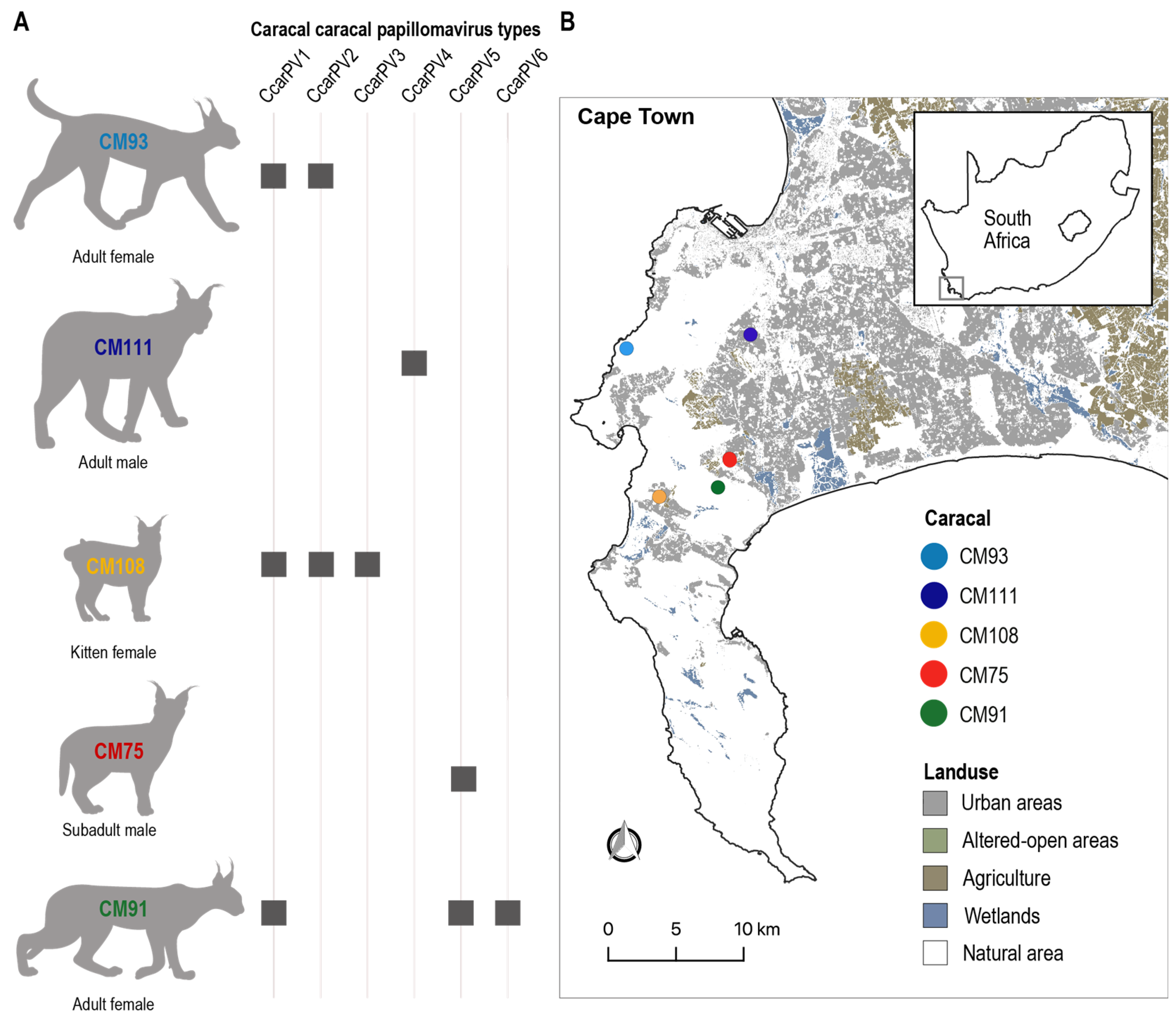
2.1. Study Site and Sample Collection

As part of a long-term study undertaken by the Urban Caracal Project (www.urbancaracal.org; accessed 12 February 2024) to monitor the health and well-being of caracals in the greater Cape Town area, rectal swab samples were collected during post-mortem of deceased animals (n = 26) between 2021 and 2023 using PurFlock ultra 6″ sterile flock swabs (Puritan Medical Products, Guilford, ME, USA). Cause of death for these caracals was determined to be motor vehicle impact when crossing urban roads, disease and/or poisoning, or poaching. The swabs were stored at −20 °C in Puritan UniTranz-RT media (Puritan Medical Products, Guilford, ME, USA) for downstream nucleic acid extraction.

2.2. Sample Processing and Papillomavirus Genome Identification

High Pure Viral Nucleic Acid Kit (Roche Diagnostics, Indianapolis, IN, USA) was used to isolate viral nucleic acid from 200 µL of the UTM buffer in which the swabs were stored. Then, 1 µL of the viral nucleic acid was enriched for circular molecules by rolling circle amplification (RCA) using the Illustra TempliPhi Kit (Cytiva Lifesciences, Marlborough, MA, USA). An aliquot of viral nucleic acid was combined with the RCA product and high-throughput sequencing (HTS) libraries were generated using an Illumina DNA prep (M) tagmentation kit (Illumina, San Diego, CA, USA). Libraries were sequenced on Illumina NovaSeq X plus sequencer at Psomagen Inc. (Rockville, MD, USA). The raw paired-end reads (2 × 150 bp) were trimmed using Trimmomatic −0.39 [40] and de novo assembled with MEGAHIT v1.2.9 [41]. The de novo assembled contigs of >1000 nts were screened against a viral RefSeq protein sequence database (release 220) using DIAMOND BLASTx [42]. We also screened the contigs for host mitochondrial genomes using Diamond BLASTx [42] with a mitochondrial RefSeq database (release 220). Contigs were determined as circular based on terminal redundancy. Read mapping to confirm adequate depth and coverage of full genomes was performed using BBmap [43].
The complete papillomavirus and mitochondrial genome sequences are deposited in GenBank with accession # OR915584-OR915593 and PP566117-PP566121, respectively. The SRA data are deposited under BioProject #PRJNA1045660, BioSample # SAMN38451862-SAMN38451866 and SRA # SRR26982246-SRR26982250 for the PV sequences. For the caracal mitochondrial genomes under BioProject # PRJNA1033669-PRJNA1033673, BioSample # SAMN38451862-SAMN38451866, and SRA # SRR28492289-SRR28492293.

2.3. Genome Characterization, Pairwise Comparison, and Phylogenetic Analyses

The PV genomes were annotated with Cenote-Taker2 [44], and then manually checked with annotation of PVs from PAVE [4]. The mitochondrial genomes of the hosts were annotated with the MITOS server [45,46], and then manually checked.
Pairwise similarity identities were determined for the full genome, and the E1, E2, E3, E6, E7, L1, and L2 genes and protein sequences of the PVs from this study and of those most closely related using SDT v1.2 [47].
Dataset of L1, E1, and E2 protein sequences of all PV types referenced at PAVE [4], as well as those from this study, were assembled. We opted to use the protein sequences of L1, E1, and E2 as these are the most conserved amongst the papillomaviruses and can be more credibly aligned than the corresponding nucleotide sequences. These were aligned using MAFFT [48] and trimmed using TrimAL [49] with the 0.2 gap option and concatenated (L1 + E1 + E2). The best-fit amino acid substitution models LG + I+G for the E1, LG + I + G + F for the E2, and LG + I + G + F for the L1 were determined using ProtTest3 [50]. A partitioned phylogenetic tree was inferred using IQ-TREE2 [51] with aLRT branch support and rooted with the L1 + E1 + E2 of avian papillomaviruses. The phylogenetic tree was visualized in iTOLv6 [52]. Mitochondrial genomes from caracals together with those available from other members of the Caracal genus available in GenBank were aligned using MAFFT [48] and a neighbor-joining tree was constructed using FastTree [53], implemented in Geneious Prime 2024.0.4.
The motif discovery and comparison tools MEME [54] and Tomtom [55] were used to identify conserved motifs in the non-coding regions of the genomes of feline-infecting Lambdapapillomaviruses.

3. Results and Discussion

3.1. Identification of Papillomavirus Genomes in Wild Caracal

As part of an ongoing effort to monitor caracal health and survival in the greater Cape Town area (Western Cape, South Africa), rectal swabs were collected from deceased caracals as a result of being hit by motor vehicles, and/or suspected disease or pesticide poisoning between the period 2021–2023 (Table 2). Ten novel PV genomes were determined from the rectal swabs of five caracals (Figure 1, Table 2). No obvious lesions or papillomas were observed during post-mortem sample collection.
Ten PV genomes were de novo assembled from high-throughput sequencing data with an average read depth ranging from 21- to 108,899-fold. All ten PV genomes have identifiable E1, E2, E6, E7, L1, and L2 genes and range in genome size from 7566 to 8176 bp (Figure 2A). Based on the papillomavirus ICTV species demarcation determination of <70% pairwise identity for the L1 gene sequences and <10% pairwise identity for PV types, these ten PV represent six novel types; Caracal caracal papillomavirus (CcarPV) 1–6, (Figure 2, Table 2). CcarPV1, -2, -3, and -4 share >70% L1 gene sequence identity with other felid PVs, i.e., LwiePV1 [22], LrPV1, PlpPV1, UuPV1 and PcPV1 [20,21], and FcaPV1 [11], and, therefore, these all belong to the same species. CcarPV5 and -6 share <70% L1 gene nucleotide sequence identity with other PVs and each other, and, therefore, will represent two new species. All CcarPV isolates belonging to the same type share 100% L1 gene nucleotide sequence pairwise identity.
Caracals CM93 and CM91, both adult females, were found to have multiple PVs, with CM93 harboring two PVs, CcarPV1 (OR915585) and CcarPV2 (OR915587), and CM91 three PVs, CcarPV1 (OR915584), CcarPV5 (OR915592), and CcarPV6 (OR915593) (Figure 1 and Figure 2). Caracal CM108, a female kitten, harbored three PVs: CcarPV1 (OR915586), CcarPV2 (OR915588), and CcarPV3 (OR915589). Lastly, Caracal CM75 and CM111, both males, a subadult and an adult, both harbored one PV, CcarPV5 (OR915591) and CCarPV4 (OR915590), respectively. CM93, CM108, and CM91 were all infected with CcarPV1 and one or two other PV types, showing a high rate of PV coinfection which is commonly seen in mammals [6,56]. Further, CcarPV1 and -2 were present in both CM93 and CM108. Taking into consideration the fact that these three individuals share at least one PV type and were found deceased within a 10 km radius from each other, this may indicate these cats were related, and/or interacted with other caracal(s) not sampled infected with these PV types.

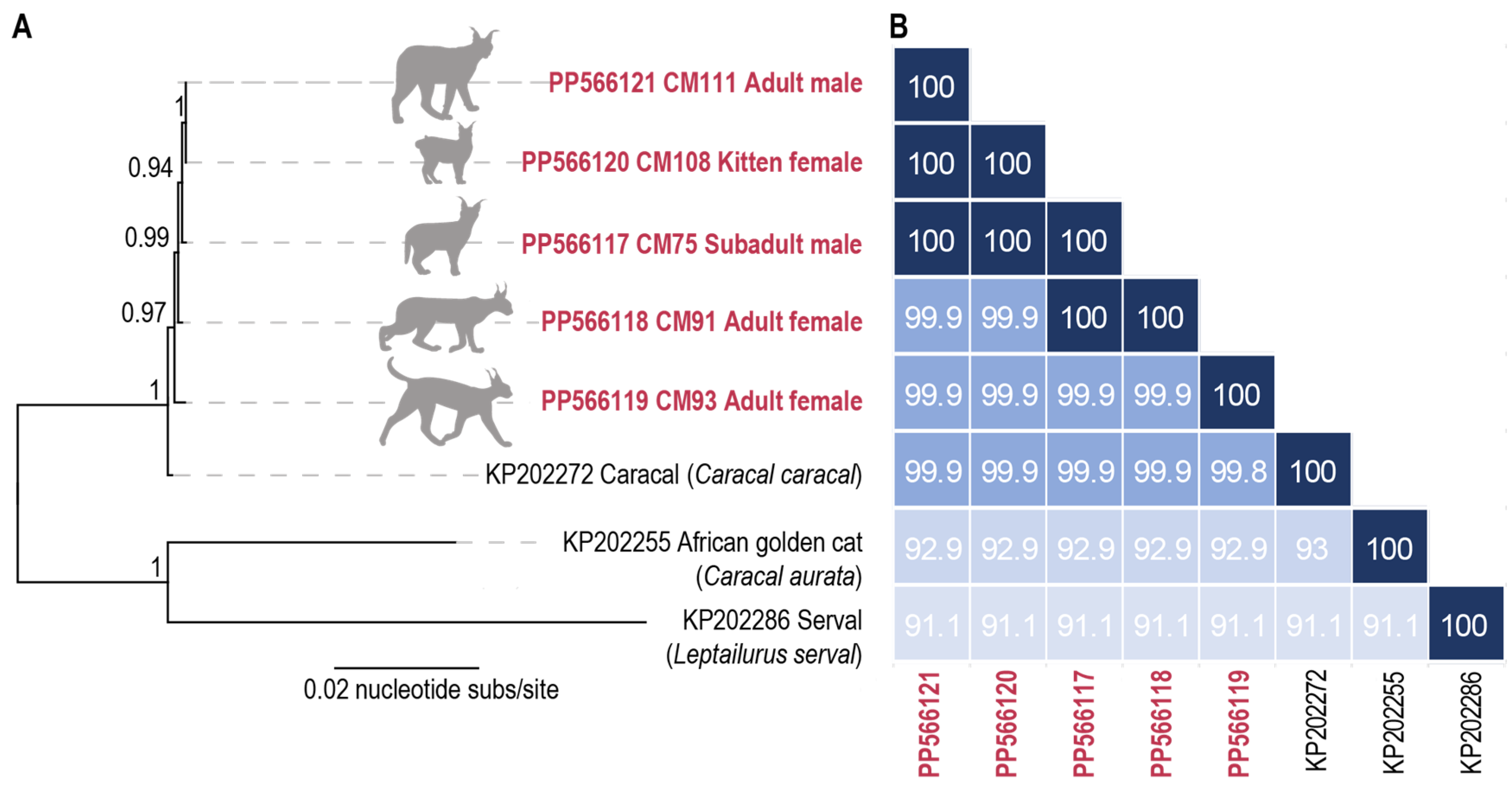
3.2. Caracal Mitochondrial Genomes

An advantage to using rolling circle amplification for the enrichment of circular DNA molecules is that this enables the simultaneous identification of host mitochondrial genomes that are also circular. From the PV-positive caracal samples, we were able to determine the full mitochondrial genomes. Phylogenetically (based on the mitochondrial sequences), all five caracals are closely related and sit within the Caracal clade, forming a sister lineage to the caracal mitochondrial genome (KP202272) available in GenBank (Figure 3). The mitochondrial genomes of caracals CM75, CM91, CM93, CM108, and CM111 share 99.9–100% pairwise nucleotide identity with each other and 99.8–99.9% pairwise nucleotide identity with a caracal mitochondrial genome (KP202272) [57] (Figure 3). This high level of similarity is not surprising given the recent study which showed that the Caracal population in Cape Town have elevated levels of inbreeding [58]. A comparison with mitochondrial genomes of two other members of the caracal lineage, an African golden cat (Caracal aurata) (KP202255) and a serval (Leptailurus serval) (KP202286) [57], showed they share 91.1–93% pairwise nucleotide identity.

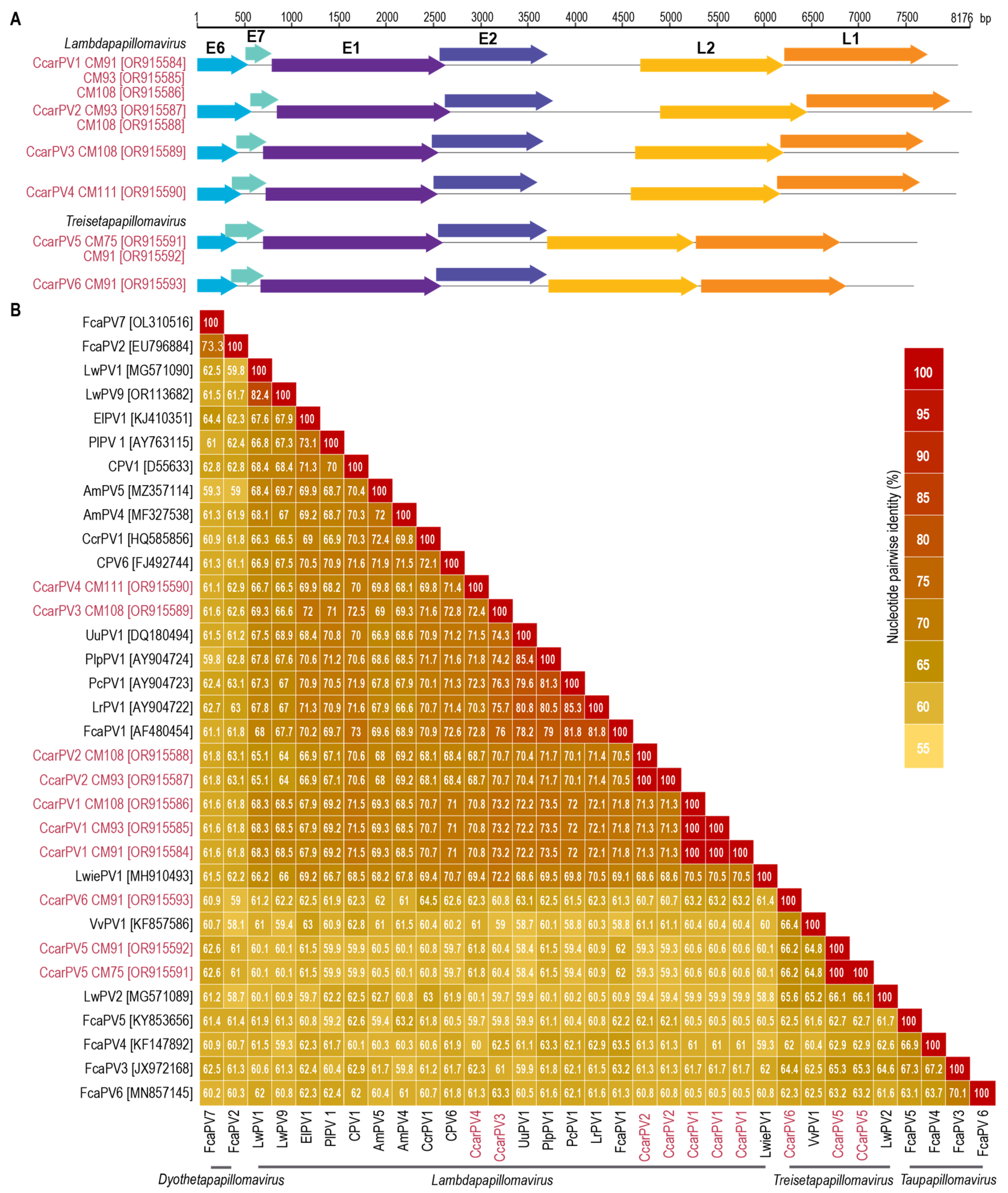
3.3. Sequence Comparison of Caracal PVs

For the six CcarPV types, their genomes share 59.4–71.1% pairwise identity (Supplementary Data S1), showing a significant diversity amongst these genomes. For CcarPV1, CcarPV2, and CcarPV5, however, multiple isolates were identified from more than one individual, and, within each type, the isolate sequences are identical. PVs can be very slow-evolving, and, therefore, it is not uncommon to find identical sequences in samples from different individuals, even for PVs sampled decades apart [59,60]. A full-genome pairwise comparison of the CcarPVs with the PVs most closely related reveals that they share 58.8–72.6% pairwise identity, with CcarPV3 and PcPV1 (AY904723) from a puma [20] sharing the highest pairwise identity of 72.6%. A pairwise comparison of the protein sequences of E1, E2, E3, E6, E7, L1, and L2 for CcarPVs with those of PVs most closely related show that the L1 and E1 proteins share the highest pairwise identities ranging from 47.2–87.1% and 45.1–76.8%, respectively. Overall, the E6 protein has the lowest pairwise identity (23.1–59.1%) for the CcarPVs and those of the most closely related PVs.

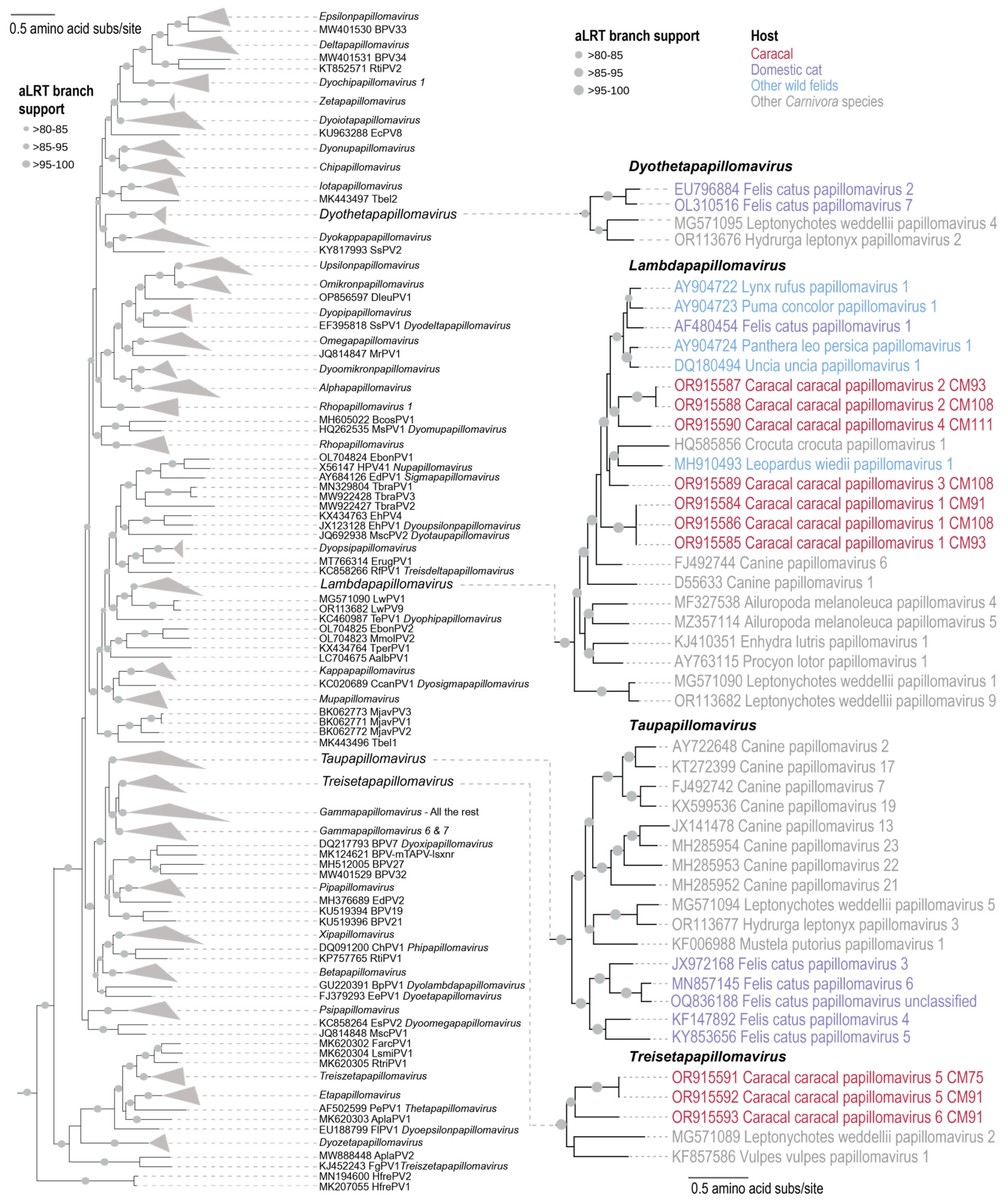
3.4. Caracal PV L1 + E1 + E2 Phylogeny

A maximum-likelihood phylogenetic tree was constructed from the concatenated L1 + E1 + E2 protein sequences of the caracal PVs and those of representative PV sequences from GenBank. This analysis showed these six CcarPV types are part of two genera, Treisetapapillomavirus and Lambdapapillomavirus (Figure 4). Treisetapapillomavirus currently comprises two PVs; one identified from a Weddel seal (Leptonychotes weddellii) [5] and one from a red fox (Vulpes vulpes) [61]. Lambdapapillomavirus comprises PVs from felid species (wild and domestic) [20,22], Weddel seal [5], giant panda (Ailuropoda melanoleuca) [32], sea otter (Enhydra lutris) [62], raccoon (Procyon lotor) [63], spotted hyena (Crocuta crocuta) [64], and domestic dog (Canis familiaris) [65,66]. Previously identified PVs from wild felids, puma and bobcat [20,22], all cluster with members of the Lambdapapillomavirus genus, whereas those from domestic cats are distributed across three genera, i.e., Lambdapapillomavirus, Taupapillomavirus, and Dyothetapapillomavirus. The CcarPVs from this study that are part of the Lambdapapillomavirus genus are CcarPV1, -2, -3, and -4, all grouped in a felid-PV-dominant subclade with one non-felid PV, CcrPV1 (HQ585856) from a spotted hyena [64]. CcarPV1 is basal in this clade, whereas CcarPV3 is most closely related to LwiePV1 (MH910493) [22] from a margay and CcrPV1 (HQ585856) from a spotted hyena [64]. CcarPV2 and -4 cluster in a clade that is basal to the other felid PVs in the genus Lambdapapillomavirus [11,20,22]. It should be noted that some of these subclades do not have strong branch support, and, therefore, as more PVs are identified and added to this group, it will likely help resolve these phylogenetic relationships more robustly. CcarPV5 and -6 group with the PVs in the genus Treisetapapillomavirus as a sister clade to Leptonychotes weddellii papillomavirus 2 (MG571089) [5] from a Weddell seal and Vulpes vulpes papillomavirus 1 (KF857586) [61] from a red fox. The polyphylogenetic distribution of the CcarPVs is similar to that noted for the domestic cat PVs, and, therefore, with the increased sampling of wild felids, a similar pattern may emerge. This is significant as it indicates a more complex evolutionary history than what was previously thought.

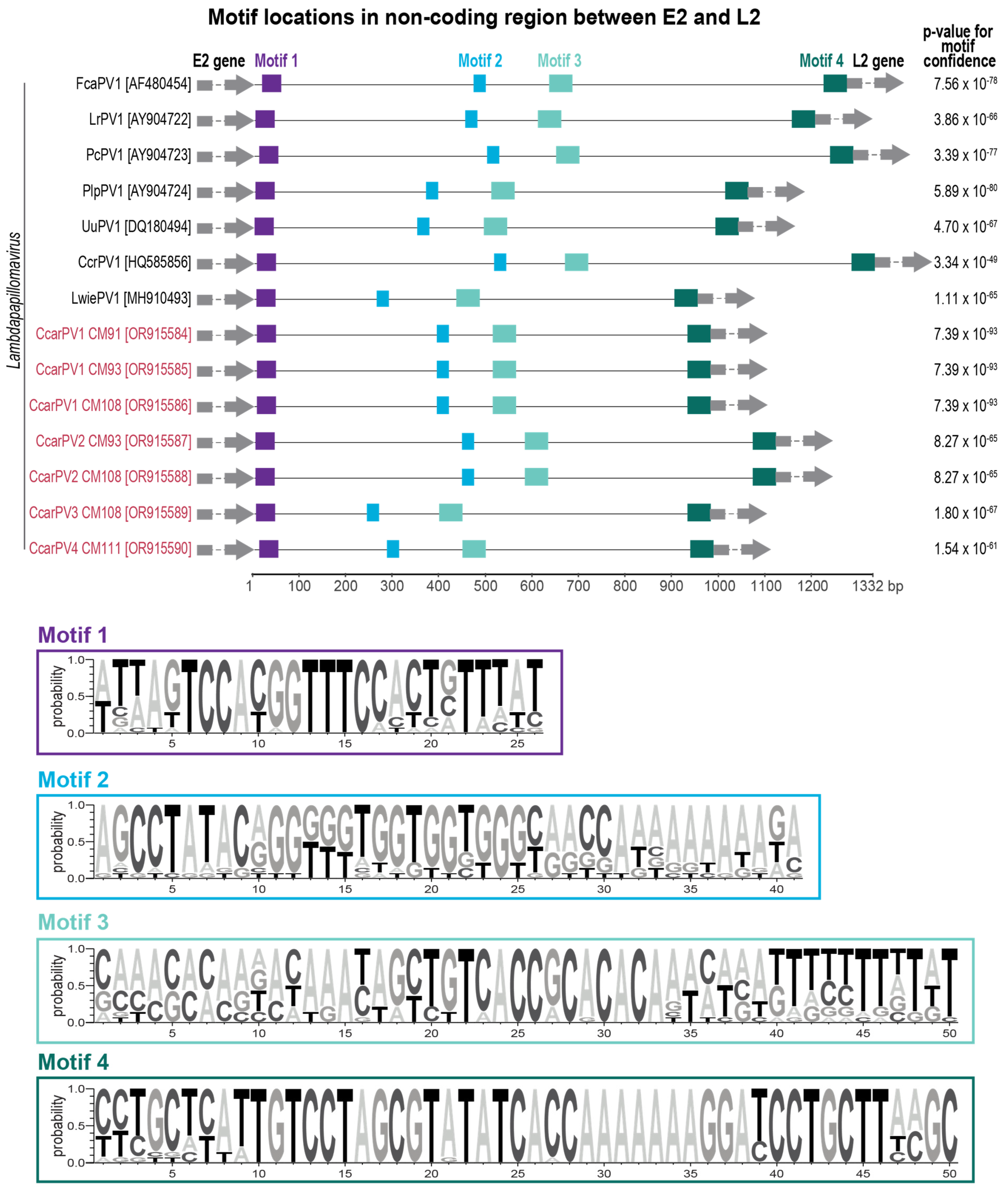
3.5. Large Non-Coding Region in the Genomes of Lambdapapillomaviruses

Treisetapapillomavirus genomes are up to 1215 bp smaller (7392–7598 bp; [5,61] than those of lambdapapillomaviruses (7944–8607 bp) [11,20,64]. This difference in genome size appears to be due, at least in part, to a stretch of a non-coding region between the E2 and the L2 coding open reading frames (ORFs) in lambdapapillomaviruses, with the exception of Leptonychotes weddellii papillomavirus 1 (MG571090) from a Weddell seal [5]. This non-coding region has previously been noted and discussed in Rector et al. (2007). They noted there are several conserved regions that are likely to be of regulatory or other functional importance. To investigate this further, we used the motif discovery tool MEME [54] to scan this region, revealing four conserved motifs that were present in all the felid PVs and the CcrPV1 from spotted hyena (HQ585856) [64] in the Lambdapapillomavirus genus (Figure 5). A comparison of these regions with motif analyses tools such as Tomtom [55] indicates that these are possibly single-stranded DNA-binding motifs sharing the highest similarities to those associated with transcription factors in humans [67]. Although these findings support that this region has conserved motifs that may be involved in the DNA binding of transcription factors, in vitro molecular studies are needed to investigate this further.

3.6. E6 and E7 Protein Motifs

The E6 and E7 are two early proteins that are encoded in most mammalian PVs. These oncoproteins have largely been studied in human PVs and play an important role in regulating the cell cycle in order to sustain cellular replication activity and viral proliferation [68]. Further, it is the ability of E6 and E7 proteins to bind tumor suppressors p53 and pRB, respectively, which is thought to drive tumor production [69]. The E6 of the CcarPVs contains two zinc-binding domains which show conservation with other felid PVs as well as other Carnivora species that are most closely related (Figure 6). The C-X-F-C-X29-C-X2-C motif is the conserved for the first domain; however, the second domain has one less amino acid in the lambdapapillomavirus E6 proteins compared with those of treisetapapillomaviruses C-X2-C-X3-L-X21/23-R-X3-R-X2C-X2-C. The E7 protein L-X-C/S-X-E motif which binds the pRB in the lambdapapillomaviruses has a conserved L-X-C-X-E, unlike the treisetapapillomaviruses where it is L-X-S-X-E (Figure 6). The zinc-binding domain in the E7 for members of these two genera varies in the number of residues from 34–37 nts (C-X2-C-X26/28/29-C-X2-C). This domain, for all of those in the E7 proteins of PVs in the Treisetapapillomavirus genus, and that of LwiePV1 (MH910493) [22] and CcrPV1 (HQ585856) [64] from the Lambdapapillomavirus genus, has a 37 residue zinc-binding motif. On the other hand, the E7 of other members of the lambdapapillomaviruses have 36 residues, except for UuPV1 (DQ180494) from snow leopard [20] which has 35 residues.

4. Conclusions

Through the sampling of deceased caracals in the greater Cape Town region of South Africa, we identified ten novel PV genomes from five individuals. These represent six diverse caracal PV types (CcarPV1–6) that belong to two genera, Treisetapapillomavirus and Lambdapapillomavirus. Lambdapapillomavirus comprises members of PVs from other felids [20,22] and the Carnivora species, whereas Treisetapapillomavirus previously only comprised Leptonychotes weddellii papillomavirus 2 (MG571089) [5] and Vulpes vulpes papillomavirus 1 (KF857586) [61]. Although these were identified from rectal swabs of deceased caracals, no obvious pathology typical of PV infections were noted. Not all PVs are associated with lesions or papillomas; for example, several of the human-infecting PV types in the Betapapillomavirus genus are symptomless [70].
Three of the five caracals were identified to have mixed infections of 2–3 CcarPV types. Additionally, four out of the five caracals harbored at least one CcarPV type whose genome is identical to that from another caracal. Given that PV transmission requires close direct contact, this may be representative of social interactions and family connections between the four caracals (CM75, CM91, CM93, and CM108). Alternatively, given the slow evolutionary rate of papillomaviruses, these types may have been circulating in this population for some time and transmitted through intermediary interaction partners that were not sampled in this study.
We were able to determine the host mitochondrial genomes for the five caracals which all share a 99.9–100% genome-wide pairwise identity with each other. Although we were unable to determine relatedness, a more extensive host genomic investigation of these caracals would help to shed some light on any family relationships. A recent study has shown that the Cape Peninsula caracal population has limited inward migration and appears to have high levels of inbreeding [58] which is supported in the lack of diversity seen in the mitochondrial genomes described here.
Sequence and phylogenetic analyses of these CcarPVs shows that, although these do share similarities to other PVs at a nucleotide and protein level, they are still diverse and distinct from other PVs. The identification of two lineages of the CcarPVs shows that there are diverse PVs circulating within an individual as well as within this caracal population. Notably, caracal CM91 from which CcarPV1, CcarPV5, and CcarPV6 were recovered harbored PV types belonging to the two lineages (Lambdapapillomavirus and Treisetapapillomavirus). This is similar to the pattern seen for the domestic cat PVs [18] as well as some other mammal PVs [1,5,71,72].
A unique non-coding region between the E2 and L2 ORFs is present in CcarPV1–4, other felid PV members, as well as CcrPV1 [64] from a spotted hyena (in the Lambdapapillomavirus genus), likely resulting from an expansion event that may have occurred in a shared ancestor. This region likely plays a regulatory or functional role, given the conserved nucleotide motifs present across members of this PV lineage. Four conserved motifs were identified in this region that are likely single-stranded DNA-binding domains; however, more research is needed to elucidate the biological importance of these and this insertion/expansion region. Since Rector et al. [20] demonstrated evidence of a long co-speciation history for the feline PVs, several new lineages of domestic cat PVs have been identified. It is, therefore, possible that several lineages have coevolved with their felid hosts for some time. These findings, together with the diverse caracal PVs from this study, also highlights possible host switching and/or recombination, leading to the emergence of these polyphyletic lineages.
Overall, the findings in this study expand the known felid PV diversity, demonstrate the utility of using rectal sampling for identifying PVs, as well as host mitochondrial genomes, and provide broader insights into PV dynamics in wild felid populations.

Supplementary Materials

The following supporting information can be downloaded at: https://www.mdpi.com/article/10.3390/v16050701/s1 Supplementary Data S1: Pairwise identity matrices for the full genome and E1, E2, E3, E6, E7, L1, and L2 protein sequences of the CcarPVs with their most closely related PV representative sequences.

Author Contributions

Conceptualization, S.K., L.E.K.S., J.M.B. and A.V.; methodology, S.K., L.E.K.S., G.R.M.L., J.M.B. and A.V.; validation, S.K. and A.V.; formal analysis, S.K. and A.V.; investigation, S.K. and A.V.; resources, S.K., L.E.K.S., G.R.M.L., J.M.B. and A.V.; data curation, S.K. and A.V.; writing—original draft preparation, S.K. and A.V.; writing—review and editing, S.K., L.E.K.S., G.R.M.L., M.D.D.K., J.S.M., J.M.B. and A.V.; visualization, S.K. and A.V.; supervision, A.V.; project administration, S.K. and A.V.; funding acquisition, J.M.B. and A.V. All authors have read and agreed to the published version of the manuscript.

Funding

The molecular work was supported by philanthropic funds from AV. Field sampling of Cape Town’s caracal has been funded by the Claude Leon Foundation, the University of Cape Town Research Council, Cape Leopard Trust, Botanica Wines, Stellenbosch University, the National Research Foundation, Wildlife ACT, CoCT, Experiment, Big Cat Rescue, and numerous private donors.

Institutional Review Board Statement

This study did not need ethical approval as all these animals are deceased animals. The work presented in this manuscript is from autopsy analysis—that animal has died as a result of car accidents or other causes in the wild. This manuscript does describe any work with live animals.

Informed Consent Statement

Not applicable.

Data Availability Statement

The PV genome sequences were deposited under accession # OR915584-OR915593 and the caracal mitochondrial genomes under accession # PP566117- PP566121. The mapped raw reads to these PV genomes are deposited under BioProject #PRJNA1045660, BioSample # SAMN38451862-SAMN38451866 and SRA # SRR26982246-SRR26982250, and the caracal mitochondrial genomes under BioProject PRJNA1033669-PRJNA1033673, BioSample # SAMN38451862-SAMN38451866, and SRA # SRR28492289-SRR28492293.

Conflicts of Interest

The authors declare no conflicts of interest.

References

  1. Frias-De-Diego, A.; Jara, M.; Escobar, L.E. Papillomavirus in Wildlife. Front. Ecol. Evol. 2019, 7, 406. [Google Scholar] [CrossRef]
  2. Van Doorslaer, K.; Chen, Z.; Bernard, H.U.; Chan, P.K.S.; DeSalle, R.; Dillner, J.; Forslund, O.; Haga, T.; McBride, A.A.; Villa, L.L.; et al. ICTV Virus Taxonomy Profile: Papillomaviridae. J. Gen. Virol. 2018, 99, 989–990. [Google Scholar] [CrossRef] [PubMed]
  3. Kraberger, S.; Austin, C.; Farkas, K.; Desvignes, T.; Postlethwait, J.H.; Fontenele, R.S.; Schmidlin, K.; Bradley, R.W.; Warzybok, P.; Van Doorslaer, K.; et al. Discovery of novel fish papillomaviruses: From the Antarctic to the commercial fish market. Virology 2022, 565, 65–72. [Google Scholar] [CrossRef] [PubMed]
  4. Van Doorslaer, K.; Li, Z.; Xirasagar, S.; Maes, P.; Kaminsky, D.; Liou, D.; Sun, Q.; Kaur, R.; Huyen, Y.; McBride, A.A. The Papillomavirus Episteme: A major update to the papillomavirus sequence database. Nucleic Acids Res. 2017, 45, D499–D506. [Google Scholar] [CrossRef]
  5. Smeele, Z.E.; Burns, J.M.; Van Doorsaler, K.; Fontenele, R.S.; Waits, K.; Stainton, D.; Shero, M.R.; Beltran, R.S.; Kirkham, A.L.; Berngartt, R.; et al. Diverse papillomaviruses identified in Weddell seals. J. Gen. Virol. 2018, 99, 549–557. [Google Scholar] [CrossRef] [PubMed]
  6. Padilla-Mendoza, J.R.; Gomez-Lopez, L.A.; Lopez-Casamichana, M.; Azuara-Liceaga, E.I.; Cortes-Malagon, E.M.; Lopez-Canovas, L.; Reyes-Hernandez, O.D.; Rodriguez, M.A.; Bonilla-Delgado, J.; Lopez-Reyes, I. Human Papillomavirus Coinfection in the Cervical Intraepithelial Lesions and Cancer of Mexican Patients. Biomed. Res. Int. 2020, 2020, 4542320. [Google Scholar] [CrossRef] [PubMed]
  7. Munday, J.S. Papillomaviruses in felids. Vet. J. 2014, 199, 340–347. [Google Scholar] [CrossRef] [PubMed]
  8. Sundberg, J.P.; Van Ranst, M.; Montali, R.; Homer, B.L.; Miller, W.H.; Rowland, P.H.; Scott, D.W.; England, J.J.; Dunstan, R.W.; Mikaelian, I.; et al. Feline papillomas and papillomaviruses. Vet. Pathol. 2000, 37, 1–10. [Google Scholar] [CrossRef] [PubMed]
  9. Munday, J.S.; Dunowska, M.; Hills, S.F.; Laurie, R.E. Genomic characterization of Felis catus papillomavirus-3: A novel papillomavirus detected in a feline Bowenoid in situ carcinoma. Vet. Microbiol. 2013, 165, 319–325. [Google Scholar] [CrossRef]
  10. Munday, J.S.; Dittmer, K.E.; Thomson, N.A.; Hills, S.F.; Laurie, R.E. Genomic characterisation of Felis catus papillomavirus type 5 with proposed classification within a new papillomavirus genus. Vet. Microbiol. 2017, 207, 50–55. [Google Scholar] [CrossRef]
  11. Tachezy, R.; Duson, G.; Rector, A.; Jenson, A.B.; Sundberg, J.P.; Van Ranst, M. Cloning and genomic characterization of Felis domesticus papillomavirus type 1. Virology 2002, 301, 313–321. [Google Scholar] [CrossRef] [PubMed]
  12. Dunowska, M.; Munday, J.S.; Laurie, R.E.; Hills, S.F. Genomic characterisation of Felis catus papillomavirus 4, a novel papillomavirus detected in the oral cavity of a domestic cat. Virus Genes 2014, 48, 111–119. [Google Scholar] [CrossRef] [PubMed]
  13. Carrai, M.; Van Brussel, K.; Shi, M.; Li, C.X.; Chang, W.S.; Munday, J.S.; Voss, K.; McLuckie, A.; Taylor, D.; Laws, A.; et al. Identification of A Novel Papillomavirus Associated with Squamous Cell Carcinoma in A Domestic Cat. Viruses 2020, 12, 124. [Google Scholar] [CrossRef] [PubMed]
  14. Lange, C.E.; Tobler, K.; Markau, T.; Alhaidari, Z.; Bornand, V.; Stockli, R.; Trussel, M.; Ackermann, M.; Favrot, C. Sequence and classification of FdPV2, a papillomavirus isolated from feline Bowenoid in situ carcinomas. Vet. Microbiol. 2009, 137, 60–65. [Google Scholar] [CrossRef] [PubMed]
  15. Munday, J.S.; Kiupel, M.; French, A.F.; Howe, L.; Squires, R.A. Detection of papillomaviral sequences in feline Bowenoid in situ carcinoma using consensus primers. Vet. Dermatol. 2007, 18, 241–245. [Google Scholar] [CrossRef] [PubMed]
  16. Graham, E.H.; Adamowicz, M.S.; Angeletti, P.; Clarke, J.; Fernando, S.; Herr, J.R. Genome Sequence of Feline Papillomavirus Strain P20 Assembled from Metagenomic Data from the Skin of a House Cat Owner. Microbiol. Resour. Announc. 2022, 11, e0107021. [Google Scholar] [CrossRef] [PubMed]
  17. Munday, J.S.; Gedye, K.; Knox, M.A.; Pfeffer, H.; Lin, X. Genetic characterisation of Felis catus papillomavirus type 7, a rare infection of cats that may be associated with skin cancer. Vet. Microbiol. 2023, 284, 109813. [Google Scholar] [CrossRef] [PubMed]
  18. Munday, J.S.; Thomson, N.A. Papillomaviruses in Domestic Cats. Viruses 2021, 13, 1664. [Google Scholar] [CrossRef]
  19. Munday, J.S.; Thomson, N.; Dunowska, M.; Knight, C.G.; Laurie, R.E.; Hills, S. Genomic characterisation of the feline sarcoid-associated papillomavirus and proposed classification as Bos taurus papillomavirus type 14. Vet. Microbiol. 2015, 177, 289–295. [Google Scholar] [CrossRef]
  20. Rector, A.; Lemey, P.; Tachezy, R.; Mostmans, S.; Ghim, S.J.; Van Doorslaer, K.; Roelke, M.; Bush, M.; Montali, R.J.; Joslin, J.; et al. Ancient papillomavirus-host co-speciation in Felidae. Genome Biol. 2007, 8, R57. [Google Scholar] [CrossRef]
  21. Sundberg, J.P.; Montali, R.J.; Bush, M.; Phillips, L.G.; O’Brien, S.J.; Jenson, A.B.; Burk, R.D.; Van Ranst, M. Papillomavirus-Associated Focal Oral Hyperplasia in Wild and Captive Asian Lions (Panthera leo persica). J. Zoo Wildl. Med. 1996, 27, 61–70. [Google Scholar]
  22. Dolz, G.; Lecis, R.; Solorzano-Morales, A.; Aguilar-Vargas, F.; Solorzano-Scott, T.; Pena, R.; Zobba, R.; Tore, G.; Pittau, M.; Alberti, A. Leopardus wiedii Papillomavirus type 1, a novel papillomavirus species in the tree ocelot, suggests Felidae Lambdapapillomavirus polyphyletic origin and host-independent evolution. Infect. Genet. Evol. 2020, 81, 104239. [Google Scholar] [CrossRef] [PubMed]
  23. Steenkamp, G.; Tordiffe, A.S.W.; Suleman, E.; Oosthuizen, A.; Brettschneider, H.; Boy, S.C. Sublingual papillomas of cheetahs in southern Africa. Vet. Pathol. 2022, 59, 997–1002. [Google Scholar] [CrossRef] [PubMed]
  24. Johansson, O.; Ullman, K.; Lkhagvajav, P.; Wiseman, M.; Malmsten, J.; Leijon, M. Detection and Genetic Characterization of Viruses Present in Free-Ranging Snow Leopards Using Next-Generation Sequencing. Front. Vet. Sci. 2020, 7, 645. [Google Scholar] [CrossRef] [PubMed]
  25. Womble, M.; Georoff, T.A.; Helmick, K.; Carpenter, N.A.; Joslin, J.; Tupa, L.; Tetzloff, J.; McAloose, D. Mortality Review for the North American Snow Leopard (Panthera Uncia) Zoo Population from January 1999 to December 2019. J. Zoo Wildl. Med. 2021, 52, 145–156. [Google Scholar] [CrossRef] [PubMed]
  26. Munday, J.S.; Knight, C.G.; Howe, L. The same papillomavirus is present in feline sarcoids from North America and New Zealand but not in any non-sarcoid feline samples. J. Vet. Diagn. Investig. 2010, 22, 97–100. [Google Scholar] [CrossRef] [PubMed]
  27. Ito, S.; Chambers, J.K.; Mori, C.; Sumi, A.; Omachi, T.; Nakayama, H.; Uchida, K. Comparative In Vitro and In Vivo Studies on Feline, Canine, and Human Merkel Cell Carcinoma. Vet. Pathol. 2021, 58, 276–287. [Google Scholar] [CrossRef] [PubMed]
  28. Alberti, A.; Tore, G.; Scagliarini, A.; Gallina, L.; Savini, F.; Caporali, C.; Abramo, F. Regressing Multiple Viral Plaques and Skin Fragility Syndrome in a Cat Coinfected with FcaPV2 and FcaPV3. Case Rep. Vet. Med. 2015, 2015, 469317. [Google Scholar] [CrossRef]
  29. Chu, S.; Wylie, T.N.; Wylie, K.M.; Johnson, G.C.; Skidmore, Z.L.; Fleer, M.; Griffith, O.L.; Bryan, J.N. A virome sequencing approach to feline oral squamous cell carcinoma to evaluate viral causative factors. Vet. Microbiol. 2020, 240, 108491. [Google Scholar] [CrossRef]
  30. Yamashita-Kawanishi, N.; Sawanobori, R.; Matsumiya, K.; Uema, A.; Chambers, J.K.; Uchida, K.; Shimakura, H.; Tsuzuki, M.; Chang, C.Y.; Chang, H.W.; et al. Detection of felis catus papillomavirus type 3 and 4 DNA from squamous cell carcinoma cases of cats in Japan. J. Vet. Med. Sci. 2018, 80, 1236–1240. [Google Scholar] [CrossRef]
  31. Munday, J.S.; Peters-Kennedy, J. Consistent detection of Felis domesticus papillomavirus 2 DNA sequences within feline viral plaques. J. Vet. Diagn. Investig. 2010, 22, 946–949. [Google Scholar] [CrossRef]
  32. Zhao, M.; Yue, C.; Yang, Z.; Li, Y.; Zhang, D.; Zhang, J.; Yang, S.; Shen, Q.; Su, X.; Qi, D.; et al. Viral metagenomics unveiled extensive communications of viruses within giant pandas and their associated organisms in the same ecosystem. Sci. Total Environ. 2022, 820, 153317. [Google Scholar] [CrossRef]
  33. Munday, J.S.; French, A.F.; Gibson, I.R.; Knight, C.G. The presence of p16 CDKN2A protein immunostaining within feline nasal planum squamous cell carcinomas is associated with an increased survival time and the presence of papillomaviral DNA. Vet. Pathol. 2013, 50, 269–273. [Google Scholar] [CrossRef]
  34. Kok, M.K.; Yamashita-Kawanishi, N.; Chambers, J.K.; Haritani, M.; Ushigusa, T.; Haga, T.; Nakayama, H.; Uchida, K. Pathologic characterization of Felis catus papillomavirus type 5 (FcaPV-5)-associated viral plaques and Bowenoid in situ carcinoma in a Domestic Shorthair cat. J. Vet. Med. Sci. 2019, 81, 660–666. [Google Scholar] [CrossRef] [PubMed]
  35. Munday, J.S.; Hunt, H.; Orbell, G.; Pfeffer, H. Detection of a Novel Papillomavirus Type within a Feline Cutaneous Basal Cell Carcinoma. Vet. Sci. 2022, 9, 671. [Google Scholar] [CrossRef] [PubMed]
  36. Munday, J.S.; Kiupel, M.; French, A.F.; Howe, L. Amplification of papillomaviral DNA sequences from a high proportion of feline cutaneous in situ and invasive squamous cell carcinomas using a nested polymerase chain reaction. Vet. Dermatol. 2008, 19, 259–263. [Google Scholar] [CrossRef]
  37. Munday, J.S.; Knight, C.G.; French, A.F. Evaluation of feline oral squamous cell carcinomas for p16CDKN2A protein immunoreactivity and the presence of papillomaviral DNA. Res. Vet. Sci. 2011, 90, 280–283. [Google Scholar] [CrossRef] [PubMed]
  38. Anis, E.A.; Frank, L.A.; Francisco, R.; Kania, S.A. Identification of canine papillomavirus by PCR in Greyhound dogs. PeerJ 2016, 4, e2744. [Google Scholar] [CrossRef]
  39. Munday, J.S.; Hanlon, E.M.; Howe, L.; Squires, R.A.; French, A.F. Feline cutaneous viral papilloma associated with human papillomavirus type 9. Vet. Pathol. 2007, 44, 924–927. [Google Scholar] [CrossRef]
  40. Bolger, A.M.; Lohse, M.; Usadel, B. Trimmomatic: A flexible trimmer for Illumina sequence data. Bioinformatics 2014, 30, 2114–2120. [Google Scholar] [CrossRef]
  41. Li, D.; Luo, R.; Liu, C.M.; Leung, C.M.; Ting, H.F.; Sadakane, K.; Yamashita, H.; Lam, T.W. MEGAHIT v1.0: A fast and scalable metagenome assembler driven by advanced methodologies and community practices. Methods 2016, 102, 3–11. [Google Scholar] [CrossRef] [PubMed]
  42. Buchfink, B.; Reuter, K.; Drost, H.G. Sensitive protein alignments at tree-of-life scale using DIAMOND. Nat. Methods 2021, 18, 366–368. [Google Scholar] [CrossRef] [PubMed]
  43. Bushnell, B. BBMap: A Fast, Accurate, Splice-Aware Aligner; LBNL-7065E; Lawrence Berkeley National Lab. (LBNL): Berkeley, CA, USA, 2014. [Google Scholar]
  44. Tisza, M.J.; Belford, A.K.; Dominguez-Huerta, G.; Bolduc, B.; Buck, C.B. Cenote-Taker 2 democratizes virus discovery and sequence annotation. Virus Evol. 2021, 7, veaa100. [Google Scholar] [CrossRef] [PubMed]
  45. Donath, A.; Juhling, F.; Al-Arab, M.; Bernhart, S.H.; Reinhardt, F.; Stadler, P.F.; Middendorf, M.; Bernt, M. Improved annotation of protein-coding genes boundaries in metazoan mitochondrial genomes. Nucleic Acids Res. 2019, 47, 10543–10552. [Google Scholar] [CrossRef]
  46. Bernt, M.; Donath, A.; Juhling, F.; Externbrink, F.; Florentz, C.; Fritzsch, G.; Putz, J.; Middendorf, M.; Stadler, P.F. MITOS: Improved de novo metazoan mitochondrial genome annotation. Mol. Phylogenet Evol. 2013, 69, 313–319. [Google Scholar] [CrossRef] [PubMed]
  47. Muhire, B.M.; Varsani, A.; Martin, D.P. SDT: A virus classification tool based on pairwise sequence alignment and identity calculation. PLoS ONE 2014, 9, e108277. [Google Scholar] [CrossRef]
  48. Katoh, K.; Standley, D.M. MAFFT multiple sequence alignment software version 7: Improvements in performance and usability. Mol. Biol. Evol. 2013, 30, 772–780. [Google Scholar] [CrossRef] [PubMed]
  49. Capella-Gutierrez, S.; Silla-Martinez, J.M.; Gabaldon, T. trimAl: A tool for automated alignment trimming in large-scale phylogenetic analyses. Bioinformatics 2009, 25, 1972–1973. [Google Scholar] [CrossRef]
  50. Darriba, D.; Taboada, G.L.; Doallo, R.; Posada, D. ProtTest 3: Fast selection of best-fit models of protein evolution. Bioinformatics 2011, 27, 1164–1165. [Google Scholar] [CrossRef]
  51. Minh, B.Q.; Schmidt, H.A.; Chernomor, O.; Schrempf, D.; Woodhams, M.D.; von Haeseler, A.; Lanfear, R. IQ-TREE 2: New Models and Efficient Methods for Phylogenetic Inference in the Genomic Era. Mol. Biol. Evol. 2020, 37, 1530–1534. [Google Scholar] [CrossRef]
  52. Letunic, I.; Bork, P. Interactive Tree Of Life (iTOL) v5: An online tool for phylogenetic tree display and annotation. Nucleic Acids Res. 2021, 49, W293–W296. [Google Scholar] [CrossRef] [PubMed]
  53. Price, M.N.; Dehal, P.S.; Arkin, A.P. FastTree 2–approximately maximum-likelihood trees for large alignments. PLoS ONE 2010, 5, e9490. [Google Scholar] [CrossRef]
  54. Bailey, T.L.; Elkan, C. Fitting a mixture model by expectation maximization to discover motifs in biopolymers. Proc. Int. Conf. Intell. Syst. Mol. Biol. 1994, 2, 28–36. [Google Scholar]
  55. Gupta, S.; Stamatoyannopoulos, J.A.; Bailey, T.L.; Noble, W.S. Quantifying similarity between motifs. Genome Biol. 2007, 8, R24. [Google Scholar] [CrossRef]
  56. Daudt, C.; da Silva, F.R.C.; Streck, A.F.; Weber, M.N.; Mayer, F.Q.; Cibulski, S.P.; Canal, C.W. How many papillomavirus species can go undetected in papilloma lesions? Sci. Rep. 2016, 6, 36480. [Google Scholar] [CrossRef] [PubMed]
  57. Li, G.; Davis, B.W.; Eizirik, E.; Murphy, W.J. Phylogenomic evidence for ancient hybridization in the genomes of living cats (Felidae). Genome Res. 2016, 26, 1–11. [Google Scholar] [CrossRef] [PubMed]
  58. Kyriazis, C.C.; Serieys, L.E.K.; Bishop, J.M.; Drouilly, M.; Viljoen, S.; Wayne, R.K.; Lohmueller, K.E. The influence of gene flow on population viability in an isolated urban caracal population. bioRxiv 2023. [Google Scholar] [CrossRef]
  59. Kocjan, B.J.; Gale, N.; Hocevar Boltezar, I.; Seme, K.; Fujs Komlos, K.; Hosnjak, L.; Maver, P.J.; Jelen, M.M.; Zupanic Pajnic, I.; Balazic, J.; et al. Identical human papillomavirus (HPV) genomic variants persist in recurrent respiratory papillomatosis for up to 22 years. J. Infect. Dis. 2013, 207, 583–587. [Google Scholar] [CrossRef] [PubMed]
  60. Ahola, H.; Stenlund, A.; Moreno-Lopez, J.; Pettersson, U. Sequences of bovine papillomavirus type 1 DNA--functional and evolutionary implications. Nucleic Acids Res. 1983, 11, 2639–2650. [Google Scholar] [CrossRef]
  61. Mengual-Chulia, B.; Wittstatt, U.; Bravo, I.G. The First Papillomavirus Isolated from Vulpes vulpes (VvulPV1) Is Basal to the Gammapapillomavirus Genus. Genome Announc. 2015, 3, e00111-15. [Google Scholar] [CrossRef]
  62. Ng, T.F.; Miller, M.A.; Kondov, N.O.; Dodd, E.M.; Batac, F.; Manzer, M.; Ives, S.; Saliki, J.T.; Deng, X.; Delwart, E. Oral papillomatosis caused by Enhydra lutris papillomavirus 1 (ElPV-1) in southern sea otters (Enhydra lutris nereis) in California, USA. J. Wildl. Dis. 2015, 51, 446–453. [Google Scholar] [CrossRef]
  63. Rector, A.; Van Doorslaer, K.; Bertelsen, M.; Barker, I.K.; Olberg, R.A.; Lemey, P.; Sundberg, J.P.; Van Ranst, M. Isolation and cloning of the raccoon (Procyon lotor) papillomavirus type 1 by using degenerate papillomavirus-specific primers. J. Gen. Virol. 2005, 86 Pt 7, 2029–2033. [Google Scholar] [CrossRef]
  64. Stevens, H.; Heylen, E.; De Keyser, K.; Maes, R.; Kiupel, M.; Wise, A.; Nelson, K.; Holekamp, K.; Engh, A.; McKnight, C.; et al. Complete Genome Sequence of the Crocuta crocuta Papillomavirus Type 1 (CcrPV1) from a Spotted Hyena, the First Papillomavirus Characterized in a Member of the Hyaenidae. Genome Announc. 2013, 1, e00062-12. [Google Scholar] [CrossRef] [PubMed]
  65. Isegawa, N.; Nakano, K.; Ohta, M.; Shirasawa, H.; Tokita, H.; Simizu, B. Cloning and sequencing of the L1 gene of canine oral papillomavirus. Gene 1994, 146, 261–265. [Google Scholar] [PubMed]
  66. Lange, C.E.; Tobler, K.; Ackermann, M.; Panakova, L.; Thoday, K.L.; Favrot, C. Three novel canine papillomaviruses support taxonomic clade formation. J. Gen. Virol. 2009, 90 Pt 11, 2615–2621. [Google Scholar] [CrossRef]
  67. Jolma, A.; Yan, J.; Whitington, T.; Toivonen, J.; Nitta, K.R.; Rastas, P.; Morgunova, E.; Enge, M.; Taipale, M.; Wei, G.; et al. DNA-binding specificities of human transcription factors. Cell 2013, 152, 327–339. [Google Scholar] [CrossRef]
  68. Tomaic, V. Functional Roles of E6 and E7 Oncoproteins in HPV-Induced Malignancies at Diverse Anatomical Sites. Cancers 2016, 8, 95. [Google Scholar] [CrossRef] [PubMed]
  69. Yim, E.K.; Park, J.S. The role of HPV E6 and E7 oncoproteins in HPV-associated cervical carcinogenesis. Cancer Res. Treat. 2005, 37, 319–324. [Google Scholar] [CrossRef] [PubMed]
  70. Antonsson, A.; Forslund, O.; Ekberg, H.; Sterner, G.; Hansson, B.G. The ubiquity and impressive genomic diversity of human skin papillomaviruses suggest a commensalic nature of these viruses. J. Virol. 2000, 74, 11636–11641. [Google Scholar] [CrossRef]
  71. Paietta, E.N.; Kraberger, S.; Regney, M.; Custer, J.M.; Ehmke, E.; Yoder, A.D.; Varsani, A. Interspecies Papillomavirus Type Infection and a Novel Papillomavirus Type in Red Ruffed Lemurs (Varecia rubra). Viruses 2023, 16, 37. [Google Scholar] [CrossRef]
  72. Vanmechelen, B.; Lahoreau, J.; Dendauw, P.; Nicolier, A.; Maes, P. Co-infection of distinct papillomavirus types in a captive North American porcupine. Virol. J. 2023, 20, 12. [Google Scholar] [CrossRef] [PubMed]
Figure 1. (A) Summary of caracal host and papillomavirus type. (B) Land-use map showing sampling locations of caracals that were found to be positive for papillomavirus.
Viruses 16 00701 g001
Figure 2. (A) Genome organization of CcarPV genomes described in this study. (B) L1 gene nucleotide pairwise comparisons of the CcarPVs with those of PVs that are members of Dyothetapapillomavirus, Lambdapapillomavirus, Taupapillomavirus (felid PVs only), and Treisetapapillomaviruss genera. PVs from caracals are shown in red. Abbreviations are as follows: Caracal caracal papillomavirus (CcarPV), Felis catus papillomavirus (FCaPV), Canis familiaris papillomavirus (CPV) Leptonychotes weddellii papillomavirus (LwPV), Crocuta crocuta papillomavirus (CcrPV), Vulpes vulpes papillomavirus (VvPV), Lynx rufus papillomavirus (LrPV), Puma concolor papillomavirus (PcPV), Panthera leo persica papillomavirus (PlpPV), Panthera uncia papillomavirus (UuPV), Leopardus wiedii papillomavirus (LwiePV), Ailuropoda melanoleuca papillomavirus (AmPV), Procyon lotor papillomavirus (PlPV), and Enhydra lutris papillomavirus (ElPV).
Viruses 16 00701 g002
Figure 3. (A) Phylogeny of mitochondrial genomes from caracals in this study together with those in the caracal lineage available in GenBank. (B) Pairwise similarity of the mitochondrial genomes.
Viruses 16 00701 g003
Figure 4. Maximum-likelihood phylogenetic tree of concatenated L1 + E1 + E2 protein sequences of the CcarPVs and representative PVs. PVs from caracals are shown in red font, those from domestic cats in purple, other wild felids in blue, and other Carnivora species in grey.
Viruses 16 00701 g004
Figure 5. Conserved motifs identified in the non-coding region between the E2 and the L2 of the felid PVs and CcrPV1 from spotted hyena in the Lambdapapillomavirus genus. p-value indicates motif confidence.
Viruses 16 00701 g005
Figure 6. Zinc-binding motifs identified in the E6 proteins of treisetapapillomaviruses and lambdapapillomaviruses, and the pRB-binding motif (Lx[C/S]xE) and zinc-binding motif in E7 protein of the CcarPVs (red font) and those of the most closely related PVs. Hydrophobicity of amino acids is shown as follows: blue (hydrophilic), black (hydrophobic), and green (neutral).
Viruses 16 00701 g006
Table 1. Summary of PVs (full genome and partial sequences available in GenBank) that have been identified in felids. N/A—not avaliable/unknown.
Type/Species/GenusSourceAccession
Number
Collection DateCountryNucleotide
Completeness
Isolation SourceReference
Acinonyx jubatus papillomavirus 1 (AjuPV1)/unclassifiedCheetah (Acinonyx jubatus)MG552617-MG552632, MG552634-MG552638, MG552640-MG552642, MG552644-MG552651, MG552653-MG5526682014/2015NamibiaPartialOral lesion[23]
Leopardus wiedii papillomavirus 1 (LwiePV1)/unclassified/LambdapapillomavirusMargay (Leopardus wiedii)MH9104932017Costa RicaCompleteSkin lesion[22]
Lynx rufus papillomavirus 1 (LrPV1)/Lambdapapillomavirus 1/LambdapapillomavirusBobcat (Lynx rufus)AY904722N/AUSACompleteOral lesion[20]
Panthera leo persica papillomavirus 1 (PlpPV1)/Lambdapapillomavirus 1/LambdapapillomavirusAsiatic lion (Panthera leo persica)AY904724N/AUSACompleteOral lesion[20]
CheetahKP760482, KP7604832014NamibiaPartialOral lesion[23]
African lion (Panthera leo)KP7604812014NamibiaPartialOral lesion[23]
MG5526162014South AfricaPartialOral lesion[23]
MG5526332014South AfricaPartialOral lesion[23]
MG5526392014NamibiaPartialOral lesion[23]
MG5526522017South AfricaPartialOral lesion[23]
MG5526692017South AfricaPartialOral lesion[23]
Asian tiger (Panthera tigris tigris)MG5526432015South AfricaPartialOral lesionUnpublished
Uncia uncia papillomavirus/unclassifiedSnow leopard (Panthera uncia)OR355483N/AUSAPartialSkin lesion[25]
MT7997832012MongoliaPartialRectal swab[24]
Uncia uncia papillomavirus 1 (UuPV1)/Lambdapapillomavirus 1/LambdapapillomavirusSnow leopard (Panthera uncia)DQ180494N/AUSACompleteOral lesion[20]
Puma concolor papillomavirus 1 (PcPV1)/Lambdapapillomavirus 1/LambdapapillomavirusPuma (Puma concolor)AY904723N/AUSACompleteOral lesion[20]
Bos taurus papillomavirus 14 (BVP14) /Deltapapillomavirus 4/DeltapapillomavirusDomestic cat (Felis catus)KP2763432012USACompleteSkin lesion[19]
Feline sarcoid-associated papillomavirus/unclassifiedDomestic cat (Felis catus)FJ9776162008USAPartialSkin lesion[26]
Felis catus papillomavirus 1 (FcaPV 1)/Lambdapapillomavirus 1/LambdapapillomavirusDomestic cat (Felis catus)AF480454N/AN/ACompleteSkin lesion[11]
Felis catus papillomavirus 2 (FcaPV 2)/Dyothetapapillomavirus 1/DyothetapapillomavirusDomestic cat (Felis catus)EU7968842007GermanyCompleteSkin lesion[14]
LC612600N/AJapanCompleteSkin lesion[27]
KP8686172014ItalyPartialSkin lesion[28]
Felis catus papillomavirus 3 (FcaPV 3)/Taupapillomavirus 3/TaupapillomavirusDomestic cat (Felis catus)JX9721682010New ZealandCompleteSkin lesion[9]
KY8251882012USACompleteOral lesion[29]
KP8686182014ItalyPartialSkin lesion[28]
LC3334182013JapanPartialSkin lesion[30]
OP3212662019TurkeyPartialSkin lesionUnpublished
HM1307361997USAPartialSkin lesion[31]
Felis catus papillomavirus 4 (FcaPV 4)/Taupapillomavirus 3/TaupapillomavirusDomestic cat (Felis catus)KF1478922011New ZealandCompleteOral lesion[12]
LC3334122013JapanCompleteSkin lesion[30]
EF4472842007USAPartialSkin lesion[15]
LC3334132014JapanCompleteSkin lesion[30]
MZ3571152020ChinaPartialOral swab[32]
HM8021392010New ZealandPartialSkin lesion[33]
Felis catus papillomavirus 5 (FcaPV 5)/unclassified Taupapillomavirus/TaupapillomavirusDomestic cat (Felis catus)KY8536562016New ZealandCompleteSkin lesion[10]
LC432492, LC4324932017JapanPartialSkin lesion[34]
Felis catus papillomavirus 6 (FcaPV 6)/unclassified Taupapillomavirus/TaupapillomavirusDomestic cat (Felis catus)MN8571452020AustraliaCompleteSkin lesion[13]
Felis catus papillomavirus 7 (FcaPV 7)/unclassified Dyothetapapillomavirus/DyothetapapillomavirusHuman skin of domestic cat ownerOL310516N/AUSACompleteN/A[16]
Felis catus papillomavirus unclassified/unclassified/TaupapillomavirusDomestic cat (Felis catus)OQ8361882022New ZealandCompleteSkin lesion[17]
OP7626042022New ZealandPartialSkin lesion[35]
Felis catus papillomavirus unclassifiedDomestic cat (Felis catus)FJ2223272008New ZealandPartialSkin lesion[36]
Felis catus papillomavirus unclassifiedDomestic cat (Felis catus)KX3459342016New ZealandPartialSkin lesion[35]
Felis catus papillomavirus unclassifiedDomestic cat (Felis catus)ON0177882022New ZealandPartialSkin lesion[35]
Felis catus papillomavirus unclassifiedDomestic cat (Felis catus)GU7246832009New ZealandPartialOral lesion[37]
Human papillomavirus 182 (HPV182)/Betapapillomavirus 2/BetapapillomavirusDomestic cat (Felis catus)GQ9166462001USAPartialN/A[38]
Human papillomavirus 9 (HPV9)/Betapapillomavirus 2/BetapapillomavirusDomestic cat (Felis catus)EF608232N/ANew ZealandPartialSkin lesion[39]
Table 2. Sample information for PV-positive caracals from this study.
Animal IDSampling DateAge ClassSexCause of MortalityLatitudeLongitudeCaracal Caracal Papillomavirus (CcarPV) [Accession Number]
CM9323 April 2022AdultFemaleHit by car−34.00908318.348333CcarPV1 [OR915585]
CcarPV2 [OR915587]
CM10818 May 2023KittenFemaleDisease or pesticides−34.106693718.3710802CcarPV1 [OR915586]
CcarPV2 [OR915588]
CcarPV3 [OR915589]
CM751 March 2021SubadultMaleHit by car−34.08305418.427006CcarPV5 [OR915591]
CM9110 April 2022AdultFemaleHit by car−34.10158018.417405CcarPV1 [OR915584]
CcarPV5 [OR915592]
CcarPV6 [OR915593]
CM1112 June 2023AdultMalePoached−34.00203718.445959CcarPV4 [OR915590]
Disclaimer/Publisher’s Note: The statements, opinions and data contained in all publications are solely those of the individual author(s) and contributor(s) and not of MDPI and/or the editor(s). MDPI and/or the editor(s) disclaim responsibility for any injury to people or property resulting from any ideas, methods, instructions or products referred to in the content.

