
Sudden Infant Death Associated with Rhinovirus Infection


Abstract
:1. Introduction
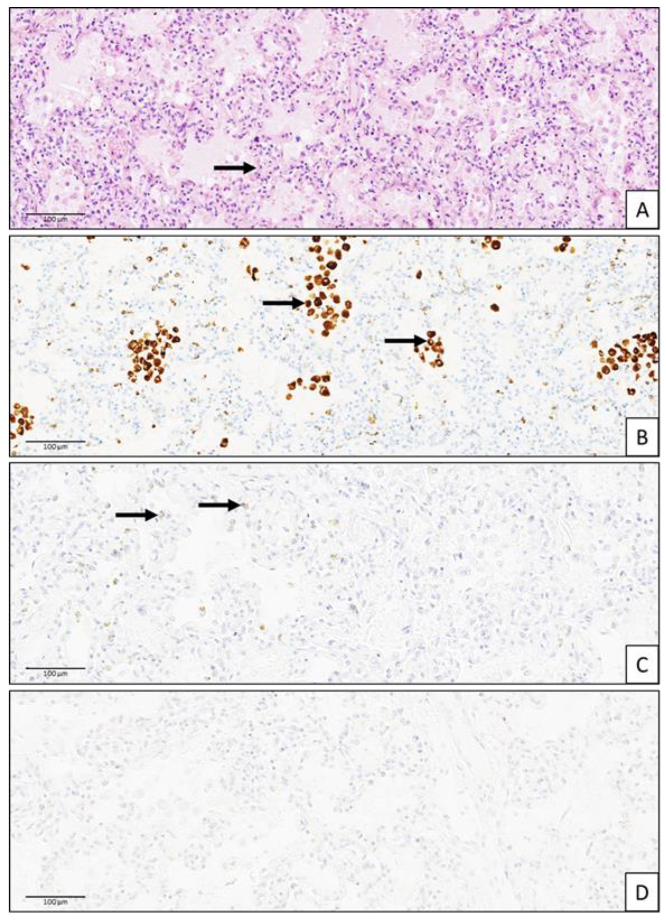
2. Case Report
3. Discussion
4. Conclusions
Supplementary Materials
Author Contributions
Funding
Institutional Review Board Statement
Informed Consent Statement
Data Availability Statement
Acknowledgments
Conflicts of Interest
References
- Jacobs, S.E.; Lamson, D.M.; St George, K.; Walsh, T.J. Human rhinoviruses. Clin. Microbiol. Rev. 2013, 26, 135–162. [Google Scholar] [CrossRef] [PubMed]
- van Benten, I.; Koopman, L.; Niesters, B.; Hop, W.; van Middelkoop, B.; de Waal, L.; van Drunen, K.; Osterhaus, A.; Neijens, H.; Fokkens, W. Predominance of rhinovirus in the nose of symptomatic and asymptomatic infants. Pediatr. Allergy Immunol. 2003, 14, 363–370. [Google Scholar] [CrossRef]
- Kieninger, E.; Fuchs, O.; Latzin, P.; Frey, U.; Regamey, N. Rhinovirus infections in infancy and early childhood. Eur. Respir. J. 2013, 41, 443–452. [Google Scholar] [CrossRef]
- Miller, E.K.; Lu, X.; Erdman, D.D.; Poehling, K.A.; Zhu, Y.; Griffin, M.R.; Hartert, T.V.; Anderson, L.J.; Weinberg, G.A.; Hall, C.B.; et al. Rhinovirus-associated hospitalizations in young children. J. Infect. Dis. 2007, 195, 773–781. [Google Scholar] [CrossRef] [PubMed]
- Miller, E.K.; Williams, J.V.; Gebretsadik, T.; Carroll, K.N.; Dupont, W.D.; Mohamed, Y.A.; Morin, L.L.; Heil, L.; Minton, P.A.; Woodward, K.; et al. Host and viral factors associated with severity of human rhinovirus-associated infant respiratory tract illness. J. Allergy Clin. Immunol. 2011, 127, 883–891. [Google Scholar] [CrossRef]
- Comte, A.; Bour, J.B.; Darniot, M.; Pitoiset, C.; Aho-Glele, L.S.; Manoha, C. Epidemiological characteristics and clinical outcomes of human rhinovirus infections in a hospitalized population. Severity is independently linked to RSV coinfection and comorbidities. J. Clin. Virol. 2020, 125, 104290. [Google Scholar] [CrossRef] [PubMed]
- Ortega, H.; Nickle, D.; Carter, L. Rhinovirus and asthma: Challenges and opportunities. Rev. Med. Virol. 2021, 31, e2193. [Google Scholar] [CrossRef] [PubMed]
- Coriolani, G.; Ferranti, S.; Biasci, F.; Lotti, F.; Grosso, S. Acute flaccid myelitis temporally associated with rhinovirus infection: Just a coincidence? Neurol. Sci. 2020, 41, 457–458. [Google Scholar] [CrossRef] [PubMed]
- Hazama, K.; Shiihara, T.; Tsukagoshi, H.; Matsushige, T.; Dowa, Y.; Watanabe, M. Rhinovirus-associated acute encephalitis/encephalopathy and cerebellitis. Brain Dev. 2019, 41, 551–554. [Google Scholar] [CrossRef]
- Holzel, A.; Smith, P.A.; Tobin, J.O. A New Type of Meningo-Encephalitis Associated with a Rhinovirus. Acta Paediatr. Scand. 1965, 54, 168–174. [Google Scholar] [CrossRef] [PubMed]
- Soma, N.; Aizawa, Y.; Matsunaga, M.; Saitoh, A. Clinically Mild Encephalitis/Encephalopathy with a Reversible Splenial Lesion Associated with Rhinovirus. Pediatr. Infect. Dis. J. 2021, 40, e122–e125. [Google Scholar] [CrossRef] [PubMed]
- Savolainen, C.; Blomqvist, S.; Mulders, M.N.; Hovi, T. Genetic clustering of all 102 human rhinovirus prototype strains: Serotype 87 is close to human enterovirus 70. J. Gen. Virol. 2002, 83 Pt 2, 333–340. [Google Scholar] [CrossRef] [PubMed]
- Mirand, A.; Henquell, C.; Archimbaud, C.; Chambon, M.; Charbonne, F.; Peigue-Lafeuille, H.; Bailly, J.L. Prospective identification of enteroviruses involved in meningitis in 2006 through direct genotyping in cerebrospinal fluid. J. Clin. Microbiol. 2008, 46, 87–96. [Google Scholar] [CrossRef] [PubMed]
- Nix, W.A.; Oberste, M.S.; Pallansch, M.A. Sensitive, seminested PCR amplification of VP1 sequences for direct identification of all enterovirus serotypes from original clinical specimens. J. Clin. Microbiol. 2006, 44, 2698–2704. [Google Scholar] [CrossRef]
- Oza, S.; Lawn, J.E.; Hogan, D.R.; Mathers, C.; Cousens, S.N. Neonatal cause-of-death estimates for the early and late neonatal periods for 194 countries: 2000–2013. Bull. World Health Organ. 2015, 93, 19–28. [Google Scholar] [CrossRef]
- Las Heras, J.; Swanson, V.L. Sudden death of an infant with rhinovirus infection complicating bronchial asthma: Case report. Pediatr. Pathol. 1983, 1, 319–323. [Google Scholar] [CrossRef]
- Urquhart, G.E.; Grist, N.R. Virological studies of sudden, unexplained infant deaths in Glasgow 1967-70. J. Clin. Pathol. 1972, 25, 443–446. [Google Scholar] [CrossRef]
- Pierres-Surer, N.; Beby-Defaux, A.; Bourgoin, A.; Venot, C.; Berthier, M.; Grollier, G.; Oriot, D.; Agius, G. Rhinovirus infections in hospitalized children: A 3-year study. Arch. Pediatr. 1998, 5, 9–14. [Google Scholar]
- Martin Perceval, L.; Scherdel, P.; Jarry, B.; de Visme, S.; Levieux, K.; Gras-Le Guen, C. Sudden Unexpected Death in Infancy: Current Practices in Virological Investigations and Documentation in the French Registry. J. Pediatr. 2023, 257, 113324. [Google Scholar] [CrossRef] [PubMed]
- Cai, X.Y.; Lu, X.D.; Lin, G.Y.; Cai, Z.W.; Lin, C.X.; Chen, P.Z.; Zheng, Y.L.; Zhou, X.H.; Feng, X.Y.; Xiao, Z.X. Monitoring of viral pathogens in pediatric intensive care unit and analysis of clinical significance. Zhonghua Er Ke Za Zhi 2013, 51, 453–459. [Google Scholar]
- Pelkonen, T.; Roine, I.; Anjos, E.; Kaijalainen, S.; Roivainen, M.; Peltola, H.; Pitkaranta, A. Picornaviruses in cerebrospinal fluid of children with meningitis in Luanda, Angola. J. Med. Virol. 2012, 84, 1080–1083. [Google Scholar] [CrossRef]
- Angel-Ambrocio, A.H.; Bautista-Carbajal, P.; Garcia-Leon, M.L.; Gomora-Herrera, M.J.; Pedernera-Astegiano, E.A.; Wong-Chew, R.M. Microglia HMC3 cells are highly susceptible to Rhinovirus infection. Virus Res. 2020, 288, 198110. [Google Scholar] [CrossRef] [PubMed]
- Li, C.X.; Burrell, R.; Dale, R.C.; Kesson, A.; Blyth, C.C.; Clark, J.E.; Crawford, N.; Jones, C.A.; Britton, P.N.; Holmes, E.C. Diagnosis and analysis of unexplained cases of childhood encephalitis in Australia using metatranscriptomic sequencing. J. Gen. Virol. 2022, 103, 001736. [Google Scholar] [CrossRef] [PubMed]
- Liu, J.; Zhao, H.; Feng, Z.; Liu, Y.; Feng, Q.; Qian, S.; Xu, L.; Gao, H.; Xie, Z. A severe case of human rhinovirus A45 with central nervous system involvement and viral sepsis. Virol. J. 2022, 19, 72. [Google Scholar] [CrossRef] [PubMed]
- Harvala, H.; McIntyre, C.L.; McLeish, N.J.; Kondracka, J.; Palmer, J.; Molyneaux, P.; Gunson, R.; Bennett, S.; Templeton, K.; Simmonds, P. High detection frequency and viral loads of human rhinovirus species A to C in fecal samples; diagnostic and clinical implications. J. Med. Virol. 2012, 84, 536–542. [Google Scholar] [CrossRef] [PubMed]
- Liu, J.M.; Tan, B.H.; Wu, S.; Gui, Y.; Suo, J.L.; Li, Y.C. Evidence of central nervous system infection and neuroinvasive routes, as well as neurological involvement, in the lethality of SARS-CoV-2 infection. J. Med. Virol. 2021, 93, 1304–1313. [Google Scholar] [CrossRef] [PubMed]
- van Riel, D.; Verdijk, R.; Kuiken, T. The olfactory nerve: A shortcut for influenza and other viral diseases into the central nervous system. J. Pathol. 2015, 235, 277–287. [Google Scholar] [CrossRef] [PubMed]
- Lupo, J.; Schuffenecker, I.; Morel-Baccard, C.; Bardet, J.; Payen, V.; Kaiser, L.; Constant, S.; Lobrinus, J.A.; Lin-Marq, N.; Lina, B.; et al. Disseminated rhinovirus C8 infection with infectious virus in blood and fatal outcome in a child with repeated episodes of bronchiolitis. J. Clin. Microbiol. 2015, 53, 1775–1777. [Google Scholar] [CrossRef]
- Bryant, V.A.; Sebire, N.J. Natural Diseases Causing Sudden Death in Infancy and Early Childhood. In SIDS Sudden Infant and Early Childhood Death: The Past, the Present and the Future; Duncan, J.R., Byard, R.W., Eds.; University of Adelaide Press: Adelaide, Australia, 2018. [Google Scholar]
- Goldwater, P.N. Current SIDS research: Time to resolve conflicting research hypotheses and collaborate. Pediatr. Res. 2023, 94, 1273–1277. [Google Scholar] [CrossRef]
- Qu, D.; Engelmann, T.A.; Preuss, V.; Hagemeier, L.; Radomsky, L.; Beushausen, K.; Keil, J.; Vennemann, B.; Falk, C.S.; Klintschar, M. Pulmonary immune profiling of SIDS: Impaired immune maturation and age-related cytokine imbalance. Pediatr. Res. 2023, 93, 1239–1249. [Google Scholar] [CrossRef] [PubMed]
- Morichi, S.; Urabe, T.; Morishita, N.; Takeshita, M.; Ishida, Y.; Oana, S.; Yamanaka, G.; Kashiwagi, Y.; Kawashima, H. Pathological analysis of children with childhood central nervous system infection based on changes in chemokines and interleukin-17 family cytokines in cerebrospinal fluid. J. Clin. Lab. Anal. 2018, 32, e22162. [Google Scholar] [CrossRef] [PubMed]
- Cerar, S.; Kucan, R.; Paro-Panjan, D.; Nosan, G. The burden of viral lower respiratory tract infections during the neonatal period: Six-year experience at a tertiary referral hospital. Croat. Med. J. 2022, 63, 343–351. [Google Scholar] [CrossRef]
- Mage, D.T.; Donner, E.M. Is excess male infant mortality from sudden infant death syndrome and other respiratory diseases X-linked? Acta Paediatr. 2014, 103, 188–193. [Google Scholar] [CrossRef] [PubMed]
- Huang, L.Y.; Chen, W.J.; Yang, Y.N.; Wu, C.Y.; Wu, P.L.; Tey, S.L.; Yang, S.N.; Liu, H.K. Maternal Age, the Disparity across Regions and Their Correlation to Sudden Infant Death Syndrome in Taiwan: A Nationwide Cohort Study. Children 2021, 8, 771. [Google Scholar] [CrossRef] [PubMed]
- Bednarczuk, N.; Milner, A.; Greenough, A. The Role of Maternal Smoking in Sudden Fetal and Infant Death Pathogenesis. Front. Neurol. 2020, 11, 586068. [Google Scholar] [CrossRef]
- Jiang, C.; Chen, Q.; Xie, M. Smoking increases the risk of infectious diseases: A narrative review. Tob. Induc. Dis. 2020, 18, 60. [Google Scholar] [CrossRef]


| Blood Parameters (Day of Sudden Death) | Normal Range | |
|---|---|---|
| WBC (×103/μL) | 8.6 | 5.0–20.0 |
| RBC (×1012/L) | 3.5 | 3.5–6.1 |
| Hemoglobin (g/100 mL) | 12 | 12.0–20.5 |
| Platelets (×103/μL) | 210 | 150–450 |
| NEU (×103/μL); % | 1.3; 15.2% | [1.0–9.0] |
| LYM (×103/μL); % | 6.51; 75.8% | [2.2–16.8] |
| MONO (×103/μL); % | 0.65; 7.6% | [0.05–1.1] |
| EOS (×103/μL); % | 0.06; 0.8% | [0.0–0.85] |
| CRP (mg/mL) | <4 | < 4 |
| Pre-Autopsy Samples | At-Autopsy Samples | |||||||||||||
|---|---|---|---|---|---|---|---|---|---|---|---|---|---|---|
| Throat | BAL | NS | NP Swab | Rectal Swab | Muscle | Heart | Blood | Serum | CSF | Liver | Heart | Lung | Kidney | |
| Film array meningitis/encephalitis panel * (Biofire, bioMérieux) | EV | |||||||||||||
| EV (RIDA®gene, R- Biopharm) | ||||||||||||||
| Bocavirus, Rotavirus A, Aichivirus | ||||||||||||||
| Norovirus, Sapovirus, Astrovirus | ||||||||||||||
| Parechovirus (R-gene, bioMérieux) | ||||||||||||||
| CMV (R-gene, BioMérieux) | ||||||||||||||
| HSV-1, HSV-2 (R-gene, bioMérieux) | ||||||||||||||
| EBV (R-gene, BioMérieux) | ||||||||||||||
| Parvovirus B19 (R-gene, BioMérieux) | ||||||||||||||
| HHV-6 (R-gene, BioMérieux) | ||||||||||||||
| Influenza A and B (Panther, Hologic) | ||||||||||||||
| RSV, hMPV (Panther, Hologic) | ||||||||||||||
| Parainfluenza 1, 2 3, and 4 (Panther, Hologic) | ||||||||||||||
| RV/EV (Panther, Hologic) | RV | |||||||||||||
| SARS-CoV2 (Panther, Hologic) | ||||||||||||||
| AdV (R-gene, BioMérieux) | ||||||||||||||
| Rubella IgM (VIDAS® BioMérieux) | ||||||||||||||
| M. pneumoniae IgM (Virclia) | ||||||||||||||
| Film array respiratory panel RP2 plus ** (Biofire, bioMérieux) | RV | |||||||||||||
| RV/EV Typing and sequencing | RV | RV | RV | |||||||||||
| RV&EV/Ctrl cell (R-gene, BioMérieux) | RV | RV | RV-neg | |||||||||||
Disclaimer/Publisher’s Note: The statements, opinions and data contained in all publications are solely those of the individual author(s) and contributor(s) and not of MDPI and/or the editor(s). MDPI and/or the editor(s) disclaim responsibility for any injury to people or property resulting from any ideas, methods, instructions or products referred to in the content. |
© 2024 by the authors. Licensee MDPI, Basel, Switzerland. This article is an open access article distributed under the terms and conditions of the Creative Commons Attribution (CC BY) license (https://creativecommons.org/licenses/by/4.0/).
Share and Cite
Auvray, C.; Perez-Martin, S.; Schuffenecker, I.; Pitoiset, C.; Tarris, G.; Ambert-Balay, K.; Martin, L.; Dullier-Taillefumier, N.; Bour, J.-B.; Manoha, C. Sudden Infant Death Associated with Rhinovirus Infection. Viruses 2024, 16, 518. https://doi.org/10.3390/v16040518
Auvray C, Perez-Martin S, Schuffenecker I, Pitoiset C, Tarris G, Ambert-Balay K, Martin L, Dullier-Taillefumier N, Bour J-B, Manoha C. Sudden Infant Death Associated with Rhinovirus Infection. Viruses. 2024; 16(4):518. https://doi.org/10.3390/v16040518
Chicago/Turabian StyleAuvray, Christelle, Stéphanie Perez-Martin, Isabelle Schuffenecker, Cécile Pitoiset, Georges Tarris, Katia Ambert-Balay, Laurent Martin, Nathalie Dullier-Taillefumier, Jean-Baptiste Bour, and Catherine Manoha. 2024. "Sudden Infant Death Associated with Rhinovirus Infection" Viruses 16, no. 4: 518. https://doi.org/10.3390/v16040518