
Arboviral Risk Associated with Solid Organ and Hematopoietic Stem Cell Grafts: The Prophylactic Answers Proposed by the French High Council of Public Health in a National Context
 , , and
Abstract
:1. Introduction
2. West Nile Virus (WNV)
- Human cases of WNV infection diagnosed on French territory must be informed as a notifiable disease to SpF [31];
- Regarding labile blood products, from the first confirmed autochthonous human case, an individual NAT screening (ID-NAT) must be implemented without delay for all blood donations collected in the affected Department; blood components prepared from donations already collected in the affected Department between the date of the alert and that of the effective implementation of the ID-NAT must be quarantined pending the results of the testing, with the exception of platelet concentrates that are all pathogen-reduced (Intercept Blood System®) and can be released without delay. Blood donors having stayed (at least one night) in an affected Department must be deferred until 28 days after their return or tested by ID-NAT. Candidates for donation reporting a diagnosis of WNV infection must be deferred 120 days after the end of symptoms;
- As for SOT and HSCT, in case of the identification of a new autochthonous case, donors living or having stayed in the Region or Department concerned must be tested by WNV NAT and IgM/IgG serology must be performed, ideally before transplantation. The decision of whether to use the organs or cells from donors testing positive either by NAT or IgM serology is submitted to the benefit–risk balance, with the recommendation of delaying the grafts that are not urgently needed;
- A list of countries at risk for WNV during the period of circulation of the virus (namely, June to November) is updated each year [32]. Candidates for blood, organ, or cell donations originating from or having traveled in these countries must be either tested for WNV (NAT for blood and NAT + serology for grafts) or their donation postponed for 28 days after returning from the risk area;
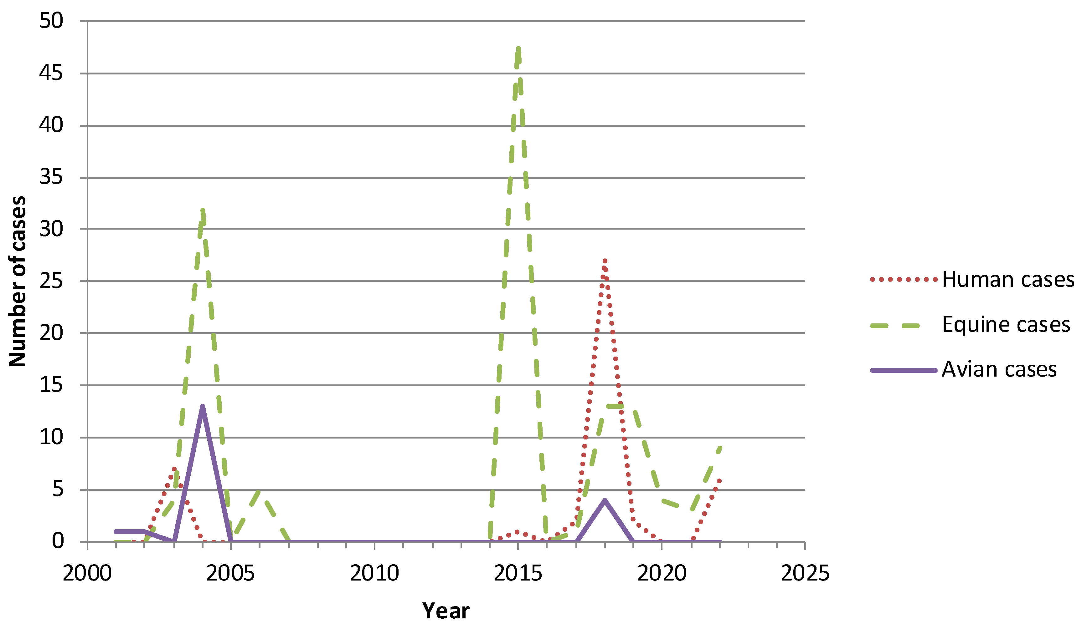
- In parallel, a surveillance of the circulation of WNV is recommended in horses and birds.
3. Dengue Virus
- When a case or a cluster of dengue fever is identified in an endemic area, ID-NAT screening must be performed for blood donations at the Department level. When positive, the products must be discarded and the donor deferred 28 days from the date of the end of the symptoms. For donors of solid organs and bone marrow present in the cluster areas or having stayed there, PCR and IgM/IgG serology must be performed close to the time of the donation. In case of the positivity of one of these markers, it is recommended that living donors postpone the graft or select another donor if available; in deceased donors, it is recommended to discard the organs, except in case of vital emergency for the recipient for which a benefit–risk evaluation must be performed. If organs are transplanted, a specific follow-up of the recipient is required [62].
- In clusters of autochthonous dengue in mainland France, blood collections must be postponed in the area, donors living or having stayed in this area must be excluded from blood donation for 28 days, and blood products already collected in this area and not treated by the Intercept® process must be placed in quarantine in order to be tested by ID-NAT. Each transmission event is considered closed at the end of 45 days following the onset of clinical symptoms of the last detected human case. In contrast to blood products, due to the very low risk of selecting a positive donor of solid organ or bone marrow, no specific information is provided in this context [63].
4. Tick-Borne Encephalitis Virus
- For blood products, donors having experienced a tick bite in the 28 days preceding the donation in an area known to be at risk for TBEV (in or out of France) during the period of virus circulation (March to November) must be excluded for 28 days after the tick bite’s date. In addition, blood collection is interrupted in areas where a source of foodborne outbreak of TBEV is recognized, with quarantine of blood products already collected and not secured by the Intercept® process, until testing negative by TBEV NAT;
- For SOT and HSCT, living donors staying or traveling in at-risk areas for TBEV must be made aware of the risks of tick bites and of the consumption of unpasteurized milk and milk products from March to November to avoid contamination. All living donors should be questioned for a tick bite that had occurred less than one month before the donation in a zone at risk for TBEV when completing the pre-donation check-up list. In case of a positive answer to this question, living donors must be tested for TBEV (NAT and IgM/IgG serology) prior to the gift; if at least one of these tests is positive, it is recommended to postpone the graft or to select another donor if available. For deceased donors recently exposed to a tick bite, when this information can be recorded from his/her relatives or after skin inspection, as it may be difficult to obtain virological tests prior to the transplantation, it is recommended to inform the recipient(s) and their medical team of the potential risk of TBEV infection and to perform specific virological tests in case of fever or neurological symptoms in the two months following the transplantation.
5. Other Arboviruses Circulating in France
5.1. Usutu Virus
5.2. Chikungunya Virus
- The French Territories in the Americas also experienced an important CHIKV outbreak in 2013–2014 with respectively 72,500, 81,200, and 15,000 cases in Martinique, Guadeloupe, and French Guiana. No significant re-emergence of the virus was further observed.
- In French Polynesia, a CHIKV outbreak was reported in 2014–2015.
- Finally, three clusters of autochthonous CHIKV infections were observed in the south of mainland France: 2 cases in 2010 [83], 12 cases in 2014, and 17 cases in 2017. These emerging cases must be taken into consideration as the capacity of adaptation of this virus to its vector is illustrated by the size of the first outbreak that occurred in Europe in 2007 and that had involved 217 cases in eastern Italy [84,85].
5.3. Zika Virus
6. Concluding Remarks
Author Contributions
Funding
Institutional Review Board Statement
Informed Consent Statement
Data Availability Statement
Acknowledgments
Conflicts of Interest
References
- Giménez-Richarte, Á.; Ortiz de Salazar, M.I.; Giménez-Richarte, M.P.; Collado, M.; Fernández, P.L.; Clavijo, C.; Navarro, L.; Arbona, C.; Marco, P.; Ramos-Rincon, J.M. Transfusion-transmitted arboviruses: Update and systematic review. PLoS Negl. Trop. Dis. 2022, 16, e0010843. [Google Scholar] [CrossRef] [PubMed]
- Cheng, V.C.C.; Sridhar, S.; Wong, S.C.; Wong, S.C.Y.; Chan, J.F.W.; Yip, C.C.Y.; Chau, C.H.; Au, T.W.K.; Hwang, Y.Y.; Yau, C.; et al. Japanese encephalitis virus transmitted via blood transfusion, Hong Kong, China. Emerg. Infect. Dis. 2018, 24, 49–57. [Google Scholar] [CrossRef] [PubMed]
- Taylor, L.; Condon, T.; Destrampe, E.M.; Brown, J.A.; McGavic, J.; Gould, C.V.; Chambers, T.V.; Kosoy, O.I.; Burkhalter, K.L.; Annambhotla, P.; et al. Powassan virus infection likely acquired through blood transfusion presenting as encephalitis in a kidney transplant recipient. Clin. Infect. Dis. 2021, 72, 1051–1054. [Google Scholar] [CrossRef] [PubMed]
- Venkat, H.; Adams, L.; Sunenshine, R.; Krow-Lucal, E.; Levy, C.; Kafenbaum, T.; Sylvester, T.; Smith, K.; Townsend, J.; Dosmann, M.; et al. St. Louis encephalitis virus possibly transmitted through blood transfusion, Arizona, 2015. Transfusion 2017, 57, 2987–2994. [Google Scholar] [CrossRef] [PubMed]
- Centers for Disease Control and Prevention. Transfusion-related transmission of yellow fever vaccine virus, California, 2009. Morb. Mortal. Wkly. Rep. 2010, 59, 34–37. [Google Scholar]
- Centers for Disease Control and Prevention. Transmission of Colorado tick fever virus by blood transfusion, Montana. Morb. Mortal. Wkly. Rep. 1975, 24, 422–427. [Google Scholar]
- Couderc, T.; Gangneux, N.; Chrétien, F.; Caro, V.; Le Luong, T.; Ducloux, B.; Tolou, H.; Lecuit, M.; Grandadam, M. Chikungunya virus infection of corneal grafts. J. Infect. Dis. 2012, 206, 851–859. [Google Scholar] [CrossRef] [PubMed]
- Hoad, V.C.; Speers, D.J.; Keller, A.J.; Dowse, G.K.; Seed, C.R.; Lindsay, M.D.; Faddy, H.M.; Pink, J. First reported case of transfusion-transmitted Ross River virus infection. Med. J. Aust. 2015, 202, 267–270. [Google Scholar] [CrossRef]
- Devillers, J.; David, J.P.; Barrès, B.; Alout, H.; Lapied, B.; Chouin, S.; Dusfour, I.; Billault, C.; Mekki, F.; Attig, I.; et al. Integrated plan of insecticide resistance surveillance in mosquito vectors in France. Insects 2023, 14, 457. [Google Scholar] [CrossRef]
- Robert, M.A.; Stewart-Ibarra, A.M.; Estallo, E.L. Climate change and viral emergence: Evidence from Aedes- borne arboviruses. Curr. Opin. Virol. 2020, 40, 41–47. [Google Scholar] [CrossRef]
- Chancey, C.; Grinev, A.; Volkova, E.; Rios, M. The global ecology and epidemiology of West Nile virus. Biomed. Res. Int. 2015, 2015, 376230. [Google Scholar] [CrossRef] [PubMed]
- Petersen, L.R.; Brault, A.C.; Nasci, R.S. West Nile virus: Review of the literature. JAMA 2013, 310, 308–315. [Google Scholar] [CrossRef] [PubMed]
- Nash, D.; Mostashari, F.; Fine, A.; Miller, J.; O’Leary, D.; Murray, K.; Huang, A.; Rosenberg, A.; Greenberg, A.; Sherman, M.; et al. The outbreak of West Nile virus infection in the New York City area in 1999. N. Engl. J. Med. 2001, 344, 1807–1814. [Google Scholar] [CrossRef]
- Pealer, L.N.; Marfin, A.A.; Petersen, L.R.; Lanciotti, R.S.; Page, P.L.; Stramer, S.L.; Stobierski, M.G.; Signs, K.; Newman, B.; Kapoor, H.; et al. Transmission of West Nile virus through blood transfusion in the United States in 2002. N. Engl. J. Med. 2003, 349, 1236. [Google Scholar] [CrossRef] [PubMed]
- Centers for Disease Control and Prevention. Update: Investigations of West Nile virus infections in recipients of organ transplantation and blood transfusion, Michigan, 2002. Morb. Mortal. Wkly. Rep. 2002, 51, 879. [Google Scholar]
- Iwamoto, M.; Jernigan, D.B.; Guasch, A.; Trepka, M.J.; Blackmore, C.G.; Hellinger, W.C.; Pham, S.M.; Zaki, S.; Lanciotti, R.S.; Lance-Parker, S.E.; et al. Transmission of West Nile virus from an organ donor to four transplant recipients. N. Engl. J. Med. 2003, 348, 2196. [Google Scholar] [CrossRef] [PubMed]
- Centers for Disease Control and Prevention. Fatal West Nile virus infection after probable transfusion-associated transmission, Colorado, 2012. Morb. Mortal. Wkly. Rep. 2013, 62, 622–624. [Google Scholar]
- Pervanidou, D.; Detsis, M.; Danis, K.; Mellou, K.; Papanikolaou, E.; Terzaki, I.; Baka, A.; Veneti, L.; Vakali, A.; Dougas, G.; et al. West Nile virus outbreak in humans, Greece, 2012: Third consecutive year of local transmission. Eurosurveillance 2014, 19, 20758. [Google Scholar] [CrossRef]
- Anesi, J.A.; Silveira, F.P.; AST Infectious Diseases Community of Practice. Arenaviruses and West Nile Virus in solid organ transplant recipients: Guidelines from the American Society of Transplantation Infectious Diseases Community of Practice. Clin. Transplant. 2019, 33, e13576. [Google Scholar] [CrossRef]
- Centers for Disease Control and Prevention. West Nile virus infections in organ transplant recipients, New York and Pennsylvania, August-September, 2005. Morb. Mortal. Wkly. Rep. 2005, 54, 1021–1023. [Google Scholar]
- Centers for Disease Control and Prevention. West Nile virus transmission via organ transplantation and blood transfusion, Louisiana, 2008. Morb. Mortal. Wkly. Rep. 2009, 58, 1263–1267. [Google Scholar]
- Rhee, C.; Eaton, E.F.; Concepcion, W.; Blackburn, B.G. West Nile virus encephalitis acquired via liver transplantation and clinical response to intravenous immunoglobulin: Case report and review of the literature. Transpl. Infect. Dis. 2011, 13, 312–317. [Google Scholar] [CrossRef] [PubMed]
- Nett, R.J.; Kuehnert, M.J.; Ison, M.G.; Orlowski, J.P.; Fischer, M.; Staples, J.E. Current practices and evaluation of screening solid organ donors for West Nile virus. Transpl. Infect. Dis. 2012, 14, 268–277. [Google Scholar] [CrossRef] [PubMed]
- Morelli, M.C.; Sambri, V.; Grazi, G.L.; Gaibani, P.; Pierro, A.; Cescon, M.; Ercolani, G.; Cavrini, F.; Rossini, G.; Capobianchi, M.R.; et al. Absence of neuroinvasive disease in a liver transplant recipient who acquired West Nile virus (WNV) infection from the organ donor and who received WNV antibodies prophylactically. Clin. Infect. Dis. 2010, 51, e34–e37. [Google Scholar] [CrossRef] [PubMed]
- Inojosa, W.O.; Scotton, P.G.; Fuser, R.; Giobbia, M.; Paolin, A.; Maresca, M.C.; Brunello, A.; Nascimben, E.; Sorbara, C.; Rigoli, R.; et al. West Nile virus transmission through organ transplantation in north-eastern Italy: A case report and implications for pre-procurement screening. Infection 2012, 40, 557–562. [Google Scholar] [CrossRef] [PubMed]
- Winston, D.J.; Vikram, H.R.; Rabe, I.B.; Dhillon, G.; Mulligan, D.; Hong, J.C.; Busuttil, R.W.; Nowicki, M.J.; Mone, T.; Civen, R.; et al. Donor-derived West Nile virus infection in solid organ transplant recipients: Report of four additional cases and review of clinical, diagnostic, and therapeutic features. Transplantation 2014, 97, 881–889. [Google Scholar] [CrossRef] [PubMed]
- Barzon, L.; Montarsi, F.; Quaranta, E.; Monne, I.; Pacenti, M.; Michelutti, A.; Toniolo, F.; Danesi, P.; Marchetti, G.; Gobbo, F.; et al. Early start of seasonal transmission and co-circulation of West Nile virus lineage 2 and a newly introduced lineage 1 strain, northern Italy, June 2022. Eurosurveillance 2022, 27, 2200548. [Google Scholar] [CrossRef] [PubMed]
- Haut Conseil de la Santé Publique. Avis du 7 Novembre 2022 sur la Sécurisation des Produits du Corps humain dans un Contexte de Circulation du Virus West Nile en France Métropolitaine. Available online: https://www.hcsp.fr/explore.cgi/avisrapportsdomaine?clefr=1259 (accessed on 15 August 2023).
- Haut Conseil de la Santé Publique. Statement on Measures of Security for Human Body Products Owing to a Human Case Report of West Nile Virus (WNV) Infection in Mainland France Outside the Seasonal Alert Period. 13 January 2023. Available online: https://www.hcsp.fr/explore.cgi/avisrapportsdomaine?clefr=1270 (accessed on 15 August 2023).
- Haut Conseil de la Santé Publique. Avis du 28 Mars 2023 sur la Sécurisation des Eléments et Produits Issus du Corps Humain en Prévision de la Circulation du Virus West Nile en France Métropolitaine. Available online: https://www.hcsp.fr/explore.cgi/avisrapportsdomaine?clefr=1284 (accessed on 15 August 2023).
- Haut Conseil de la Santé Publique. Avis du 7 Février 2020 Relatif à l’Inscription à la Liste des Maladies à Déclaration Obligatoire de l’Infection due au Virus West Nile. Available online: https://www.hcsp.fr/explore.cgi/avisrapportsdomaine?clefr=843 (accessed on 15 August 2023).
- Haut Conseil de la Santé Publique. Liste des Pays à Risque de Transmission du Virus West Nile (WNV) pour les Produits du Corps Humain, Saison 2023. Avis du 24 Mai 2023. Available online: https://www.hcsp.fr/explore.cgi/avisrapportsdomaine?clefr=1307 (accessed on 15 August 2023).
- Bhatt, S.; Gething, P.W.; Brady, O.J.; Messina, J.P.; Farlow, A.W.; Moyes, C.L.; Drake, J.M.; Brownstein, J.S.; Hoen, A.G.; Sankoh, O.; et al. The global distribution and burden of dengue. Nature 2013, 496, 504–507. [Google Scholar] [CrossRef]
- Gwee, X.W.S.; Chua, P.E.Y.; Pang, J. Global dengue importation: A systematic review. BMC Infect. Dis. 2021, 21, 1078. [Google Scholar]
- Pozzetto, B.; Memmi, M.; Garraud, O. Is transfusion-transmitted dengue fever a potential public health threat? World J. Virol. 2015, 4, 113–123. [Google Scholar] [CrossRef]
- Chuang, V.W.M.; Wong, T.Y.; Leung, Y.H.; Ma, E.; Law, Y.L.; Tsang, O.; Chan, K.M.; Tsang, I.; Que, T.L.; Yung, R.; et al. Review of dengue fever cases in Hong Kong during 1998 to 2005. Hong Kong Med. J. 2008, 14, 170–177. [Google Scholar] [PubMed]
- Tambyah, P.A.; Koay, E.S.C.; Poon, M.L.M.; Lin, R.V.T.P.; Ong, B.K.C.; Transfusion-Transmitted Dengue Infection Study Group. Dengue hemorrhagic fever transmitted by blood transfusion. N. Engl. J. Med. 2008, 359, 1526–1527. [Google Scholar] [CrossRef] [PubMed]
- Stramer, S.L.; Linnen, J.M.; Carrick, J.M.; Foster, G.A.; Krysztof, D.E.; Zou, S.; Dodd, R.Y.; Tirado-Marrero, L.M.; Hunsperger, E.; Santiago, G.A.; et al. Dengue viremia in blood donors identified by RNA and detection of dengue transfusion transmission during the 2007 dengue outbreak in Puerto Rico. Transfusion 2012, 52, 1657–1666. [Google Scholar] [CrossRef] [PubMed]
- Matos, D.; Tomashek, K.M.; Perez-Padilla, J.; Muñoz-Jordán, J.; Hunsperger, E.; Horiuchi, K.; Noyd, D.; Winton, C.; Foster, G.; Lanteri, M.; et al. Probable and possible transfusion-transmitted dengue associated with NS1 antigen-negative but RNA confirmed-positive red blood cells. Transfusion 2016, 56, 215–222. [Google Scholar] [CrossRef] [PubMed]
- Oh, H.B.; Muthu, V.; Daruwalla, Z.J.; Lee, S.Y.; Koay, E.S.; Tambyah, P.A. Bitten by a bug or a bag? Transfusion- transmitted dengue: A rare complication in the bleeding surgical patient. Transfusion 2015, 55, 1655–1661. [Google Scholar] [CrossRef] [PubMed]
- Sabino, E.C.; Loureiro, P.; Lopes, M.E.; Capuani, L.; McClure, C.; Chowdhury, D.; Di-Lorenzo-Oliveira, C.; Oliveira, L.C.; Linnen, J.M.; Lee, T.H.; et al. Transfusion-transmitted dengue and associated clinical symptoms during the 2012 epidemic in Brazil. J. Infect. Dis. 2016, 213, 694–702. [Google Scholar] [CrossRef] [PubMed]
- Levi, J.E.; Nishiya, A.; Félix, A.C.; Salles, N.A.; Sampaio, L.R.; Hangai, F.; Sabino, E.C.; Mendrone, A., Jr. Real-time symptomatic case of transfusion-transmitted dengue. Transfusion 2015, 55, 961–964. [Google Scholar] [CrossRef]
- Rigau-Pérez, J.G.; Vorndam, A.V.; Clark, G.G. The dengue and dengue hemorrhagic fever epidemic in Puerto Rico, 1994–1995. Am. J. Trop. Med. Hyg. 2001, 64, 67–74. [Google Scholar] [CrossRef]
- Tan, F.L.-S.; Loh, D.L.S.K.; Prabhakaran, K.; Tambyah, P.A.; Yap, H.-K. Dengue haemorrhagic fever after living donor renal transplantation. Nephrol. Dial. Transplant. 2005, 20, 447–448. [Google Scholar] [CrossRef]
- Tangnararatchakit, K.; Tirapanich, W.; Tapaneya-Olarn, W.; Sumethkul, V.; Sirachainan, N.; Watcharananan, S.; Leenanupunth, C.; Yoksan, S.; Chuansumrit, A. Severe nonfebrile dengue infection in an adolescent after postoperative kidney transplantation: A case report. Transplant. Proc. 2012, 44, 303–306. [Google Scholar] [CrossRef]
- Saigal, S.; Choudhary, N.S.; Saraf, N.; Kataria, S.; Mohanka, R.; Soin, A.S. Transmission of dengue virus from a donor to a recipient after living donor liver transplantation. Liver Transpl. 2013, 19, 1413–1414. [Google Scholar] [CrossRef] [PubMed]
- Punzel, M.; Korukluoğlu, G.; Caglayik, D.Y.; Menemenlioglu, D.; Bozdag, S.C.; Tekgündüz, E.; Altuntaş, F.; Campos, R.d.M.; Burde, B.; Günther, S.; et al. Dengue virus transmission by blood stem cell donor after travel to Sri Lanka; Germany, 2013. Emerg. Infect. Dis. 2014, 20, 1366–1369. [Google Scholar]
- Gupta, R.K.; Gupta, G.; Chorasiya, V.K.; Bag, P.; Shandil, R.; Bhatia, V.; Wadhawan, M.; Vij, V.; Kumar, A. Dengue virus transmission from living donor to recipient in liver transplantation: A case report. J. Clin. Exp. Hepatol. 2016, 6, 59–61. [Google Scholar] [CrossRef] [PubMed]
- Rosso, F.; Pineda, J.C.; Sanz, A.M.; Cedano, J.A.; Caicedo, L.A. Transmission of dengue virus from deceased donors to solid organ transplant recipients: Case report and literature review. Braz. J. Infect. 2018, 22, 63–69. [Google Scholar] [CrossRef] [PubMed]
- Shaji Mathew, J.; Menon, V.P.; Menon, V.P.; Mallick, S.; Sivasankara Pillai Thankamony Amma, B.; Balakrishnan, D.; Gopalakrishnan, U.; Narayana Menon, R.; Athira, P.P.; Jagan, O.A.; et al. Dengue virus transmission from live donor liver graft. Am. J. Transplant. 2019, 19, 1838–1846. [Google Scholar] [CrossRef] [PubMed]
- Lecadieu, A.; Teysseyre, L.; Larsen, K.; Vidal, C.; Caron, M.; Traversier, N.; Di Ascia, L.; Aujoulat, T.; Allyn, J.; Allou, N. Case report: Transmission of dengue virus from a deceased donor to a kidney transplant recipient previously infected by dengue virus. Am. J. Trop. Med. Hyg. 2021, 104, 2199–2201. [Google Scholar] [CrossRef] [PubMed]
- Janani, M.K.; Durgadevi, P.; Padmapriya, J.; Malathi, J.; Kulandai, L.T.; Rao Madhavan, H.N. First report on detection of dengue virus in the donor cornea. Cornea 2018, 37, 1586–1589. [Google Scholar] [CrossRef]
- L’Azou, M.; Taurel, A.-F.; Flamand, C.; Quenel, P. Recent epidemiological trends of dengue in the French Territories of the Americas (2000–2012): A systematic literature review. PLoS Negl. Trop. Dis. 2014, 8, e3235. [Google Scholar] [CrossRef]
- L’Azou, M.; Jean-Marie, J.; Bessaud, M.; Cabié, A.; Césaire, R.; de Lamballerie, X.; Courbil, R.; Richard, P. Dengue seroprevalence in the French West Indies: A prospective study in adult blood donors. Am. J. Trop. Med. Hyg. 2015, 92, 1137–1140. [Google Scholar] [CrossRef]
- World Health Organization. Dengue Fever—Region of the Americas (PAHO)—French Guiana, Guadeloupe, Martinique, Saint-Martin, and Saint-Barthélemy. March 2020. Available online: https://www.who.int/emergencies/disease-outbreak-news/item/2020-DON251 (accessed on 15 August 2023).
- Hafsia, S.; Haramboure, M.; Wilkinson, D.A.; Baldet, T.; Yemadje-Menudier, L.; Vincent, M.; Tran, A.; Atyame, C.; Mavingui, P. Overview of dengue outbreaks in the southwestern Indian Ocean and analysis of factors involved in the shift toward endemicity in Reunion Island: A systematic review. PLoS Negl. Trop. Dis. 2022, 16, e0010547. [Google Scholar] [CrossRef]
- Richard, V.; Cao-Lormeau, V.M. Mosquito vectors of arboviruses in French Polynesia. New Microbes New Infect. 2019, 31, 100569. [Google Scholar] [CrossRef] [PubMed]
- La Ruche, G.; Souarès, Y.; Armengaud, A.; Peloux-Petiot, F.; Delaunay, P.; Desprès, P.; Lenglet, A.; Jourdain, F.; Leparc- Goffart, I.; Charlet, F.; et al. First two autochthonous dengue virus infections in metropolitan France, September 2010. Eurosurveillance 2010, 15, 19676. [Google Scholar] [CrossRef] [PubMed]
- Cochet, A.; Calba, C.; Jourdain, F.; Grard, G.; Durand, G.A.; Guinard, A.; Investigation Team; Noël, H.; Paty, M.C.; Franke, F. Autochthonous dengue in mainland France, 2022: Geographical extension and incidence increase. Eurosurveillance 2022, 27, 2200818. [Google Scholar] [CrossRef] [PubMed]
- Roche, B.; Léger, L.; L’Ambert, G.; Lacour, G.; Foussadier, R.; Besnard, G.; Barré-Cardi, H.; Simard, F.; Fontenille, D. The spread of Aedes albopictus in Metropolitan France: Contribution of environmental drivers and human activities and predictions for a near future. PLoS ONE 2015, 10, e0125600. [Google Scholar] [CrossRef] [PubMed]
- Semenza, J.C.; Rocklöv, J.; Ebi, K.L. Climate change and cascading risks from infectious disease. Infect. Dis. Ther. 2022, 11, 1371–1390. [Google Scholar] [CrossRef] [PubMed]
- Haut Conseil de la Santé Publique. Avis du 28 Novembre 2020 Relatif à l’Actualisation des Mesures de Prévention vis-à-vis du Virus de la Dengue à Appliquer aux Produits Issus du Corps Humain dans les Antilles Françaises. Available online: https://www.hcsp.fr/explore.cgi/avisrapportsdomaine?clefr=953 (accessed on 15 August 2023).
- Haut Conseil de la Santé Publique. Avis du 19 Octobre 2022 sur les Mesures de Prévention pour la Sécurité Infectieuse Transfusionnelle et de la Greffe à la Suite de Cas de Dengue Autochtones dans le Sud de la France. Available online: https://www.hcsp.fr/explore.cgi/avisrapportsdomaine?clefr=1258 (accessed on 15 August 2023).
- Ličková, M.; Fumačová Havlíková, S.; Sláviková, M.; Slovák, M.; Drexler, J.F.; Klempa, B. Dermacentor reticulatus is a vector of tick-borne encephalitis virus. Ticks Tick Borne Dis. 2020, 11, 101414. [Google Scholar] [CrossRef] [PubMed]
- Kunze, M.; Banović, P.; Bogovič, P.; Briciu, V.; Čivljak, R.; Dobler, G.; Hristea, A.; Kerlik, J.; Kuivanen, S.; Kynčl, J.; et al. Recommendations to improve tick-borne encephalitis surveillance and vaccine uptake in Europe. Microorganisms 2022, 10, 1283. [Google Scholar] [CrossRef]
- Michelitsch, A.; Wernike, K.; Klaus, C.; Dobler, G.; Beer, M. Exploring the reservoir hosts of tick-borne encephalitis virus. Viruses 2019, 11, 669. [Google Scholar] [CrossRef]
- Ličková, M.; Fumačová Havlíková, S.; Sláviková, M.; Klempa, B. Alimentary infections by tick-borne encephalitis virus. Viruses 2021, 14, 56. [Google Scholar] [CrossRef]
- Martello, E.; Gillingham, E.L.; Phalkey, R.; Vardavas, C.; Nikitara, K.; Bakonyi, T.; Gossner, C.M.; Leonardi-Bee, J. Systematic review on the non-vectorial transmission of tick-borne encephalitis virus (TBEv). Ticks Tick Borne Dis. 2022, 13, 102028. [Google Scholar] [CrossRef]
- Wahlberg, P.; Saikku, P.; Brummer-Korvenkontio, M. Tick-borne viral encephalitis in Finland. The clinical features of Kumlinge disease during 1959–1987. J. Intern. Med. 1989, 225, 173–177. [Google Scholar] [PubMed]
- Lipowski, D.; Popiel, M.; Perlejewski, K.; Nakamura, S.; Bukowska-Osko, I.; Rzadkiewicz, E.; Dzieciatkowski, T.; Milecka, A.; Wenski, W.; Ciszek, M.; et al. A cluster of fatal tick-borne encephalitis virus infection in organ transplant setting. J. Infect. Dis. 2017, 215, 896–901. [Google Scholar] [CrossRef] [PubMed]
- Gonzalez, G.; Bournez, L.; Moraes, R.A.; Marine, D.; Galon, C.; Vorimore, F.; Cochin, M.; Nougairède, A.; Hennechart-Collette, C.; Perelle, S.; et al. A One-health approach to investigating an outbreak of alimentary tick-borne encephalitis in a non-endemic area in France (Ain, Eastern France): A longitudinal serological study in livestock, detection in ticks, and the first tick-borne encephalitis virus isolation and molecular characterisation. Front. Microbiol. 2022, 13, 863725. [Google Scholar] [PubMed]
- Haut Conseil de la Santé Publique. Inscription de l’Encéphalite à Tiques sur la Liste des Maladies à Déclaration Obligatoire. 5 Juin 2020. Available online: https://www.hcsp.fr/explore.cgi/avisrapportsdomaine?clefr=856 (accessed on 15 August 2023).
- Saegerman, C.; Humblet, M.F.; Leandri, M.; Gonzalez, G.; Heyman, P.; Sprong, H.; L’Hostis, M.; Moutailler, S.; Bonnet, S.I.; Haddad, N.; et al. First expert elicitation of knowledge on possible drivers of observed increasing human cases of tick-borne encephalitis in Europe. Viruses 2023, 15, 791. [Google Scholar] [CrossRef] [PubMed]
- Haut Conseil de la Santé Publique. Avis du 23 Juillet 2020 Relatif aux Risques Transfusionnels et de Contamination des Greffons Induits par le Virus de l’Encéphalite à Tiques (TBEV); Précautions à Prendre lors de Foyers Infectieux. Available online: https://www.hcsp.fr/explore.cgi/avisrapportsdomaine?clefr=895 (accessed on 15 August 2023).
- Vilibic-Cavlek, T.; Petrovic, T.; Savic, V.; Barbic, L.; Tabain, I.; Stevanovic, V.; Klobucar, A.; Mrzljak, A.; Ilic, M.; Bogdanic, M.; et al. Epidemiology of Usutu virus: The European scenario. Pathogens 2020, 9, 699. [Google Scholar] [CrossRef] [PubMed]
- Lecollinet, S.; Blanchard, Y.; Manson, C.; Lowenski, S.; Laloy, E.; Quenault, H.; Touzain, F.; Lucas, P.; Eraud, C.; Bahuon, C.; et al. Dual emergence of Usutu virus in common blackbirds, Eastern France, 2015. Emerg. Infect. Dis. 2016, 22, 2225–2227. [Google Scholar] [CrossRef] [PubMed]
- Simonin, Y.; Sillam, O.; Carles, M.J.; Gutierrez, S.; Gil, P.; Constant, O.; Martin, M.F.; Girard, G.; Van de Perre, P.; Salinas, S.; et al. Human Usutu virus infection with atypical neurologic presentation, Montpellier, France, 2016. Emerg. Infect. Dis. 2018, 24, 875–878. [Google Scholar] [CrossRef] [PubMed]
- Desdouits, M.; Kamgang, B.; Berthet, N.; Tricou, V.; Ngoagouni, C.; Gessain, A.; Manuguerra, J.C.; Nakouné, E.; Kazanji, M. Genetic characterization of chikungunya virus in the Central African Republic. Infect. Genet. Evol. 2015, 33, 25–31. [Google Scholar] [CrossRef]
- Sharif, N.; Sarkar, M.K.; Ferdous, R.N.; Ahmed, S.N.; Billah, M.B.; Talukder, A.A.; Zhang, M.; Dey, S.K. Molecular epidemiology, evolution and reemergence of Chikungunya virus in South Asia. Front. Microbiol. 2021, 12, 689979. [Google Scholar] [CrossRef]
- Fritel, X.; Rollot, O.; Gerardin, P.; Gauzere, B.A.; Bideault, J.; Lagarde, L.; Dhuime, B.; Orvain, E.; Cuillier, F.; Ramful, D.; et al. Chikungunya virus infection during pregnancy, Reunion, France, 2006. Emerg. Infect. Dis. 2010, 16, 418–425. [Google Scholar] [CrossRef]
- Josseran, L.; Paquet, C.; Zehgnoun, A.; Caillere, N.; Le Tertre, A.; Solet, J.L.; Ledrans, M. Chikungunya disease outbreak, Reunion Island. Emerg. Infect. Dis. 2006, 12, 1994–1995. [Google Scholar] [CrossRef] [PubMed]
- Borgherini, G.; Poubeau, P.; Staikowsky, F.; Lory, M.; Le Moullec, N.; Becquart, J.P.; Wengling, C.; Michault, A.; Paganin, F. Outbreak of chikungunya on Reunion Island: Early clinical and laboratory features in 157 adult patients. Clin. Infect. Dis. 2007, 44, 1401–1407. [Google Scholar] [CrossRef] [PubMed]
- Grandadam, M.; Caro, V.; Plumet, S.; Thiberge, J.M.; Souarès, Y.; Failloux, A.B.; Tolou, H.J.; Budelot, M.; Cosserat, D.; Leparc-Goffart, I.; et al. Chikungunya virus, southeastern France. Emerg. Infect. Dis. 2011, 17, 910–913. [Google Scholar] [CrossRef] [PubMed]
- Rezza, G.; Nicoletti, L.; Angelini, R.; Romi, R.; Finarelli, A.C.; Panning, M.; Cordioli, P.; Fortuna, C.; Boros, S.; Magurano, F.; et al. Infection with Chikungunya virus in Italy: An outbreak in a temperate region. Lancet 2007, 370, 1840–1846. [Google Scholar] [CrossRef] [PubMed]
- Watson, R. Chikungunya fever is transmitted locally in Europe for first time. Br. Med. J. 2007, 335, 532–533. [Google Scholar] [CrossRef]
- Haut Conseil de la Santé Publique. Dengue et Chikungunya: Mesures pour la Sécurité Transfusionnelle et des Greffes. 14 Juin 2019. Available online: https://www.hcsp.fr/explore.cgi/avisrapportsdomaine?clefr=722 (accessed on 15 August 2023).
- Pierrotti, L.C.; Lopes, M.I.B.F.; Nascimento, A.P.D.; Caiaffa-Filho, H.; Lemos, F.B.C.; Reusing, J.O., Jr.; Sejas, O.N.E.; David-Neto, E.; Azevedo, L.S. Chikungunya in kidney transplant recipients: A series of cases. Int. J. Infect. Dis. 2017, 64, 96–99. [Google Scholar] [CrossRef] [PubMed]
- Rosso, F.; Rodríguez, S.; Cedano, J.A.; Mora, B.L.; Moncada, P.A.; Velez, J.D. Chikungunya in solid organ transplant recipients, a case series and literature review. Transpl. Infect. Dis. 2018, 20, e12978. [Google Scholar] [CrossRef]
- Smithburn, K.C. Neutralizing antibodies against certain recently isolated viruses in the sera of human beings residing in East Africa. J. Immunol. 1952, 69, 22334. [Google Scholar] [CrossRef]
- Duffy, M.R.; Chen, T.H.; Hancock, W.T.; Powers, A.M.; Kool, J.L.; Lanciotti, R.S.; Pretrick, M.; Marfel, M.; Holzbauer, S.; Dubray, C.; et al. Zika virus outbreak on Yap Island, Federated States of Micronesia. N. Engl. J. Med. 2009, 360, 2536–2543. [Google Scholar] [CrossRef]
- Musso, D.; Nilles, E.J.; Cao-Lormeau, V.M. Rapid spread of emerging Zika virus in the Pacific area. Clin. Microbiol. Infect. 2014, 20, O595–O596. [Google Scholar] [CrossRef]
- Besnard, M.; Lastere, S.; Teissier, A.; Cao-Lormeau, V.; Musso, D. Evidence of perinatal transmission of Zika virus. French Polynesia. December 2013 and February 2014. Eurosurveillance 2014, 9, 20751. [Google Scholar] [CrossRef]
- Petersen, L.R.; Jamieson, D.J.; Honein, M.A. Zika virus. N. Engl. J. Med. 2016, 375, 294–295. [Google Scholar] [CrossRef] [PubMed]
- Cao-Lormeau, V.M.; Blake, A.; Mons, S.; Lastère, S.; Roche, C.; Vanhomwegen, J.; Dub, T.; Baudouin, L.; Teissier, A.; Larre, P.; et al. Guillain-Barre syndrome outbreak associated with Zika virus infection in French Polynesia: A case-control study. Lancet 2016, 387, 1531–1539. [Google Scholar] [CrossRef] [PubMed]
- Campos, G.S.; Bandeira, A.C.; Sardi, S.L. Zika vírus outbreak, Bahia, Brazil. Emerg. Infect. Dis. 2015, 21, 1005–1006. [Google Scholar] [CrossRef] [PubMed]
- Faria, N.R.; Azevedo, R.; Kraemer, M.U.G.; Souza, R.; Cunha, M.S.; Hill, S.C.; Thézé, J.; Bonsall, M.B.; Bowden, T.A.; Rissanen, I.; et al. Zika virus in the Americas: Early epidemiological and genetic findings. Science 2016, 352, 345–349. [Google Scholar] [CrossRef] [PubMed]
- Foy, B.D.; Kobylinski, K.C.; Chilson Foy, J.L.; Blitvich, B.J.; Travassos da Rosa, A.; Haddow, A.D.; Lanciotti, R.S.; Tesh, R.B. Probable non-vector-borne transmission of Zika virus, Colorado, USA. Emerg. Infect. Dis. 2011, 17, 880–882. [Google Scholar] [CrossRef] [PubMed]
- Moreira, J.; Peixoto, T.M.; Siqueira, A.M.; Lamas, C.C. Sexually acquired Zika virus: A systematic review. Clin. Microbiol. Infect. 2017, 23, 296–305. [Google Scholar] [CrossRef]
- Funk, A.L.; Hoen, B.; Vingdassalom, I.; Ryan, C.; Kadhel, P.; Schepers, K.; Gaete, S.; Tressières, B.; Fontanet, A. Reassessment of the risk of birth defects due to Zika virus in Guadeloupe, 2016. PLoS Negl. Trop. Dis. 2021, 15, e0009048. [Google Scholar] [CrossRef]
- Moore, C.A.; Staples, J.E.; Dobyns, W.B.; Pessoa, A.; Ventura, C.V.; Fonseca, E.B.; Ribeiro, E.M.; Ventura, L.O.; Neto, N.N.; Arena, J.F.; et al. Characterizing the pattern of anomalies in congenital Zika syndrome for pediatric clinicians. JAMA Pediatr. 2017, 171, 288–295. [Google Scholar] [CrossRef]
- Hoen, B.; Schaub, B.; Funk, A.L.; Ardillon, V.; Boullard, M.; Cabié, A.; Callier, C.; Carles, G.; Cassadou, S.; Césaire, R.; et al. Pregnancy outcomes after ZIKV infection in French Territories in the Americas. N. Engl. J. Med. 2018, 378, 985–994. [Google Scholar] [CrossRef]
- Nielsen-Saines, K.; Brasil, P.; Kerin, T.; Vasconcelos, Z.; Gabaglia, C.R.; Damasceno, L.; Pone, M.; Abreu de Carvalho, L.M.; Pone, S.M.; Zin, A.A.; et al. Delayed childhood neurodevelopment and neurosensory alterations in the second year of life in a prospective cohort of ZIKV-exposed children. Nat. Med. 2019, 25, 1213–1217. [Google Scholar] [CrossRef] [PubMed]
- Simonin, Y.; van Riel, D.; van de Perre, P.; Rockx, B.; Salinas, S. Differential virulence between Asian and African lineages of Zika virus. PLoS Negl. Trop. Dis. 2017, 11, e0005821. [Google Scholar] [CrossRef] [PubMed]
- Lustig, Y.; Mendelson, E.; Paran, N.; Melamed, S.; Schwartz, E. Detection of Zika virus RNA in whole blood of imported Zika virus disease cases up to 2 months after symptom onset, Israel, December 2015 to April 2016. Eurosurveillance 2016, 21, 30269. [Google Scholar] [CrossRef] [PubMed]
- Herriman, R. Transfusion-Associated Zika Virus Reported in Brazil. Outbreak News Today. 18 December 2015. Available online: http://outbreaknewstoday.com/transfusion-associated-zika-virus-reported-in-brazil-76935/ (accessed on 15 August 2023).
- Barjas-Castro, M.L.; Angerami, R.N.; Cunha, M.S.; Suzuki, A.; Nogueira, J.S.; Rocco, I.M.; Maeda, A.Y.; Vasami, F.G.; Katz, G.; Boin, I.F.; et al. Probable transfusion-transmitted Zika virus in Brazil. Transfusion 2016, 56, 1684–1688. [Google Scholar] [CrossRef] [PubMed]
- Motta, I.J.; Spencer, B.R.; Cordeiro da Silva, S.G.; Arruda, M.B.; Dobbin, J.A.; Gonzaga, Y.B.; Arcuri, I.P.; Tavares, R.C.; Atta, E.H.; Fernandes, R.F.; et al. Evidence for transmission of Zika virus by platelet transfusion. N. Engl. J. Med. 2016, 375, 1101–1103. [Google Scholar] [CrossRef] [PubMed]
- Beau, F.; Lastère, S.; Mallet, H.P.; Mauguin, S.; Broult, J.; Laperche, S. Impact on blood safety of the last arboviruses outbreaks in French Polynesia (2012–2018). Transfus. Clin. Biol. 2020, 27, 4–9. [Google Scholar] [CrossRef] [PubMed]
- Giron, S.; Franke, F.; Decoppet, A.; Cadiou, B.; Travaglini, T.; Thirion, L.; Durand, G.; Jeannin, C.; L’Ambert, G.; Grard, G.; et al. Vector-borne transmission of Zika virus in Europe, southern France, August 2019. Eurosurveillance 2019, 24, 1900655. [Google Scholar] [CrossRef]
- Brady, O.G.; Hay, S. The first local cases of Zika virus in Europe. Lancet 2019, 394, 1991–1992. [Google Scholar] [CrossRef]
- Haut Conseil de la Santé Publique. Infection par le Virus Zika: Inscription sur la Liste des Maladies à Déclaration Obligatoire. 2 Février 2016. Available online: https://www.hcsp.fr/explore.cgi/avisrapportsdomaine?clefr=539 (accessed on 15 August 2023).
- Haut Conseil de la Santé Publique. Mesures de Prévention pour la Sécurité Infectieuse Transfusionnelle et de la Greffe Résultant de la Circulation de Virus Zika à la Suite de Cas Autochtones en France Métropolitaine. 22 Novembre 2019. Available online: https://www.hcsp.fr/explore.cgi/avisrapportsdomaine?clefr=752 (accessed on 15 August 2023).

| Virological Data Family/Genus | Vectors | Main Vertebrate Hosts | Geographic Distribution | Incubation (Days) | Percent of Asymptomatic Forms | Main Clinical Symptoms | Current Vaccine Prophylaxis | Documented Transmission by Blood or Grafts (Reference) |
|---|---|---|---|---|---|---|---|---|
| Flaviviridae/Flavivirus (single-stranded RNA, enveloped) | ||||||||
| Dengue virus (DENV, serotypes 1 to 4) | Mosquitoes (Aedes aegypti and Aedes albopictus) | Humans Non-human primates | World (mainly intertropical regions) | 2–14 | 50–75 | Fever, hemorrhagic dengue, shock | Yes | Yes (numerous cases, see text and tables) |
| Japanese encephalitis virus (JEV) | Mosquitoes (genus Culex) | Pigs Water birds | South-East Asia and Western Pacific | 4–15 | >99 | Fever, headache, encephalitis | Yes | Yes [2] |
| Powassan virus (POWN) | Ticks (genera Ixodes and Dermacentor) | Rodents, deer, and other wild mammals | North America Russia | 7–30 | Frequent | Fever, headache, vomiting, weakness, (meningo-)encephalitis | No | Yes [3] |
| Saint-Louis encephalitis virus (SLEV) | Mosquitoes (genus Culex) | Birds | Americas | 4–21 | >99 | Fever, encephalitis | No | Yes [4] |
| Tick-borne encephalitis virus (TBEV) | Ticks (genera Ixodes and Dermacentor) | Rodents Deer | Europe, Asia | 2–28 | 80 | Fever, encephalitis | Yes | Yes (see text) |
| Usutu virus (USUV) | Mosquitoes (genus Culex but also Aedes) | Birds | Africa, Israel, Europe | 3–6 | Frequent | Fever, rash, (meningo-)encephalitis | No | No |
| West Nile virus (WNV) | Mosquitoes (genus Culex but also Aedes albopictus) | Birds | Asia, Africa, Europe, Americas | 2–14 | 80 | Fever, encephalitis | No | Yes (numerous cases, see text and tables) |
| Yellow fever virus (YFV) | Mosquitoes (Aedes aegypti) | Humans Non-human primates | Africa Central and South America | 3–6 | 55 | Fever, jaundice Hemorrhagic fever, shock | Yes | Yes with YFV vaccine [5] |
| Zika virus (ZIKV) | Different mosquitoes (genera Aedes, Anopheles, Mansonia) | Humans Non-human primates | Africa, Oceania, India, South-East Asia, Western Indies, Central and South America, Europe | 3–12 | 30–80 | Fever, rash, conjunctivitis, arthralgia, myalgia, Guillain–Barré syndrome, microcephaly | No | Yes (see text) |
| Reoviridae/Coltivirus (double-stranded RNA, naked) | ||||||||
| Colorado tick fever virus (CTFV) | Ticks (Dermacentor andersoni) | Humans | Western USA, Canada | 3–6 | Low | Fever, encephalitis | No | Yes [6] |
| Togaviridae/Alphavirus (single-stranded RNA, enveloped) | ||||||||
| Chikungunya virus (CHIKV) | Mosquitoes (Aedes aegypti and Aedes albopictus) | Humans Non-human primates | Africa, Asia, Europe, Indian and Pacific oceans, Western Indies, Americas | 1–12 | 15 | Fever, fatigue, arthralgia | No | Yes [7] |
| Ross river virus (RRV) | Different mosquitoes (genera Culex, Aedes, Anopheles, Mansonia) | Kangaroos and wallabies | Oceania, South Pacific | 5–15 | 50–75 | Fever, rash, arthralgia | No | Yes [8] |
| Year/Country (Reference) | Donor | Recipient(s) | |||||||
|---|---|---|---|---|---|---|---|---|---|
| Infection Route | Serum Testing | Organ | OSPT 1 | Serum Testing | CSF Testing | Treatment | Symptoms | Outcome | |
| 2002/USA [15,16] | Blood transfusion | NAT 2 + | Kidney Kidney Heart Liver | 13 17 8 7 | IgM+ IgM− NAT+ IgM+ | IgM+ IgM− IgM+ NoNAt tested | None None None None | Neuroinvasive disease Neuroinvasive disease Neuroinvasive diseaseFever | Survived Died 3 Survived Survived |
| 2005/USA [20] | Probable mosquito bite | NAT– IgM+ IgG+ | Liver Lung Kidney Kidney | 13 17 AS 4 AS | IgM+ IgM+; IgG+ NAT+; IgM−; IgG+ NAT−; IgM−; IgG− | NAT+; IgM+ NAT+; IgM+ Not tested Not tested | Immunotherapy Immunotherapy Immunotherapy Immunotherapy | Neuroinvasive disease Neuroinvasive disease Asymptomatic Not infected | Coma Coma Survived Survived |
| 2008/USA [21] | Blood transfusion | NAT−; IgM+ | Heart | 8 | IgM+ | IgM+ | Supportive care | Neuroinvasive disease | Survived |
| 2009/USA [22] | Probable mosquito bite | NAT+; IgM− | Liver | 15 | NAT−; IgM+; IgG- | IgM+ | Immunotherapy | Neuroinvasive disease | Survived |
| 2009/USA [23] | Mosquito bite | NAT+; IgM+; IgG equivocal | Kidney Kidney Liver | NA 5 AS AS | NA NA NA | NA NA NA | NA NA NA | Neuroinvasive disease AsymptomaticA symptomatic | Survived Survived Survived |
| 2009/Italy [24] | Mosquito bite | NAT+ | Liver | AS | NAT+; IgM+ | Not tested | Immunotherapy | Asymptomatic | Survived |
| 2010/USA [23] | Mosquito bite | NAT+; IgM−; IgG+ | Kidney Kidney Liver | 8 AS AS | IgM+; IgG+ NAT+; IgM+; IgG+ NAT−; IgM−; IgG+ | NAT−; IgM+ Not tested Not tested | Supportive care None None | Neuroinvasive disease Asymptomatic Asymptomatic | Died Survived Survived |
| 2011/Italy [25] | Mosquito bite | NAT−; IgM+; IgG+ | Kidney Kidney Liver Lung Heart | 10 10 AS AS AS | NAT+; IgM+; IgG+ NAT+; IgM+; IgG+ NAT−; IgM+; IgG+ NAT+; IgM+; IgG+ NAT−; IgM−; IgG− | NAT+; IgM+; IgG+ NAT+; IgM+; IgG+ Not tested Not tested Not tested | Immunotherapy None None None None | Neuroinvasive disease Neuroinvasive disease Asymptomatic Asymptomatic Not infected | Coma Survived Survived Survived Survived |
| 2011/USA [26] | Increased WNV activity in the donor region | NAT−; IgM+; IgG+ (NAT+ in lymph nodes and spleen) | Kidney Kidney Lungs Liver | 10 17 20 AS 7 | NAT+ NAT+; IgM+ NAT+ NAT−; IgM−; IgG+ | NAT+; IgM− NAT+; IgM− NAT+; IgM+ NAT+; IgM− | Immunotherapy + IFNα2b 6 Immunotherapy + IFNα2b Immunotherapy + IFNα2b Immunotherapy + oral ribavirin | Neuroinvasive disease Neuroinvasive disease Neuroinvasive disease No sign of WNV infection | Died Survived Survived Survived |
| Country (Reference) | Period | Donor | Recipient(s) | Accountability | ||||||
|---|---|---|---|---|---|---|---|---|---|---|
| Sex/Age/Status | Viral Diagnosis | Clinical Picture | Graft | Sex/Age | Viral Diagnosis | Clinical Picture | Evolution | |||
| Puerto Rico [43] | 1994 | NA 1/NA/Alive | Not tested | Fever 2 days after gift | Bone marrow | NA/6 | Not tested | Not reported | Deceased | Low |
| Singapore [44] | Not reported | F/NA/Alive | Not tested | Fever | Kidney | M/23 | PCR+ (DENV-1) | Hemorrhagic dengue | Survived | Intermediate |
| Thailand [45] | Not reported | F/NA/Alive | Positive serology without details | Fever one month before gift | Kidney | F/16 | NS1 Ag+ PCR+ (DENV-1) Culture+ | Hemorrhagic dengue | Survived | Low |
| India [46] | Not reported | M/19/Alive | NS1 Ag+ PCR+ (type?) | Not reported | Liver | M/38 | NS1Ag+ PCR+ (type?) | Fever + hepatitis | Survived | Strong |
| Germany [47] | 2013 | F/24/Alive | IgM weakly + IgG weakly + NS1 Ag+ PCR+ (DENV-1) | Fever one day before gift, 8 days after return from Sri Lanka | Bone marrow | M/51 | IgM+/IgG+ NS1 Ag+ PCR+ (DENV-1) | Leukemia worsening Enterocolitis Hepatic veno-occlusive disease | Death not dependent of dengue | Very strong (same sequence) |
| India [48] | Not reported | M/29/Alive | NS1 Ag+ | Fever 3 days after gift | Liver | M/40 | NS1 Ag+ | Fever | Survived | Strong |
| Columbia [49] | 2007 | M/40/Deceased | IgM+/IgG+ | Fever | Liver | M/53 | IgM+/IgG− PCR+ (DENV-3) | Fever + transient encephalopathy + hepatitis | Survived | Strong |
| Heart | M/41 | IgM−/IgG− PCR+ (DENV-3) | Hemorrhagic dengue + shock | Survived | Strong | |||||
| 2010 | M/32/Deceased | IgM−/IgG+ NS1+ | Asymptomatic | Kidney | F/31 | IgM−/IgG− NS1 Ag+ PCR+ (DENV-4) | Hemorrhagic dengue | Survived | Strong | |
| Kidney | F/48 | IgM+/IgG− NS1 Ag−, PCR- | Fever | Survived | Strong | |||||
| India [50] | 2016 | 58/M/Alive | IgM+/IgG+ NS1 Ag− PCR+ (DENV-1) | Fever 6 days after gift Encephalopathy | Liver | M/58 | IgM−/IgG− NS1 Ag+ PCR+ (DENV-1) | Fever + encephalopathy + liver and kidney failure | Deceased | Very strong (same sequence) |
| La Reunion, France [51] | 2020 | 62/M/Deceased | IgM+/IgG+ PCR− | Asymptomatic | Kidney Kidney | M/58 M/61 | PCR+ (DENV-1) IgM+/IgG+ PCR+ (DENV-1) IgM+/IgG+ | Pancytopenia + hepatic cytolysis + hemorrhagic shockIntraabdominal collection operated infected by S. epidermidis Thrombopenia + hepatic cytolysis | Survived Survived | Strong Strong |
Disclaimer/Publisher’s Note: The statements, opinions and data contained in all publications are solely those of the individual author(s) and contributor(s) and not of MDPI and/or the editor(s). MDPI and/or the editor(s) disclaim responsibility for any injury to people or property resulting from any ideas, methods, instructions or products referred to in the content. |
© 2023 by the authors. Licensee MDPI, Basel, Switzerland. This article is an open access article distributed under the terms and conditions of the Creative Commons Attribution (CC BY) license (https://creativecommons.org/licenses/by/4.0/).
Share and Cite
Pozzetto, B.; Grard, G.; Durand, G.; Paty, M.-C.; Gallian, P.; Lucas-Samuel, S.; Diéterlé, S.; Fromage, M.; Durand, M.; Lepelletier, D.; et al. Arboviral Risk Associated with Solid Organ and Hematopoietic Stem Cell Grafts: The Prophylactic Answers Proposed by the French High Council of Public Health in a National Context. Viruses 2023, 15, 1783. https://doi.org/10.3390/v15091783
Pozzetto B, Grard G, Durand G, Paty M-C, Gallian P, Lucas-Samuel S, Diéterlé S, Fromage M, Durand M, Lepelletier D, et al. Arboviral Risk Associated with Solid Organ and Hematopoietic Stem Cell Grafts: The Prophylactic Answers Proposed by the French High Council of Public Health in a National Context. Viruses. 2023; 15(9):1783. https://doi.org/10.3390/v15091783
Chicago/Turabian StylePozzetto, Bruno, Gilda Grard, Guillaume Durand, Marie-Claire Paty, Pierre Gallian, Sophie Lucas-Samuel, Stéphanie Diéterlé, Muriel Fromage, Marc Durand, Didier Lepelletier, and et al. 2023. "Arboviral Risk Associated with Solid Organ and Hematopoietic Stem Cell Grafts: The Prophylactic Answers Proposed by the French High Council of Public Health in a National Context" Viruses 15, no. 9: 1783. https://doi.org/10.3390/v15091783