Article

Mechanisms of Action of the Host-Targeting Agent Cyclosporin A and Direct-Acting Antiviral Agents against Hepatitis C Virus

1
CS Bond Life Sciences Center, Department of Molecular Microbiology & Immunology, University of Missouri, Columbia, MO 65201, USA
2
CS Bond Life Sciences Center, Department of Veterinary Pathobiology, University of Missouri, Columbia, MO 65201, USA
3
Laboratory of Virology and Infectious Disease, The Rockefeller University, New York, NY 10065, USA
4
Laboratory of Biochemical Pharmacology, Center for ViroScience and Cure, Department of Pediatrics, Emory University School of Medicine and Children’s Healthcare of Atlanta, Atlanta, GA 30322, USA
*
Author to whom correspondence should be addressed.
Viruses 2023, 15(4), 981; https://doi.org/10.3390/v15040981
Submission received: 10 February 2023 / Revised: 30 March 2023 / Accepted: 4 April 2023 / Published: 17 April 2023
(This article belongs to the Special Issue Boosting Flavivirus Research: A Pandengue Net Initiative)

Abstract

:
Several direct-acting antivirals (DAAs) are available, providing interferon-free strategies for a hepatitis C cure. In contrast to DAAs, host-targeting agents (HTAs) interfere with host cellular factors that are essential in the viral replication cycle; as host genes, they are less likely to rapidly mutate under drug pressure, thus potentially exhibiting a high barrier to resistance, in addition to distinct mechanisms of action. We compared the effects of cyclosporin A (CsA), a HTA that targets cyclophilin A (CypA), to DAAs, including inhibitors of nonstructural protein 5A (NS5A), NS3/4A, and NS5B, in Huh7.5.1 cells. Our data show that CsA suppressed HCV infection as rapidly as the fastest-acting DAAs. CsA and inhibitors of NS5A and NS3/4A, but not of NS5B, suppressed the production and release of infectious HCV particles. Intriguingly, while CsA rapidly suppressed infectious extracellular virus levels, it had no significant effect on the intracellular infectious virus, suggesting that, unlike the DAAs tested here, it may block a post-assembly step in the viral replication cycle. Hence, our findings shed light on the biological processes involved in HCV replication and the role of CypA.

1. Introduction

Chronic hepatitis C virus (HCV) infection is a major cause of severe liver diseases, including liver cirrhosis and hepatocellular carcinoma, with an estimated 58 million people infected worldwide, and approximately 290,000 people dying from HCV in 2019 [1,2]. Until recently, the standard of care for HCV infection comprised pegylated interferon-alpha (pegIFN-α) and ribavirin (RBV), which elicit a sustained virologic response (SVR) of less than 50% in the most common genotype (genotype 1) viruses [3]. IFN/RBV therapy also gives rise to severe side effects, including fatigue, depression, and anemia, leading many patients to discontinue therapy [4]. This drove major efforts in the development of potent direct-acting antivirals (DAAs) that target essential HCV viral proteins. Drugs targeting the viral protease NS3/4A, the pleiotropic RNA binding protein NS5A, and the RNA-dependent RNA polymerase (RdRp) NS5B have been approved by the Food and Drug Administration (FDA), and their use in combination therapy is associated with high SVR rates, leading to cures in over 95% of the treated patients [5,6,7].
HCV is a Hepacivirus that possesses a positive single-strand RNA genome of ~9.6 kb. The genome is translated as a single polyprotein of ~3030 amino acids, which is proteolytically cleaved by cellular and viral proteases into ten mature viral proteins [8]. The viral structural proteins comprise a core and the envelope glycoproteins (E1 and E2), and the viral nonstructural proteins are the viroporin p7, the autoprotease NS2, the protease/helicase NS3/4A, the membrane-reorganizing NS4B, the multi-functional RNA binding phosphoprotein NS5A, and the RdRp NS5B [9].
NS5A is a protein consisting of approximately 450 amino acids that localizes to the endoplasmic reticulum (ER) and ER-derived membranes [10]. It forms a dimer [11,12,13] and has pleiotropic functions, including RNA binding activity required for efficient RNA replication [14]. A key function of NS5A is to drive the localization of replication complexes at lipid droplets (LDs) for viral assembly; LD localization depends on RNA binding, the PTPPL motif (residues 100–104), and diacylglycerol acyltransferase-1 [14,15,16,17]. NS5A comprises three domains (DI, DII, and DIII). DI and DII are indispensable for RNA replication [18,19], whereas DIII plays an important role in infectious virus assembly [20]. Both DII and DIII have been shown to interact with cyclophilin A (CypA) in order to regulate HCV RNA replication and virus production [21,22,23]. More recently, the modification of NS5A by interferon stimulated gene 15 (ISG15) has been suggested to enhance the interaction between NS5A and CypA [24]. Despite this progress, a complete understanding of the biological importance of the interactions between NS5A and CypA remains elusive.
Cyclophilin proteins are highly abundant, and cytosolic peptidyl-prolyl isomerases [25] are some of the most studied host factors in HCV replication. CypA has been shown to be essential for HCV replication in biochemical studies [26] in gene silencing experiments [27,28], as well as via the successful treatment of HCV-infected individuals with CypA inhibitors [29]; CypA compatibility has also been proposed to play a role in determining the host range of HCV [30]. An important inhibitor of CypA is the immunosuppressive drug cyclosporin A (CsA), which binds to CypA and inhibits the interaction with calcineurin [31]. The role of CypA in HCV replication, however, is unrelated to its function as an immune modulator, as demonstrated by the anti-HCV activity of CsA analogues which lack immunosuppressive properties [32,33], and may involve the peptidyl-prolyl isomerase activity of CypA, with NS5A as the substrate [23]. Although the activity of CypA has been associated with HCV NS2, NS5A, and NS5B proteins [34,35,36], NS5A appears to be the key binding partner for CypA [23] and CsA resistance mutations predominantly map to NS5A [27,37].
The first DAAs to be approved were boceprevir and telaprevir in 2011 [38,39]. Danoprevir (DNV) is a second-generation protease inhibitor with high specificity and antiviral potency, currently approved for use in China [40]. In 2013, the protease inhibitors were joined by sofosbuvir (SOF), a nucleoside analog targeting the HCV polymerase. The final class of anti-HCV DAAs to be approved comprised the highly potent NS5A inhibitors ledipasvir (LDV) and daclatasvir (DCV), approved by the FDA in 2014 and 2015, respectively [41,42,43,44]. They have been proposed to block RNA replication by acting on the replication complex (RC) and viral production by redistributing NS5A from the ER to LDs [45,46,47,48]. Although highly effective DAA regimens have been designed to combine one DAA with rapid antiviral effects with a second DAA with a high barrier to resistance, treatment failure due to viral resistance remained a problem, particularly for some HCV genotypes and for patients who failed prior therapy. This has in large part been addressed by the development of other more potent and better tolerated drugs and drug combinations, such as the NS3 protease and the NS5A inhibitor combination of glecaprevir–pibrentasvir, which can induce the SVR of 95% or higher with few side effects, leading to high adherence [49]. Similarly positive results have been described for combinations of sofosbuvir and the NS5A inhibitor velpatasvir [50,51,52], and triple therapy combining sofosbuvir–velpatasvir with the NS3 protease inhibitor voxilaprevir has proven to be efficacious for treatment-experienced patients [53,54]. Host-targeting antivirals (HTAs) have also been proposed as potential contributors to combination therapies, as they may provide a high genetic barrier to drug resistance, given the low rate of mutation of human genes compared to viral genes and the broad genotype coverage. The non-immunosuppressive CsA analogue alisporivir showed promise in clinical trials with pegIFN-α and RBV [55,56,57], although this promise has been surpassed by DAAs. Even without an immediate clinical need, it is nevertheless of interest to understand the essential roles played by host factors in viral replication.
In this study, we compared the inhibition kinetics, mechanisms of action, and antiviral efficacy levels of CsA to DAAs in Huh7.5.1 cells using a multiplex assay approach that we previously employed to illustrate the rapid anti-viral activity of NS5A inhibitors [58]. Our results show that CsA induced the rapid suppression of viral protein levels. In contrast, LDV was the fastest to suppress viral RNA levels. We found that NS5A inhibitors LDV and DCV rapidly suppressed both the infectivity of the intracellular virus and the extracellular virus, suggesting that they inhibit viral assembly. However, while CsA rapidly suppressed the infectivity of extracellular virus, it had no significant effect on the infectivity of the intracellular virus, indicating that it may block a step in the viral life cycle after assembly, but before, or at the step of, virus release.

2. Materials and Methods

2.1. Cells and Viruses

Huh-7.5.1 cells have been previously described [59]. Cells were propagated in DMEM (Invitrogen), supplemented with 10% fetal bovine serum (FBS). Jc1-FLAG2(p7-nsGluc2A) [60], hereafter referred to as Jc1/Gluc2A, is a chimera of the J6 and JFH-1 HCV isolates and expresses Gaussia luciferase.

2.2. Compounds and Antibodies

DCV (BMS-790052) and DNV (RG7227) were purchased from Selleck Chemicals, (Houston, TX, USA). LDV (GS-5885) was purchased from MedChem Express (Monmouth Junction, NJ, USA). SOF (GS-7977) was purchased from Acme Bioscience (Palo Alto, CA, USA). Hoechst-33258 and BODIPY 493/503 were purchased from Invitrogen (Waltham, MA, USA), and HCS LipidTOX Deep Red Neutral Lipid Stain was purchased from Thermo Fisher Scientific (Waltham, MA, USA). Cyclosporin A (CsA—30024) was purchased from Sigma-Aldrich (St. Louis, MO, USA). Mouse monoclonal primary antibody 9E10, specific for NS5A, was provided by Brett Lindenbach (Yale University) [61]. Mouse monoclonal antibodies, specific for HCV core (C7-50), dsRNA (J2), and GAPDH (G-9), were purchased from Abcam (Waltham, MA, USA), Jena Bioscience (Jena, Germany), and Santa Cruz Biotechnology (Santa Cruz, CA, USA), respectively. Secondary antibodies Alexa 488-, 647- and HRP-conjugated anti-mouse IgG antibodies were purchased from Invitrogen and Santa Cruz Biotechnology, respectively.

2.3. The In Vitro Transcription and Electroporation of HCV RNAs

The Jc1/Gluc2A plasmid was linearized using XbaI and purified with Wizard SV Gel and the PCR Clean-Up System (Promega, Madison, WI, USA). Purified template DNA (1 µg) was transcribed using the MEGAscript T7 RNA production system (Ambion, Naugatuck, CT, USA). Template DNA was removed by treating it with Turbo DNase (Ambion, Naugatuck, CT, USA) at 37 °C for 15 min. RNA was cleaned up using the RNeasy Mini Kit (Qiagen, Germantown, MD, USA), and the RNA quality was monitored by agarose gel electrophoresis. RNA (10 µg) was electroporated into 5 × 10⁵ Huh-7.5.1 cells using 4 mm gap electroporation cuvettes (Thermo Fisher Scientific, Waltham, MA, USA) [(low range: 200, high range: 500, high CAP 500V Max, volts (kV): 0.27, high CAP (μF × 1000): 0.95)]. Electroporated cells were resuspended in pre-warmed DMEM plus 10% FBS and plated in T175 flasks.

2.4. Gaussia Luciferase Reporter Assay

Next, 2 × 10⁵ cells/well were plated in a 12-well plate. The following day, cells were infected with Jc1/Gluc2A virus at a multiplicity of infection (MOI) of 0.5 for 48 h. Then, 30 µL of supernatant was added to 30 µL of lysis buffer from the Gaussia Luciferase Glow Assay Kit (Thermo Fisher Scientific, Waltham, MA, USA) in black opaque 96-well microplates and incubated at room temperature for 20 min. Afterwards, 50 µL of 1× coelenterazine was added according to manufacturer’s instructions, and luciferase activity was measured with an EnSpire 2300 Multilabel Plate Reader (PerkinElmer, Waltham, MA, USA). To determine compound potency, serially diluted compounds were added to the cells prior to infection. Luciferase activity was then plotted against the log10 transformed drug concentration, and the concentration at which 50% reduction in viral replication was achieved (EC50) was determined using Prism (Graphpad).

2.5. Western Blot Analysis

Whole-cell extracts were prepared in the RIPA buffer (150 mM NaCl, 50 mM Tris/HCl pH 7.5, 1 mM EGTA, 1 mM EDTA, 0.1% SDS, and 1% TritonX-100) containing a cocktail of protease inhibitors (Sigma) and quantitated by the Bradford assay (Bio-Rad). Next, 50 μg of protein lysates was electrophoresed on an SDS–polyacrylamide gel and transferred to a polyvinylidene difluoride Immobilon-P membrane (Millipore). Membranes were probed with an anti-NS5A antibody (1:4000), an anti-core antibody (1:8000), or an anti-GAPDH antibody (1:5000), followed by an HRP-conjugated secondary antibody. Bound antibodies were visualized by adding Luminata Forte Western HRP substrates (Millipore, Temecula, CA, USA) to the membrane and imaging was conducted with a Fuji camera system. Protein quantities were then assessed by the densitometry analysis of band intensities using Image Gauge software (FujiFilm, Edison, NJ, USA).

2.6. Quantitative Reverse Transcription Quantitative PCR (RT-qPCR)

The total RNA sample was extracted with TRIzol (Sigma-Aldrich, St. Louis, MO, USA) according to the manufacturer’s instructions. The Power SYBR Green RNA-to-CT 1-Step Kit (Applied Biosystems, Foster City, CA, USA) was used to quantify the amount of HCV RNA. Primers specific for the 5′ UTR were 5’-TGCGGAACCGGTGAGTACA-3’ (forward) and 5’-TGCGGAACCGGTGAGTACA -3’ (reverse). The PCR cycling conditions were as follows: 30 min at 48 °C for reverse transcription, 10 min at 95 °C for enzyme activation, and 40 cycles of amplification with 15 s at 95 °C for denaturation and 1 min at 60 °C for annealing and extension. Standard curve reactions were run in parallel with serially diluted Jc1/Gluc2A plasmid, ranging from 2.0 × 10⁷ to 2.0 × 100 copies.

2.7. Strand-Specific RT-qPCR

The initial reverse transcription step of the HCV 5′ UTR was carried out as previously described [62]. Briefly, 1 μg of RNA was denatured at 70 °C for 8 min with dNTPs and either the RC21 primer 5′-CTCCCGGGGCACTCGCAAGC-3′ (for the positive strand) or the tag-RC1 primer 5′-ggccgtcatggtggcgaataaGCCTAGCCATGGCGTTAGTA-3′ (for the negative strand), followed by incubation at 4 °C for 5 min. Thermoscript reverse transcriptase (Invitrogen) was added to the denatured RNA template and incubated at 60 °C for 1 h, followed by RNase H treatment for 20 min at 37 °C. Reverse-transcribed cDNA was mixed with RC1 (5′-GCCTAGCCATGGCGTTAGTA-3′) and RC21 primers for positive strand amplification and tag (5′-ggccgtcatggtggcgaataa-3′) and RC21 primers for negative strand amplification. Amplification was conducted by denaturation at 95 °C for 10 min, followed by 40 cycles of denaturation at 95 °C for 15 s and annealing/extension at 60 °C for 1 min using PerfeCTa SYBR Green FastMix (Quanta Biosciences, Gaithersburg, MD, USA). Amplification was carried out using the Applied Biosystems 7500 Fast Real-Time PCR Instrument.

2.8. Fluorescence Microscopy

Huh-7.5.1 cells were seeded into 8-well chambered coverglass (Thermo Fisher Scientific) at a density of 1 × 10⁴ cells/well. The cells were infected with Jc1/Gluc2A virus for 48 h before the virus-containing medium was replaced with the medium containing 100 × EC50 NS5A inhibitors, CsA, or the DMSO control. Infected cells were treated for 8 h before being fixed with 4% paraformaldehyde for 20 min and permeabilized with PBS supplemented with 0.1% Tween-20 (PBS-T) for 15 min. Cells were blocked with 1% BSA, 0.2% skimmed milk in PBS-T for 30 min, and stained with anti-NS5A antibody (1:1000) and dsRNA antibody J2 (1:1000) for 1 h. The secondary antibodies Alexa Fluor 488 goat anti-mouse IgG (H + L) and Alexa Fluor 647 goat anti-mouse IgG (H + L) (Invitrogen) (1:2000) were used to label the anti-NS5A antibody and the dsRNA antibody, respectively. The cells were counterstained with Hoechst-33258 to label the nuclei and/or BODIPY 493/503 (Invitrogen) or HCS LipidTOX Deep Red Neutral Lipid Stain (Thermo Fisher Scientific) to label LDs and mounted in the ProLong Gold Antifade Reagent (Life Technology, Carlsbad, CA, USA) or stored in PBS. Images were obtained using the Leica TCP SP8 MP confocal fluorescence microscope and the BioTek Cytation 5 multi-mode reader.

2.9. Limited Dilution Assay (TCID50)

Next, 6 × 10³ cells/well were seeded into a 0.1% gelatin-coated 96-well plate. Cells were infected with 50 µL of six serial dilutions of virus-containing media, ranging from undiluted to 105 dilutions. Then, 72 h post infection, cells were fixed and permeabilized with 100% methanol for 30 min at −20 °C, washed with PBS followed by PBS-T, and blocked with 1% BSA and 0.2% skim milk in PBS-T. Afterwards, 3% hydrogen peroxide was added to block endogenous peroxidase activity. Cells were stained with anti-NS5A (1:25,000), ImmPRESS anti-mouse IgG (1:3000) (Vector Laboratories), and the 3,3’-diaminobenzidine tetrahydrochloride (DAB) substrate (1 drop/mL) (Invitrogen), respectively. NS5A-positive wells were counted and recorded using a light microscope. TCID50 was calculated with a Reed & Muench Calculator, as previously described [63].

2.10. Extracellular and Intracellular Infectivity Assays

Huh-7.5.1 cells were seeded into 12-well plates at 2 × 10⁵ cells/well. Cells were infected with the Jc1/Gluc2A virus for 48 h before the virus-containing medium was replaced with a 1 mL medium containing 100 × EC50 inhibitors or DMSO control and incubated at 37 °C for 8 h. For extracellular viral infectivity, 1 mL of supernatant was clarified by centrifugation (3000 × g) and transferred to 15 mL of disposable conical centrifuge tubes. The viral RNA was extracted from 200 μL of clarified supernatant using the QIAamp Viral RNA Mini Kit (Qiagen, Germantown, MD, USA), according to the manufacturer’s instructions, and was used to quantify the amount of released viral RNA by RT-qPCR. The remaining 800 μL of clarified supernatant was subjected to viral precipitation (to overcome the typically low titers of HCV produced from infected cells) by adding one quarter of the sterile-filtered 40% (w/v) PEG-8000 in PBS (final concentration is 8% (w/v)) and performing overnight incubation at 4 °C. Viral precipitates were collected by centrifugation (4000× g, 30 min) and washed twice with PBS. PBS supernatants were removed, and the pellets were resuspended in 1 mL of DMEM media containing 10% FBS. To determine the extracellular infectivity levels, naïve Huh-7.5.1 cells were infected with the HCV-containing media, normalized by the amount of released RNA (to ensure that the specific infectivity level was measured, not the assembly efficiency and the release), and a limited dilution assay was performed, as described above. For intracellular viral infectivity, infected cells were trypsinized, washed twice with PBS, and then resuspended in the medium. The cells were subjected to four freeze–thaw cycles using a dry-ice ethanol bath and a 37 °C water bath, and were then centrifuged to remove the cell debris. The supernatant was used to infect naïve Huh-7.5.1 cells and a limited dilution assay was performed, as described above.

2.11. Statistical Analysis

The data were collated and averaged, and standard errors of the means (SEMs) were calculated using Excel (Microsoft). Graphs were plotted and comparative statistics, typically using analysis of variance (ANOVA), were calculated using Prism (Graphpad).

3. Results

3.1. Time Course of HCV Inhibition by Antiviral Inhibitors

To compare the inhibition mechanisms of CsA and DAAs, we first compared the compounds’ abilities to block HCV translation. Accordingly, Huh-7.5.1 cells were infected for 48 h with HCV (JC1/Gluc2A), and then were treated with one of the following antiviral inhibitors from distinct classes: the NS5A inhibitor DCV or LDV, the NS3/4A inhibitor DNV, the NS5B inhibitor SOF, or the CypA inhibitor CsA. The production of secreted luciferase from the HCV RNA was then measured over time. We performed these experiments using high concentrations (100 × EC50) of each compound where any reduction in viral protein would be pronounced and unambiguous. All compounds are expected to be non-cytotoxic at these doses; CsA, although a host-targeting compound, reportedly required a dose greater than 50 µM to induce 50% cell death in Huh7.5 cells [64]. A reduction in extracellular luciferase following CsA treatment was evident from 3 h post treatment (hpt), while LDV treatment led to a reduction from 8 hpt and matched the CsA response by 16 hpt (Figure 1A). The other inhibitors showed reductions after 8 hpt, although in the case of DNV, the effect was only minor (Figure 1A). Next, we compared the kinetic characteristics of antiviral inhibitors for their ability to suppress the relative levels of HCV RNA. Interestingly, LDV, DCV, and CsA exerted detectable reductions in RNA copy numbers at 3 hpt (Figure 1B). The fastest decrease was observed with the NS5A inhibitors; specifically, there were approximately 45% and 60% declines in HCV RNA copy number at 3 hpt with DCV and LDV, respectively, whereas there was approximately a 65% reduction in the HCV RNA copy number at 8 h following the addition of CsA (Figure 1B). Collectively, these results suggest that while CsA is the most rapid suppressor of protein expression, NS5A inhibitors act more rapidly to suppress viral RNA replication.

3.2. Kinetics of HCV Protein Suppression

To confirm whether reduced luciferase activity reflected a reduction in the translation of HCV proteins, we conducted Western blot analysis to detect two viral proteins: HCV core and NS5A. At 24 hpt, all antivirals caused significant decreases in the amounts of HCV NS5A and core (based on densitometry analyses applied to Western blots), presumably following the suppression of replication (Figure 2A). At 8 hpt, the levels of HCV proteins in cells treated with DCV, DNV, or SOF were ~80 to 92% of those in DMSO-treated cells; however, the levels of NS5A and core proteins were appreciably decreased by 40 to 50% with CsA and LDV treatment (Figure 2B). Hence, in agreement with a reduction in HCV translation measured by the luciferase reporter assay, the results of Western blot for viral proteins indicate that CsA and LDV treatment rapidly suppressed HCV NS5A and core protein production.

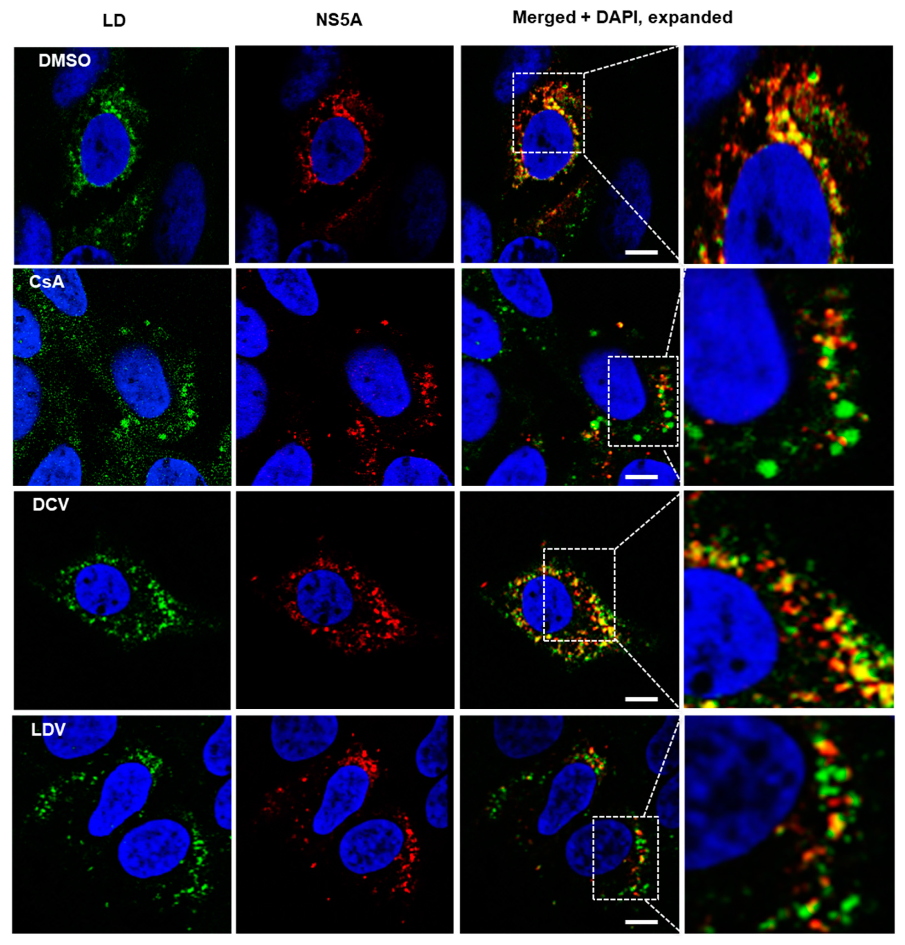
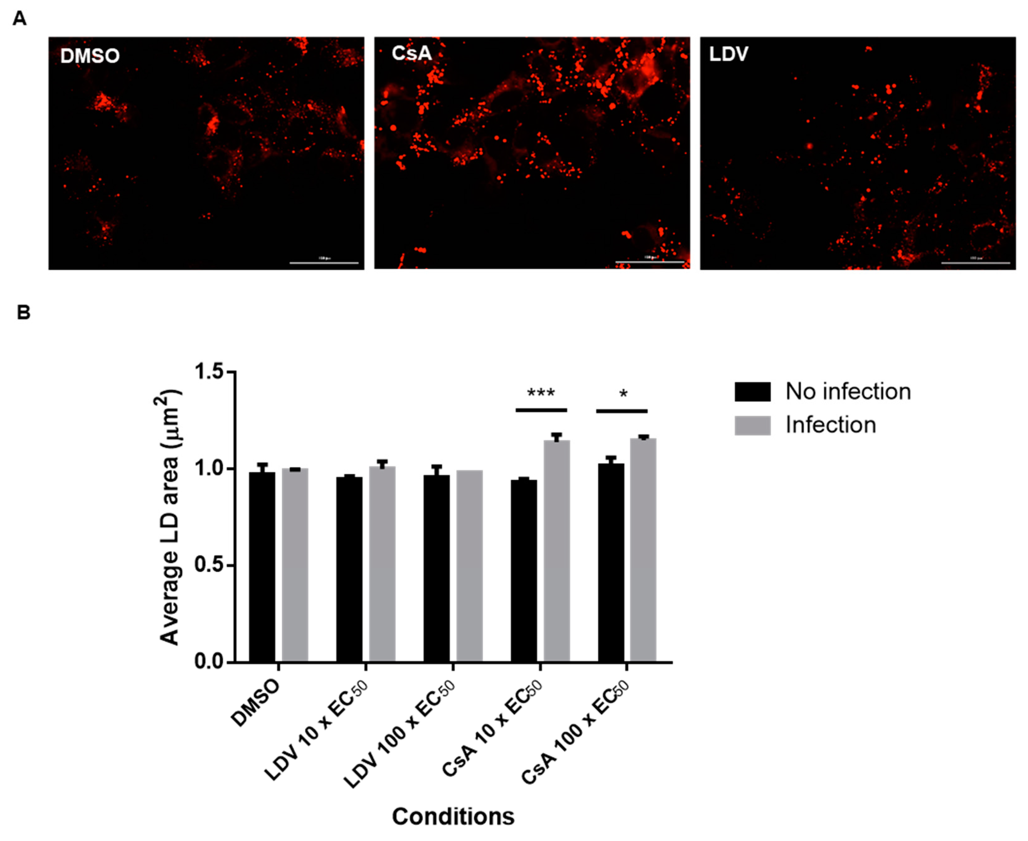
3.3. The Effect of CsA and NS5A Inhibitors on the Localization of NS5A at LDs

We and others have previously reported changes in NS5A localization following treatment with NS5A-targeting compounds [47,58]. To gain further insight into the impact of CypA inhibitors, HCV-infected cells were treated with DMSO, DCV, LDV, or CsA, and the distribution of NS5A and LDs was observed using confocal microscopy. Consistent with previous reports [47,58], the NS5A distribution appeared more punctate for all the drugs compared to DMSO (Figure 3). Furthermore, it appeared that the treatment of HCV-infected cells with CsA and LDV decreased the extent of NS5A co-localization with LDs (Figure 3). Notably, we also observed that the LDs were enlarged following treatment with CsA (Figure 3, expanded section). To quantify this effect, uninfected and HCV-infected cells were treated for 8 h with 10 × EC50 or 100 × EC50 CsA or LDV, LDs were stained with LipidTox Deep Red Neutral Lipid Stain (Figure 4A, red), automated unbiased imaging was performed, and the average area of LDs was calculated. In uninfected cells, no differences in the LD area were observed (ANOVA with Tukey’s multiple comparison test); however, the treatment of infected cells with CsA (but not LDV) induced a small but reproducible enlargement of LDs, suggesting that this change is specifically attributed to the inhibition of interactions between HCV and CypA (Figure 4B). These data suggest that CsA, but not LDV, caused LD enlargement, indicating different mechanisms of action.

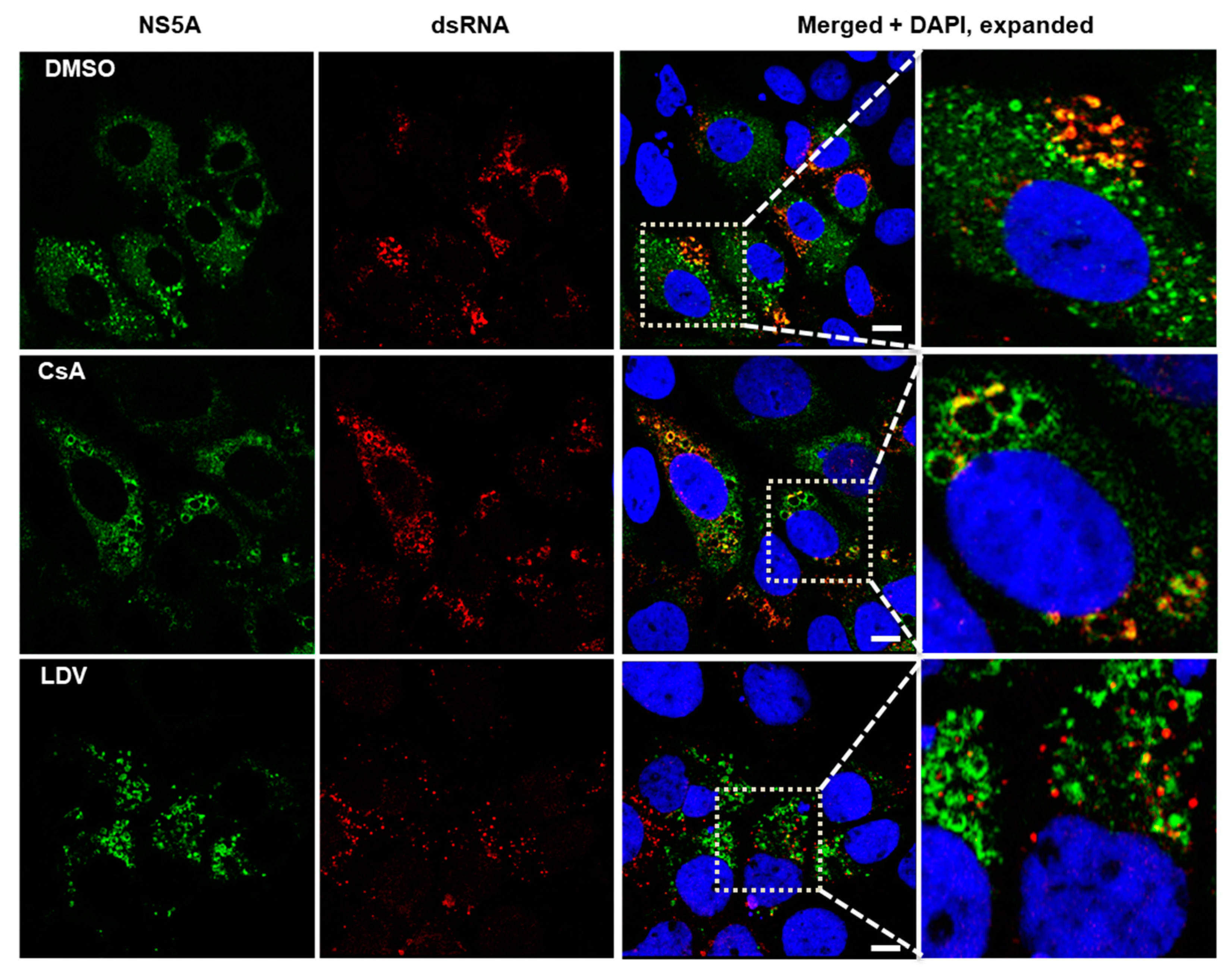
3.4. Interaction between NS5A and HCV Double-Stranded RNA

NS5A plays a critical role in HCV genome replication and assembly at LDs [14]. Double-stranded RNA (dsRNA) is formed during HCV replication and has been reported to co-localize with LDs and NS5A [65]. Using an antibody that recognizes dsRNA, we visualized HCV dsRNA and followed changes in its localization during treatment with LDV and CsA. We found that in cells treated with DMSO, NS5A was mostly diffused throughout the cytoplasm but was also observed on circular structures (presumably LDs) where it co-localized with dsRNA (Figure 5, expanded section). In cells treated with CsA, dsRNA mostly co-localized with NS5A at what appeared to be enlarged LDs (Figure 5, expanded section). Finally, in cells treated with LDV, dsRNA appeared to be restricted to discrete foci that did not co-localize with NS5A (Figure 5, expanded section). Collectively, these data suggest that CsA did not disrupt the co-localization of NS5A with dsRNA, although it may have perturbed the structure of lipid droplets. Conversely, LDV did appear to interrupt the interaction between NS5A and dsRNA.

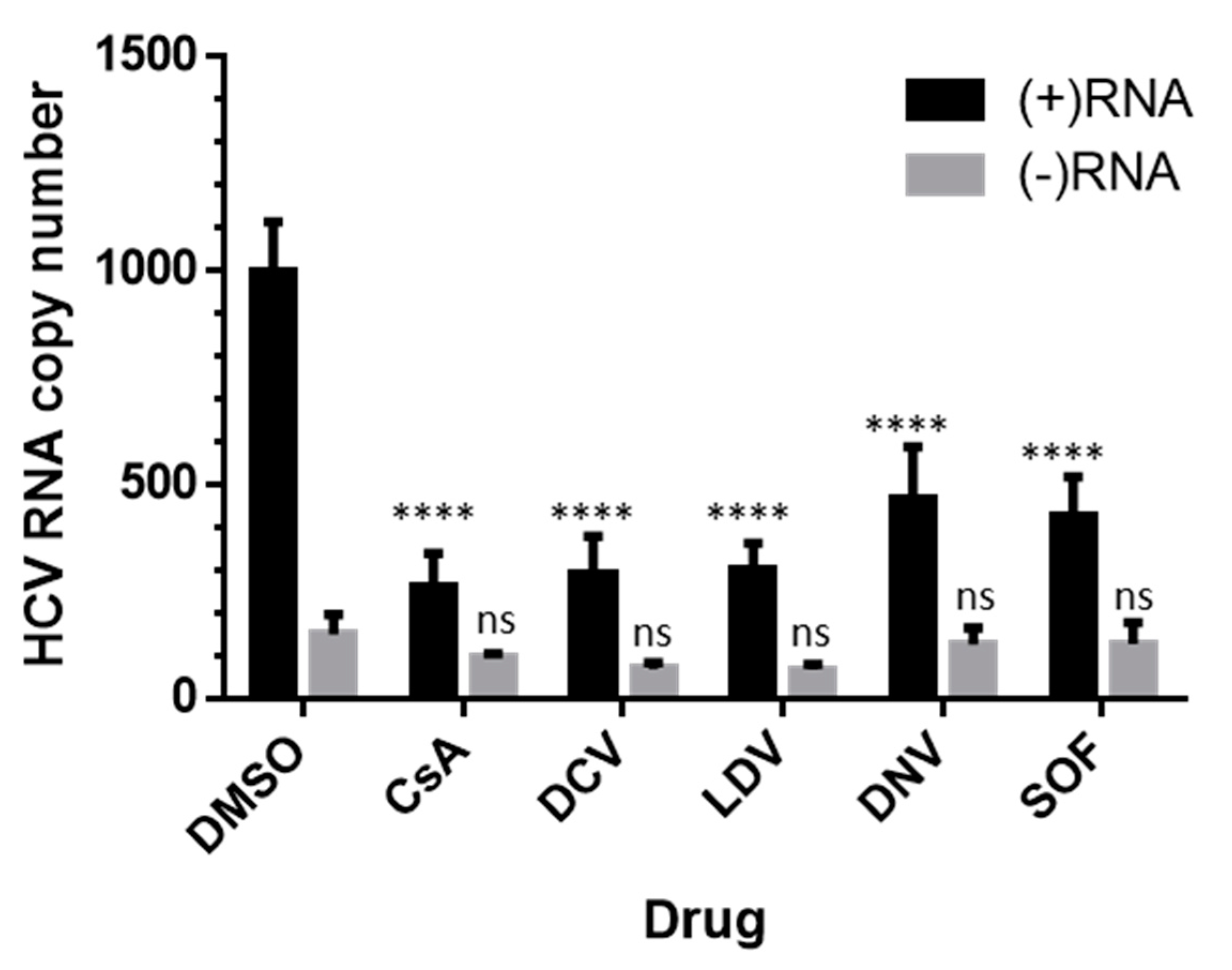
3.5. Effects on HCV RNA Assessed by Strand-Specific RT-qPCR

In order to dissect the inhibitory effects on HCV (+) or (−) strand RNA, we performed strand-specific RT-qPCR, as previously described [62,66]. Huh-7.5.1 cells were infected for 48 h and treated with DCV, LDV, DNV, CsA, or SOF at a 100 × EC50 concentration. All the inhibitors appeared to suppress HCV (+)RNA relative to the corresponding DMSO control (Figure 6). In contrast, the decreases in (−)RNA following the administration of inhibitors were less profound and not significant relative to DMSO (Figure 6).

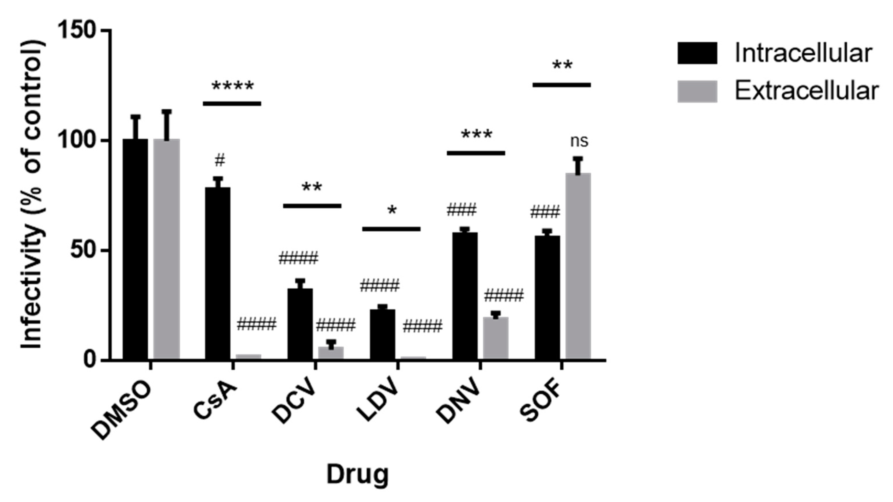
3.6. The Infectivity of Intracellular and Extracellular Viruses

Finally, as we documented the effects of CsA and NS5A inhibitors on viral RNA and cellular LDs, as well as the site of virion assembly, we examined the effects of the inhibitors on HCV assembly and release. We performed limited dilution assays to assess intracellular and extracellular viral infectivity. Cells were infected with Jc1/Gluc2A for 48 h followed by 8 h of exposure to inhibitors at 100 × EC50 concentration. RNA was extracted from cell lysates or media. Following normalization based on the genome copy number, naïve Huh-7.5.1 cells were infected with these viral stocks, and viral infectivity was determined with a limited dilution assay after 72 h. We observed a significant reduction in both intracellular and extracellular viral infectivity (relative to the corresponding DMSO control) for all conditions except SOF (extracellular infectivity) (Figure 7). We additionally compared effects on intracellular versus extracellular infectivity and found that there was a significant difference in all cases. In the SOF-treated cells, intracellular viral infectivity was preferentially suppressed relative to extracellular viral infectivity, whereas for CsA and all other DAA treatments, extracellular viral infectivity was preferentially suppressed relative to intracellular viral infectivity (Figure 7); this effect was particularly strong (~50-fold) in cells treated with CsA. The ability of CsA to induce the strong inhibition of extracellular infectivity, without a corresponding impact on intracellular infectivity, suggests that CsA may act to impair the release of infectious viral particles.

4. Discussion

With the advent of DAAs that target essential HCV nonstructural proteins, tremendous progress has been made in developing highly effective combination therapies with low cost, high genetic barrier to resistance, and broad genotype coverage [7,51,52,53,54,56,57]. Therapeutics that target host cellular factors have the potential to increase the barrier to resistance even further. CypA is a host factor that has been the subject of great interest because it plays a critical role in HCV replication and is a potential binding partner of several HCV proteins. Hence, this component of basic virology is important in order to understand the role of CypA in HCV infection. Here, we examined the effect of CsA (presumed to be acting via CypA inhibition) on the HCV replication cycle and compared the mechanism of action of CsA to those of DAAs.
We found that CsA exerted faster suppression of HCV protein expression than any DAA included in the study. In contrast, the NS5A-targeting compounds LDV and DCV induced the fastest suppression of HCV RNA levels. This suggests that CsA can act post-transcriptionally; this is because, at a given time point post treatment, CsA-treated cells contained more viral RNA, but less viral protein, relative to the effect on cells treated with DAAs.
We further pursued the mechanisms of CsA and NS5A-targetting drugs through studies on their interactions with LDs, an important site for HCV assembly and release [67]. Indeed, HCV release and specific infectivity were reduced following interference with apolipoprotein synthesis and secretion [68,69,70,71]; these similarities between the effects of interference with the apolipoprotein release pathway and those following CsA treatment suggest a potential mechanistic relationship. We found that CsA increased the LD size in infected cells and reduced NS5A co-localization with LDs. The NS5A inhibitor LDV also reduced NS5A co-localization with LDs, and additionally reduced dsRNA colocalization with NS5A, a phenotype consistent with reports that NS5A inhibitors can prevent NS5A-RNA binding [72], but that was not observed with CsA. NS5A inhibitors were found to suppress infectious virus levels in both intracellular and extracellular compartments, consistent with its activity against HCV replication and association with LDs. In contrast, CsA only suppressed the extracellular virus, consistent with the model that CsA impairs the interaction between NS5A and LDs, but not the interaction with dsRNA. These results suggest that the activity of CsA is related to NS5A, consistent with extensive reports on the CypA interactions with NS5A [13,18,21,22,23,24,73,74,75,76], but is distinct from the activity of NS5A inhibitors.
In recent years, many investigators have uncovered mechanisms by which CypA contributes to HCV replication. CypA interacts with NS5A to enhance its RNA binding activity, and that function is abolished by CsA [23,77]. More broadly, Cyp inhibitors suppress HCV replication by blocking the de novo formation of the membranous web (the NS4B rearranged cellular membranes upon which HCV replication takes place) [78,79,80] and displacing NS5B from the HCV RC [81]. Similarly, MxB has been reported to inhibit HCV replication by impairing the NS5A-CypA interaction [73,82]. Perhaps most pertinently, the non-immunosuppressive Cyp inhibitor NIM811 causes the accumulation of neutral lipids into LDs in the cell and decreases apolipoprotein B secretion through the VLDL pathway [83], as apolipoprotein secretion has been shown to play an essential role in HCV assembly and release; thus, these data provide a mechanism that is consistent with the observations presented here, whereby CypA may be an important factor for viral particle secretion.
The HCV genomic RNA has multiple functions: it is a replication template, an mRNA, and the packaged genome of infectious particles. The potential of CsA/CypA to influence HCV replication and the release of infectious particles is implied by two earlier studies. A S168A mutation in NS2 leads to the inhibition of HCV particle assembly, without affecting HCV RNA replication or protein synthesis; this assembly defect can be partially rescued through a mutation in NS5A V464L [84]. Intriguingly, the NS5A V464L mutation also confers the ability to replicate independently of CypA, thus allowing resistance to CsA [28]. These observations suggest that NS5A plays a key role in modulating the fate of HCV RNA, which agrees with extensive evidence demonstrating NS5A as a substrate for CypA [22,23,85,86] and the essential role of NS2 in particle assembly [84,87]. Together with our data, these observations are consistent with the close interdependency of genome replication and particle assembly in HCV, and the critical importance of CypA-NS5A interaction for the regulation of these processes.
In conclusion, we found that CsA both rapidly suppresses viral protein synthesis and potently inhibits the release of infectious HCV. While there is little demand for CsA-derived molecules as therapeutics for HCV, our findings support those of earlier studies and provide additional insight into the mechanism of action of CsA, and illustrate the virtues of exploiting pleiotropic proteins, such as NS5A, and virus–host interactions as antiviral targets.

Author Contributions

Conceptualization, C.M.R., R.R. and S.G.S.; methodology, D.L., T.P.N. and R.R.; formal analysis, D.L., T.P.N., A.D.H. and P.R.T.; investigation, D.L., T.P.N. and J.J.; resources, E.M. and C.M.R.; writing—original draft preparation, D.L.; writing—review and editing, P.R.T. and S.G.S.; supervision, S.G.S.; funding acquisition, C.M.R. and S.G.S. All authors have read and agreed to the published version of the manuscript.

Funding

This work was supported by the NIH, the National Institute of Allergy and Infectious Disease, and the National Institute of General Medical Sciences (AI099284 to C.M.R. and AI120860, AI132833 to S.G.S.).

Institutional Review Board Statement

Not applicable.

Informed Consent Statement

Not applicable.

Data Availability Statement

The data presented in this study are contained within the article.

Acknowledgments

The authors acknowledge the gift of mouse monoclonal primary antibody 9E10 by Brett Lindenbach, Yale University.

Conflicts of Interest

The authors declare no conflict of interest. The funders had no role in the design of the study; in the collection, analyses, or interpretation of the data; in the writing of the manuscript; or in the decision to publish the results.

References

  1. World Health Organization. Hepatitis C. Available online: https://www.who.int/news-room/fact-sheets/detail/hepatitis-c (accessed on 16 June 2022).
  2. Manns, M.P.; Buti, M.; Gane, E.; Pawlotsky, J.M.; Razavi, H.; Terrault, N.; Younossi, Z. Hepatitis C virus infection. Nat. Rev. Dis. Prim. 2017, 3, 17006. [Google Scholar] [CrossRef]
  3. Feld, J.J.; Hoofnagle, J.H. Mechanism of action of interferon and ribavirin in treatment of hepatitis C. Nature 2005, 436, 967–972. [Google Scholar] [CrossRef]
  4. Sulkowski, M.S.; Cooper, C.; Hunyady, B.; Jia, J.; Ogurtsov, P.; Peck-Radosavljevic, M.; Shiffman, M.L.; Yurdaydin, C.; Dalgard, O. Management of adverse effects of Peg-IFN and ribavirin therapy for hepatitis C. Nat. Rev. Gastroenterol. Hepatol. 2011, 8, 212–223. [Google Scholar] [CrossRef] [PubMed]
  5. Gotte, M.; Feld, J.J. Direct-acting antiviral agents for hepatitis C: Structural and mechanistic insights. Nat. Rev. Gastroenterol. Hepatol. 2016, 13, 338–351. [Google Scholar] [CrossRef]
  6. Schinazi, R.; Halfon, P.; Marcellin, P.; Asselah, T. HCV direct-acting antiviral agents: The best interferon-free combinations. Liver Int. 2014, 34 (Suppl. S1), 69–78. [Google Scholar] [CrossRef]
  7. Dietz, C.; Maasoumy, B. Direct-Acting Antiviral Agents for Hepatitis C Virus Infection-from Drug Discovery to Successful Implementation in Clinical Practice. Viruses 2022, 14, 1325. [Google Scholar] [CrossRef]
  8. Bartenschlager, R.; Frese, M.; Pietschmann, T. Novel insights into hepatitis C virus replication and persistence. Adv. Virus. Res. 2004, 63, 71–180. [Google Scholar] [CrossRef]
  9. Moradpour, D.; Penin, F. Hepatitis C virus proteins: From structure to function. Curr. Top. Microbiol. Immunol. 2013, 369, 113–142. [Google Scholar] [CrossRef]
  10. Penin, F.; Brass, V.; Appel, N.; Ramboarina, S.; Montserret, R.; Ficheux, D.; Blum, H.E.; Bartenschlager, R.; Moradpour, D. Structure and function of the membrane anchor domain of hepatitis C virus nonstructural protein 5A. J. Biol. Chem. 2004, 279, 40835–40843. [Google Scholar] [CrossRef]
  11. Lim, P.J.; Chatterji, U.; Cordek, D.; Sharma, S.D.; Garcia-Rivera, J.A.; Cameron, C.E.; Lin, K.; Targett-Adams, P.; Gallay, P.A. Correlation between NS5A dimerization and hepatitis C virus replication. J. Biol. Chem. 2012, 287, 30861–30873. [Google Scholar] [CrossRef]
  12. Tellinghuisen, T.L.; Marcotrigiano, J.; Rice, C.M. Structure of the zinc-binding domain of an essential component of the hepatitis C virus replicase. Nature 2005, 435, 374–379. [Google Scholar] [CrossRef] [PubMed]
  13. Shanmugam, S.; Nichols, A.K.; Saravanabalaji, D.; Welsch, C.; Yi, M. HCV NS5A dimer interface residues regulate HCV replication by controlling its self-interaction, hyperphosphorylation, subcellular localization and interaction with cyclophilin A. PLoS Pathog. 2018, 14, e1007177. [Google Scholar] [CrossRef] [PubMed]
  14. Ross-Thriepland, D.; Harris, M. Hepatitis C virus NS5A: Enigmatic but still promiscuous 10 years on! J. Gen. Virol. 2015, 96, 727–738. [Google Scholar] [CrossRef] [PubMed]
  15. Miyanari, Y.; Atsuzawa, K.; Usuda, N.; Watashi, K.; Hishiki, T.; Zayas, M.; Bartenschlager, R.; Wakita, T.; Hijikata, M.; Shimotohno, K. The lipid droplet is an important organelle for hepatitis C virus production. Nat. Cell Biol. 2007, 9, 1089–1097. [Google Scholar] [CrossRef] [PubMed]
  16. Alvisi, G.; Madan, V.; Bartenschlager, R. Hepatitis C virus and host cell lipids: An intimate connection. RNA Biol. 2011, 8, 258–269. [Google Scholar] [CrossRef] [PubMed]
  17. Camus, G.; Herker, E.; Modi, A.A.; Haas, J.T.; Ramage, H.R.; Farese, R.V., Jr.; Ott, M. Diacylglycerol acyltransferase-1 localizes hepatitis C virus NS5A protein to lipid droplets and enhances NS5A interaction with the viral capsid core. J. Biol. Chem. 2013, 288, 9915–9923. [Google Scholar] [CrossRef] [PubMed]
  18. Ross-Thriepland, D.; Amako, Y.; Harris, M. The C terminus of NS5A domain II is a key determinant of hepatitis C virus genome replication, but is not required for virion assembly and release. J. Gen. Virol. 2013, 94, 1009–1018. [Google Scholar] [CrossRef]
  19. Tellinghuisen, T.L.; Marcotrigiano, J.; Gorbalenya, A.E.; Rice, C.M. The NS5A protein of hepatitis C virus is a zinc metalloprotein. J. Biol. Chem. 2004, 279, 48576–48587. [Google Scholar] [CrossRef]
  20. Appel, N.; Zayas, M.; Miller, S.; Krijnse-Locker, J.; Schaller, T.; Friebe, P.; Kallis, S.; Engel, U.; Bartenschlager, R. Essential role of domain III of nonstructural protein 5A for hepatitis C virus infectious particle assembly. PLoS Pathog. 2008, 4, e1000035. [Google Scholar] [CrossRef]
  21. Verdegem, D.; Badillo, A.; Wieruszeski, J.M.; Landrieu, I.; Leroy, A.; Bartenschlager, R.; Penin, F.; Lippens, G.; Hanoulle, X. Domain 3 of NS5A protein from the hepatitis C virus has intrinsic alpha-helical propensity and is a substrate of cyclophilin A. J. Biol. Chem. 2011, 286, 20441–20454. [Google Scholar] [CrossRef]
  22. Dujardin, M.; Madan, V.; Gandhi, N.S.; Cantrelle, F.X.; Launay, H.; Huvent, I.; Bartenschlager, R.; Lippens, G.; Hanoulle, X. Cyclophilin A allows the allosteric regulation of a structural motif in the disordered domain 2 of NS5A and thereby fine-tunes HCV RNA replication. J. Biol. Chem. 2019, 294, 13171–13185. [Google Scholar] [CrossRef]
  23. Foster, T.L.; Gallay, P.; Stonehouse, N.J.; Harris, M. Cyclophilin A interacts with domain II of hepatitis C virus NS5A and stimulates RNA binding in an isomerase-dependent manner. J. Virol. 2011, 85, 7460–7464. [Google Scholar] [CrossRef]
  24. Abe, T.; Minami, N.; Bawono, R.G.; Matsui, C.; Deng, L.; Fukuhara, T.; Matsuura, Y.; Shoji, I. ISGylation of Hepatitis C Virus NS5A Protein Promotes Viral RNA Replication via Recruitment of Cyclophilin A. J. Virol. 2020, 94, e00532-20. [Google Scholar] [CrossRef] [PubMed]
  25. Galat, A. Function-dependent clustering of orthologues and paralogues of cyclophilins. Proteins 2004, 56, 808–820. [Google Scholar] [CrossRef]
  26. Fernandes, F.; Ansari, I.U.; Striker, R. Cyclosporine inhibits a direct interaction between cyclophilins and hepatitis C NS5A. PLoS ONE 2010, 5, e9815. [Google Scholar] [CrossRef]
  27. Yang, F.; Robotham, J.M.; Nelson, H.B.; Irsigler, A.; Kenworthy, R.; Tang, H. Cyclophilin A is an essential cofactor for hepatitis C virus infection and the principal mediator of cyclosporine resistance in vitro. J. Virol. 2008, 82, 5269–5278. [Google Scholar] [CrossRef]
  28. Kaul, A.; Stauffer, S.; Berger, C.; Pertel, T.; Schmitt, J.; Kallis, S.; Zayas, M.; Lohmann, V.; Luban, J.; Bartenschlager, R. Essential role of cyclophilin A for hepatitis C virus replication and virus production and possible link to polyprotein cleavage kinetics. PLoS Pathog. 2009, 5, e1000546. [Google Scholar] [CrossRef]
  29. Jesudian, A.B.; Gambarin-Gelwan, M.; Jacobson, I.M. Advances in the treatment of hepatitis C virus infection. Gastroenterol. Hepatol. 2012, 8, 91–101. [Google Scholar]
  30. Gaska, J.M.; Balev, M.; Ding, Q.; Heller, B.; Ploss, A. Differences across cyclophilin A orthologs contribute to the host range restriction of hepatitis C virus. Elife 2019, 8, e44436. [Google Scholar] [CrossRef]
  31. Liu, J.; Farmer, J.D., Jr.; Lane, W.S.; Friedman, J.; Weissman, I.; Schreiber, S.L. Calcineurin is a common target of cyclophilin-cyclosporin A and FKBP-FK506 complexes. Cell 1991, 66, 807–815. [Google Scholar] [CrossRef]
  32. Ma, S.; Boerner, J.E.; TiongYip, C.; Weidmann, B.; Ryder, N.S.; Cooreman, M.P.; Lin, K. NIM811, a cyclophilin inhibitor, exhibits potent in vitro activity against hepatitis C virus alone or in combination with alpha interferon. Antimicrob. Agents. Chemother. 2006, 50, 2976–2982. [Google Scholar] [CrossRef]
  33. Paeshuyse, J.; Kaul, A.; De Clercq, E.; Rosenwirth, B.; Dumont, J.M.; Scalfaro, P.; Bartenschlager, R.; Neyts, J. The non-immunosuppressive cyclosporin DEBIO-025 is a potent inhibitor of hepatitis C virus replication in vitro. Hepatology 2006, 43, 761–770. [Google Scholar] [CrossRef]
  34. Ciesek, S.; Steinmann, E.; Wedemeyer, H.; Manns, M.P.; Neyts, J.; Tautz, N.; Madan, V.; Bartenschlager, R.; von Hahn, T.; Pietschmann, T. Cyclosporine A inhibits hepatitis C virus nonstructural protein 2 through cyclophilin A. Hepatology 2009, 50, 1638–1645. [Google Scholar] [CrossRef]
  35. Liu, Z.; Robida, J.M.; Chinnaswamy, S.; Yi, G.; Robotham, J.M.; Nelson, H.B.; Irsigler, A.; Kao, C.C.; Tang, H. Mutations in the hepatitis C virus polymerase that increase RNA binding can confer resistance to cyclosporine A. Hepatology 2009, 50, 25–33. [Google Scholar] [CrossRef]
  36. Robida, J.M.; Nelson, H.B.; Liu, Z.; Tang, H. Characterization of hepatitis C virus subgenomic replicon resistance to cyclosporine in vitro. J. Virol. 2007, 81, 5829–5840. [Google Scholar] [CrossRef]
  37. Yang, F.; Robotham, J.M.; Grise, H.; Frausto, S.; Madan, V.; Zayas, M.; Bartenschlager, R.; Robinson, M.; Greenstein, A.E.; Nag, A.; et al. A major determinant of cyclophilin dependence and cyclosporine susceptibility of hepatitis C virus identified by a genetic approach. PLoS. Pathog. 2010, 6, e1001118. [Google Scholar] [CrossRef]
  38. Chatel-Chaix, L.; Baril, M.; Lamarre, D. Hepatitis C Virus NS3/4A Protease Inhibitors: A Light at the End of the Tunnel. Viruses 2010, 2, 1752–1765. [Google Scholar] [CrossRef]
  39. Marks, K.M.; Jacobson, I.M. The first wave: HCV NS3 protease inhibitors telaprevir and boceprevir. Antivir. Ther. 2012, 17, 1119–1131. [Google Scholar] [CrossRef]
  40. Miao, M.; Jing, X.; De Clercq, E.; Li, G. Danoprevir for the Treatment of Hepatitis C Virus Infection: Design, Development, and Place in Therapy. Drug. Des. Devel. Ther. 2020, 14, 2759–2774. [Google Scholar] [CrossRef]
  41. Gao, M.; Nettles, R.E.; Belema, M.; Snyder, L.B.; Nguyen, V.N.; Fridell, R.A.; Serrano-Wu, M.H.; Langley, D.R.; Sun, J.H.; O’Boyle, D.R., 2nd; et al. Chemical genetics strategy identifies an HCV NS5A inhibitor with a potent clinical effect. Nature 2010, 465, 96–100. [Google Scholar] [CrossRef]
  42. Afdhal, N.; Zeuzem, S.; Kwo, P.; Chojkier, M.; Gitlin, N.; Puoti, M.; Romero-Gomez, M.; Zarski, J.P.; Agarwal, K.; Buggisch, P.; et al. Ledipasvir and sofosbuvir for untreated HCV genotype 1 infection. N. Engl. J. Med. 2014, 370, 1889–1898. [Google Scholar] [CrossRef] [PubMed]
  43. Afdhal, N.; Reddy, K.R.; Nelson, D.R.; Lawitz, E.; Gordon, S.C.; Schiff, E.; Nahass, R.; Ghalib, R.; Gitlin, N.; Herring, R.; et al. Ledipasvir and sofosbuvir for previously treated HCV genotype 1 infection. N. Engl. J. Med. 2014, 370, 1483–1493. [Google Scholar] [CrossRef]
  44. Gamal, N.; Gitto, S.; Andreone, P. Efficacy and Safety of Daclatasvir in Hepatitis C: An Overview. J. Clin. Transl. Hepatol. 2016, 4, 336–344. [Google Scholar] [CrossRef]
  45. Fridell, R.A.; Wang, C.; Sun, J.H.; O’Boyle, D.R., 2nd; Nower, P.; Valera, L.; Qiu, D.; Roberts, S.; Huang, X.; Kienzle, B.; et al. Genotypic and phenotypic analysis of variants resistant to hepatitis C virus nonstructural protein 5A replication complex inhibitor BMS-790052 in humans: In vitro and in vivo correlations. Hepatology 2011, 54, 1924–1935. [Google Scholar] [CrossRef]
  46. Pawlotsky, J.M. NS5A inhibitors in the treatment of hepatitis C. J. Hepatol. 2013, 59, 375–382. [Google Scholar] [CrossRef]
  47. Targett-Adams, P.; Graham, E.J.; Middleton, J.; Palmer, A.; Shaw, S.M.; Lavender, H.; Brain, P.; Tran, T.D.; Jones, L.H.; Wakenhut, F.; et al. Small molecules targeting hepatitis C virus-encoded NS5A cause subcellular redistribution of their target: Insights into compound modes of action. J. Virol. 2011, 85, 6353–6368. [Google Scholar] [CrossRef] [PubMed]
  48. Cheng, G.; Peng, B.; Corsa, A.; Yu, M.; Nash, M.; Lee, Y.-J.; Xu, Y.; Kirschberg, T.; Tian, Y.; Taylor, J.; et al. 1172 Antiviral Activity and Resistance Profile of the Novel Hcv Ns5a Inhibitor GS-5885. J. Hepatol. 2012, 56, S464. [Google Scholar] [CrossRef]
  49. Zeuzem, S.; Foster, G.R.; Wang, S.; Asatryan, A.; Gane, E.; Feld, J.J.; Asselah, T.; Bourliere, M.; Ruane, P.J.; Wedemeyer, H.; et al. Glecaprevir-Pibrentasvir for 8 or 12 Weeks in HCV Genotype 1 or 3 Infection. N. Engl. J. Med. 2018, 378, 354–369. [Google Scholar] [CrossRef]
  50. Feld, J.J.; Jacobson, I.M.; Hezode, C.; Asselah, T.; Ruane, P.J.; Gruener, N.; Abergel, A.; Mangia, A.; Lai, C.L.; Chan, H.L.; et al. Sofosbuvir and Velpatasvir for HCV Genotype 1, 2, 4, 5, and 6 Infection. N. Engl. J. Med. 2015, 373, 2599–2607. [Google Scholar] [CrossRef]
  51. Curry, M.P.; O’Leary, J.G.; Bzowej, N.; Muir, A.J.; Korenblat, K.M.; Fenkel, J.M.; Reddy, K.R.; Lawitz, E.; Flamm, S.L.; Schiano, T.; et al. Sofosbuvir and Velpatasvir for HCV in Patients with Decompensated Cirrhosis. N. Engl. J. Med. 2015, 373, 2618–2628. [Google Scholar] [CrossRef]
  52. Foster, G.R.; Afdhal, N.; Roberts, S.K.; Brau, N.; Gane, E.J.; Pianko, S.; Lawitz, E.; Thompson, A.; Shiffman, M.L.; Cooper, C.; et al. Sofosbuvir and Velpatasvir for HCV Genotype 2 and 3 Infection. N. Engl. J. Med. 2015, 373, 2608–2617. [Google Scholar] [CrossRef]
  53. Llaneras, J.; Riveiro-Barciela, M.; Lens, S.; Diago, M.; Cachero, A.; Garcia-Samaniego, J.; Conde, I.; Arencibia, A.; Arenas, J.; Gea, F.; et al. Effectiveness and safety of sofosbuvir/velpatasvir/voxilaprevir in patients with chronic hepatitis C previously treated with DAAs. J. Hepatol. 2019, 71, 666–672. [Google Scholar] [CrossRef]
  54. Degasperi, E.; Spinetti, A.; Lombardi, A.; Landonio, S.; Rossi, M.C.; Pasulo, L.; Pozzoni, P.; Giorgini, A.; Fabris, P.; Romano, A.; et al. Real-life effectiveness and safety of sofosbuvir/velpatasvir/voxilaprevir in hepatitis C patients with previous DAA failure. J. Hepatol. 2019, 71, 1106–1115. [Google Scholar] [CrossRef] [PubMed]
  55. Buti, M.; Flisiak, R.; Kao, J.H.; Chuang, W.L.; Streinu-Cercel, A.; Tabak, F.; Calistru, P.; Goeser, T.; Rasenack, J.; Horban, A.; et al. Alisporivir with peginterferon/ribavirin in patients with chronic hepatitis C genotype 1 infection who failed to respond to or relapsed after prior interferon-based therapy: FUNDAMENTAL, a Phase II trial. J. Viral. Hepat. 2015, 22, 596–606. [Google Scholar] [CrossRef] [PubMed]
  56. Flisiak, R.; Jaroszewicz, J.; Flisiak, I.; Lapinski, T. Update on alisporivir in treatment of viral hepatitis C. Expert. Opin. Investig. Drugs. 2012, 21, 375–382. [Google Scholar] [CrossRef]
  57. Pawlotsky, J.M.; Flisiak, R.; Sarin, S.K.; Rasenack, J.; Piratvisuth, T.; Chuang, W.L.; Peng, C.Y.; Foster, G.R.; Shah, S.; Wedemeyer, H.; et al. Alisporivir plus ribavirin, interferon free or in combination with pegylated interferon, for hepatitis C virus genotype 2 or 3 infection. Hepatology. 2015, 62, 1013–1023. [Google Scholar] [CrossRef]
  58. Liu, D.; Ji, J.; Ndongwe, T.P.; Michailidis, E.; Rice, C.M.; Ralston, R.; Sarafianos, S.G. Fast hepatitis C virus RNA elimination and NS5A redistribution by NS5A inhibitors studied by a multiplex assay approach. Antimicrob. Agents Chemother. 2015, 59, 3482–3492. [Google Scholar] [CrossRef] [PubMed]
  59. Zhong, J.; Gastaminza, P.; Cheng, G.; Kapadia, S.; Kato, T.; Burton, D.R.; Wieland, S.F.; Uprichard, S.L.; Wakita, T.; Chisari, F.V. Robust hepatitis C virus infection in vitro. Proc. Natl. Acad. Sci. USA 2005, 102, 9294–9299. [Google Scholar] [CrossRef] [PubMed]
  60. Marukian, S.; Jones, C.T.; Andrus, L.; Evans, M.J.; Ritola, K.D.; Charles, E.D.; Rice, C.M.; Dustin, L.B. Cell culture-produced hepatitis C virus does not infect peripheral blood mononuclear cells. Hepatology 2008, 48, 1843–1850. [Google Scholar] [CrossRef]
  61. Lindenbach, B.D.; Evans, M.J.; Syder, A.J.; Wolk, B.; Tellinghuisen, T.L.; Liu, C.C.; Maruyama, T.; Hynes, R.O.; Burton, D.R.; McKeating, J.A.; et al. Complete replication of hepatitis C virus in cell culture. Science 2005, 309, 623–626. [Google Scholar] [CrossRef]
  62. Komurian-Pradel, F.; Perret, M.; Deiman, B.; Sodoyer, M.; Lotteau, V.; Paranhos-Baccala, G.; Andre, P. Strand specific quantitative real-time PCR to study replication of hepatitis C virus genome. J. Virol. Methods. 2004, 116, 103–106. [Google Scholar] [CrossRef] [PubMed]
  63. Reed, L.J.; Meunch, H.A. A Simple Method of Estimating Fifty per Cent Endpoints. Am. J. Epidemiol. 1938, 27, 493–497. [Google Scholar] [CrossRef]
  64. Dittmar, M.; Lee, J.S.; Whig, K.; Segrist, E.; Li, M.; Kamalia, B.; Castellana, L.; Ayyanathan, K.; Cardenas-Diaz, F.L.; Morrisey, E.E.; et al. Drug repurposing screens reveal cell-type-specific entry pathways and FDA-approved drugs active against SARS-CoV-2. Cell Rep. 2021, 35, 108959. [Google Scholar] [CrossRef]
  65. Targett-Adams, P.; Boulant, S.; McLauchlan, J. Visualization of double-stranded RNA in cells supporting hepatitis C virus RNA replication. J. Virol. 2008, 82, 2182–2195. [Google Scholar] [CrossRef] [PubMed]
  66. Liu, D.; Tedbury, P.R.; Lan, S.; Huber, A.D.; Puray-Chavez, M.N.; Ji, J.; Michailidis, E.; Saeed, M.; Ndongwe, T.P.; Bassit, L.C.; et al. Visualization of Positive and Negative Sense Viral RNA for Probing the Mechanism of Direct-Acting Antivirals against Hepatitis C Virus. Viruses. 2019, 11, 1039. [Google Scholar] [CrossRef] [PubMed]
  67. Filipe, A.; McLauchlan, J. Hepatitis C virus and lipid droplets: Finding a niche. Trends. Mol. Med. 2015, 21, 34–42. [Google Scholar] [CrossRef] [PubMed]
  68. Hueging, K.; Weller, R.; Doepke, M.; Vieyres, G.; Todt, D.; Wolk, B.; Vondran, F.W.; Geffers, R.; Lauber, C.; Kaderali, L.; et al. Several Human Liver Cell Expressed Apolipoproteins Complement HCV Virus Production with Varying Efficacy Conferring Differential Specific Infectivity to Released Viruses. PLoS ONE 2015, 10, e0134529. [Google Scholar] [CrossRef] [PubMed]
  69. Gastaminza, P.; Kapadia, S.B.; Chisari, F.V. Differential biophysical properties of infectious intracellular and secreted hepatitis C virus particles. J. Virol. 2006, 80, 11074–11081. [Google Scholar] [CrossRef]
  70. Huang, H.; Sun, F.; Owen, D.M.; Li, W.; Chen, Y.; Gale, M., Jr.; Ye, J. Hepatitis C virus production by human hepatocytes dependent on assembly and secretion of very low-density lipoproteins. Proc. Natl. Acad. Sci. USA 2007, 104, 5848–5853. [Google Scholar] [CrossRef]
  71. Chang, K.S.; Jiang, J.; Cai, Z.; Luo, G. Human apolipoprotein e is required for infectivity and production of hepatitis C virus in cell culture. J. Virol. 2007, 81, 13783–13793. [Google Scholar] [CrossRef] [PubMed]
  72. Ascher, D.B.; Wielens, J.; Nero, T.L.; Doughty, L.; Morton, C.J.; Parker, M.W. Potent hepatitis C inhibitors bind directly to NS5A and reduce its affinity for RNA. Sci. Rep. 2014, 4, 4765. [Google Scholar] [CrossRef] [PubMed]
  73. Yi, D.R.; An, N.; Liu, Z.L.; Xu, F.W.; Raniga, K.; Li, Q.J.; Zhou, R.; Wang, J.; Zhang, Y.X.; Zhou, J.M.; et al. Human MxB Inhibits the Replication of Hepatitis C Virus. J. Virol. 2019, 93, e01285-18. [Google Scholar] [CrossRef] [PubMed]
  74. Waller, H.; Chatterji, U.; Gallay, P.; Parkinson, T.; Targett-Adams, P. The use of AlphaLISA technology to detect interaction between hepatitis C virus-encoded NS5A and cyclophilin A. J. Virol. Methods. 2010, 165, 202–210. [Google Scholar] [CrossRef] [PubMed]
  75. Goto, K.; Watashi, K.; Inoue, D.; Hijikata, M.; Shimotohno, K. Identification of cellular and viral factors related to anti-hepatitis C virus activity of cyclophilin inhibitor. Cancer. Sci. 2009, 100, 1943–1950. [Google Scholar] [CrossRef] [PubMed]
  76. von Hahn, T.; Schiene-Fischer, C.; Van, N.D.; Pfaender, S.; Karavul, B.; Steinmann, E.; Potthoff, A.; Strassburg, C.; Hamdi, N.; Abdelaziz, A.I.; et al. Hepatocytes that express variants of cyclophilin A are resistant to HCV infection and replication. Gastroenterology 2012, 143, 439–447. [Google Scholar] [CrossRef]
  77. Nag, A.; Robotham, J.M.; Tang, H. Suppression of viral RNA binding and the assembly of infectious hepatitis C virus particles in vitro by cyclophilin inhibitors. J. Virol. 2012, 86, 12616–12624. [Google Scholar] [CrossRef]
  78. Madan, V.; Paul, D.; Lohmann, V.; Bartenschlager, R. Inhibition of HCV replication by cyclophilin antagonists is linked to replication fitness and occurs by inhibition of membranous web formation. Gastroenterology 2014, 146, 1361–1372.e1–9. [Google Scholar] [CrossRef]
  79. Chatterji, U.; Bobardt, M.; Tai, A.; Wood, M.; Gallay, P.A. Cyclophilin and NS5A inhibitors, but not other anti-hepatitis C virus (HCV) agents, preclude HCV-mediated formation of double-membrane-vesicle viral factories. Antimicrob. Agents. Chemother. 2015, 59, 2496–2507. [Google Scholar] [CrossRef] [PubMed]
  80. Chatterji, U.; Bobardt, M.; Schaffer, L.; Wood, M.; Gallay, P.A. Cyclophilin Inhibitors Remodel the Endoplasmic Reticulum of HCV-Infected Cells in a Unique Pattern Rendering Cells Impervious to a Reinfection. PLoS ONE 2016, 11, e0159511. [Google Scholar] [CrossRef] [PubMed]
  81. Liu, Z.; Yang, F.; Robotham, J.M.; Tang, H. Critical role of cyclophilin A and its prolyl-peptidyl isomerase activity in the structure and function of the hepatitis C virus replication complex. J. Virol. 2009, 83, 6554–6565. [Google Scholar] [CrossRef] [PubMed]
  82. Li, Q.; An, N.; Yin, X.; Zhang, R.; Shao, H.; Yi, D.; Cen, S. MxB Disrupts Hepatitis C Virus NS5A-CypA Complex: Insights From a Combined Theoretical and Experimental Approach. Front. Microbiol. 2022, 13, 849084. [Google Scholar] [CrossRef] [PubMed]
  83. Anderson, L.J.; Lin, K.; Compton, T.; Wiedmann, B. Inhibition of cyclophilins alters lipid trafficking and blocks hepatitis C virus secretion. Virol. J. 2011, 8, 329. [Google Scholar] [CrossRef] [PubMed]
  84. Yi, M.; Ma, Y.; Yates, J.; Lemon, S.M. Trans-complementation of an NS2 defect in a late step in hepatitis C virus (HCV) particle assembly and maturation. PLoS Pathog. 2009, 5, e1000403. [Google Scholar] [CrossRef]
  85. Badillo, A.; Receveur-Brechot, V.; Sarrazin, S.; Cantrelle, F.X.; Delolme, F.; Fogeron, M.L.; Molle, J.; Montserret, R.; Bockmann, A.; Bartenschlager, R.; et al. Overall Structural Model of NS5A Protein from Hepatitis C Virus and Modulation by Mutations Confering Resistance of Virus Replication to Cyclosporin A. Biochemistry 2017, 56, 3029–3048. [Google Scholar] [CrossRef] [PubMed]
  86. Ngure, M.; Issur, M.; Shkriabai, N.; Liu, H.W.; Cosa, G.; Kvaratskhelia, M.; Gotte, M. Interactions of the Disordered Domain II of Hepatitis C Virus NS5A with Cyclophilin A, NS5B, and Viral RNA Show Extensive Overlap. ACS Infect. Dis. 2016, 2, 839–851. [Google Scholar] [CrossRef]
  87. Jones, C.T.; Murray, C.L.; Eastman, D.K.; Tassello, J.; Rice, C.M. Hepatitis C virus p7 and NS2 proteins are essential for production of infectious virus. J. Virol. 2007, 81, 8374–8383. [Google Scholar] [CrossRef]
Figure 1. Time course of HCV inhibition by antivirals. Huh-7.5.1 cells were infected with Jc1/Gluc2A at an MOI of 0.5 for 48 h and then treated with 100 × EC50 of the indicated compounds. (A) Culture supernatants were analyzed for luciferase activity at the indicated time points. (B) The total cellular RNA sample was extracted using TRIzol reagent and analyzed for HCV 5′ UTR RNA by RT-qPCR at the indicated time points. The data were normalized to a DMSO-treated control. Each data point represents an average value of 3 individual experiments. The error bars represent the standard errors of the mean (SEMs). DCV, daclatasvir; DNV, danoprevir; CsA, cyclosporin A; SOF, sofosbuvir; LDV, ledipasvir.
Viruses 15 00981 g001
Figure 2. Suppression of HCV protein expression. Jc1/Gluc2A-virus-infected Huh-7.5.1 cells were treated with 100 × EC50 of DCV, DNV, SOF, CsA, or LDV. Cell lysates were harvested 24 hpt (A) or 8 hpt (B), separated by SDS-PAGE and immunoblotted for NS5A or core. NS5A and core intensities were normalized to GAPDH and quantified as a relative fold change (FC) with respect to DMSO-treated samples, as shown in the blots below. Each value represents the average value of 3 individual experiments. In (A), the arrow indicates NS5A and the asterisk indicates a non-specific host protein. NS5A and Core were probed on the same membrane, thus sharing a GAPDH control. DCV, daclatasvir; DNV, danoprevir; CsA, cyclosporin A; SOF, sofosbuvir; LDV, ledipasvir. * indicated non-specific band.
Viruses 15 00981 g002
Figure 3. The distribution of NS5A and lipid droplets (LDs) following treatment with CsA and NS5A inhibitors. HCV-infected cells were treated for 8 h with DMSO, or 100 × EC50 DCV, CsA, and LDV. Cells were then fixed and stained with BODIPY 493/503 neutral lipid stain to label the LDs (green) and anti-NS5A antibodies (red), and with Hoechst-33258 to label the nuclei (blue). Images were acquired on a Leica TCS SP8 microscope with a 63× oil objective. Merged images and expanded merged images show the localization of NS5A and LDs. The scale bar represents 10 µm. DCV, daclatasvir; CsA, cyclosporin A; LDV, ledipasvir.
Viruses 15 00981 g003
Figure 4. Changes in the LD area following treatment with CsA. Jc1/Gluc2A-virus-infected Huh-7.5.1 cells were treated with DMSO (10 × EC50 or 100 × EC50) of LDV or CsA for 8 h. The cells were fixed and stained with HCS LipidTox Deep Red Neutral Lipid Stain to label the LDs. Images were acquired on a BioTek Cytation 5 cell imaging multi-mode reader with 40× objective and analyzed by BioTek Gen5 software. (A) Representative images of LDs in cells treated with 100 × EC50 LDV or CsA for 8 h. The scale bar represents 100 µm. (B) The average area of LDs for each treatment. Each data point represents the average value of 2 individual experiments. The error bars represent the SEMs. Statistical analysis was performed using 2-way ANOVA with Šidák’s multiple comparisons test. * p < 0.05, *** p < 0.001. CsA, cyclosporin A; LDV, ledipasvir.
Viruses 15 00981 g004
Figure 5. Localization of NS5A and dsRNA following treatment with CsA and LDV. Confocal immunofluorescence microscopy showing the cellular distribution of NS5A and dsRNA in HCV-infected cells treated for 8 h with DMSO (first row), 100 × EC50 CsA (second row), and LDV (third row). The cells were fixed and stained with 9E10 to label NS5A (green, first column), J2 to label dsRNA (red, second column), and DAPI to label nuclei (blue). Images were acquired on a Leica TCS SP8 microscope with a 63× oil objective. Merged images and expanded merged images show the localization of NS5A and dsRNA. The scale bar represents 10 µm. CsA, cyclosporin A; LDV, ledipasvir.
Viruses 15 00981 g005
Figure 6. Strand-specific suppression of HCV RNA. Jc1/Gluc2A-virus-infected Huh-7.5.1 cells were treated with DMSO or 100 × EC50 of compounds for 8 h. Total cellular RNA was extracted by TRIzol reagent and analyzed for HCV 5′ UTR RNA by RT-qPCR. The cycle threshold (CT) values were converted to copy numbers based on the standard curve of the Jc1/Gluc2A-genome-encoded plasmid. The data are shown for drug effects on HCV (+)RNA (black) and HCV (−) RNA (grey). Each data point represents the average value of 3 individual experiments and the error bars represent the SEMs. Statistical analysis was performed using 2-way ANOVA with Tukey’s post hoc test to compare samples within the (+) RNA group or (−) RNA group. ns, not significant; **** p < 0.0001. DCV, daclatasvir; DNV, danoprevir; CsA, cyclosporin A; SOF, sofosbuvir; LDV, ledipasvir.
Viruses 15 00981 g006
Figure 7. Impact of antivirals on intracellular and extracellular virus production. Jc1/Gluc2A-virus-infected Huh-7.5.1 cells were treated with DMSO or 100 × EC50 of antiviral inhibitors for 8 h. Intracellular virus was obtained from four cycles of freeze–thaw cell lysate, whereas extracellular virus was precipitated by PEG from culture supernatant. Viral RNA was extracted and the viral copy number was determined by RT-qPCR. Huh-7.5.1 cells were infected with a normalized amount of the virus and analyzed for viral titers by the limited dilution (TCID50) assay. Data were normalized to DMSO controls. Each data point represents the average value of 2 individual experiments. The error bars represent the SEMs. Statistical analysis was performed using 2-way ANOVA with Šidák’s multiple comparisons test to compare the effects on intra- vs. extracellular infectivity, p-values shown over bars: * p < 0.05; ** p < 0.01; *** p < 0.001; **** p < 0.0001. Analysis was also performed to compare compound treatments to corresponding DMSO controls. p-values indicate the columns above. ns, not significant; # p < 0.05; ### p < 0.001; #### p < 0.0001. DCV, daclatasvir; DNV, danoprevir; CsA, cyclosporin A; SOF, sofosbuvir; LDV, ledipasvir.
Viruses 15 00981 g007
Disclaimer/Publisher’s Note: The statements, opinions and data contained in all publications are solely those of the individual author(s) and contributor(s) and not of MDPI and/or the editor(s). MDPI and/or the editor(s) disclaim responsibility for any injury to people or property resulting from any ideas, methods, instructions or products referred to in the content.

