Article

T Cell Transcriptional Signatures of Influenza A/H3N2 Antibody Response to High Dose Influenza and Adjuvanted Influenza Vaccine in Older Adults

by
Iana H. Haralambieva
1,
Huy Quang Quach
1,
Inna G. Ovsyannikova
1,
Krista M. Goergen
2,
Diane E. Grill
2,
Gregory A. Poland
1 and
Richard B. Kennedy
1,*,†
1
Mayo Clinic Vaccine Research Group, Mayo Clinic, Rochester, MN 55905, USA
2
Department of Quantitative Health Sciences, Mayo Clinic, Rochester, MN 55905, USA
*
Author to whom correspondence should be addressed.
Current address: Co-Director, Mayo Clinic Vaccine Research Group, Mayo Clinic, 200 First Street SW, Rochester, MN 55905, USA.
Viruses 2022, 14(12), 2763; https://doi.org/10.3390/v14122763
Submission received: 21 October 2022 / Revised: 6 December 2022 / Accepted: 7 December 2022 / Published: 11 December 2022
(This article belongs to the Special Issue Advances in Universal Influenza Vaccines and Therapies)

Abstract

:
Older adults experience declining influenza vaccine-induced immunity and are at higher risk of influenza and its complications. For this reason, high dose (e.g., Fluzone) and adjuvanted (e.g., Fluad) vaccines are preferentially recommended for people age 65 years and older. However, T cell transcriptional activity shaping the humoral immune responses to Fluzone and Fluad vaccines in older adults is still poorly understood. We designed a study of 234 older adults (≥65 years old) who were randomly allocated to receive Fluzone or Fluad vaccine and provided blood samples at baseline and at Day 28 after immunization. We measured the humoral immune responses (hemagglutination inhibition/HAI antibody titer) to influenza A/H3N2 and performed mRNA-Seq transcriptional profiling in purified CD4+ T cells, in order to identify T cell signatures that might explain differences in humoral immune response by vaccine type. Given the large differences in formulation (higher antigen dose vs adjuvant), our hypothesis was that each vaccine elicited a distinct transcriptomic response after vaccination. Thus, the main focus of our study was to identify the differential gene expression influencing the antibody titer in the two vaccine groups. Our analyses identified three differentially expressed, functionally linked genes/proteins in CD4+ T cells: the calcium/calmodulin dependent serine/threonine kinase IV (CaMKIV); its regulator the TMEM38B/transmembrane protein 38B, involved in maintenance of intracellular Ca2+ release; and the transcriptional coactivator CBP/CREB binding protein, as regulators of transcriptional activity/function in CD4+ T cells that impact differences in immune response by vaccine type. Significantly enriched T cell-specific pathways/biological processes were also identified that point to the importance of genes/proteins involved in Th1/Th2 cell differentiation, IL-17 signaling, calcium signaling, Notch signaling, MAPK signaling, and regulation of TRP cation Ca2+ channels in humoral immunity after influenza vaccination. In summary, we identified the genes/proteins and pathways essential for cell activation and function in CD4+ T cells that are associated with differences in influenza vaccine-induced humoral immunity by vaccine type. These findings provide an additional mechanistic perspective for achieving protective immunity in older adults.

1. Introduction

Influenza causes considerable morbidity and mortality, with an annual illness burden of 9–41 million cases, leading to 140,000–710,000 hospitalizations and 12,000–52,000 deaths during the 2010–2020 time period in the United States [1]. Although the influenza virus can infect people of any age group and disease severity varies, a significantly higher risk of serious influenza complications is evident in older adults. As a result, older adults constitute 50 to 70% of influenza-associated hospitalizations and 70–85% of influenza-linked deaths [2,3,4,5]. Seasonal influenza vaccination remains the best way to prevent influenza and its complications [6]. However, older adults are often insufficiently protected by vaccination [7], due to the gradual decline of immune system response mechanisms with aging, often referred to as immunosenescence [8].
In the United States, two influenza vaccines are recommended for adults ≥65 years old: high-dose inactivated influenza vaccine (Fluzone) and adjuvanted inactivated influenza vaccine (Fluad). During the period of our study, both vaccines contained the influenza A/Michigan/45/2015 (H1N1)pdm09-like virus hemagglutinin (HA), the A/Singapore/INFIMH-16-0019/2016 A(H3N2)-like virus HA, and the B/Colorado/06/2017-like (Victoria lineage) virus HA. Each dose of the Fluzone (high-dose) vaccine contains 60 µg of HA per strain (4-fold higher than the standard dose influenza vaccine), and Fluad (adjuvanted vaccine) contains 15 µg HA (equivalent to the standard dose vaccine), formulated with MF59 adjuvant. Both the Fluzone and Fluad vaccines elicit superior antibody responses, compared to standard dose influenza vaccines [9,10,11]. The high-dose Fluzone vaccine induces higher HAI titers than the standard-dose vaccine and demonstrates better protection and a reduced risk of respiratory-related hospitalization in older adults of ≥65 years old [9,12]. Similarly, the Fluad adjuvanted vaccine was also found to be more effective in protecting older adults from pneumonia and influenza-related hospitalization, compared to the standard-dose, non-adjuvanted vaccine [13,14]. The relative effectiveness (in subjects ≥ 65 years old) of adjuvanted influenza vaccine was found to be greater, compared to the high-dose influenza vaccine, during the 2017–2018 and 2018–2019 influenza seasons [15]. Hence, a better understanding of the immunologic mechanisms driving influenza vaccine immunogenicity and efficacy is critical not only to enhance vaccine effectiveness, but also to design better influenza vaccines for older individuals. Herein, we comprehensively investigate the transcriptional and immune responses to the Fluzone and Fluad vaccines in 234 adults ≥ 65 years old and identify differential transcriptional immune signatures in CD4+ T cells, underlying the differences in antibody titers in vaccine recipients of the Fluzone and Fluad vaccines. This work sheds further light on mechanistic explanations, regarding the the observed differences in vaccine-induced humoral immunity (influenza A/H3N2 HAI antibody titer) by vaccine type.

2. Methods

The following methods are similar or identical to our previously published studies [16,17,18,19,20].

2.1. Study Subjects

This study was approved by The Mayo Clinic Institutional Review Board (IRB# 17-010601).
During the period of August to December 2018, we enrolled 234 healthy subjects, aged ≥ 65 years, who were randomly allocated to receive either high-dose influenza vaccine (Fluzone) or MF59-adjuvanted influenza vaccine (Fluad). Both inactivated vaccines contained antigens of the: (i) H1N1 (A/Michigan/45/2015/pdm09-like strain), (ii) H3N2 (A/Singapore/INFIMH-16-0019/2016-like strain), and (iii) B lineage (Colorado/06/2017-like Victoria lineage strain) strains. Blood draws were obtained on all subjects at baseline (prior to vaccination) and 28 days following vaccination. Written informed consent was obtained from each subject at the time of enrollment. All subjects were asked to provide their vaccination histories and report their health status. During the study, study subjects were excluded if they exhibited symptoms related to influenza.

2.2. Processing Blood Samples

Blood samples were collected and processed as previously published [19]. Briefly, blood (100 mL) was collected from each subject at two different time points: day 0 (baseline before immunization) and day 28 after immunization (peak of humoral immune responses). Each blood sample included: (i) 90 mL of blood collected in 9 BD Vacutainer® tubes containing lithium heparin (anticoagulant) that was used for isolation of peripheral bone mononuclear cells (PBMCs), and (ii) 10 mL of blood collected in 2 BD Vacutainer® tubes (containing clot activator) that was used for the preparation of serum. PBMCs were isolated by gradient centrifugation using Ficoll Paque Plus tube (GE Healthcare Life Sciences, Uppsala, Sweden), following the manufacturer’s instructions. Isolated PBMCs were resuspended in freezing media (20% heat-inactivated FCS, 10% DMSO in RPMI) at 1 × 10⁷ cells/mL and stored in liquid nitrogen for future use. For serum, blood tubes were centrifuged at 2000× g for 15 min at room temperature. Serum was collected from the supernatant after centrifugation and stored at −80 °C.

2.3. Influenza Hemagglutination Inhibition (HAI) Assay

Hemagglutination inhibition (HAI) assays were performed using the influenza A/H3N2 virus (A/Singapore/INFIMH-16-0019/2016/H3N2 strain), in accordance with WHO guidelines [20] and following an optimized protocol, as previously published [17]. Briefly, sera were treated with receptor-destroying enzyme (RDE) from Vibrio cholerae filtrate (Sigma-Aldrich, St. Louis, MO, USA) to eliminate non-specific inhibitors of hemagglutination. Treated sera were two-fold serially diluted with PBS in a V-bottom 96-well plate. Diluted sera (25 µL) were mixed with 25 µL of standardized influenza A/H3N2 virus solution containing 4 hemagglutination units (HAU) and incubated for 30 min at room temperature. Then, 0.5% turkey red blood cell suspension (50 µL) was added to the mixture. The HAI titer were determined after 45 min incubation and defined as the reciprocal of the highest serum dilution that completely inhibited hemagglutination. All dilutions were tested in triplicate. A positive pooled control serum was run with each batch of samples. The coefficient of variation (CV) of the HAI assay in our laboratory was 2.9% [21].

2.4. mRNA Sequencing

mRNA next-generation sequencing was performed using similar protocols, as described in our previous publications [16,18,22]. Briefly, untouched CD4+ Tcells were first isolated from PBMCs via negative selection (depletion of non-target cells expressing CD8, CD14, CD15, CD16, CD19, CD34, CD36, CD56, CD123, and CD235a), using the Miltenyi Biotec’s Pan-T cell isolation kit, CD8 specific magnetic microbeads, LS columns, and a MidiMACS™ Separator. Total RNA was extracted from isolated cells using the RNeasy Plus Mini Kit and RNAprotect reagent (Qiagen, Valencia, CA, USA). RNA quantity and quality were assessed by Nanodrop (Thermo Fisher Scientific, Wilmington, DE, USA) and Agilent 2010 Bioanalyzer (Agilent, Palo Alto, CA, USA) assays. The TruSeq® Stranded mRNA Library Prep v2 (Illumina, San Diego, CA, USA) was used to create cDNA libraries, following the manufacturer’s protocol at Mayo Clinic’s Gene Sequencing Core Facility. Illumina’s NovaSeq 6000 S2 Reagent Kit (100 cycles) was used to perform paired-end read sequencing on the Illumina NovaSeq 6000 Instrument. The MAP-RSeq version 3.0 pipeline was used to align reads using STAR to the hg38 human reference genome, and gene expression counts were obtained using featureCounts, utilizing the gene definition files from Ensembl v78 [23]. Conditional quantile regression was used for normalization [24]. All gene expression values within each gene were scaled to have a mean of zero and a standard deviation of one prior to analyses.

2.5. Statistical Analysis

Differences between vaccine types were tested with Wilcoxon rank sum test for continuous variables and Pearson’s chi-squared test for discrete variables. To assess the effect of both gene expression and vaccine type on the immune outcome, we fit linear regression models with each gene and immune response variable. The dependent variables in the models were scaled gene expression, vaccine type, and the interaction between vaccine type and gene expression (i.e., interaction model). An interaction model with a significant interaction term/finding indicates that the predicted immune response trajectory is different for each vaccine for the specified gene. Interaction plots are presented, showing the predicted immune response separately for each vaccine type across the scaled gene expression. In all our models, the immune outcome was defined as the log2 influenza A/H3N2 HAI titer (Day 28–Day 0). Due to the large number of tests, false discovery rate q-values (for the per gene comparison analyses) were calculated using Storey’s method [25]. In the per-gene analyses, we set significance thresholds at p ≤ 0.005/q ≤ 0.2. Gene set enrichment analysis was conducted using the ClusterProfiler package in R, and the online KEGG module and pathway data sets [26]. We restricted the geneset size to be between 3 and 800. FDR for the pathway analysis were calculated using Benjamini–Hochberg method [27].

3. Results

3.1. Demographic and Immune Response Characteristics for the Study Cohort

Of 250 subjects initially enrolled for this study, 9 subjects were excluded due to the lack of vaccination history and 7 subjects withdrew during the study. Of the 234 subjects who completed all study visits for blood sampling, 146 (62.4%) were females and 88 (37.6%) were males (Figure 1, Table 1). The racial/ethnicity characteristics of the study subjects reflected the Olmsted County population, with Hispanics and African Americans constituting only 1.7% and 0.4% of the study subjects, respectively (Table 1). An equal number of subjects (n = 117) were randomly allocated to receive either high-dose Fluzone or MF59-adjuvated Fluad vaccine (Table 1). Demographic variables were comparable between the two groups, including height, weight, and BMI measurements (Table 1). In our previous studies, demographic variables have been found to significantly impact vaccine-induced immune responses [8,19,28]. Since demographic/clinical variables in this study were not significantly different between our two vaccine groups, this allowed us to investigate the impact of the influenza vaccine type on immune responses, without these confounding differences between groups. Both vaccines were immunogenic and resulted in a rise in influenza A/H3N2 HAI antibody titer, relative to the baseline (Supplementary Figure S1), with no statistically significant differences in antibody response between males and females (data not shown). The HAI Ab titer change against influenza A/H3N2 virus (log2 change in HAI titer from Day 0 to Day 28, Table 1, Supplementary Figure S1) demonstrated a slightly higher response in the Fluzone vaccine recipients, although this difference did not reach statistical significance between the two cohorts (p = 0.06, Table 1).

3.2. Humoral Immune Response to Fluad and Fluzone Vaccination Is Shaped by Differential CD4+ T Cell Gene Expression

We tested the hypothesis that vaccine-induced immunity/immunogenicity occurs through different activation of T cell help by vaccine type, and these differences are influenced by gene expression in the CD4+ T cell population. To do this, we conducted three interaction modeling analyses, allowing for the effect of gene expression to vary by vaccine type, in order to assess the effect on immune outcome. These included: interaction model 1, where we assessed the effect of the Day 28/Day 0 gene expression change; interaction model 2, where we assessed the effect of the baseline/Day 0 gene expression; and interaction model 3, where we assessed the effect of Day 28 gene expression. The immune outcome in all analyses is defined as the log2 influenza A/H3N2 HAI titer (Day 28–Day 0). Significance thresholds at p ≤ 0.005/q ≤ 0.2 were used in these analyses.
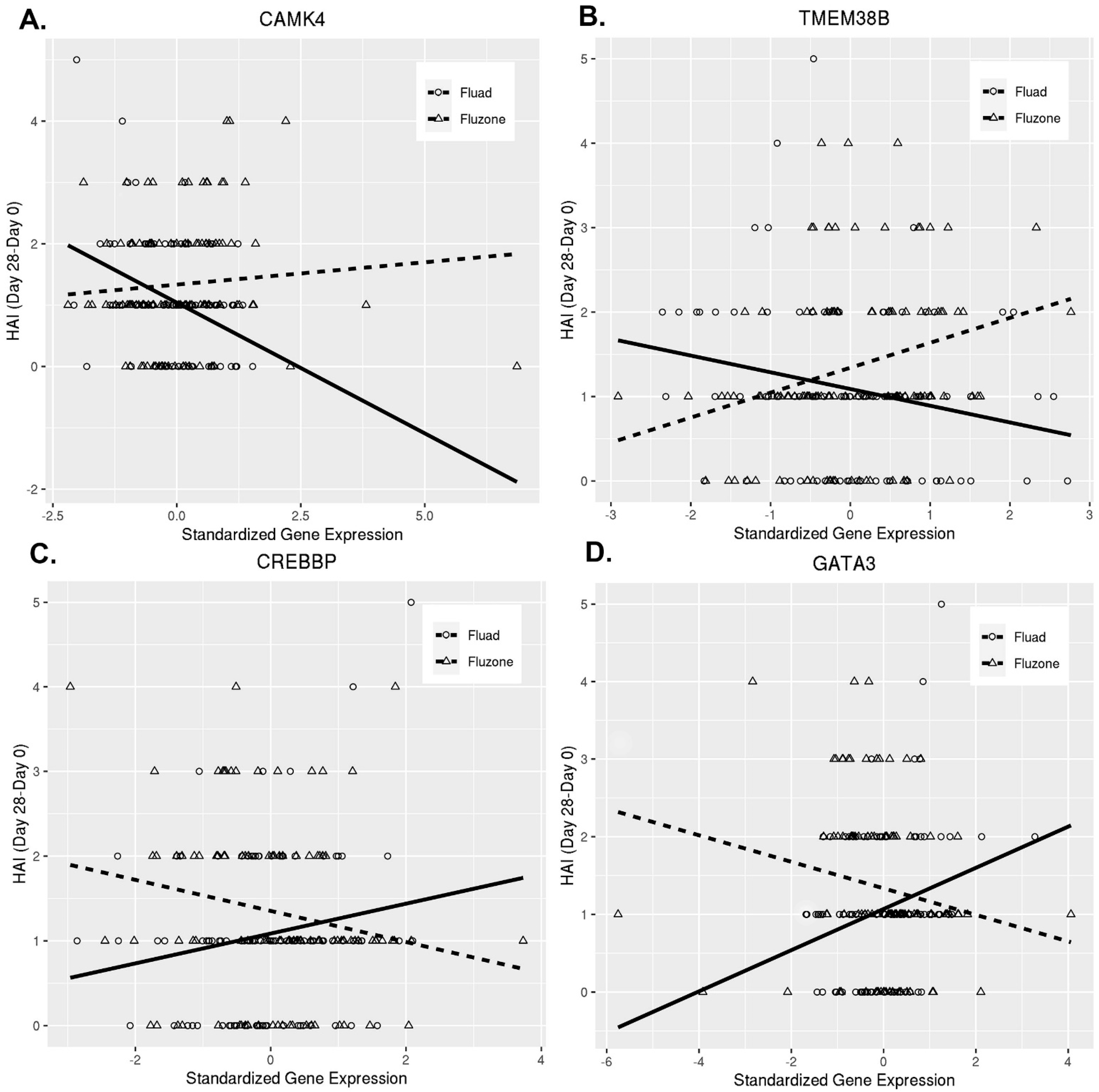
First, we explored the influence of CD4+ T cell transcriptomic changes over time (Day 28/Day 0 gene expression changes—interaction model 1) on HAI antibody titer following vaccination. This model identified 128 genes that meet our significance criteria (Table 2, Supplementary Figure S2, Supplementary Table S1). The majority of these genes/factors have no known direct immune function, but are involved in cellular processes in T cells, such as cell signaling, metabolism, transcription, and membrane trafficking, with a plausible or established link to immune processes (Table 2, Supplementary Table S1). Among our major significant findings, we identified CAMK4, which encodes the calcium-/calmodulin-dependent serine/threonine kinase IV (CaMKIV), (p = 0.0008, q = 0.1575, Table 2). Another significant gene of interest (TMEM38B) is linked to CAMKIV function and encodes a cation channel protein involved in the regulation of intracellular calcium [29]. These observations are consistent with the reported role of CAMKIV in the development of humoral immunity after influenza vaccination in previous system vaccinology studies [30,31,32]. Similar to these previous reports, we found that CAMK4 gene expression was negatively associated with antibody titer in our overall study cohort (gene estimate −0.4262, p = 0.0006, data not shown), but also demonstrated that this gene/factor is likely involved in the differential activation of T cell help by vaccine type (Figure 2). Figure 2 visualizes the differential effect of CAMK4 and several functionally-related genes on the immune outcome in the two vaccine groups. These significant genes were selected for display from interaction model 1, because they were functionally linked and previously reported (for CAMK4) to influence immune responses following influenza vaccination [30,31,32]. The negative association of the CAMK4 gene expression change with the immune outcome was quite pronounced for the Fluad vaccine. Conversely, there was a very slight positive association for the Fluzone vaccine (Figure 2). Of note, similar differential patterns were observed for other important genes, as well (Figure 2). Slightly below our significance/false discovery rate thresholds (with p = 0.0086, q = 0.2562, Table 2), we also identified a third CAMKIV-linked gene/factor, the CREBBP/CREB binding protein (CBP), which is a transcriptional coactivator of CREB (a master regulator of transcription in T cells) regulated by nuclear Ca2+ and CaMKIV [33].
Among the top 20 genes (Table 2), we also identified four members of the solute carrier family proteins (SLC2A11, SLC38A5, SLC25A53, SLC23A3) that are metabolic regulators of T cell differentiation and function [29] and have a role in innate and inflammatory responses in macrophages [34,35]. Other interesting genes with a role in T cell differentiation/activation/function from interaction model 1 include: PLEKHG5/pleckstrin homology and RhoGEF domain containing G5 (a protein that activates the NF-kappa-B signaling pathway); PIAS3/protein inhibitor of activated STAT 3; TRAF7/TNF receptor associated factor 7; GATA3/GATA binding protein 3 (transcriptional regulator of Th2 differentiation/function); and IL36A/interleukin 36 alpha (a protein that can activate NF-kappa-B and MAPK signaling pathways, involved in inflammation (Table 2). All significant genes from interaction model 1 are listed in Supplementary Table S1.
Second, we explored the interaction of the genes by vaccine type, for Day 0 (baseline) gene expression (interaction model 2) or Day 28 gene expression (interaction model 3) on the antibody response/HAI titer (Day 28–Day 0) (Table 3). Although we observed a few interesting immune signaling and regulatory factors among our top findings from these models (i.e., PIAS3/protein inhibitor of activated STAT 3 (also identified in interaction model 1, described above); NLRX1/NLR family member X1, a versatile immune regulatory protein [36]; and POGLUT1/ protein O-glucosyltransferase 1, an important regulator of Notch signaling, see Table 3), the resultant findings did not meet our statistical threshold and must be interpreted with caution.

3.3. Biological Pathways and Immune Functions in T Cells Associated with the Immune Response by Vaccine Type

To better understand the biological processes and pathways controlled by the differentially expressed genes in CD4+ T cells between the Fluad and Fluzone vaccine recipients, we performed pathway enrichment analysis on all the genes from interaction model 1. This analysis identified pathways/biological functions that are significantly represented in our results, capturing the effects of genes/factors below the significance threshold. We used pathway definitions from MSigDB’s index of KEGG canonical pathways (see Statistical Methods) [37]. In the enrichment analysis, we used a significance/false discovery rate threshold of q ≤ 0.05 (Table 4) for pathway identification. We found 42 significantly enriched pathways, of which 7 have been associated with non-influenza viral infections and mostly included overlapping inflammatory and innate genes (Table 4). A total of 11 of the 42 significantly enriched pathways were directly related to T cell differentiation, activation, and/or function, with a variety of signal transduction, transcription-related, effector, and other genes involved in Th1/Th2 cell differentiation, TNF signaling pathway, Notch signaling pathway, MAPK signaling pathway, IL-17 signaling pathway, calcium signaling pathway, regulation of transient receptor potential (TRP) cation channels (stress-sensitive Ca2+-permeable ion channels), rap1/GTPase signaling pathway (important for the interaction between T cells and APCs, finetuning TCR signaling, and T cell responses) [38], and tight junction and apoptosis (Table 4, T cell-related pathways highlighted in bold). Significantly enriched pathways based on genes from interaction models 2 and 3 are presented in Supplementary Table S2. Pathways linked to T cell activity, such as Th1/Th2 cell differentiation, Th17 cell differentiation, and multiple signaling pathways, were also found to be enriched (Supplementary Table S2).

4. Discussion

High-dose Fluzone and adjuvanted Fluad vaccines are known to induce a more robust humoral immune response in older adults than conventional influenza vaccines, with a consequent increase in protection against influenza. However, little is known about T cell transcriptional signatures shaping the humoral response to these vaccines in older adults, who are at risk of immunosenescence-related decrease in immune function (i.e., have suboptimal vaccine-induced immunity) and at higher risk of influenza and influenza-linked complications. The major focus of our study was to identify transcriptional immune signatures in the T helper cell compartment and the possible mechanisms underlying differences in vaccine-induced humoral immunity between the Fluzone and Fluad vaccines in older adults (≥65 years old). We hypothesized that T cell signaling and function play an essential role in those differences and employed an immune response modeling approach using the interaction of CD4+ T cell gene expression by vaccine type.
The goal of our research efforts was to find T cell genes differentially influencing the immune response/antibody titer in the two vaccine groups. Our per-gene modeling efforts identified at least three functionally-linked proteins, i.e., the multifunctional CaMKIV kinase, one of its regulators/activators (transmembrane protein 38B, involved in maintenance of intracellular Ca2+ release), and its immediate interacting partner (the CREB binding protein/CBP), as pivotal players in the T-cell signaling events and transcriptional regulation in CD4+ T cells, thus resulting in antigen-specific activation of T cells and differential functional activity impacting humoral immunity. Importantly, we did observe a differential effect of other identified genes in each vaccine group, which significantly influenced the HAI antibody titer (Figure 2), as expected from our appied modeling strategies.
A different approach (microarray-based gene expression on RNA extracted from PBMCs) has been previously used to compare the immune responses in subjects immunized with either trivalent inactivated influenza vaccine or live attenuated influenza vaccine and led to the discovery of the role of CaMKIV kinase (gene expression at day 3) in the negative regulation of influenza vaccine humoral immunity [30,31,32]. The role of CaMKIV in influenza vaccine immunity was validated in vivo, in a CaMKIV-deficient mouse model, in which influenza vaccination led to higher antibody titers [30]. Furthermore, CAMK4 was identified as one of the top genes of a geneset/cluster (linked to T cell activation) that was associated with reduced cellular immune responses (IFN-γ, IL-2 cytokine secretion) after influenza vaccination in 138 healthy older adults [39]. Gene signatures (derived from gene expression in PBMCs) linked to innate immune function (such as the Toll-like receptor-linked immune signature, inflammatory monocyte-linked signature, antiviral interferon signature, and dendritic cell function-linked signature) were found to be correlated with HAI antibody titer at day 28, when comparing trivalent inactivated influenza vaccine with MF59-adjuvanted trivalent inactivated vaccine in 14- to 24-months old children [40]. Importantly, the MF59-adjuvanted vaccine was demonstrated to induce a higher frequency of antigen-specific polyfunctional CD4+ T cells in the latter study [40], although the underlying mechanism and/or link to humoral immunity remains unknown. Our study identified pathways and functions linked to adaptive immunity/T cell immune fuinction, rather than innate immunity, (e.g., T cell differentiation, T cell activation, and T cell-linked signaling) to influnece the antibody response induced by MF59-adjuvanted vs. high-dose influenza vaccine in older adults. This suggests that differences of induced immune responses to these vaccines may vary by age, most likely due to differences in immune system components/function and immune memory in different age groups (older adults vs. very young children). Furthermore, non-specific vaccine components, such as adjuvants, may have differential effect on immune function dependent on age.
Our findings (CAMKIV, TMEM38B, CBP) are functionally linked to the downstream regulation/activation of the CREB gene, which encodes for the cAMP-responsive element binding protein (CREB), a transcription factor involved in different cellular responses, such as cell proliferation, cell survival, cell differentiation, and effector functions [41]. It is often triggered by extracellular stimuli (e.g., glutamate, growth, and inflammatory stimuli), activated by phosphorylation to initiate, carry forward, and facilitate transcriptional programmes of immune genes (among other genes) containing cAMP-responsive elements. Many T cell function-specific genes also contain cAMP-responsive elements (e.g., IL2, IL10, TNF, TCRA/TCRα, TCRB/TCR Vβ, CD3D/CD3δ, CD8A/CD8α, CD25/IL2RA, IL2RG), suggesting that CREB is a master regulator of transcriptional activity in T cells, with a major role in Th1, Th2, and Th17 differentiation, the generation/maintenance of Tregs, and T cell effector function [41]. Its role in T cells is illuminated in transgenic CREB mutant mice with dominant negative/transcriptionally inactive CREB, where defects in T cell proliferation, T cell function (IL-2 production), and apoptosis were demonstrated [42]. Similar models demonstrate defects in T helper effector function (IL-4 and IFN-γ production), with failure to mount an antigen-specific antibody response [43]. Recent findings in murine models also point to the instrumental role of CAMKIV for the expansion of Tfh lymphocytes and for the regulation of humoral immune responses through control over the BCL6 gene expression [44].
The optimal activation/phosphorylation and transcription factor binding of CREB during T cell activation requires (in addition to the activation of the kinase cascade PKA, PKC, MAPK ERK1/2, p38, and MSK1/2 [41,45,46]) the engagement of both CD3 and CD28, as well as the recruitment/binding of the CREB-binding protein/CBP to CREB, which, in turn, is thoroughly dependent on p38 MAPK and CaMKIV activation [46]. In addition, both kinases are engaged in CREB phosphorylation, and CaMKIV alone is able to activate/phosphorylate CBP (controlled in addition by nuclear Ca2+) at Ser301 [33,46,47]. Thus, CAMKIV and its functionally linked partners (CBP and transmembrane protein 38B) are instrumental for the CREB-based transcriptional activity in T cells.
CaMKIV kinase signals via the calcium-dependent CaMKK-CaMK4 cascade and is activated upon increase of the intracellular Ca2+ concentration (controlled by proteins such as the transmembrane protein 38B/TMEM38B) and calmodulin. As described above, its function/expression is indispensable for the optimal stimulation of CREB/CBP-activated transcriptional programs in T cells, and in particular, CD4+ T cells with a memory phenotype [48]. In addition to the above-mentioned downstream effects, transcription factor CREB is essential for innate immune function through the inhibition of NF-kB activation in monocytes/macrophages to limit inflammation and promote survival, as well as for survival of dendritic cells upon TLR stimulation [41,49].
Our results also emphasize the significance of other T cell-specific transcription factors from our per-gene interaction model 1, such as the GATA binding protein 3, a major regulator of Th2 T cell differentiation, which regulates the expression of Th2 cytokines [50], and/or other regulatory factors and signal transducers involved in MAPK and NF-kB signaling, Notch signaling, Jak-STAT signaling, and TNF signaling. Consistent with this, our pathway enrichment analysis points to most of these pathways and biological processes and, in particular, to pathway/biological functions linked to the CaMKIV-CBP-CREB activation and downstream effects in T cells (calcium signaling pathway, regulation of TRP Ca2+-permeable cation channels, IL-17 signaling pathway, and Th1/Th2 differentiation, among others).
The strengths of our study include the randomized study design, as well as the use of a high-dimensional dataset linked to a specific cell type—purified CD4+ T cells. Of the three models we used in our analysis approach, the most significant and biologically relevant findings come from interaction model 1, where we assessed the effect of transcriptomic changes over time (rather than baseline/Day 0 and Day 28 gene expression).
The major limitation is the possibility of false positive findings, which is addressed by using p-value/q-value thresholds. The potential for type II error also exists, given the relatively limited numbers of individuals studied, and our results may or may not be generalizable to other races and/or ethnicities, other than the population we studied. Regardless, our analytical approach allowed us to identify transcriptomic signatures that modify vaccine-induced immunity of the two influenza vaccines we studied, leading to a better biologic understanding. The differential effect of some of the identified genes (Figure 2) on immune function/outcome in the two vaccine groups is unclear mechanistically, but could be linked to the differential trigerring (vs. no influence for example) of specific immune pathways (e.g., calcium signaling and Ca2+ signaling-linked T cell activation) by an adjuvant or higher antigen dose. For example, it is demonstrated that MF59 induces the release of extracellular ATP, which, in turn, can influence the transport of Ca2+ against elecrochemical gradient (which is ATP-dependent) and, thus, have an impact on calcium signaling and T cell activation [51,52]. An important limitation in our study is the lack of functional validation of our major targets and pathways, which is planned for future studies.
Collectively, our study identified important T cell factors and functions regulating influenza vaccine humoral immunity by vaccine type and highlinging possible differences in their mechanism of action. Further validation and new analyses regarding these unique data are in progress, including the modeling of cellular immune response outcomes following vaccination.

Supplementary Materials

The following supporting information can be downloaded at: https://www.mdpi.com/article/10.3390/v14122763/s1, Figure S1: Antibody response by vaccine type; Figure S2: Significant findings from interaction model 1; Table S1: Influence of vaccine type and CD4+ T cell gene expression change on Ab response to influenza vaccination - full list of significant genes from interaction model 1; Table S2: Enriched biological pathways/processes involved in the immune outcome by vaccine type (from interaction model 2 [Day 0 gene expression effect] and from interaction model 3 [Day 28 gene expression effect])

Author Contributions

I.H.H., I.G.O., G.A.P., and R.B.K. were involved in the conceptualization of the study. I.H.H., H.Q.Q., I.G.O., R.B.K., and D.E.G. were involved in the data acquisition, methodology of the study and in the visualization of the study results. K.M.G. and D.E.G. performed statistical analyses. R.B.K. and G.A.P. were involved in the funding acquisition and project administration. All authors were involved in the writing, editing, and revising of the manuscript. All authors have read and agreed to the published version of the manuscript.

Funding

This research was funded by the National Institute of Allergy and Infectious Diseases of the National Institutes of Health with grant number R01AI132348, the Center for Influenza Vaccine Research for High Risk Populations (CIVR-HRP) with contract number 75N93019C00052 (CIVIC). The content is solely the responsibility of the authors and does not necessarily represent the official views of the National Institute of Health.

Institutional Review Board Statement

The study was conducted in accordance with the Declaration of Helsinki, and approved by the Mayo Clinic Institutional Review Board (IRB# 17-010601).

Informed Consent Statement

Written, informed consent was obtained from each subject at the time of enrollment for this study.

Data Availability Statement

All data included in the current study are available from the corresponding author upon reasonable request.

Acknowledgments

We would like to thank all of the participants in this study. We are grateful to financial support from NIAID and CIVR-HRP.

Conflicts of Interest

Poland is the chair of a Safety Evaluation Committee for novel investigational vaccine trials being conducted by Merck Research Laboratories. Poland provides consultative advice to: AiZtech; AstraZeneca UK Limited; Eli Lilly and Company; Emergent Biosolutions; Exelixis, Inc.; Genevant Sciences, Inc.; GlaxoSmithKline; Janssen Global Services, LLC; Krog & Partners, Inc.; Medicago USA; Merck & Co., Inc.; Moderna; Novavax; Pfizer-BNT; Regeneron Pharmaceuticals, Inc.; Sanofi; Syneos Health; Valneva and Vyriad. Poland is an adviser to the White House and World Health Organization on Covid-19 vaccines and monkeypox. Drs. Poland and Ovsyannikova hold patents related to vaccinia and measles peptide vaccines. Drs. Kennedy, Poland, and Ovsyannikova hold a patent related to vaccinia peptide vaccines. Drs. Poland, Kennedy, Ovsyannikova, and Haralambieva hold a patent related to the impact of single nucleotide polymorphisms on measles vaccine immunity. Drs. Poland, Kennedy, and Ovsyannikova have received grant funding from ICW Ventures for preclinical studies on a peptide-based COVID-19 vaccine. Kennedy has received funding from Merck Research Laboratories to study waning immunity to mumps vaccine. Kennedy also offers consultative advice on vaccine development to Merck & Co. and Sanofi Pasteur. These activities have been reviewed by the Mayo Clinic Conflict of Interest Review Board and are conducted in compliance with Mayo Clinic Conflict of Interest policies. This research has been reviewed by the Mayo Clinic Conflict of Interest Review Board and was conducted in compliance with Mayo Clinic Conflict of Interest policies.

References

  1. Available online: https://www.cdc.gov/flu/about/burden/index.html (accessed on 5 December 2022).
  2. Estimates of deaths associated with seasonal influenza—United States, 1976–2007. MMWR Morb. Mortal. Wkly. Rep. 2010, 59, 1057–1062.
  3. Reed, C.; Chaves, S.S.; Daily Kirley, P.; Emerson, R.; Aragon, D.; Hancock, E.B.; Butler, L.; Baumbach, J.; Hollick, G.; Bennett, N.M.; et al. Estimating influenza disease burden from population-based surveillance data in the United States. PLoS ONE 2015, 10, e0118369. [Google Scholar] [CrossRef] [PubMed]
  4. Zhou, H.; Thompson, W.W.; Viboud, C.G.; Ringholz, C.M.; Cheng, P.Y.; Steiner, C.; Abedi, G.R.; Anderson, L.J.; Brammer, L.; Shay, D.K. Hospitalizations associated with influenza and respiratory syncytial virus in the United States, 1993–2008. Clin. Infect. Dis. 2012, 54, 1427–1436. [Google Scholar] [CrossRef] [PubMed] [Green Version]
  5. Available online: https://www.cdc.gov/flu/highrisk/65over.htm (accessed on 5 December 2022).
  6. World Health Organization. Global Influenza Strategy 2019–2030; World Health Organization: Geneva, Switzerland, 2019. [Google Scholar]
  7. Goodwin, K.; Viboud, C.; Simonsen, L. Antibody response to influenza vaccination in the elderly: A quantitative review. Vaccine 2006, 24, 1159–1169. [Google Scholar] [CrossRef] [PubMed]
  8. Haralambieva, I.H.; Painter, S.D.; Kennedy, R.B.; Ovsyannikova, I.G.; Lambert, N.D.; Goergen, K.M.; Oberg, A.L.; Poland, G.A. The impact of immunosenescence on humoral immune response variation after influenza A/H1N1 vaccination in older subjects. PLoS ONE 2015, 10, e0122282. [Google Scholar] [CrossRef]
  9. DiazGranados, C.A.; Dunning, A.J.; Kimmel, M.; Kirby, D.; Treanor, J.; Collins, A.; Pollak, R.; Christoff, J.; Earl, J.; Landolfi, V. Efficacy of high-dose versus standard-dose influenza vaccine in older adults. N. Engl. J. Med. 2014, 371, 635–645. [Google Scholar] [CrossRef]
  10. Domnich, A.; Arata, L.; Amicizia, D.; Puig-Barberà, J.; Gasparini, R.; Panatto, D. Effectiveness of MF59-adjuvanted seasonal influenza vaccine in the elderly: A systematic review and meta-analysis. Vaccine 2017, 35, 513–520. [Google Scholar] [CrossRef]
  11. Frey, S.E.; Reyes, M.R.; Reynales, H.; Bermal, N.N.; Nicolay, U.; Narasimhan, V.; Forleo-Neto, E.; Arora, A.K. Comparison of the safety and immunogenicity of an MF59®-adjuvanted with a non-adjuvanted seasonal influenza vaccine in elderly subjects. Vaccine 2014, 32, 5027–5034. [Google Scholar] [CrossRef]
  12. Gravenstein, S.; Davidson, H.E.; Taljaard, M.; Ogarek, J.; Gozalo, P.; Han, L.; Mor, V. Comparative effectiveness of high-dose versus standard-dose influenza vaccination on numbers of US nursing home residents admitted to hospital: A cluster-randomised trial. Lancet Respir. Med. 2017, 5, 738–746. [Google Scholar] [CrossRef]
  13. Lapi, F.; Marconi, E.; Simonetti, M.; Baldo, V.; Rossi, A.; Sessa, A.; Cricelli, C. Adjuvanted versus nonadjuvanted influenza vaccines and risk of hospitalizations for pneumonia and cerebro/cardiovascular events in the elderly. Expert Rev. Vaccines 2019, 18, 663–670. [Google Scholar] [CrossRef]
  14. McConeghy, K.W.; Davidson, H.E.; Canaday, D.H.; Han, L.; Saade, E.; Mor, V.; Gravenstein, S. Cluster-randomized Trial of Adjuvanted Versus Nonadjuvanted Trivalent Influenza Vaccine in 823 US Nursing Homes. Clin. Infect. Dis. 2021, 73, e4237–e4243. [Google Scholar] [CrossRef] [PubMed]
  15. Boikos, C.; Fischer, L.; O’Brien, D.; Vasey, J.; Sylvester, G.C.; Mansi, J.A. Relative Effectiveness of Adjuvanted Trivalent Inactivated Influenza Vaccine Versus Egg-derived Quadrivalent Inactivated Influenza Vaccines and High-dose Trivalent Influenza Vaccine in Preventing Influenza-related Medical Encounters in US Adults ≥ 65 Years During the 2017–2018 and 2018–2019 Influenza Seasons. Clin. Infect. Dis. 2021, 73, 816–823. [Google Scholar] [CrossRef] [PubMed]
  16. Haralambieva, I.H.; Eberhard, K.G.; Ovsyannikova, I.G.; Grill, D.E.; Schaid, D.J.; Kennedy, R.B.; Poland, G.A. Transcriptional signatures associated with rubella virus-specific humoral immunity after a third dose of MMR vaccine in women of childbearing age. Eur. J. Immunol. 2021, 51, 1824–1838. [Google Scholar] [CrossRef] [PubMed]
  17. Kaufmann, L.; Syedbasha, M.; Vogt, D.; Hollenstein, Y.; Hartmann, J.; Linnik, J.E.; Egli, A. An optimized hemagglutination inhibition (HI) assay to quantify influenza-specific antibody titers. J. Vis. Exp. 2017, 55833. [Google Scholar] [CrossRef]
  18. Ovsyannikova, I.G.; Salk, H.M.; Kennedy, R.B.; Haralambieva, I.H.; Zimmermann, M.T.; Grill, D.E.; Oberg, A.L.; Poland, G.A. Gene signatures associated with adaptive humoral immunity following seasonal influenza A/H1N1 vaccination. Genes Immun. 2016, 17, 371–379. [Google Scholar] [CrossRef] [Green Version]
  19. Quach, H.Q.; Chen, J.; Monroe, J.M.; Ratishvili, T.; Warner, N.D.; Grill, D.E.; Haralambieva, I.H.; Ovsyannikova, I.G.; Poland, G.A.; Kennedy, R.B. The influence of sex, BMI, and age on cellular and humoral immune responses against measles after a 3rd dose of MMR vaccine. J. Infect. Dis. 2022, jiac351. [Google Scholar] [CrossRef]
  20. Webster, R.; Cox, N.; Stöhr, K. WHO Animal Influenza Manual; WHO/CDS/CSR/NCS/2002.5; World Health Organization: Geneva, Switzerland, 2002. [Google Scholar]
  21. Zimmermann, M.T.; Kennedy, R.B.; Grill, D.E.; Oberg, A.L.; Goergen, K.M.; Ovsyannikova, I.G.; Haralambieva, I.H.; Poland, G.A. Integration of Immune Cell Populations, mRNA-Seq, and CpG Methylation to Better Predict Humoral Immunity to Influenza Vaccination: Dependence of mRNA-Seq/CpG Methylation on Immune Cell Populations. Front. Immunol. 2017, 8, 445. [Google Scholar] [CrossRef] [Green Version]
  22. Haralambieva, I.H.; Ovsyannikova, I.G.; Kennedy, R.B.; Zimmermann, M.T.; Grill, D.E.; Oberg, A.L.; Poland, G.A. Transcriptional signatures of influenza A/H1N1-specific IgG memory-like B cell response in older individuals. Vaccine 2016, 34, 3993–4002. [Google Scholar] [CrossRef] [Green Version]
  23. Kalari, K.R.; Nair, A.A.; Bhavsar, J.D.; O’Brien, D.R.; Davila, J.I.; Bockol, M.A.; Nie, J.; Tang, X.; Baheti, S.; Doughty, J.B. MAP-RSeq: Mayo analysis pipeline for RNA sequencing. BMC Bioinform. 2014, 15, 224. [Google Scholar] [CrossRef] [Green Version]
  24. Hansen, K.D.; Irizarry, R.A.; Wu, Z. Removing technical variability in RNA-seq data using conditional quantile normalization. Biostatistics 2012, 13, 204–216. [Google Scholar] [CrossRef] [Green Version]
  25. Storey, J.D. False Discovery Rate. Int. Encycl. Stat. Sci. 2011, 1, 504–508. [Google Scholar]
  26. Yu, G.; Wang, L.-G.; Han, Y.; He, Q.-Y. clusterProfiler: An R package for comparing biological themes among gene clusters. OMICS J. Integr. Biol. 2012, 16, 284–287. [Google Scholar] [CrossRef] [PubMed]
  27. Benjamini, Y.; Hochberg, Y. Controlling the false discovery rate: A practical and powerful approach to multiple testing. J. R. Stat. Soc. Ser. B 1995, 57, 289–300. [Google Scholar] [CrossRef]
  28. Voigt, E.A.; Ovsyannikova, I.G.; Kennedy, R.B.; Grill, D.E.; Goergen, K.M.; Schaid, D.J.; Poland, G.A. Sex differences in older adults’ immune responses to seasonal influenza vaccination. Front. Immunol. 2019, 10, 180. [Google Scholar] [CrossRef] [Green Version]
  29. Song, W.; Li, D.; Tao, L.; Luo, Q.; Chen, L. Solute carrier transporters: The metabolic gatekeepers of immune cells. Acta Pharm. Sin. B 2020, 10, 61–78. [Google Scholar] [CrossRef]
  30. Nakaya, H.I.; Wrammert, J.; Lee, E.K.; Racioppi, L.; Marie-Kunze, S.; Haining, W.N.; Means, A.R.; Kasturi, S.P.; Khan, N.; Li, G.-M. Systems biology of vaccination for seasonal influenza in humans. Nat. Immunol. 2011, 12, 786–795. [Google Scholar] [CrossRef]
  31. Li, S.; Nakaya, H.I.; Kazmin, D.A.; Oh, J.Z.; Pulendran, B. Systems biological approaches to measure and understand vaccine immunity in humans. Semin. Immunol. 2013, 25, 209–218. [Google Scholar] [CrossRef] [Green Version]
  32. Pulendran, B. Systems vaccinology: Probing humanity’s diverse immune systems with vaccines. Proc. Natl. Acad. Sci. USA 2014, 111, 12300–12306. [Google Scholar] [CrossRef] [Green Version]
  33. Chawla, S.; Hardingham, G.E.; Quinn, D.R.; Bading, H. CBP: A signal-regulated transcriptional coactivator controlled by nuclear calcium and CaM kinase IV. Science 1998, 281, 1505–1509. [Google Scholar] [CrossRef]
  34. Filip, A.M.; Klug, J.; Cayli, S.; Fröhlich, S.; Henke, T.; Lacher, P.; Eickhoff, R.; Bulau, P.; Linder, M.; Carlsson-Skwirut, C.; et al. Ribosomal protein S19 interacts with macrophage migration inhibitory factor and attenuates its pro-inflammatory function. J. Biol. Chem. 2009, 284, 7977–7985. [Google Scholar] [CrossRef] [Green Version]
  35. Roger, T.; Ding, X.; Chanson, A.L.; Renner, P.; Calandra, T. Regulation of constitutive and microbial pathogen-induced human macrophage migration inhibitory factor (MIF) gene expression. Eur. J. Immunol. 2007, 37, 3509–3521. [Google Scholar] [CrossRef] [PubMed]
  36. Nagai-Singer, M.A.; Morrison, H.A.; Allen, I.C. NLRX1 Is a Multifaceted and Enigmatic Regulator of Immune System Function. Front. Immunol. 2019, 10, 2419. [Google Scholar] [CrossRef] [PubMed]
  37. Kanehisa, M.; Goto, S. KEGG: Kyoto encyclopedia of genes and genomes. Nucleic Acids Res. 2000, 28, 27–30. [Google Scholar] [CrossRef] [PubMed]
  38. Katagiri, K.; Hattori, M.; Minato, N.; Kinashi, T. Rap1 functions as a key regulator of T-cell and antigen-presenting cell interactions and modulates T-cell responses. Mol. Cell. Biol. 2002, 22, 1001–1015. [Google Scholar] [CrossRef] [PubMed] [Green Version]
  39. Voigt, E.A.; Grill, D.E.; Zimmermann, M.T.; Simon, W.L.; Ovsyannikova, I.G.; Kennedy, R.B.; Poland, G.A. Transcriptomic signatures of cellular and humoral immune responses in older adults after seasonal influenza vaccination identified by data-driven clustering. Sci. Rep. 2018, 8, 739. [Google Scholar] [CrossRef] [Green Version]
  40. Nakaya, H.I.; Clutterbuck, E.; Kazmin, D.; Wang, L.; Cortese, M.; Bosinger, S.E.; Patel, N.B.; Zak, D.E.; Aderem, A.; Dong, T. Systems biology of immunity to MF59-adjuvanted versus nonadjuvanted trivalent seasonal influenza vaccines in early childhood. Proc. Natl. Acad. Sci. USA 2016, 113, 1853–1858. [Google Scholar] [CrossRef] [Green Version]
  41. Wen, A.Y.; Sakamoto, K.M.; Miller, L.S. The role of the transcription factor CREB in immune function. J. Immunol. 2010, 185, 6413–6419. [Google Scholar] [CrossRef] [Green Version]
  42. Barton, K.; Muthusamy, N.; Chanyangam, M.; Fischer, C.; Clendenin, C.; Leiden, J.M. Defective thymocyte proliferation and IL-2 production in transgenic mice expressing a dominant-negative form of CREB. Nature 1996, 379, 81–85. [Google Scholar] [CrossRef]
  43. Zhang, F.; Rincon, M.; Flavell, R.A.; Aune, T.M. Defective Th function induced by a dominant-negative cAMP response element binding protein mutation is reversed by Bcl-2. J. Immunol. 2000, 165, 1762–1770. [Google Scholar] [CrossRef] [Green Version]
  44. Scherlinger, M.; Li, H.; Pan, W.; Tsokos, M.; Tsokos, G. CaMK4 Controls the Humoral Response via the T Follicular Helper Compartment and Regulates Autoimmunity. Arthritis Rheumatol. 2022, 74 (Suppl. 9), 8524. Available online: https://acrabstracts.org/abstract/camk4-controls-the-humoral-response-via-the-t-follicular-helper-compartment-and-regulates-autoimmunity/ (accessed on 5 December 2022).
  45. Kaiser, M.; Wiggin, G.R.; Lightfoot, K.; Arthur, J.S.; Macdonald, A. MSK regulate TCR-induced CREB phosphorylation but not immediate early gene transcription. Eur. J. Immunol. 2007, 37, 2583–2595. [Google Scholar] [CrossRef] [PubMed]
  46. Yu, C.T.; Shih, H.M.; Lai, M.Z. Multiple signals required for cyclic AMP-responsive element binding protein (CREB) binding protein interaction induced by CD3/CD28 costimulation. J. Immunol. 2001, 166, 284–292. [Google Scholar] [CrossRef] [PubMed] [Green Version]
  47. Hu, S.C.; Chrivia, J.; Ghosh, A. Regulation of CBP-mediated transcription by neuronal calcium signaling. Neuron 1999, 22, 799–808. [Google Scholar] [CrossRef] [PubMed]
  48. Anderson, K.A.; Means, A.R. Defective signaling in a subpopulation of CD4+ T cells in the absence of Ca2+/calmodulin-dependent protein kinase IV. Mol. Cell. Biol. 2002, 22, 23–29. [Google Scholar] [CrossRef] [PubMed] [Green Version]
  49. Illario, M.; Giardino-Torchia, M.L.; Sankar, U.; Ribar, T.J.; Galgani, M.; Vitiello, L.; Masci, A.M.; Bertani, F.R.; Ciaglia, E.; Astone, D.; et al. Calmodulin-dependent kinase IV links Toll-like receptor 4 signaling with survival pathway of activated dendritic cells. Blood 2008, 111, 723–731. [Google Scholar] [CrossRef] [Green Version]
  50. Kanwisher, N.; McDermott, J.; Chun, M.M. The fusiform face area: A module in human extrastriate cortex specialized for face perception. J. Neurosci. 1997, 17, 4302–4311. [Google Scholar] [CrossRef] [Green Version]
  51. Trebak, M.; Kinet, J.P. Calcium signalling in T cells. Nat. Rev. Immunol. 2019, 19, 154–169. [Google Scholar] [CrossRef]
  52. Vono, M.; Taccone, M.; Caccin, P.; Gallotta, M.; Donvito, G.; Falzoni, S.; Palmieri, E.; Pallaoro, M.; Rappuoli, R.; Di Virgilio, F.; et al. The adjuvant MF59 induces ATP release from muscle that potentiates response to vaccination. Proc. Natl. Acad. Sci. USA 2013, 110, 21095–21100. [Google Scholar] [CrossRef] [PubMed]
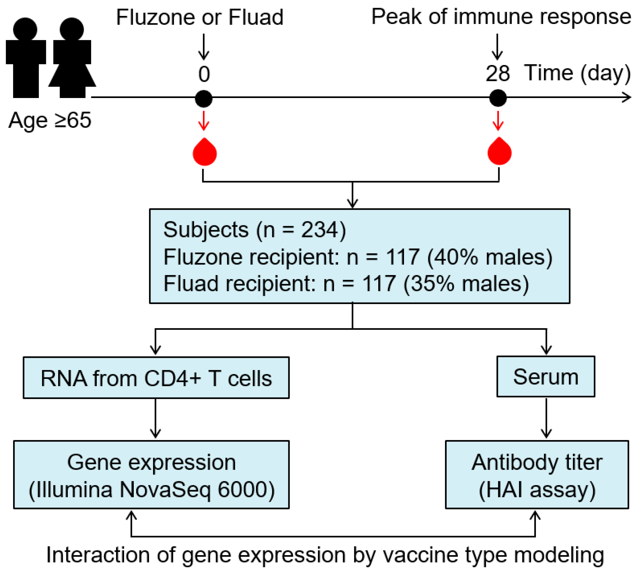
Figure 1. Study design. Of the 234 subjects enrolled (aged ≥65), an equal number (n = 117) of subjects was randomly allocated to receive either high-dose Fluzone or MF59-adjuvanted Fluad influenza vaccine. Blood was sampled before immunization (Day 0/baseline) and 28 days after immunization (Day 28). Humoral immune responses against the influenza A/H3N2 virus were assessed in serum using the hemagglutination inhibition assay (HAI). Gene expression was profiled in purified CD4+ T cells isolated from PBMCs. The effect of the interaction of CD4+ T cell gene expression by vaccine type on antibody titer was evaluated using linear regression models (see Methods).
Viruses 14 02763 g001
Figure 2. Differential effect of selected significant genes on immune outcome in the two vaccine groups. Interaction plots for CAMK4 (A), TMEM38B (B), CREBBP (C), and GATA3 gene (D) demonstrate the differential effect on the immune outcome by vaccine type (i.e., the different trajectories of association for the Fluad (solid line, circles) versus Fluzone (dashed line, triangles) vaccines for the specific genes. The horizontal axis represents the standardized gene expression Day 28/Day 0 change (gene expression minus the mean divided by the standard deviation); a one unit change in the standard gene expression is equivalent to a one standard deviation change in gene expression. The vertical axis is the log2 change in HAI titer from Day 0 to Day 28; a one unit increase in the log2 HAI measure represents a doubling of the HAI titer from Day 0 to Day 28.
Viruses 14 02763 g002
Table 1. Demographic and clinical variables of the study cohort.
VariablesFluad (n = 117)Fluzone (n = 117)Total (n = 234)p-Value
Sex 0.418 *
 Female76 (65.0%)70 (59.8%)146 (62.4%)
 Male41 (35.0%)47 (40.2%)88 (37.6%)
Age at enrollment (years) 0.556 **
 Median71.90071.10071.500
 Q1, Q368.200, 76.50067.700, 75.60067.900, 76.000
Ethnicity 1.000 *
 Non-Hispanic or Latino115 (98.3%)115 (98.3%)230 (98.3%)
 Hispanic or Latino2 (1.7%)2 (1.7%)4 (1.7%)
Race 0.316 *
 Black or African American1 (0.9%)0 (0.0%)1 (0.4%)
 White116 (99.1%)117 (100.0%)233 (99.6%)
Weight (kg) 0.977 **
 Median79.581.180.3
 Q1, Q368.9, 91.168.1, 90.768.5, 91.0
Height (cm) 0.221 **
 Median164.4165.9164.9
 Q1, Q3159.1, 173.5161.2, 175.2160.1, 174.4
BMI 0.333 **
 Median28.728.028.3
 Q1, Q325.3, 31.924.9, 31.125.2, 31.6
HAI titer (Day 28/Day 0) *** 0.062 **
 Median1.0001.0001.000
 Q1, Q30.000, 2.0001.000, 2.0000.000, 2.000
* Pearson’s Chi-squared test. ** Wilcoxon rank sum test. *** Influenza A/H3N2 HAI antibody titer (log2 change in HAI titer from Day 0 to Day 28).
Table 2. Influence of vaccine type and CD4+ T cell gene expression change on Ab response to influenza vaccination (Day 28-Day 0 HAI titer change). Full list of genes is expanded in Supplementary Table S1.
Day 28/Day 0 Gene Expression Change (Interaction Model 1) *
Gene SymbolEntrezgene DescriptionEstimatep-Value **q-Value ***
SLC2A11solute carrier family 2 member 110.60348.19 × 10−60.0701
RP11-779O18.2pre-mRNA processing factor 31−0.56042.65 × 10−50.1137
SLC38A5solute carrier family 38 member 5−0.53240.00010.1575
WDR35WD repeat domain 35 0.53320.00010.1575
APOBRapolipoprotein B receptor−0.53470.00010.1575
CC2D1Acoiled-coil and C2 domain containing 1A−0.49020.00020.1575
MYO9Bmyosin IXB −0.50590.00020.1575
PRKD2protein kinase D2 −0.50160.00020.1575
SLC25A53solute carrier family 25 member 530.48430.00030.1575
TMEM38Btransmembrane protein 38B 0.49310.00030.1575
B4GALT4beta-1,4-galactosyltransferase 4 0.49310.00030.1575
SLC23A3solute carrier family 23 member 30.48300.00030.1575
GORASP1golgi reassembly stacking protein 10.48300.00040.1575
ERLIN2ER lipid raft associated 20.48280.00040.1575
PITPNM1phosphatidylinositol transfer protein membrane-associated 1−0.47530.00040.1575
MAP4microtubule associated protein 4−0.49930.00040.1575
MVB12Bmultivesicular body subunit 12B−0.49040.00040.1575
PKIGcAMP-dependent protein kinase inhibitor gamma0.46340.00050.1575
POP5POP5 homolog, ribonuclease P/MRP subunit0.49470.00050.1575
C17orf51long intergenic non-protein coding RNA 26930.46900.00050.1575
PLEKHG5pleckstrin homology and RhoGEF domain containing G5−0.48090.00050.1575
CAMK4calcium-/calmodulin-dependent protein kinase IV 0.49910.00080.1575
PIAS3protein inhibitor of activated STAT 3−0.44580.00090.1598
TRAF7TNF receptor-associated factor 7−0.43950.00120.1684
COX6B1cytochrome c oxidase subunit 6B10.42540.00170.1783
GATA3GATA-binding protein 3−0.43580.00190.1861
IL36Ainterleukin 36 alpha0.41590.00240.1967
MAP3K13mitogen-activated protein kinase 130.42080.00250.1995
CREBBPCREB-binding protein−0.35940.00860.2562
* Top 20 genes from the model are presented, followed by other significant genes/finding (below the line), with a link to T cell differentiation/activation/function, resulting from this model. ** p-values are from the interaction term of the linear model. *** Q-values were calculated using Storey’s method (see Methods).
Table 3. Influence of CD4+ T cell gene expression (Day 0 or Day 28) on Ab response to influenza vaccination by vaccine type.
Baseline (Day 0) Gene Expression (Interaction Model 2) *
Gene SymbolEntrezgene DescriptionEstimatep-Value **q-Value ***
FAM136Afamily with sequence similarity 136 member A 0.50980.00020.4912
POLA1DNA polymerase alpha 1, catalytic subunit −0.49900.00020.4912
CCER2coiled-coil glutamate rich protein 20.48060.00040.4912
MLLT4/AFDNafadin, adherens junction formation factor 0.48120.00040.4912
AMY2Bamylase alpha 2B0.47630.00040.4912
HECTD3HECT domain E3 ubiquitin protein ligase 3 0.49350.00050.4912
C2orf66chromosome 2 open reading frame 660.46160.00060.4912
PIGWphosphatidylinositol glycan anchor biosynthesis class W 0.46050.00060.4912
SLC23A3solute carrier family 23 member 3−0.46160.00060.4912
BAIAP3BAI1-associated protein 3 0.46220.00070.4912
ITIH4inter-alpha-trypsin inhibitor heavy chain 40.45590.00070.4912
MUSTN1musculoskeletal, embryonic nuclear protein 10.45870.00070.4912
MBTPS2membrane bound transcription factor peptidase, site 2 −0.46600.00080.4912
MYO19myosin XIX0.45090.00080.4912
NLRX1NLR family member X1 −0.45880.00090.4912
RP5-966M1.6SREBF2 antisense RNA 10.45010.00090.4912
PIAS3protein inhibitor of activated STAT 30.45450.00090.4912
C14orf119chromosome 14 open reading frame 119 −0.44410.00100.4912
ALDH8A1aldehyde dehydrogenase 8 family member A1 0.44350.00110.4912
MRPS28mitochondrial ribosomal protein S28−0.45120.00120.4912
Day 28 Gene Expression (Interaction Model 3) *
Gene SymbolEntrezgene DescriptionEstimatep-Value **q-Value ***
COQ4coenzyme Q40.56982.15 × 10−50.2293
EEFSECeukaryotic elongation factor, selenocysteine tRNA-specific−0.53040.00010.3727
NEIL1nei-like DNA glycosylase 10.52430.00010.3727
SLC25A14solute carrier family 25 member 140.50000.00020.4405
CNTD2/CCNPcyclin P0.48960.00030.4405
PSMC3IPPSMC3 interacting protein0.49460.00030.4405
PPM1Nprotein phosphatase, Mg2+/Mn2+-dependent 1N0.48570.00030.4405
MYO19myosin XIX0.48000.00030.4405
BAIAP3BAI1-associated protein 30.46680.00070.7302
BIVMbasic, immunoglobulin-like variable motif containing0.46140.00080.7302
ECHDC2enoyl-CoA hydratase domain containing 20.45860.00080.7302
TTC8tetratricopeptide repeat domain 8 0.44930.00080.7364
POGLUT1protein O-glucosyltransferase 1 0.44510.00090.7598
TRIP6thyroid hormone receptor interactor 60.43160.00110.7658
SAPCD1suppressor APC domain containing 10.44720.00120.7658
CYP4F12cytochrome P450 family 4 subfamily F member 12 0.45410.00140.7658
MFI2/MELTFmelanotransferrin 0.44050.00140.7658
LPPR2/PLPPR2phospholipid phosphatase-related 2 0.42820.00140.7658
FN3KRPfructosamine 3 kinase-related protein 0.41980.00160.7658
RABL2ARAB, member of RAS oncogene family-like 2A0.41910.00160.7658
* Top 20 genes from the model are presented. ** p-values are from the interaction term of the linear model. *** Q-values were calculated using the Storey’s method (see Methods). In summary, our most significant and biologically meaningful transcriptomic signatures resulted from interaction model 1, which uses CD4+ gene expression change (Day 28/Day 0 gene expression change), rather than Day 0 gene expression or Day 28 gene expression.
Table 4. Enriched biological pathways/processes involved in the CD4+ T cell activation by vaccine type (from interaction model 1: Day 28/Day 0 gene expression effect on immune outcome by vaccine type).
KEGG Pathway *Set Sizep-ValueFDR ***
Herpes simplex virus 1 infection3881.48 × 10−60.001
MAPK signaling pathway **1503.11 × 10−60.001
Endocytosis1422.60 × 10−50.003
Human papillomavirus infection1583.77 × 10−50.003
Thyroid hormone signaling pathway694.57 × 10−50.003
Kaposi sarcoma-associated herpesvirus infection1013.19 × 10−40.011
Tight junction **813.38 × 10−40.011
C-type lectin receptor signaling pathway563.54 × 10−40.011
Folate biosynthesis144.15 × 10−40.011
PD-L1 expression and PD-1 checkpoint pathway in cancer454.18 × 10−40.011
Glucagon signaling pathway514.18 × 10−40.011
Rap1 signaling pathway **1004.56 × 10−40.011
Relaxin signaling pathway554.59 × 10−40.011
TNF signaling pathway **626.22 × 10−40.013
GnRH signaling pathway446.59 × 10−40.013
NOD-like receptor signaling pathway **959.54 × 10−40.017
Hepatitis B841.08 × 10−30.018
Glycosaminoglycan biosynthesis—chondroitin/dermatan sulfate161.24 × 10−30.019
Notch signaling pathway **401.34 × 10−30.020
Human T-cell leukemia virus 1 infection1281.45 × 10−30.020
Glutamatergic synapse441.67 × 10−30.022
IL-17 signaling pathway **401.78 × 10−30.023
Transcriptional misregulation in cancer1032.01 × 10−30.025
Human cytomegalovirus infection1122.52 × 10−30.028
Epstein-Barr virus infection1142.66 × 10−30.028
Phospholipase D signaling pathway692.72 × 10−30.028
Viral carcinogenesis1102.96 × 10−30.029
Calcium signaling pathway **902.96 × 10−30.029
Neutrophil extracellular trap formation873.05 × 10−30.029
Estrogen signaling pathway563.14 × 10−30.029
AGE-RAGE signaling pathway in diabetic complications523.50 × 10−30.030
Growth hormone synthesis, secretion, and action554.56 × 10−30.036
Circadian entrainment394.63 × 10−30.036
Serotonergic synapse414.94 × 10−30.038
Insulin signaling pathway725.22 × 10−30.039
RNA polymerase296.12 × 10−30.044
Inflammatory mediator regulation of TRP channels **496.28 × 10−30.044
Fatty acid elongation166.55 × 10−30.045
Th1 and Th2 cell differentiation **507.04 × 10−30.046
Necroptosis767.04 × 10−30.046
Dopaminergic synapse557.21 × 10−30.047
Apoptosis **907.35 × 10−30.047
* From the enriched pathways list, we removed pathways associated with diseases that arose from combinations of other pathways. ** Pathways related to T cell differentiation, activation, and function are bolded. *** FDRs were calculated using Benjamini–Hochberg method (Methods).
Publisher’s Note: MDPI stays neutral with regard to jurisdictional claims in published maps and institutional affiliations.