Article

Influence of Starvation on the Structure of Gut-Associated Bacterial Communities in the Chinese White Pine Beetle (Dendroctonus armandi)

1
College of Forestry, Fujian Agriculture and Forestry University, Fuzhou 350002, Fujian, China
2
College of Forestry, Northwest A&F University, Yangling 712100, Shaanxi, China
*
Author to whom correspondence should be addressed.
Forests 2016, 7(6), 126; https://doi.org/10.3390/f7060126
Submission received: 3 January 2016 / Revised: 27 May 2016 / Accepted: 12 June 2016 / Published: 20 June 2016
(This article belongs to the Special Issue Tree Defense Physiology to Bark Beetles)

Abstract

:
This study investigated the influence of starvation on the structure of the gut bacterial community in the Chinese white pine beetle (Dendroctonus armandi). A total of 14 operational taxonomic units (OTUs0.03) clusters belonging to nine genera were identified. Denaturing gradient gel electrophoresis (DGGE) profiles of bacterial PCR-amplified 16S rRNA gene fragments from the guts of starved male and female adults revealed that the bacterial community diversity increased after starvation. The dominant genus Citrobacter decreased significantly, whereas the genus Serratia increased in both starved female and starved male adults. The most predominant bacterial genus in D. armandi adults was Citrobacter, except for starved male adults, in which Serratia was the most abundant genus (27%). Our findings reveal that starvation affects gut bacterial dynamics in D. armandi, as has been observed in other insect species.

1. Introduction

Bacteria are known to play important roles in the life history of bark beetles [1]. Symbiotic associations have important implications in development, reproduction, and pheromone production, and influence community associations in complex ways [1,2,3,4,5]. Bacterial communities may help bark beetle colonize host trees by providing nutritional supplements that are absent from the host’s diet and lead to cellulose absorption and nitrogen fixation [6,7]. In addition, Bacteria may help bark beetles overcome host resistance through the metabolism of toxins and can enhance protection against natural enemies by keeping out potentially harmful microbes via competition for nutrients [8,9]. Bark beetles with a rapid food throughput can harbor an indigenous microbiota [5]. The composition of the intestinal microbiota co-develops with the host from birth and is subject to a complex interplay that involves genetics, diet, and lifestyle [10].
Bark beetles, in particular those of the genus Dendroctonus, are very important tree mortality agents in coniferous forests [5]. One of the most destructive Dendroctonus species in Asia is the Chinese white pine beetle (Dendroctonus armandi Tsai and Li, Curculionidae: Scolytinae), which attacks live Pinus armandi trees that are over 30 years old, and its outbreaks result in the destruction of millions of P. armandi dating from 1954 in the Qinling and Bashan Mountains in Northern China [11]. Due to the importance of D. armandi, various aspects of the species have been researched, including its intestinal microecology. We recently reported the structure of the gut-associated bacterial communities present during different D. armandi developmental stages [12], and the cellulolytic bacterial community in the intestines of D. armandi larvae was also clarified [6]. However, the effects of harsh environmental conditions, such as starvation, on the gut microbiota of D. armandi remain poorly understood.
Starved insects are often more prone to disease, likely because they have compromised immune defenses. Dillon and colleagues observed increased bacterial diversity in starved locusts, which might improve host defense against enteric threats due to the role of gut bacteria in colonization resistance [13]. Gut bacteria also have the potential to be used as a biological agent in pine forest protection. It has been reported that bacterial symbionts could be genetically modified by genetic engineering techniques to synthesize pest killing proteins or substances [14]. Hence, this study could be useful in the development of biological control agents for bark beetles, which have been understudied [15]. Defining members and clarifying effect factors of gut microflora is the first step to understand the exact role of bacterial communities in bark beetles. We investigated the effects of starvation on the gut-associated bacterial community structure in D. armandi using a method that involves PCR-denaturing gradient gel electrophoresis (DGGE).

2. Materials and Methods

2.1. Insect Collection and Gut Dissection

All of the D. armandi specimens were collected from the bark of infested P. armandi trees at the Huoditang Experimental Forest Station of Northwest A&F University in July 2014. The station is located on the southern slope of the middle Qinling Mountains (33°18′–33°28′ N, 108°21′–108°39′ E) in Shaanxi, China.
Female and male D. armandi adult samples were collected manually using fine forceps. The insect samples were transferred directly from galleries of infested Chinese white pine trees to the laboratory in sterile vials containing sterile moist paper. To separate gut-associated microorganisms from food microorganisms, all insects were maintained at 28 °C under a 12:12 h light/dark cycle for 48 h, as nearly all members of the Dendroctonus genus, including D. armandi, cannot be reared artificially. To investigate the influence of starvation on gut-associated bacteria, another 72-h starvation treatment was applied to half of the insects.
The D. armandi samples were rinsed in 70% ethanol for 3 min for surface sterilization and then rinsed twice in sterile water before dissection. After being placed in 10 mM sterilized phosphate-buffered saline (PBS), the insects were dissected under a stereomicroscope with the aid of insect pins and fine forceps to excise the mid-guts and hindguts from the abdominal segment [16]. Forty beetles in each sample (female, male, starved female and starved male adults) were pooled to be one sample respectively. Guts were excised from each D. armandi sample and transferred to a 1.5 mL microcentrifuge tube respectively, ground 30–50 times with a plastic pestle in liquid nitrogen, vortexed with 500 μL Tris-EDTA (pH 8.0) for 3 min at maximum speed and then centrifuged at low speed (4000 r/min) for 15 s to separate microbial cells from gut wall tissues. The supernatant (containing the bacterial community) was transferred to new tubes for bacterial DNA extraction. Each sample was analyzed in triplicate. All of the procedures used in this experiment were performed in a sterile environment.

2.2. Bacterial DNA Extraction

Bacterial DNA was extracted using an E.Z.N.A. Bacteria DNA Kit (Omega Biotech, Doraville, GA, USA) according to the manufacturer’s instructions and stored at −20 °C until use.

2.3. Nested PCR

Nested PCR was used to increase the resolution yield of DGGE [17,18,19]. The highly variable V3 region of the bacterial 16S rRNA gene was amplified using the fD1 and rP1 primer pair in the first round of PCR and the 341F-GC and 534R primer pair in the second round [20]. Nested PCR amplifications were performed as described by Hu et al. [12], and the PCR products were stored at −20 °C until DGGE analysis.

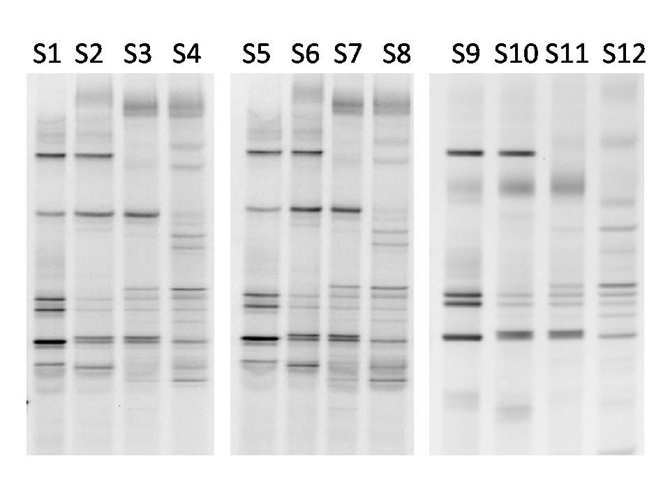
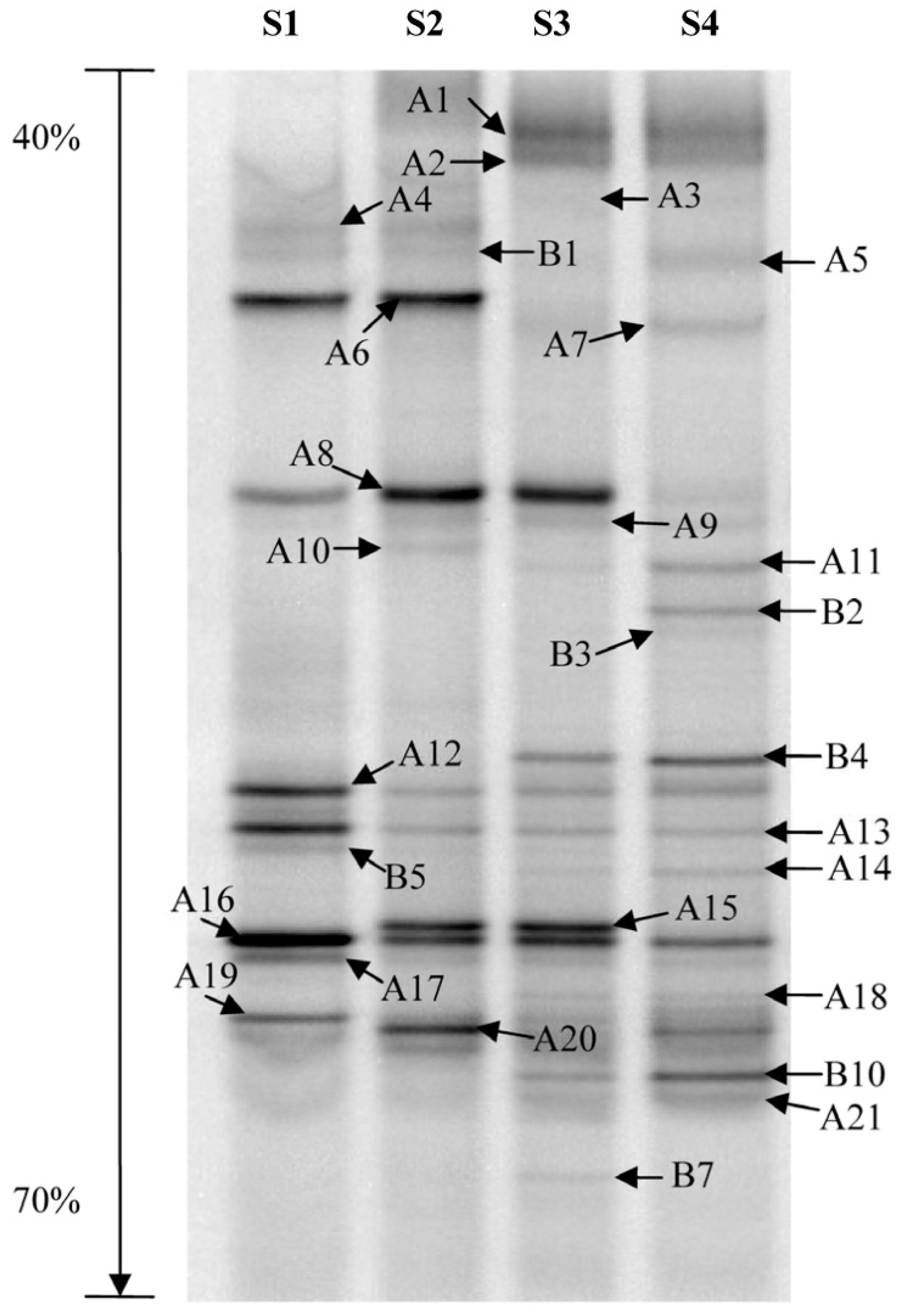
2.4. Denaturing Gradient Gel Electrophoresis (DGGE)

DGGE was performed according to previously described methods [12]. The DCode™ Universal Mutation Detection System (Bio-Rad, Hercules, CA, USA) was used for DGGE analysis. Thirty-five microliters of bacterial nested PCR products from each sample was loaded onto an 8% (w/v) poly-acrylamide (37.5:1 acrylamide/bio-acrylamide) gel containing a linear denaturing gradient of 40% to 70%. A 100% denaturing acrylamide solution is equivalent to 7 M urea and 40% (v/v) formamide [21]. Gel electrophoresis was performed initially at 120 V for 10 min and then at 70 V for 11 h at 58 °C. The polyacrylamide gel was stained with an ethidium bromide solution for 10 min and viewed under UV light using the Gel Doc™ XR System (Bio-Rad, CA, USA).

2.5. DGGE Band Profile Analysis

The dominant DGGE bands were excised immediately from the poly-acrylamide gel with sterile razor blades, and the DNA in the gel bands was eluted using an E.Z.N.A. Mag-Bind Poly-Gel DNA Extraction Kit (Omega, Doraville, GA, USA). The eluted DNA (1 μL) was re-amplified with primer pair 534R and 341F without a GC clamp at the 5′ end, and the products were evaluated by agarose gel electrophoresis. The PCR products were purified using a Gel Extraction Kit (Baitaike Biological Technology, Xi’an, China) and cloned by transformation into Escherichia coli (strain DH5α). Confirmed positive clones were sequenced (Jinsirui Biotechnology, Nanjing, China).
To determine the classification status of gut-associated bacteria, the NCBI and EzTaxon-e databases were examined for sequence matches [22,23]. To analyze the phylogenetic relationships among the gut-associated bacteria, the reliable sequences with a high sequence similarity were selected and downloaded from the databases. All of the sequences were then aligned using MUSCLE [24], and the phylogenetic trees were constructed using the maximum likelihood and neighbor-joining methods in the MEGA 5 software [25]. Bootstrap analysis was performed with 1000 replications to calculate the support for each clade. The bacterial sequences obtained during this study were deposited in the NCBI database under accession numbers KF501438 to KF501472.
The DGGE band profiles were analyzed using Quantity One software (Bio-Rad, USA). Each band was digitized using auto-detection of peak density. Based on the transferred data, the diversity indices were calculated to investigate the dominant bacterial communities and determine how the communities changed in female and male adults and starved female and male adults. Various indices of biodiversity, such as the Shannon-Wiener index (H′), richness (S) and evenness (EH), were calculated from the DGGE patterns according to the following equations:
H = i = 1 s p i ln p i = i = 1 s ( N i / N ) ln ( N i / N )
E H = H / H max = H / ln S
in which S is the number of bands in a lane, Ni is the peak density of the ith band and N is the total peak density of all of the bands in a lane [17,18,19]. Significant differences between means were analyzed using the t-test in SPSS Version 19.0.

2.6. Operational Taxonomic Units and Richness Estimation

The sequences in each phylogenetic tree were formatted as FASTA files and used to construct distance matrices for each library with MOTHUR Version 1.29.0. The distance matrices were used as the input files to define operational taxonomic units (OTUs) based on a similarity distance cutoff of 0.03. OTUs defined by distances of 0.03 generally corresponded to a bacterial species [26]. Sequences that belonged to the same cluster based on the reference of OTUs0.03 were circumscribed with braces in the phylogenic trees and labeled R1–R14 (Figure 1). The Chao index [27] was calculated to measure the absolute value of species richness. The rarefaction curve methodology was used [28,29] to estimate the relationship between the expected OTU richness and sampling depth. Finally, the rarefaction curves were generated using SigmaPlot Version 10.1.

3. Results

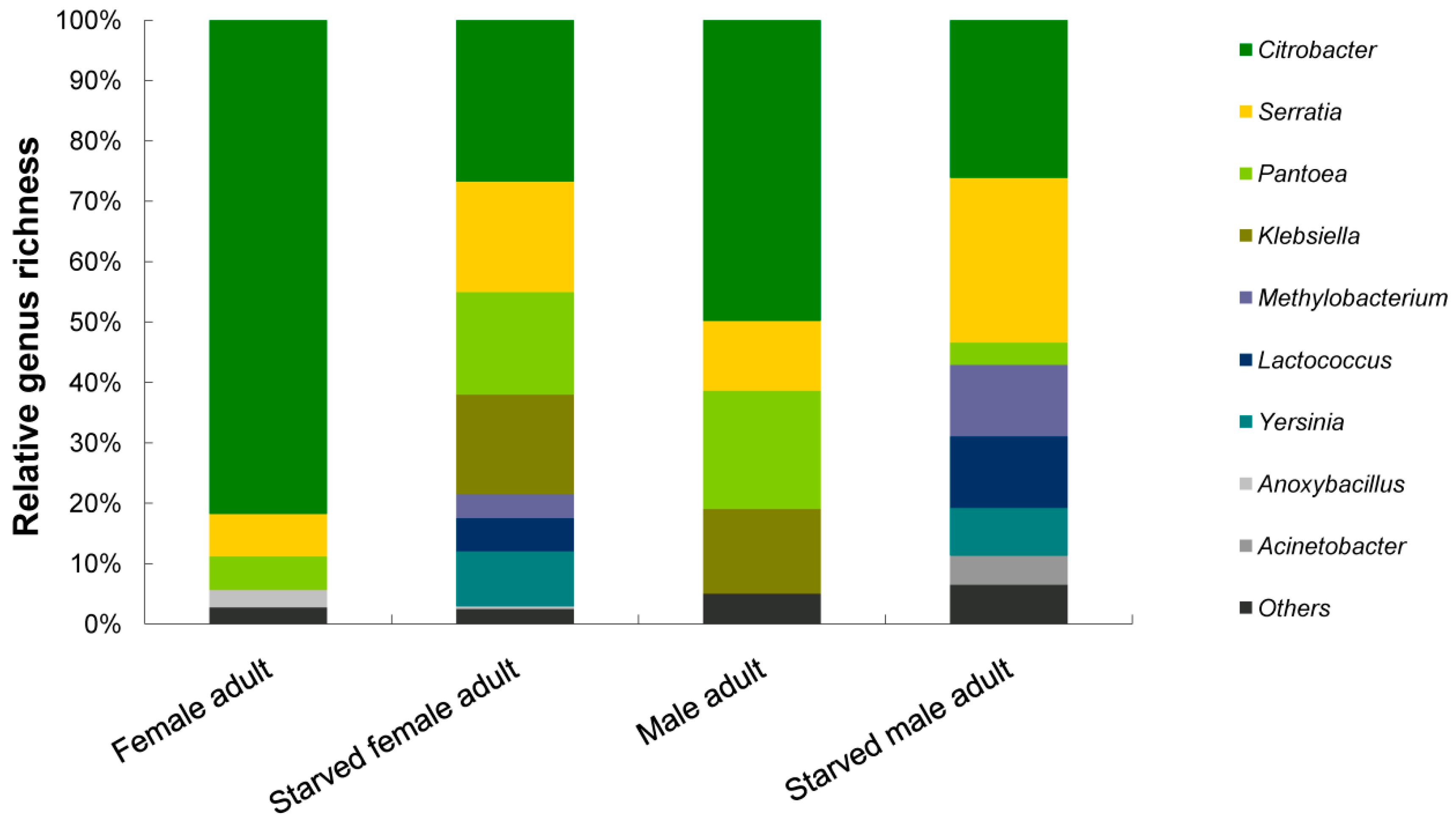
3.1. Bacterial Diversity Analysis

The number, density and composition of bacterial DGGE bands differed between fed and starved adult samples (Figure 2, Figure S1). Bacterial diversity was analyzed based on the peak density of the bands in this DGGE profile. Diversity indices are useful for estimating diversity in microbial communities (i.e., a higher Shannon-Wiener index (H′) indicates greater diversity in the bacterial community). The results presented in Table 1 show the bacterial diversity of all of the D. armandi samples analyzed.
The band number and H′ value increased significantly for both female and male adults after 48 h of starvation (p < 0.05, Student’s t-test). The total numbers of sequenced bands were 22 (starved female adult) and 21 (starved male adult), respectively. When comparing the H′ value, there was also no significant difference between the starved female adult (S3) (2.653363) and male adult samples (S4) (2.804207) (p > 0.05, Student’s t-test).

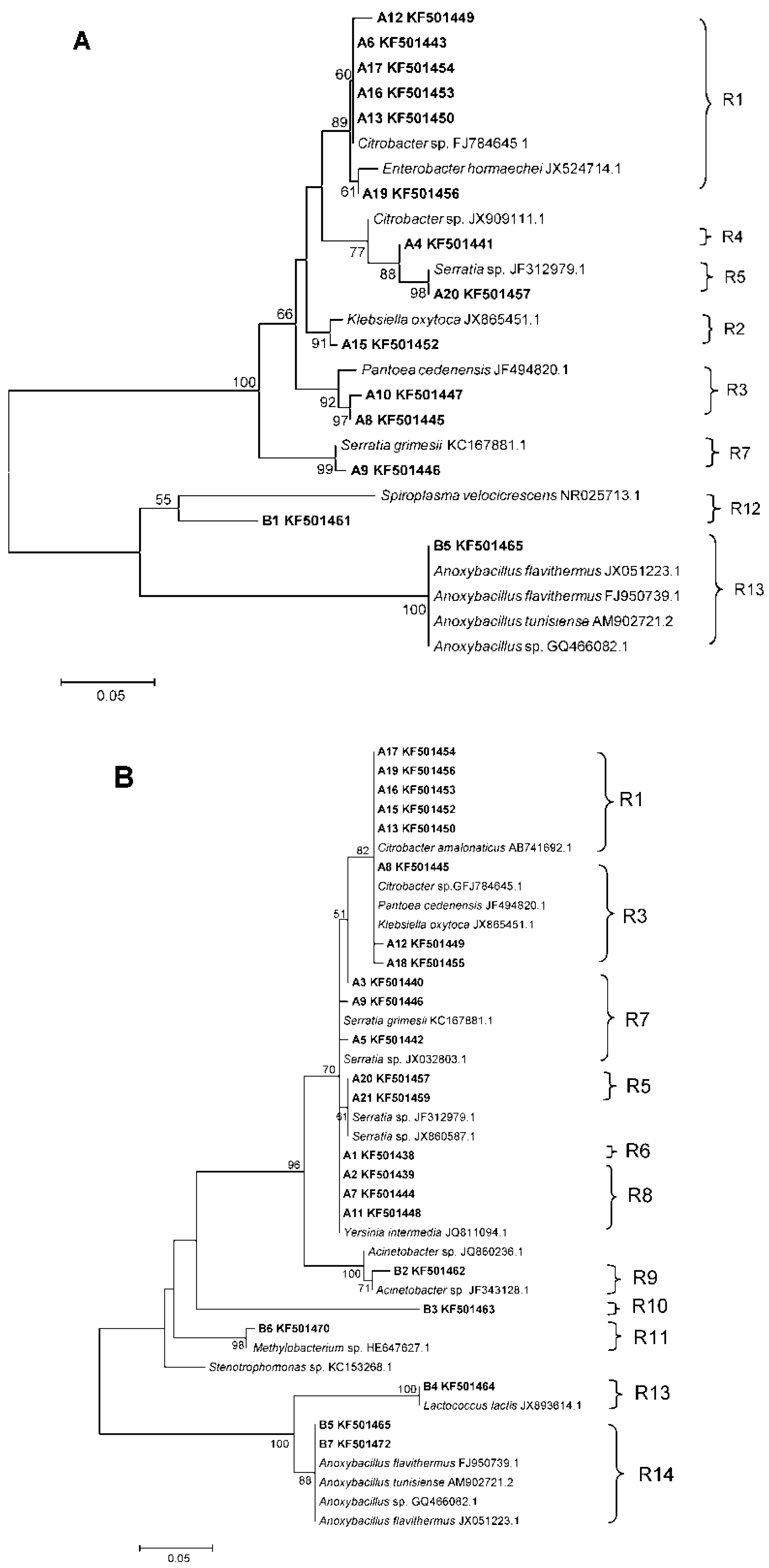
3.2. Phylogenetic Analyses and Dominant Taxa
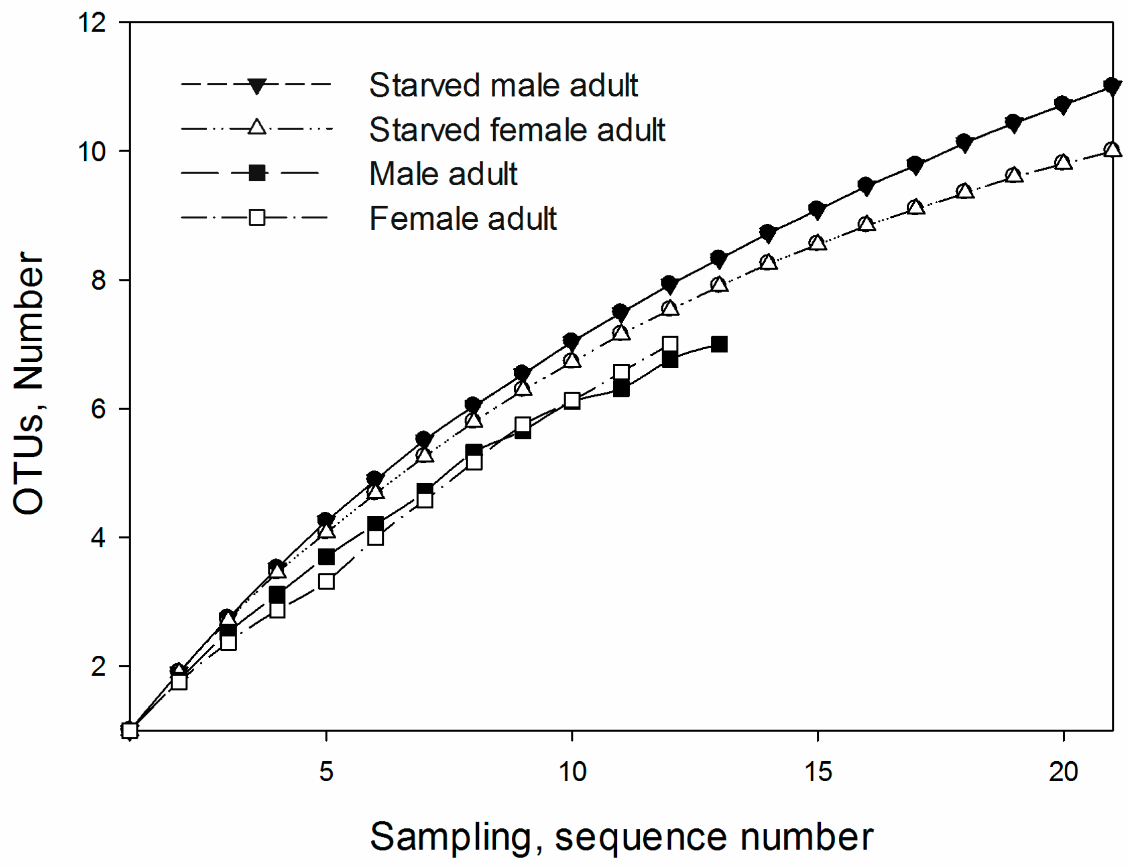
Each of the distinguishable bands in the separation pattern represented an individual bacterial species [30]. In total, 28 different bands were sequenced successfully, and two phylogenetic trees were constructed to represent D. armandi female and male adults and starved female and male adults (Figure 1). Of the 28 sequences, the major species were Proteobacteria (γ-Proteobacteria A1–A21, B2, and B3; α-Proteobacteria B6); the other species were Firmicutes (B1, B4, B5, and B7). Overall, 21 species belong to γ-Proteobacteria, Enterobacteriaceae (A1–A21), as based on the identified bands.
Combining the relative abundance of the bands in each lane with the results of the phylogenetic analysis, nine genera were identified. The most predominant bacterial genus in D. armandi adult guts was Citrobacter; the exception was starved male adults, in which Serratia was the most abundant genus (27%), slightly more abundant than Citrobacter (26%) (Figure 3). Five genera were identified in both female and male adult guts. The composition of the bacterial genera increased after starvation, with nine and eight genera found in the guts of starved female and male adults, respectively.

3.3. OTUs

Fourteen OTUs0.03 clusters (R1‒R14) were defined at a > 97% sequence identity threshold. Most of the OTUs belong to γ-Proteobacteria (10 OTUs) (R1‒R10). Other groups were less abundant, with three OTUs for Firmicutes (R12‒R14) and only one representative OTU for α-Proteobacteria (R11). The members of the largest group, γ-Proteobacteria, belong primarily to the Enterobacteriaceae family, followed by Moraxellaceae and Flavobacteriaceae. Cluster R1 was the most dominant group in all of the samples, with seven species; the closest relatives of those species were γ-Proteobacteria, Enterobacteriaceae, and Citrobacter. The R5 OTUs0.03 cluster was also observed in the gut-associated bacterial communities from all of the samples (Figure 1). The sequences belonging to the R5 cluster were grouped together with R1 on the same phylogenetic branch and were found to be affiliated with Serratia (γ-Proteobacteria, Enterobacteriaceae) including two species. The Chao index was used to better describe the relative bacterial species diversity of the female and male D. armandi adults and the starved female and male D. armandi adults. Data for the OTUs0.03 cluster and estimated relative richness values revealed that the bacterial community structure was simpler in female and male adult guts (Figure 4), with seven OTUs0.03 clusters for female adults (R1, R3, R4, R5, R7, R12, and R13) and seven OTUs0.03 clusters for male adults (R1, R2, R3, R4, R5, R7, and R12). These clusters belong to γ-Proteobacteria (R1-R7) and Firmicutes (R12 and R13) (Figure 1A). The most complex bacterial structure was observed in the guts of starved male adults, with 11 OTUs0.03 clusters related to γ-Proteobacteria (R1, R3, and R5‒R10), α-Proteobacteria (R11) and Firmicutes (R13 and R14). Starved female adults showed 10 OTUs0.03 clusters affiliated with γ-Proteobacteria (R1-R3 and R5-R8), α-Proteobacteria (R11) and Firmicutes (R13, R14) (Figure 1B).

4. Discussion

Bacterial Community Structure

This study provides new insights into the effects of starvation on gut bacterial diversity in D. armandi. Starvation positively impacted the bacterial diversity of both female and male adults. The molecular analysis indicated that the predominant species in the D. armandi bacterial community formed a low complexity group with a different structure after starvation. Low-complexity gut microbial communities have been reported for D. rhizophagus, D. valens and D. ponderosae [31,32,33]. Additionally, as found in all of the D. armandi developmental stages, some taxa were shared among the bacterial communities [12]. Citrobacter (R1 OTUs0.03 clusters) and Serratia (R5 OTUs0.03 clusters), which belong to γ-Proteobacteria, Enterobacteriaceae and are present in D. armandi guts at life different stages, were observed in the gut-associated bacterial communities of both starved female and male adults.
Compared to control adults, the complexity of gut bacteria increased after starvation (Table 1). Ten and 11 OTUs0.03 clusters were observed in starved females and males, respectively, and might be caused by reduced gut peristalsis, which increased bacterial colonization [13,34]. Furthermore, some bacteria originally present in low abundance likely bloomed sufficient amounts to be detected by DGGE. Starvation changes the mid-gut and hind-gut pH values in insects [35], and gut pH can influence bacterial community structure. The predominant bacterial species and relative richness of the predominant taxa also changed after starvation. Citrobacter was the most abundant genus in both female and male adults; the richness of this genus in females and males was 82% and 50%, respectively. In contrast, the richness of the genus Citrobacter was 27% in starved female adults and 26% in starved male adults, and the richness of the genus Serratia was as high as 27% in the guts of starved male adults.
Citrobacter spp. were reported as symbiotic microbes involved in the maintenance of normal nitrogen levels in the guts of termites and fruit flies (Ceratitis capitata) using different metabolic mechanisms, including nitrogen fixation [36,37,38]. Citrobacter freundii was also isolated from D. micans [39]. These potential functions of the Citrobacter genus might be beneficial for D. armandi, which belongs to a group of beetles that spend most of their lives between tree bark and sapwood, where they feed on phloem, which is a poor substrate with low nutritional nitrogen content [7]. Thus, the dominance of Citrobacter spp. in our study suggests that nitrogen fixation might be an important dietary supplement for D. armandi in terms of assimilable nitrogen. In addition, Citrobacter sp. has also been shown to affect insect oviposition and larval development [40], which also explained why Citrobacter was present in female guts at the highest abundance levels. The richness of Citrobacter decreased sharply after starvation treatment in D. armandi adults, which might have occurred because the environment in starved D. armandi guts is too harsh for Citrobacter growth, and the nitrogen-fixing process cannot proceed smoothly without food.
In addition, a notable increase in the abundance of the Serratia genus was observed in the guts of starved D. armandi adults. Members of Serratia secrete secondary metabolites and other biomolecules with antibacterial, antifungal, and antiprotozoal activities to outcompete other microbes. Serratia proteomaculans is associated with D. valens and D. rhizophagus guts, which contribute to insect N balance by using uric acid as sole nitrogen source [32]. This ability is crucial for colonizing niches, especially in poor conditions including poor nutrition, competing microbes and so on [41]. In contrast, Serratia is an opportunistic pathogen and typically inhabits the guts of bark beetles [42,43]. Pathogenic interactions are also an important type of insect-bacteria interactions, similar to symbiotic interactions and typically maintain the balance between the bacteria and insect host in terms of fitness, reproduction, feeding, and other parameters [44]. However, under the stress of starvation, increases in the abundance of the opportunistic pathogen Serratia spp. occurred. Starved insects may be more prone to disease, but the increased diversity of gut bacteria observed in the starved D. armandi adults in this study would improve the insect host defense against enteric threats due to the role of gut bacteria in colonization resistance, as reported for starved locusts [45].
As shown above, the predominant bacterial species in the gut of D. armandi formed a low-complexity group with a bacterial community composition that differed and diversity indices that were significantly increased after starvation. However, no significant effect of sex on gut bacterial diversity or community structure was observed. Gut bacteria have been known to represent one factor that might interfere with D. armandi development, but the nutritional deficiency of the insect host also impact gut-associated bacterial communities. Knowledge of the composition of those communities during periods of nutritional deficiency in the host insect will help us to understand the relationships between gut-associated bacteria and their host D. armandi.
Gut bacteria are a potential contender for bark beetle biocontrol strategies. Based on the recent studies, it has been hypothesized that these symbiotic gut bacteria could be genetically modified by genetic engineering techniques as a host organism to express insect killing toxins or proteins to kill specific pest [46]. Bacterial symbionts can also be used to transform genes changing the insect physiological characteristics. For example, gut bacterium Enterobacter cloacae was used to control Glyphodes pyloalis by transforming the ice nucleation gene, which increased supercooling point and resulted in an increased mortality rate in G. pyloalis [47]. The information presented here should be beneficial in the future bark beetle biocontrol.

Supplementary material

The following are available online at https://www.mdpi.com/1999-4907/7/6/126/s1. Figure S1: DGGE profiles of nested PCR-amplified 16S rRNA gene fragments of bacteria from the D. armandi guts. Lanes S1, S5, S9 female adult; S2, S6, S10 male adult; S3, S7, S11 starved female adult; S4, S8, S12 starved male adult.

Acknowledgments

We acknowledge financial support from the National Natural Science Foundation of China (31170607, 31170567) and, the program for the key discipline of Fujian Agriculture and Forestry University (6112C035005).

Author Contributions

Conceived and designed the experiments: Xia Hu, Ming Li, Feiping Zhang, Hui Chen. Performed the experiments: Xia Hu, Ming Li. Analyzed the data: Xia Hu, Ming Li, Feiping Zhang. Contributed reagents/materials/analysis tools: Xia Hu. Wrote the paper: Xia Hu.

Conflicts of Interest

The authors declare no conflict of interest.

References

  1. Douglas, A.E. The microbial dimension in insect nutritional ecology. Funct. Ecol. 2009, 23, 38–47. [Google Scholar] [CrossRef]
  2. Xu, L.; Lou, Q.; Cheng, C.; Lu, M.; Sun, J. Gut-Associated bacteria of Dendroctonus valens and their involvement in verbenone production. Microb. Ecol. 2015, 70, 1012–1023. [Google Scholar] [CrossRef] [PubMed]
  3. Therrien, J.; Mason, C.J.; Cale, J.A.; Adams, A.; Aukema, B.H.; Currie, C.R.; Raffa, K.F.; Erbilgin, N. Bacteria influence mountain pine beetle brood development through interactions with symbiotic and antagonistic fungi: Implications to climate-driven host range expansion. Oecologia 2015, 2, 1–19. [Google Scholar] [CrossRef] [PubMed]
  4. Audrey-Anne, D.; Amélie, B.; Philippe, C.; Jean-Philippe, B.; Eric, D.; Claude, G. Surveying the endomicrobiome and ectomicrobiome of bark beetles: The case of Dendroctonus simplex. Sci. Rep.-UK 2015, 5, 17190. [Google Scholar]
  5. Six, D.L.; Klepzig, K.D. Dendroctonus bark beetles as model systems for studies on symbiosis. Symbiosis 2004, 37, 1–26. [Google Scholar]
  6. Hu, X.; Yu, J.; Wang, C.; Chen, H. Cellulolytic bacteria associated with the gut of Dendroctonus armandi larvae (Coleoptera: Curculionidae: Scolytinae). Forests 2014, 5, 455. [Google Scholar] [CrossRef]
  7. Morales-Jimenez, J.; Zuniga, G.; Villa-Tanaca, L.; Hernandez-Rodriguez, C. Bacterial community and nitrogen fixation in the red turpentine beetle, Dendroctonus valens Leconte (Coleoptera: Curculionidae: Scolytinae). Microb. Ecol. 2009, 58, 879–891. [Google Scholar] [CrossRef] [PubMed]
  8. Dillon, R.J.; Dillon, V.M. The gut bacteria of insects: Nonpathogenic interactions. Annu. Rev. Entomol. 2004, 49, 71–92. [Google Scholar] [CrossRef] [PubMed]
  9. Cardoza, Y.J.; Klepzig, K.D.; Raffa, K.F. Bacteria in oral secretions of an endophytic insect inhibit antagonistic fungi. Ecol. Entomol. 2006, 31, 636–645. [Google Scholar] [CrossRef]
  10. Nicholson, J.K.; Holmes, E.; Kinross, J.; Burcelin, R.; Gibson, G.; Jia, W.; Pettersson, S. Host-gut microbiota metabolic interactions. Science 2012, 336, 1262–1267. [Google Scholar] [CrossRef] [PubMed]
  11. Chen, H.; Tang, M. Spatial and temporal dynamics of bark beetles in Chinese white pine in Qinling mountains of Shaanxi province, China. Environ. Entomol. 2007, 36, 1124–1130. [Google Scholar] [CrossRef] [PubMed]
  12. Hu, X.; Wang, C.; Chen, H.; Ma, J. Differences in the structure of the gut bacteria communities in development stages of the Chinese white pine beetle (Dendroctonus armandi). Int. J. Mol. Sci. 2013, 14, 21006–21020. [Google Scholar] [CrossRef] [PubMed]
  13. Dillon, R.J.; Webster, G.; Weightman, A.J.; Charnley, A.K. Diversity of gut microbiota increases with aging and starvation in the desert locust. Anton. Leeuw. Int. J. G. 2010, 97, 69–77. [Google Scholar] [CrossRef] [PubMed]
  14. Çelebi, Ö.; Sevim, E.; Sevim, A. Investigation of the internal bacterial flora of Eurygaster integriceps (Hemiptera: Scutelleridae) and pathogenicity of the flora members. Biologia 2014, 69, 1365–1375. [Google Scholar] [CrossRef]
  15. Fettig, C.J.; Hilszczański, J. Management strategies for bark beetles in conifer forests. In Bark Beetles: Biology and Ecology of Native and Invasive Species; Vega, F.E., Hofstetter, R.W., Eds.; Springer: London, UK, 2015; pp. 555–584. [Google Scholar]
  16. Delalibera, I.; Handelsman, J.; Raffa, K.F. Contrasts in cellulolytic activities of gut microorganisms between the wood borer, Saperda vestita (Coleoptera: Cerambycidae), and the bark beetles, Ips pini and Dendroctonus frontalis (Coleoptera: Curculionidae). Environ. Entomol. 2005, 34, 541–547. [Google Scholar] [CrossRef]
  17. Galand, P.E.; Fritze, H.; Yrjala, K. Microsite-Dpendent changes in methanogenic populations in a boreal oligotrophic fen. Environ. Microbiol. 2003, 5, 1133–1143. [Google Scholar] [CrossRef] [PubMed]
  18. Lü, D.; Li, Z.; Qin, S.; Ma, H.; Liu, G. Bacterial community structure in the Cerasus sachalinensis kom. rhizosphere based on the polymerase chain reaction-Dnaturing gradient gel electrophoresis (pcr-dgge) method. Afr. J. Biotechnol. 2013, 10, 13430–13438. [Google Scholar]
  19. Xu, Z.Y.; Tang, M.; Chen, H.; Ban, Y.H.; Zhang, H.H. Microbial community structure in the rhizosphere of Sophora viciifolia grown at a lead and zinc mine of northwest China. Sci. Total Environ. 2012, 435–436. [Google Scholar] [CrossRef] [PubMed]
  20. Muyzer, G.; Hottenträger, S.; Teske, A.; Wawer, C. Denaturing Gradient Gel Electrophoresis of PCR-Amplified 16S rDNA—A New Molecular Approach to Analyse the Genetic Diversity of Mixed Microbial Communities. In Molecular Microbial Ecology Manual; Akkermans, A.D.L., van Elsas, J.D., De Bruijn, F.J., Eds.; Kluwer Academic Publishers: Dordrecht, The Netherlands, 1996; pp. 1–23. [Google Scholar]
  21. Muyzer, G.; de Waal, E.C.; Uitterlinden, A.G. Profiling of complex microbial populations by denaturing gradient gel electrophoresis analysis of polymerase chain reaction-amplified genes coding for 16S rRNA. Appl. Environ. Microbiol. 1993, 59, 695–700. [Google Scholar] [PubMed]
  22. Cole, J.R.; Wang, Q.; Cardenas, E.; Fish, J.; Chai, B.; Farris, R.J.; Kulam-Syed-Mohideen, A.S.; McGarrell, D.M.; Marsh, T.; Garrity, G.M.; et al. The ribosomal database project: Improved alignments and new tools for rRNA analysis. Nucleic Acids Res. 2009, 37, 141–145. [Google Scholar] [CrossRef] [PubMed]
  23. Chun, J.; Lee, J.H.; Jung, Y.; Kim, M.; Kim, S.; Kim, B.K.; Lim, Y.W. Eztaxon: A web-Based tool for the identification of prokaryotes based on 16S ribosomal RNA gene sequences. Int. J. Syst. Evol. Microbiol. 2007, 57, 2259–2261. [Google Scholar] [CrossRef] [PubMed]
  24. Edgar, R.C. Muscle: Multiple sequence alignment with high accuracy and high throughput. Nucleic Acids Res. 2004, 32, 1792–1797. [Google Scholar] [CrossRef] [PubMed]
  25. Tamura, K.; Peterson, D.; Peterson, N.; Stecher, G.; Nei, M.; Kumar, S. Mega5: Molecular evolutionary genetics analysis using maximum likelihood, evolutionary distance, and maximum parsimony methods. Mol. Biol. Evol. 2011, 28, 2731–2739. [Google Scholar] [CrossRef] [PubMed]
  26. Schloss, P.D.; Handelsman, J. Introducing dotur, a computer program for defining operational taxonomic units and estimating species richness. Appl. Environ. Microbiol. 2005, 71, 1501–1506. [Google Scholar] [CrossRef] [PubMed]
  27. Chao, A.; Shen, T.-J. Nonparametric estimation of shannon’s index of diversity when there are unseen species in sample. Environ. Ecol. Stat. 2003, 10, 429–443. [Google Scholar] [CrossRef]
  28. Gotelli, N.J.; Colwell, R.K. Quantifying biodiversity: Procedures and pitfalls in the measurement and comparison of species richness. Ecology letters 2001, 4, 379–391. [Google Scholar] [CrossRef]
  29. Colwell, R.K.; Mao, C.X.; Chang, J. Interpolating, extrapolating, and comparing incidence-Based species accumulation curves. Ecology 2004, 85, 2717–2727. [Google Scholar] [CrossRef]
  30. Cocolin, L.; Aggio, D.; Manzano, M.; Cantoni, C.; Comi, G. An application of PCR-Dgge analysis to profile the yeast populations in raw milk. Int. Dairy J. 2002, 12, 407–411. [Google Scholar] [CrossRef]
  31. Adams, A.S.; Aylward, F.O.; Adams, S.M.; Erbilgin, N.; Aukema, B.H.; Currie, C.R.; Suen, G.; Raffa, K.F. Mountain pine beetles colonizing historical and naive host trees are associated with a bacterial community highly enriched in genes contributing to terpene metabolism. Appl. Environ. Microbiol. 2013, 79, 3468–3475. [Google Scholar] [CrossRef] [PubMed]
  32. Morales-Jimenez, J.; Vera-Ponce de Leon, A.; Garcia-Dominguez, A.; Martinez-Romero, E.; Zuniga, G.; Hernandez-Rodriguez, C. Nitrogen-Fixing and uricolytic bacteria associated with the gut of Dendroctonus rhizophagus and Dendroctonus valens (Curculionidae: Scolytinae). Microb. Ecol. 2013, 66, 200–210. [Google Scholar] [CrossRef] [PubMed]
  33. Winder, R.S.; Macey, D.E.; Cortese, J. Dominant bacteria associated with broods of mountain pine beetle, Dendroctonus ponderosae (Coleoptera: Curculionidae, Scolytinae). J. Entomol. Soc. Br. Columbia 2010, 107. [Google Scholar]
  34. Santo Domingo, J.W.; Kaufman, M.G.; Klug, M.J.; Holben, W.E.; Harris, D.; Tiedje, J.M. Influence of diet on the structure and function of the bacterial hindgut community of crickets. Mol. Ecol. 1998, 7, 761–767. [Google Scholar] [CrossRef]
  35. Heimpel, A.M. The pH in the gut and blood of the larch sawfly, Pristiphora erichsonii (htg.), and other insects with reference to the pathogenicity of Bacillus cereus fr. And fr. Can. J. Zool. 1955, 33, 99–106. [Google Scholar] [CrossRef]
  36. French, J.R.; Turner, G.L.; Bradbury, J.F. Nitrogen fixation by bacteria from the hindgut of termites. J. Gen. Microbiol. 1976, 96, 202–206. [Google Scholar] [CrossRef] [PubMed]
  37. Behar, A.; Yuval, B.; Jurkevitch, E. Enterobacteria-mediated nitrogen fixation in natural populations of the fruit fly Ceratitis capitata. Mol. Ecol. 2005, 14, 2637–2643. [Google Scholar] [CrossRef] [PubMed]
  38. Lilburn, T.G.; Kim, K.S.; Ostrom, N.E.; Byzek, K.R.; Leadbetter, J.R.; Breznak, J.A. Nitrogen fixation by symbiotic and free-living spirochetes. Science 2001, 292, 2495–2498. [Google Scholar] [CrossRef] [PubMed]
  39. Yaman, M.; Ertürk, Ö.; Aslan, İ. Isolation of some pathogenic bacteria from the great spruce bark beetle, Dendroctonus micans and its specific predator, Rhizophagus grandis. Folia Microbiol. 2010, 55, 35–38. [Google Scholar] [CrossRef] [PubMed]
  40. Brand, J.M.; Bracke, J.W.; Markovetz, A.J.; Wood, D.L.; Browne, L.E. Production of verbenol pheromone by a bacterium isolated from bark beetles. Nature 1975, 254, 136–137. [Google Scholar] [CrossRef] [PubMed]
  41. Petersen, L.M.; Tisa, L.S. Friend or foe? A review of the mechanisms that drive Serratia towards diverse lifestyles. Can. J. Microbiol. 2013, 59, 627–640. [Google Scholar] [CrossRef] [PubMed]
  42. Brand, J.M.; Bracke, J.W.; Markovetz, A.J.; Wood, D.L.; Browne, L.E. Production of verbenol pheromone by a bacterium isolated from bark beetles. Nature 1975, 254, 136–137. [Google Scholar] [CrossRef] [PubMed]
  43. Murdoch, S.L.; Trunk, K.; English, G.; Fritsch, M.J.; Pourkarimi, E.; Coulthurst, S.J. The opportunistic pathogen Serratia marcescens utilizes type vi secretion to target bacterial competitors. J. Bacteriol. 2011, 193, 6057–6069. [Google Scholar] [CrossRef] [PubMed]
  44. Sanchez-Contreras, M.; Vlisidou, I. The diversity of insect-bacteria interactions and its applications for disease control. Biotechnol. Genet. Eng. Rev. 2008, 25, 203–243. [Google Scholar] [CrossRef]
  45. Dillon, R.; Vennard, C.; Buckling, A.; Charnley, A. Diversity of locust gut bacteria protects against pathogen invasion. Ecol. Lett. 2005, 8, 1291–1298. [Google Scholar] [CrossRef]
  46. Sevim, A.; Gökçe, C.; Erbaş, Z.; Ozkan, F. Bacteria from Ips sexdentatus (Coleoptera: Curculionidae) and their biocontrol potential. J. Basic Microbiol. 2012, 52, 695–704. [Google Scholar] [CrossRef] [PubMed]
  47. Watanabe, K.; Abe, K.; Sato, M. Biological control of an insect pest by gut-colonizing Enterobacter cloacae transformed with ice nucleation gene. J. Appl. Microbiol. 2000, 88, 90–97. [Google Scholar] [CrossRef] [PubMed]
Figure 1. Phylogenetic trees for the bacterial community in D. armandi guts based on 16S rRNA gene fragments. (A) Neighbor-joining phylogenetic tree for the bacterial community in female and male adult guts using the model (Kimura 2-parameter + G). (B) Maximum likelihood phylogenetic tree of the bacterial community in starved female and male adult guts using the model (Kimura 2-parameter + I). R1–R14 represent different operational taxonomic units (OTUs0.03) clusters obtained with MOTHUR.
Forests 07 00126 g001
Figure 2. Denaturing gradient gel electrophoresis (DGGE) profiles of nested PCR-amplified 16S rRNA gene fragments of bacteria from the D. armandi guts. Lane S1 female adult, S2 male adult, S3 starved female adult, S4 starved male adult. Bands A1–A21 and B1–B7 represent the 16S rRNA gene regions from different bacteria.
Forests 07 00126 g002
Figure 3. Results of genus-level phylogenetic binning of the D. armandi gut-associated bacterial community. Only those genera with greater than 1% representation are shown.
Forests 07 00126 g003
Figure 4. Rarefaction curves for bacterial 16S rRNA sequences in the gut of D. armandi female and male adults and starved female and male adults calculated using MOTHUR OTU0.03. The plot shows the number of new bacterial species as a function of the number of clones sequenced.
Forests 07 00126 g004
Table 1. Richness (S), evenness (EH) and Shannon-Wiener index (H′) for the gut-associated bacteria identified in different samples of D. armandi.
LaneD. armandi sampleSEHH′
S1Female adult120.8542552.122745
S2Male adult140.8853122.336389
S3Starved female adult220.8584042.653363
S4Starved male adult210.9210662.804207

