Article

Synthesis of New RE3+ Doped Li1+xTa1−xTixO3 (RE: Eu, Sm, Er, Tm, and Dy) Phosphors with Various Emission Colors

1
Toyohashi University of Technology, Tempaku, Toyohashi 441-8580, Japan
2
KRI, Inc., Chudoji Minami-machi, Shimogyo-ku, Kyoto 600-8813, Japan
3
Keio University, Yokohama 223-8522, Japan
*
Author to whom correspondence should be addressed.
Materials 2013, 6(7), 2768-2776; https://doi.org/10.3390/ma6072768
Submission received: 19 April 2013 / Revised: 5 May 2013 / Accepted: 8 June 2013 / Published: 11 July 2013
(This article belongs to the Special Issue Luminescent Materials 2013)

Abstract

:
New phosphors with various emission colors for RE3+ doped Li1+xTa1−xTixO3 (LTT) (RE: Eu, Sm, Er, Tm, and Dy) were synthesized by electric furnace at 1423 K for 15 h. The microstructure of the host material and the photoluminescence (PL) property were determined and compared to those of RE3+ doped Li1+xNb1−xTixO3 (LNT). In the LTT phosphor, the highest PL intensity was achieved for the mixture composition Li1.11Ta0.89Ti0.11O3 with a LiTaO3 structure, although it has an M-phase superstructure. In the LTT host material, the effective activators were Eu3+ and Sm3+ ions, in contrast to the LNT host material. Here, we discuss the relationship between PL property and the host material’s structure.

1. Introduction

In the ternary Li2O-M2O5-TiO2 system, Li1+xyM1−x−3yTix+4yO3 (M: Nb or Ta, 0.06 ≤ x ≤ 0.33, 0 ≤ y ≤ 0.17) forms with a superstructure, and this is known as the M-phase. Since the discovery of the M-phase for Li1+xyNb1−x−3yTix+4yO3 (LNT) by Castrejon et al. [1,2], such structures have been investigated [3,4,5,6]. The M-phase superstructure is formed by periodical insertion of an intergrowth layer in the matrix having a trigonal structure. The relationship between dielectric property and period of the intergrowth layer of the M-phase has been studied [7,8]. To apply this unique structure to the host material of a phosphor, new phosphors have been investigated based on LNT synthesized by a conventional electric furnace [9,10]. A homogeneous material was, however, synthesized at 1373 K for 24 h. We have succeeded in synthesizing an LNT solid solution having a superstructure by millimeter-wave heating for only 1 h above 1173 K [11]. The technique has also been applied to the synthesis of rare-earth doped LNT phosphor [12,13]. The photoluminescence (PL) intensity of LNT:Eu3+ at 625 nm was much higher than that of LiNbO3:Eu. [12] Rare-earth-doped LiTaO3 (RE3+: Pr3+ [14], Er3+ [15], Tb3+ [16] Eu3+ [17] and Tm3+ [18]) have also been reported by other groups. Recently, we have reported a new red phosphor based on the quaternary Li1+x (Ta1−zNbz)1−xTixO3 (LTNT, 0.05 ≤ x ≤ 0.25, 0 ≤ z ≤ 0.625 ) solid solution as a host material. The PL intensity of the LTNT:Eu3+ phosphor was found to be dependent on the concentration of Eu3+, Ti4+, Nb5+, and Ta5+ ions [19].
In this work, rare-earth doped Li1+xTa1−xTixO3 (LTT) phosphors with various emission colors were synthesized by an electric furnace and compared to LNT phosphors for annealing condition, host material’s structure, and PL property.

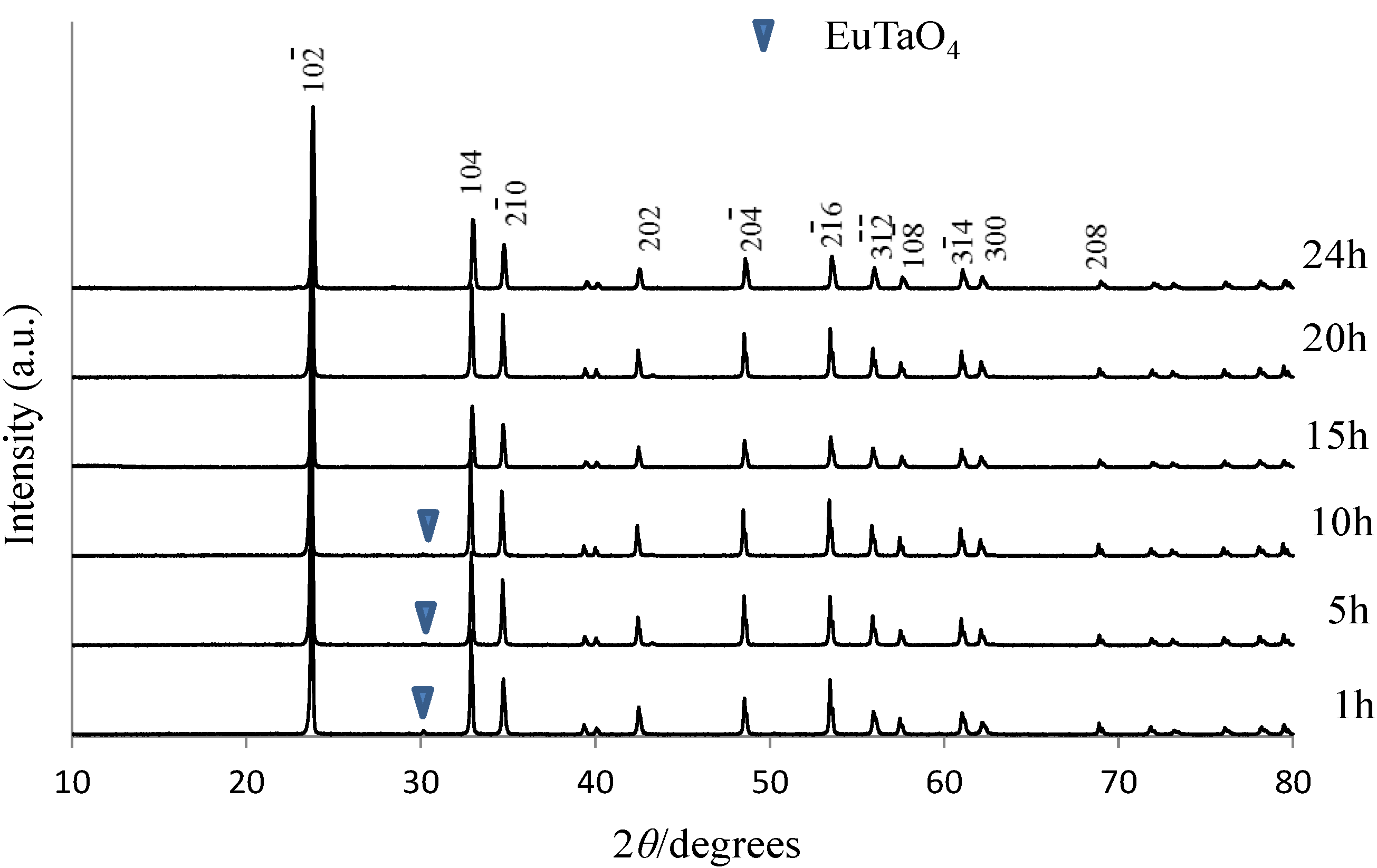
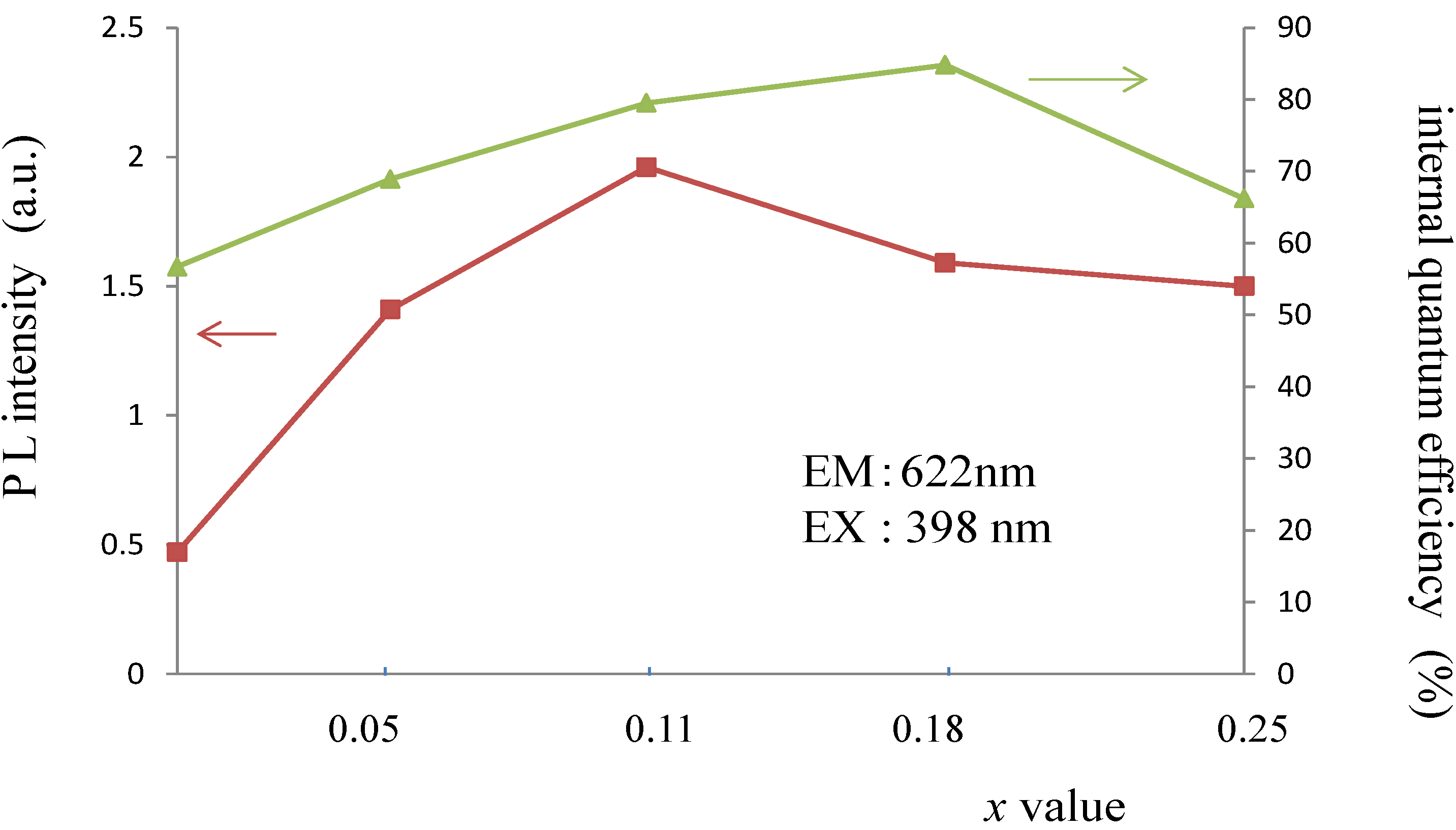
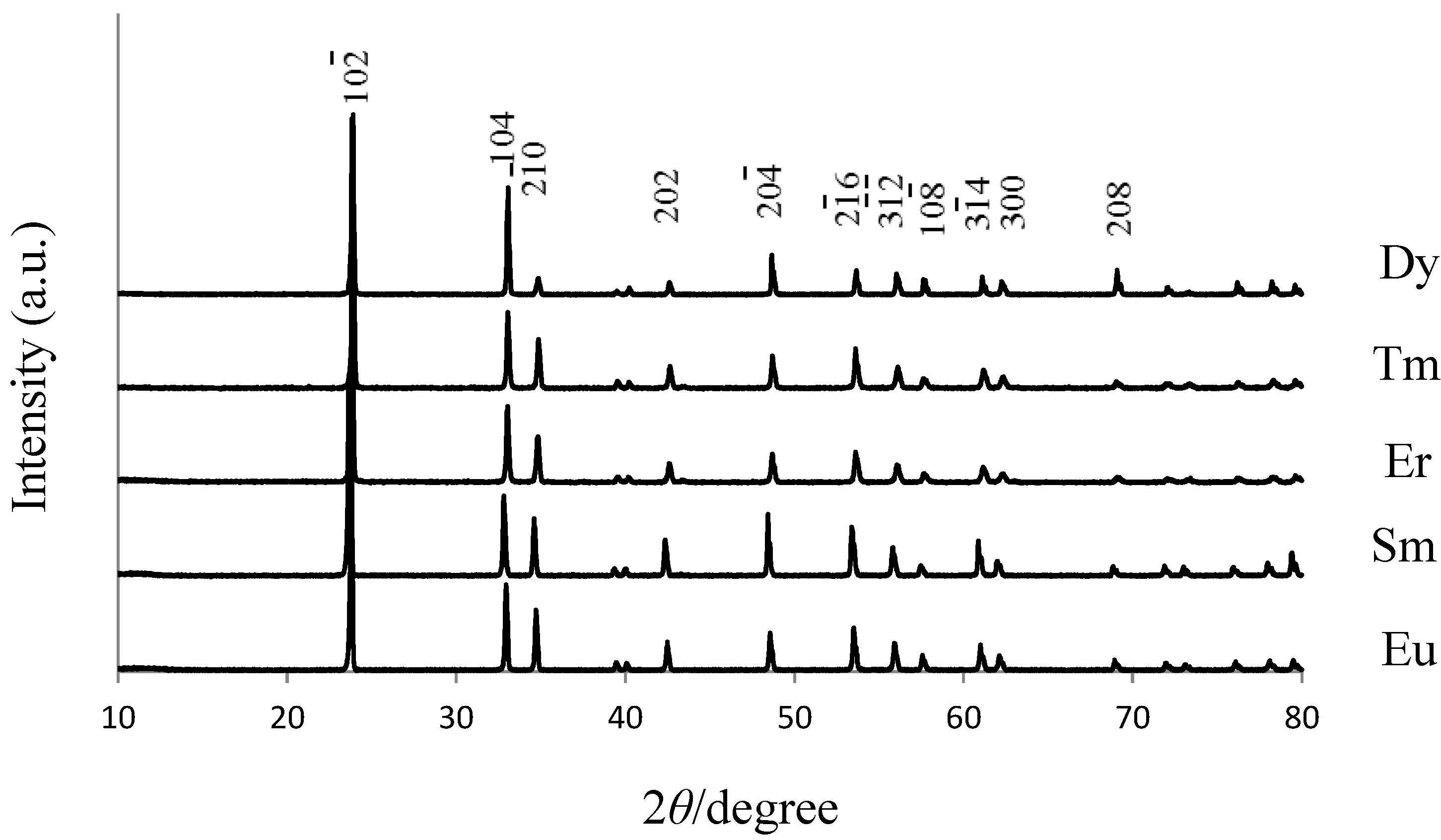
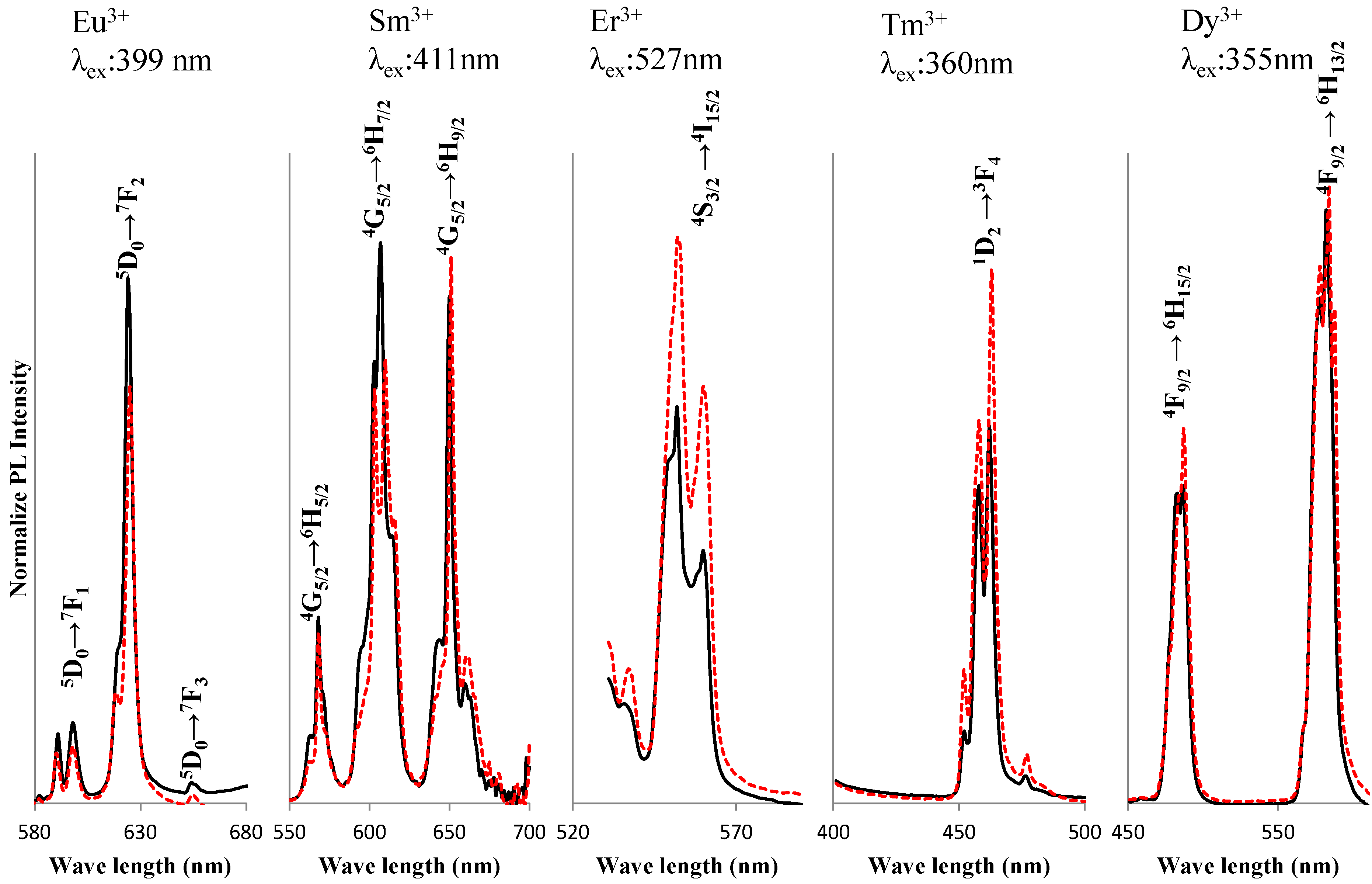
2. Results and Discussion
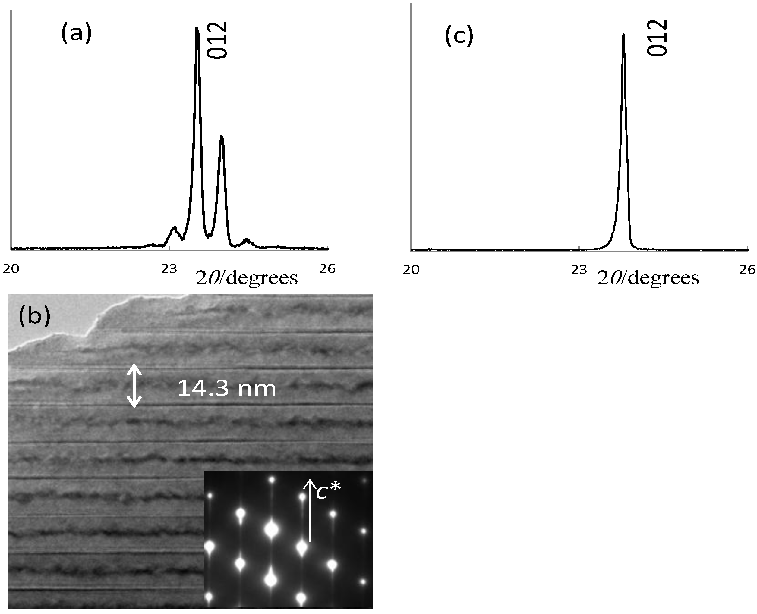
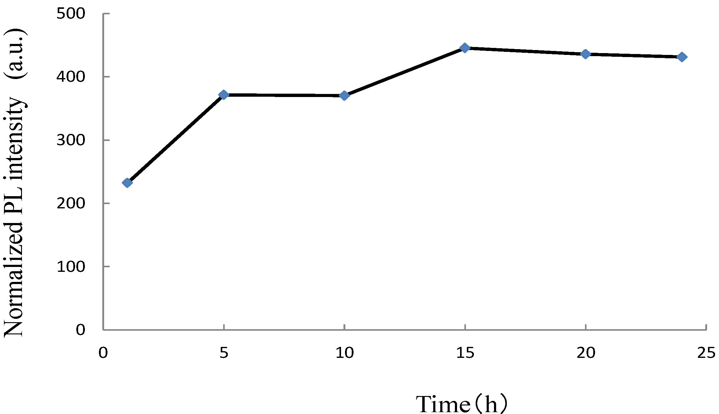
As we reported previously for the Li1+xyNb1−x−3yTix+4yO3 (LNT, 0 ≤ x ≤ 0.25, y = 0) system [12], the PL intensity was clearly improved by the addition of TiO2, which could affect the coordination state of the Eu3+ ion and/or induce a structural distortion around this ion. The most important factor is thus expected to be the TiO2 content in the Li1+xTa1−xTixO3 (LTT) solid solution. The optimal TiO2 concentration was then determined at the maximum emission peak upon excitation at around 399 nm. Red emission of the Eu3+-doped LTT was observed at an excitation wavelength of 399 nm due to the intraconfigurational 7F0-5L6 transition. The maximum emission peak at around 624 nm is associated with the hypersensitive electric-dipole 5D0-7F2 transition in the Eu3+ ion. Figure 1 shows the relationships among the PL intensity, internal quantum efficiency, and the TiO2 content in the ternary Li-Ta-Ti-O system upon varying the TiO2 content from x = 0 to 0.25 at a fixed Eu2O3 concentration of 2.5 wt %. The highest PL intensity is found for the composition of Li1.11Ta0.89Ti0.11O3 (x = 0.11, y = 0), where the chemical composition is based on the mixture ratio. The internal quantum efficiency revealed a high value of 84.8%, but the highest value appeared in Li1.18Ta0.82Ti0.18O3 with x = 0.18. The discrepancy in the x values that give the highest PL intensity and the highest quantum efficiency may come from the different measurement apparatus. Because the integrating sphere was used in the internal quantum efficiency measurement, the “x = 0.18” value is supposed to be more appropriate to discuss the relationship between the host crystal composition and the emission properties. The incorporation of Eu3+ ions in the crystal structure of LTT would cause the overlapping of orbitals with the adjacent anions, resulting in efficient red-light emission due to the hypersensitive 5D0-7F2 transition in Eu3+.
The LNT phosphor with composition of Li1.11Nb0.89Ti0.11O3: x = 0.11 was also found to have the highest PL intensity [9]. Here, the host material’s structure was compared between LNT and LTT. In Li1.11Nb0.89Ti0.11O3, the superstructure is formed by periodical insertion of the intergrowth layer with 14.3 nm spacing into a matrix with a trigonal structure (Figure 2b), and satellite reflections appeared around the (012) in the XRD pattern (Figure 2a). The superstructure was formed by doping of the Ti4+ ion, and the period was controlled by the Ti content, as described in our previous paper [11]. On the other hand, Li1.11Ta0.89Ti0.11O3 has no superstructure but only a basic structure of LiTaO3 (Figure 2c). The results revealed that the composition area of the M-phase with a superstructure is different between LNT and LTT. In our recent paper [20], the refined structure was compared between LiTaO3 and Li1.11Ta0.89Ti0.11O3 by the Rietveld method from powder X-ray diffraction data. The composition was determined as Li(Ta0.89Ti0.11)O2.945 from the final structure model, in which the Ti ion is located at the Ta site. By doping of the Ti4+ ion into LiTaO3, the <Li-O> distance of the (LiO12) polyhedron in the LiTaO3 structure (R3c) changed from 0.274 nm to 0.270 nm, and the [(Ta,Ti)O6] octahedra are all comparable with each other. As a result, the structure is stable without a superstructure.
Figure 1. Relationships among PL intensity, internal quantum efficiency, and TiO2 content in Li1+xTa1−xTixO3 host material.
Materials 06 02768 g001
Figure 2. Structures of Li1.11M0.89Ti0.11O3 (M: Nb or Ta) host material: (ab) LNT; and (c) LTT.
Materials 06 02768 g002
In Eu3+-doped LNT phosphor, an annealing time of 24 h is preferable to that of 10 h because the superstructure forms during long-time annealing [10]. In the previous work, PL intensity might have been related to the structural distortion due to the superstructure. Indeed, PL intensity increased with increasing sintering time. In the LTT, the relationship between the annealing time and PL intensity should be clear. Figure 3 shows this relationship in Li1.11Ta0.89Ti0.11O3:Eu3+ phosphor. The results show that the best annealing time is 15 h. For shorter annealing time, a small amount of EuTaO4 was detected around 30 degrees in Figure 4. To diffuse the Eu3+ ion into the LiTaO3 structure homogeneously as an activator, the annealing time needs to be 15 h. We confirmed that the Eu3+ ion is randomly distributed over the Li site because the electron-density peak at the Li/Eu site of Eu-doped LTT was higher than that at the Li site of LTT [20]. The composition determined by the Rietveld method was (Li0.977Eu0.023)(Ta0.89Ti0.11)O2.968, which has a small amount of oxygen vacancies. The crystal structure of LiTaO3 is flexible with respect to the substitutions of Eu for Li and Ti for Ta, in which the <Li-O> distance of the (LiO12) polyhedron is 0.270 nm and 0.272 nm in Li(Ta0.89Ti0.11)O2.945 and (Li0.977Eu0.023)(Ta0.89Ti0.11)O2.968, respectively.
Figure 3. Relationship between PL intensity and annealing time in Li1.11Ta0.89Ti0.11O3:Eu3+ phosphor.
Materials 06 02768 g003
Figure 4. XRD patterns of Li1.11Ta0.89Ti0.11O3:Eu3+ phosphors for various annealing times.
Materials 06 02768 g004
The LTT phosphors with various emission colors were synthesized while doping with trivalent ions of Sm, Er, Tm and Dy. The host material of Li1.11Ta0.89Ti0.11O3 was used at a fixed RE2O3 concentration of 2.5 wt %. Figure 5 shows the XRD patterns of LTT:RE3+ phosphors annealed for 15 h. All structures were LiTaO3-type, and no impurity phase was detected. The results show that the rare-earth ion diffused into the host material homogeneously and that rare-earth ions might also be located at the Li site. Emission peaks were compared between LNT:RE3+ and LTT:RE3+ phosphor, as shown in Figure 6. It should be mentioned here that the respective f-f excitations were much stronger than any possible charge-transfer excitations for the present LNT:RE3+ and LTT:RE3+ phosphors. The excitation wavelengths were then chosen with the strongest f-f excitations observed in each phosphor. Red emission of the Eu3+-doped LTT was obviously brighter than that of LNT phosphor. Red emission was also observed for the Sm3+-doped LNT upon excitation at around 411 nm due to the 6H5/2-6P3/2 transition. Upon excitation at 411 nm, the photoluminescence spectra showed emission peaks at 568 nm (4G5/2-6H5/2), 607 nm (4G5/2-6H7/2), and 651 nm (4G5/2-6H9/2). The splitting of the 607 nm emission is prominent in LNT phosphor. Therefore it seems that the peak intensity ratio of LTT phosphor at 607/651 nm is larger than that of LNT phosphor. This would be caused by the difference in the overlapping of orbitals with the adjacent anions (Ta/Nb). A green emission peak at 527 nm (4S3/2-4I15/2) was observed for LNT:Er3+ upon excitation at 552 nm (4I15/2-2H11/2). Blue emission was observed at 360 nm (1D2-3F4 transition) for the LNT:Tm3+ phosphor upon excitation at 463 nm (3H6-1D2). Yellow emission peaks at 584 nm (4F9/2-6H13/2) were observed for LNT:Dy3+ upon excitation at 355 nm (6H15/2 -4M15/2). The highest emission peak intensity was compared between LNT:RE3+ and LTT:RE3+. Green and Blue emissions of LNT phosphor were brighter than those of LTT phosphor. The internal quantum efficiency of these phosphors were low level; 23% for LTT:Sm3+, 22% for LTT:Er3+, 35% for LTT:Tm3+, and 28% for LTT:Dy3+.
Figure 5. XRD patterns of Li1.11Ta0.89Ti0.11O3:RE3+ (RE: Dy. Tm, Er, Sm, Eu) for 15 h annealing.
Materials 06 02768 g005
Figure 6. Comparison of PL intensity between LNT:RE3+ and LTT:RE3+. Dotted line: LNT:RE3+, solid line: LTT:RE3+.
Materials 06 02768 g006
In contrast, the most effective activator was the Eu3+ ion in the LTT host material. The red emission intensity excited at 339 nm of LTT: Eu3+ phosphor is three times higher than that of Y2O2S: Eu3+ (4.3 at %), which is well known as a red phosphor. It is assumed that small differences in the lattice site between LNT and LTT host materials would affect the transition probability of the respective RE3+ ions. The combination of the host material and the emitting RE3+ ion is of great significance in designing new phosphors. A more detailed structural analysis, and the relevant theoretical calculations, will be performed in the near future.
Table 1 shows the chromaticity (x, y) of the LNT:RE3+ and LTT:RE3+ phosphors in the red, green, blue and yellow color regions. In general, the PL behavior of the RE3+-doped phosphors is not affected by the host material’s structure due to the 4f-4f transitions. In Sm phosphor, however, the difference in the emission intensity ratio at 606/650 nm affected the chromaticity.
Table 1. Chromaticity (x, y) of LNT and LTT phosphors.
ActivatorLTT:RE3+LNT:RE3+
XYXY
Eu0.6730.3270.6760.324
Sm0.6220.3780.6300.370
Er0.3070.6850.3470.647
Tm0.1440.0330.1430.033
Dy0.4180.4250.4330.423

3. Experimental Procedure

The starting materials used were Li2CO3, Ta2O5 and TiO2 (>99.9% grade) to prepare the solid solution of LTT. The compositions of the LTT solid solutions prepared in this work followed the formula Li1+xyTa1−x−3yTix+4yO3 with 0.05 < x < 0.25, y = 0 as a host material. Rare earths (Eu2O3, Sm2O3, Er2O3, Tm2O3 and Dy2O3 > 99.9% grade) were doped in the host material. These powders were mixed and pressed in air at 1423 K for 1 h to 24h in a conventional electric furnace. As comparison materials, the rare earth doped Li1+xNb1−xTixO3 (LNT) phosphors were also sintered at 1373 K for 24 h [13].
Structural analysis was carried out by X-ray diffraction (XRD) using a RINT 2500 (Rigaku Co., Ltd., Tokyo, Japan) operating at 40 kV and 200 mA. Microstructure images were observed by a scanning electron microscope (SEM) (SU8000, Hitachi Co., Ltd., Tokyo, Japan) operating at 3 kV. High-resolution TEM (HRTEM) images were observed by a device (2100 F, JEOL Co., Ltd., Tokyo, Japan) operating at 200 kV and equipped with energy-dispersed spectroscopy (EDS).
Excitation and emission spectra of the obtained samples were measured by a spectrometer (model FP-6500, JASCO international Co., Ltd., Tokyo, Japan). Quantum efficiency was measured by a spectral radiometer (MCPD-7000, Otsuka Electronics Co., Ltd., Osaka, Japan).

4. Conclusions

New phosphors have been successfully synthesized by doping rare-earth materials into a Li1+xTa1−xTixO3 solid solution as a host material. The host material’s structure and photoluminescence (PL) property was compared to the RE3+-doped Li1+xNb1−xTixO3 (LNT). The resulting materials showed various color emissions, with LNT:Eu3+ and LNT:Sm3+ exhibiting red emission, LNT:Tm3+ blue emission, LNT:Er3+ green emission, and LTT:Dy3+ yellow emission. The host material’s structure of LTT phosphor with the optimal composition Li1.11Nb0.89Ti0.11O3 was not a superstructure. Therefore, the distortion due to superstructure did not affect the PL intensity in the LTT phosphor. The annealing time to synthesize the homogeneous phosphor was reduced from 24 h to 15 h. By doping Ti4+ and Eu3+ ions into the LiTaO3, the <Li-O> distance of the [LiO12] polyhedron in the LiTaO3 structure (R3c) changed without a superstructure. This change affected the interaction with the adjacent anions, and the emission from the Eu3+ ion in the Li site achieved a high value of internal quantum efficiency. In the LTT host material, the most effective activator was the Eu3+ ion. On the other hand, the emission intensities of Er- and Tm-doped LTT phosphors were lower that of LNT phosphor. We conclude that small differences in the lattice site between LNT and LTT host materials would affect the emission energy of the RE3+ ions.

Acknowledgments

We thank S. Yamada of Denki Kagaku Kogyo Co., Ltd. for help wtih our experiment. This work was partially supported by a research granted from The Murata Science Foundation (2012).

References

  1. Villafuerte-Castrejon, M.E.; Gracia, J.A.; Cisneros, E.; Valenzuela, R.; West, A.R. New rutile solid solutions Ti1−4xLixM3xO2:M = Nb, Ta, Sb. J. Brit. Ceram. Soc. 1984, 83, 143–145. [Google Scholar]
  2. Villafuerte-Castrejon, M.E.; Aragon-Pina, A.; Valenzuela, R.; West, A.R. Compound and solid-solution formation in the system Li2O-Nb2O5-TiO2. J. Solid State Ceram. 1987, 71, 103–108. [Google Scholar] [CrossRef]
  3. Smith, R.I.; West, A.R. Characterization of an incommensurate LiTiNb oxide. Mater. Res. Bull. 1992, 27, 277–285. [Google Scholar] [CrossRef]
  4. Hayashi, H.; Nakano, H.; Suzumura, K.; Urabe, K.; West, A.R. Superstructure in LiTiNb oxides. Forth Ceram. Soc. 1995, 2, 391–398. [Google Scholar]
  5. Hayashi, H.; Urabe, K.; Niihara, K. Preparation of stoichiometric crystalline Li(Nb,Ti)O3 solid solutions by sol-gel processing with metal alkoxides. Key Eng. Mater. 1999, 501, 161–163. [Google Scholar]
  6. Farber, L.; Levin, I.; Borisevichi, A.; Grey, I.E.; Roth, R.S.; Davies, P.K. Structural study of Li1+xyNb1−x−3yTix+4yO3 solid solutions. J. Solid State Chem. 2002, 166, 81–90. [Google Scholar] [CrossRef]
  7. Borisevich, A.Y.; Davies, P.K. Crystalline structure and dielectric properties of Li1+xyNb1−x−3yTix+4yO3 M-phase solid solutions. J. Am. Ceram. Soc. 2002, 85, 573–578. [Google Scholar] [CrossRef]
  8. Yamamoto, Y.; Hayashi, H.; Sekino, T.; Nakayama, T.; Kondo, T.; Wada, M.; Adachi, T.; Niihara, K. Microstructure and dielectric properties of sintered Li-Nb-Ti-O solid solution ceramics having superstructure. Mater. Res. Innov. 2003, 7, 74–79. [Google Scholar]
  9. Hayashi, H.; Nakano, H. Evolution and preparation of Li1+xyNb1−x−3yTix+4yO3 solid solution with superstructure as new phosphor. J. Alloy. Compd. 2010, 502, 360–364. [Google Scholar] [CrossRef]
  10. Hayashi, H.; Nakano, H.; Jones, M.I. Microstructure and luminescence of Eu-doped Li1+xyNb1−x−3yTix+4yO3 solid solutions with superstructure. J. Ceram. Soc. Jpn. 2010, 118, 226–230. [Google Scholar] [CrossRef]
  11. Nakano, H.; Saji, T.; Yuasa, M.; Shoji, M.; Mamoru, M. Rapid synthesis and structural analysis of Li-Nb-Ti-O solid solutions with superstructure by millimeter-wave heating. J. Ceram. Soc. Jpn. 2011, 119, 808–812. [Google Scholar] [CrossRef]
  12. Naknao, H.; Ozono, K.; Saji, T.; Miyake, S.; Hiroyuki, H. Rapid synthesis of Eu3+-doped LNT (Li-Nb-Ti-O) phosphor by millimeter-wave heating. Opt. Mater. 2012, in press. [Google Scholar]
  13. Nakano, H.; Suehrio, S.; Saji, T.; Miyake, S. Synthesis of a rare-erath doped LNT (Li-Nb-Ti-O) phosphor by millimemetr-wave heating. J. Alloy. Compd. 2013, 475–479. [Google Scholar] [CrossRef]
  14. Gryk, D.; Dyl, D.; Ryba-Romanowski, W.; Grinberg, M. Spectral properties of LiTaO3:Pr3+ under high hydrostatic pressure. J. Phys. Condens. Matter 2005, 17, 5383–5395. [Google Scholar] [CrossRef]
  15. Kostritskii, S.M.; Maring, D.B.; Tavlykasev, R.F.; Ramaswamy, R.V. Energy transfer upconversion in Er-doped LiTaO3. Appl. Phys. Lett. 2000, 76, 2161–2163. [Google Scholar] [CrossRef]
  16. Ryba-Romanowski, W.; Golab, S.; Dominiak-Dzik, G.; Paltnikov, M.N.; Sidorov, N.V. Influence of temperature on luminescence of terbium ions in LiNbO3. J. Appl. Lett. 2001, 78, 3610–3611. [Google Scholar] [CrossRef]
  17. Gasparotto, G.; Cebin, M.A.; Goes, M.S.; Lima, S.A.M.; Davolos, M.R.; Varela, J.A.; Paiva-Santos, C.O.; Zaghete, M.A. Correlation between the spectroscopic and structural properties with the occupation of Eu3+ sites in powdered Eu3+-doped LiTaO3 prepared by the pechini method. J. Appl. Phys. 2009, 106, 3506–3509. [Google Scholar] [CrossRef]
  18. Sokolska, I.; Ryba-Romanowski, W.; Golab, S.; Baba, M.; Swirkowicz, M.; Lukasiewicz, T. Spectroscopy of LiTaO3:Tm3+ crystals. J. Phys. Chem. Solids 2000, 61, 1573–1581. [Google Scholar]
  19. Nakano, H.; Ozono, K.; Hayashi, H.; Fujihara, S. Synthesis and luminescent properties of a new Eu3+-doped Li1+x (Ta1−zNbz)1−xTixO3 red phosphor. J. Am. Ceram. Soc. 2012, 2795–2797. [Google Scholar] [CrossRef]
  20. Uchida, T.; Suehiro, S.; Asaka, T.; Nakano, H.; Fukuda, K. Syntheses and crystal structures of Li(Ta0.89Ti0.11)O2.945 and (Li0.977Eu0.023)(Ta0.89Ti0.11)O2.968. Powder Diffr. 2013, in press. [Google Scholar]

