Influence of Implant Neck Design on Peri-Implant Tissue Dimensions: A Comparative Study in Dogs

 ,
,  , and
, and
Abstract
:1. Introduction
2. Materials and Methods
2.1. Animal Protocol
2.2. Sample Selection
2.3. Histological Preparation
2.4. Histometric Evaluation
2.5. Data Analysis
3. Results
Histomorphometric Evaluation
4. Discussion
5. Conclusions
Authors Contributions
Abbreviations Definition
| mm | millimeters |
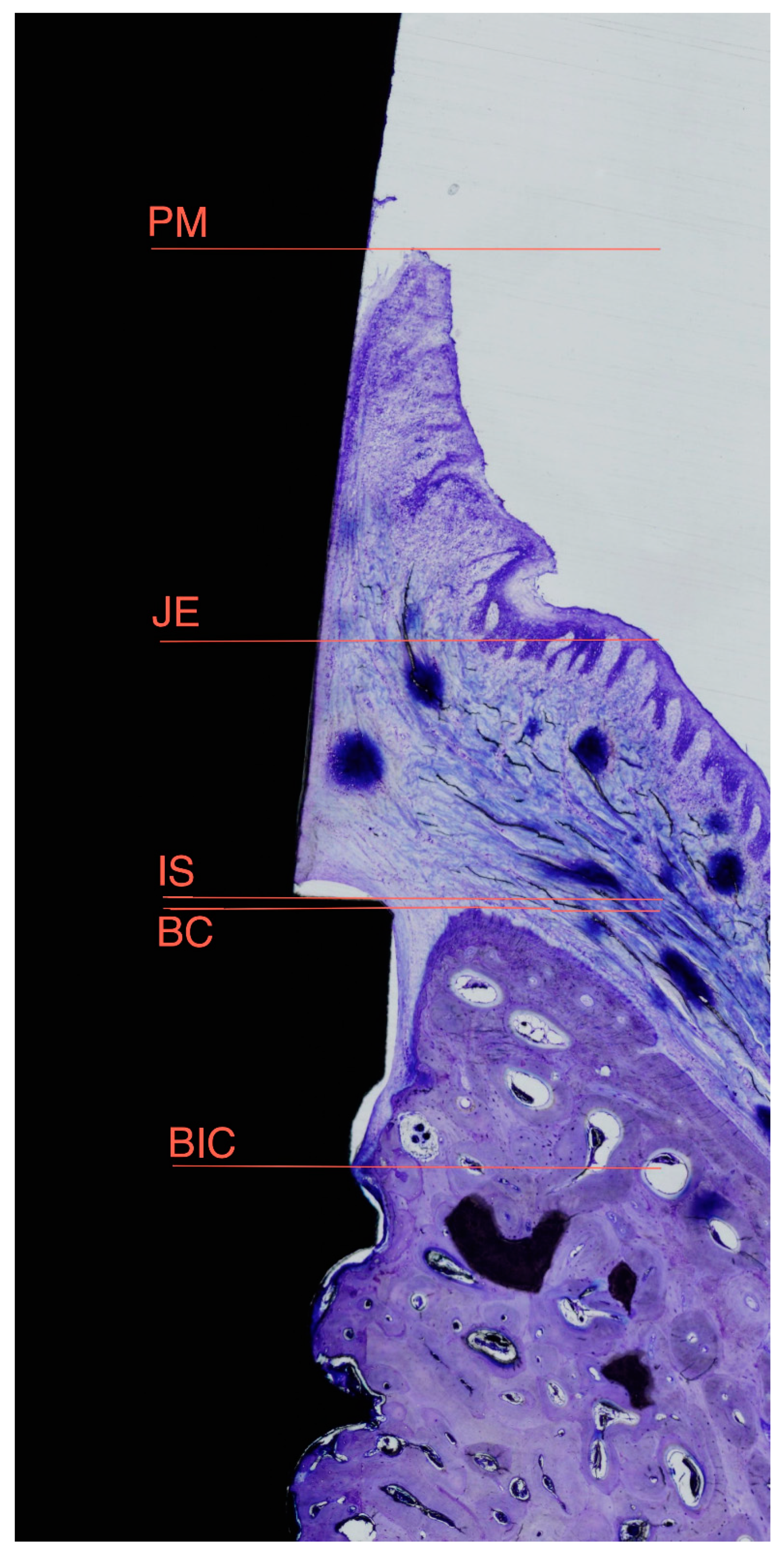
| IS-BIC | distance from the top of the implant shoulder to the first bone to implant contact |
| IS-BC | distance from the top of the implant shoulder to the bone crest |
| PM-BC | distance from the peri-implant mucosa to the bone crest |
| PM-JE | distance from the peri-implant mucosa to the apical portion of the barrier epithelium |
| PM-BIC | distance from the peri-implant mucosa to the first bone to implant contact |
| JE-BIC | distance from the apical portion of the barrier epithelium to the first bone to implant contact |
| PM-IS | distance from peri-implant mucosa to the Implant shoulder |
Funding
Acknowledgments
Conflicts of Interest
References
- Oh, T.J.; Yoon, J.; Misch, C.E.; Wang, H.L. The causes of early implant bone loss: Myth or science? J. Periodontol. 2002, 73, 322–333. [Google Scholar] [CrossRef] [PubMed]
- Den Hartog, L.; Raghoebar, G.M.; Slater, J.J.; Stellingsma, K.; Vissink, A.; Meijer, H.J. Single-tooth implants with different neck designs: A randomized clinical trial evaluating the aesthetic outcome. Clin. Implant Dent. Relat. Res. 2013, 15, 311–321. [Google Scholar] [CrossRef] [PubMed]
- Pirc, M.; Dragan, I.F. The key points of maintenance therapy for dental implants: A literature review. Compend. Contin. Educ. Dent. 2017, 38, e5–e8. [Google Scholar] [PubMed]
- Kronström, M.; Svenson, B.; Hellman, M.; Persson, G.R. Early implant failures in patients treated with Brånemark System titanium dental implants: A retrospective study. Int. J. Oral Maxillofac. Implants 2001, 16, 201–207. [Google Scholar] [PubMed]
- Canullo, L.; Camacho-Alonso, F.; Tallarico, M.; Meloni, S.M.; Xhanari, E.; Penarrocha-Oltra, D. Mucosa thickness and peri-implant crestal bone stability: A clinical and histologic prospective cohort trial. Int. J. Oral Maxillofac. Implants 2017, 32, 675–681. [Google Scholar] [CrossRef] [PubMed]
- Predecki, P.; Stephan, J.E.; Auslaender, B.A.; Mooney, V.L.; Kirkland, K. Kinetics of bone growth into cylindrical channels in aluminum oxide and titanium. J. Biomed. Mater. Res. 1972, 6, 375–400. [Google Scholar] [CrossRef] [PubMed]
- Koodaryan, R.; Hafezeqoran, A. Evaluation of implant neck surfaces for marginal bone loss: A systematic review and meta-analysis. Biomed. Res. Int. 2016, 2016, 4987526. [Google Scholar] [CrossRef] [PubMed]
- Calvo-Guirado, J.L.; Satorres, M.; Negri, B.; Ramirez-Fernandez, P.; Maté-Sánchez de Val, J.E.; Delgado-Ruiz, R.; Gomez-Moreno, G.; Abboud, M.; Romanos, G.E. Biomechanical and histological evaluation of four different titanium implant surface modifications: An experimental study in the rabbit tibia. Clin. Oral Investig. 2014, 18, 1495–1505. [Google Scholar] [CrossRef] [PubMed]
- Hansson, S. The implant neck: Smooth or provided with retention elements. A biomechanical approach. Clin. Oral Implants Res. 1999, 10, 394–405. [Google Scholar] [CrossRef] [PubMed]
- Chowdhary, R.; Halldin, A.; Jimbo, R.; Wennerberg, A. Influence of microthreads alteration on osseointegration and primary stability of implants: An FEA and in vivo analysis in rabbits. Clin. Implant Dent. Relat. Res. 2016, 17, 562–569. [Google Scholar] [CrossRef] [PubMed]
- Lee, D.W.; Choi, Y.S.; Park, K.H.; Kim, C.S.; Moon, I.S. Effect of microthread on the maintenance of marginal bone level: A 3-year prospective study. Clin. Oral Implants Res. 2007, 18, 465–470. [Google Scholar] [CrossRef] [PubMed]
- Akca, K.; Cehreli, M.C. A photoelastic and strain-gauge analysis of interface force transmission of internal-cone implants. Int. J. Periodontics Restor. Dent. 2008, 28, 391–399. [Google Scholar]
- Zanatta, L.C.; Dib, L.L.; Gehrke, S.A. Photoelastic stress analysis surrounding different implant designs under simulated static loading. J. Craniofac. Surg. 2014, 25, 1068–1071. [Google Scholar] [CrossRef] [PubMed]
- Hudieb, M.I.; Wakabayashi, N.; Kasugai, S. Magnitude and direction of mechanical stress at the osseointegrated interface of the microthread implant. J. Periodontol. 2011, 82, 1061–1070. [Google Scholar] [CrossRef] [PubMed]
- Calvo-Guirado, J.L.; López-López, P.J.; Pérez-Albacete Martínez, C.; Javed, F.; Granero-Marín, J.M.; Maté Sánchez de Val, J.E.; Ramírez Fernández, M.P. Peri-implant bone loss clinical and radiographic evaluation around rough neck and microthread implants: A 5-year study. Clin. Oral Implants Res. 2018, 29, 635–643. [Google Scholar] [CrossRef] [PubMed]
- Calvo Guirado, J.L.; Lucero-Sánchez, A.F.; Boquete Castro, A.; Abboud, M.; Gehrke, S.; Fernández Dominguez, M.; Delgado Ruiz, R.A. Peri-implant behavior of sloped shoulder dental implants used for all-on-four protocols: An histomorphometric analysis in dogs. Materials (Basel) 2018, 11, 119. [Google Scholar] [CrossRef] [PubMed]
- Jung, Y.C.; Han, C.H.; Lee, K.W. A 1-year radiographic evaluation of marginal bone around dental implants. Int. J. Oral Maxillofac. Implants 1996, 11, 811–818. [Google Scholar] [PubMed]
- Albrektsson, T.; Berglundh, T.; Lindhe, J. Osseointegration: Historic background and current concepts. Clin. Periodontol. Implant Dent. 2003, 12, 809–820. [Google Scholar]
- Petechia, L.; Usai, C.; Vassalli, M.; Gavazzo, P. Biophysical characterization of nanostructured TiO2 as a good substrate for hBM-MSC adhesion, growth and differentiation. Exp. Cell Res. 2017, 358, 111–119. [Google Scholar]
- Karlsson, U.; Gotfredsen, K.; Olsson, C. Single-tooth replacement by osseointegrated Astra Tech dental implants: A 2-year report. Int. J. Prosthodont. 1997, 10, 318–324. [Google Scholar] [PubMed]
- Smeets, R.; Stadlinger, B.; Schwarz, F.; Beck-Broichsitter, B.; Jung, O.; Precht, C.; Kloss, F.; Gröbe, A.; Heiland, M.; Ebker, T. Impact of dental implant surface modifications on osseointegration. BioMed Res. Int. 2016, 2016, 6285620. [Google Scholar] [CrossRef] [PubMed]
- CamargosGde, V.; Sotto-Maior, B.S.; Silva, W.J.; Lazari, P.C.; Del Bel Cury, A.A. Prosthetic abutment influences bone biomechanical behavior of immediately loaded implants. Braz. Oral Res. 2016, 30. [Google Scholar] [CrossRef] [Green Version]
- Donath, K.; Breuner, G. A method for the study of undecalcified bones and teeth with attached soft tissues. The Säge-Schliff (sawing and grinding) technique. J. Oral Pathol. 1982, 11, 318–326. [Google Scholar] [CrossRef] [PubMed]
- Hsu, Y.T.; Chan, H.L.; Rudek, I.; Bashutski, J.; Oh, W.S.; Wang, H.L.; Oh, T.J. Comparison of Clinical and Radiographic Outcomes of Platform-Switched Implants with a Rough Collar and Platform-Matched Implants with a Smooth Collar: A 1-Year Randomized Clinical Trial. Int. J. Oral Maxillofac. Implants 2016, 31, 382–390. [Google Scholar] [CrossRef] [PubMed]
- Khorsand, A.; Rasouli-Ghahroudi, A.A.; Naddafpour, N.; Shayesteh, Y.S.; Khojasteh, A. Effect of Microthread Design on Marginal Bone Level Around Dental Implants Placed in Fresh Extraction Sockets. Implant Dent. 2016, 25, 90–96. [Google Scholar] [CrossRef] [PubMed]
- Rasmusson, L.; Kahnberg, K.E.; Tan, A. Effects of implant design and surface on bone regeneration and implant stability: An experimental study in the dog mandible. Clin. Implant Dent. Relat. Res. 2001, 3, 2–8. [Google Scholar] [CrossRef] [PubMed]
- Abrahamsson, I.; Berglundh, T. Tissue characteristics at microthreaded implants: An experimental study in dogs. Clin. Implant Dent. Relat. Res. 2006, 8, 107–113. [Google Scholar] [CrossRef] [PubMed]
- Berglundh, T.; Abrahamsson, I.; Lindhe, J. Bone reactions to longstanding functional load at implants: An experimental study in dogs. J. Clin. Periodontol. 2005, 32, 925–932. [Google Scholar] [CrossRef] [PubMed]
- Abuhussein, H.; Pagni, G.; Rebaudi, A.; Wang, H.L. The effect of thread pattern upon implant osseointegration. Clin. Oral Implants Res. 2010, 21, 129–136. [Google Scholar] [CrossRef] [PubMed]
- Calvo-Guirado, J.L.; López-López, P.J.; Mate Sanchez, J.E.; GargalloAlbiol, J.; Velasco Ortega, E.; Delgado Ruiz, R. Crestal bone loss related to immediate implants in crestal and subcrestal position: A pilot study in dogs. Clin. Oral Implants Res. 2014, 25, 1286–1294. [Google Scholar] [CrossRef] [PubMed]
- Calvo-Guirado, J.L.; Gomez Moreno, G.; Aguilar-Salvatierra, A.; Mate Sanchez de Val, J.E.; Abboud, M.; Nemcovsky, C.E. Bone remodeling at implants with different configurations and placed immediately at different depth into extraction sockets. Experimental study in dogs. Clin. Oral Implants Res. 2015, 26, 507–515. [Google Scholar] [CrossRef] [PubMed]
- Calvo-Guirado, J.L.; López-López, P.J.; Maté Sánchez de Val, J.E.; Mareque-Bueno, J.; Delgado-Ruiz, R.A.; Romanos, G.E. Influence of neck design on peri-implant tissue healing around immediate implants: A pilot study in Foxhound dogs. Clin. Oral Implants Res. 2015, 26, 851–857. [Google Scholar] [CrossRef] [PubMed]
- Calvo-Guirado, J.L.; Pérez-Albacete, C.; Aguilar-Salvatierra, A.; de Val Maté-Sánchez, J.E.; Delgado-Ruiz, R.A.; Abboud, M.; Velasco, E.; Gómez-Moreno, G.; Romanos, G.E. Narrow-versus mini-implants at crestal and subcrestalbonelevels. Experimental study in beagle dogs at three months. Clin. Oral Investig. 2015, 19, 1363–1369. [Google Scholar] [CrossRef] [PubMed]
- Delgado-Ruiz, R.A.; Calvo-Guirado, J.L.; Abboud, M.; Ramirez-Fernandez, M.P.; Maté-Sánchez de Val, J.E.; Negri, B.; Gomez-Moreno, G.; Markovic, A. Connective tissue characteristics around healing abutments of different geometries: New methodological technique under circularly polarized light. Clin. Implant Dent. Relat. Res. 2015, 17, 667–680. [Google Scholar] [CrossRef] [PubMed]
- Calvo-Guirado, J.L.; Gómez-Moreno, G.; Delgado-Ruiz, R.A.; Maté Sánchez de Val, J.E.; Negri, B.; Ramírez Fernández, M.P. Clinical and radiographic evaluation of osseotite-expanded platform implants related to crestal bone loss: A 10-year study. Clin. Oral Implants Res. 2014, 25, 352–358. [Google Scholar] [CrossRef] [PubMed]
- Rupp, F.; Scheideler, L.; Olshanska, N.; de Wild, M.; Wieland, M.; Geis-Gerstorfer, J. Enhancing surface free energy and hydrophilicity through chemical modification of microstructured titanium implant surfaces. J. Biomed. Mater. Res. 2006, 76, 323–334. [Google Scholar] [CrossRef] [PubMed]
- Song, D.W.; Lee, D.W.; Kim, C.K.; Park, K.H.; Moon, I.S. Comparative analysis of peri-implant marginal bone loss based on microthread location: A 1-year prospective study after loading. J. Periodontol. 2009, 80, 1937–1944. [Google Scholar] [CrossRef] [PubMed]
- Al-Thobity, A.M.; Kutkut, A.; Almas, K. Microthreaded implants and crestal bone loss: A systematic review. J. Oral Implantol. 2017, 43, 157–166. [Google Scholar] [CrossRef] [PubMed]
- Macedo, J.P.; Pereira, J.; Vahey, B.R.; Henriques, B.; Benfatti, C.A.; Magini, R.S.; López-López, J.; Souza, J.C. Morse taper dental implants and platform switching: The new paradigm in oral implantology. Eur. J. Dent. 2016, 10, 148–154. [Google Scholar] [PubMed]
- D’Ercole, S.; Tripodi, D.; Marzo, G.; Bernardi, S.; Continenza, M.A.; Piattelli, A.; Iaculli, F.; Mummolo, S. Microleakage of bacteria in different implant-abutment assemblies: An in vitro study. J. Appl. Biomater. Funct. Mater. 2015, 13, e174–e180. [Google Scholar] [CrossRef] [PubMed]


| Type of Implants | IS-BC | IS-BIC | ||
|---|---|---|---|---|
| B | L | B | L | |
| Grouped design Micro-rings | 1.61(1.05a | 0.89(1.10) a | 1.76(0.77) | 1.59(0.89) a |
| Open-Thread | 0.55(1.04) a | −0.08(0.67) a | 1.03(0.66) | 0.63(0.53) a |
| Micro-rings BlueSky | 1.68(0.32) | 1.00(0.62) | 1.68(0.32) | 1.05(0.56) b |
| Micro-rings C1 | 1.53(1.55) | 0.78(1.52) | 1.84(1.11) | 2.14(0.85) b |
| Open-thread IPX | 1.45(0.34) c | 0.35(0.41) | 1.49(0.28) | 0.73(0.23) |
| Open-thread Facility | 1.52(0.55) c | 0.48(0.44) | 1.51(0.53) | 0.61(0.63) |
| Type of Implants | PM-BC | PM-BIC | PM-JE | JE-BIC | PM-IS | |||||
|---|---|---|---|---|---|---|---|---|---|---|
| B | L | B | L | B | L | B | L | B | L | |
| Grouped Micro-rings | 3.23(0.88) | 2.33(0.35) | 3.38(0.77) | 3.04(0.97) | 1.50(0.36) a | 1.39(0.40) a | 1.88(0.66) | 1.65(0.86) | 1.62(1.19) | 1.44(0.92) |
| Open-Thread | 2.57(0.93) | 1.69(0.80) | 3.03(0.80) | 2.41(0.84) | 1.08(0.42) a | 0.92(0.46) a | 1.94(0.73) | 1.49(0.75) | 2.00(0.61) | 1.78(0.65) |
| Micro-rings Blue Sky | 3.40(0.90) | 2.29(0.13) | 3.40(0.90) | 2.34(0.11) b | 1.49(0.20) | 1.32(0.16) | 1.90(0.89) | 1.01(0.25) b | 1.71(0.92) | 1.29(0.66) |
| Micro-rings C1 | 3.06(0.92) | 2.38(0.51) | 3.37(0.72) | 3.73(0.16) b | 1.51(0.51) | 1.45(0.56) | 1.85(0.43) | 2.28(0.78) b | 1.52(1.53) | 1.59(1.20) |
| Open-thread IPX | 3.15(0.68) | 1.79(0.79) | 3.19(0.63) | 2.38(0.68) | 1.34(0.20) c | 1.00(0.32) | 1.85(0.16) | 1.37(0.40) | 1.69(0.52) | 1.44(0.47) |
| Open-thread Facility | 2.20(0.91) | 1.68(1.09) | 3.00(1.00) | 2.55(1.14) | 0.86(0.39) c | 0.86(0.57) | 2.13(0.99) | 1.69(1.15) | 2.30(0.57) | 2.01(0.53) |
© 2018 by the authors. Licensee MDPI, Basel, Switzerland. This article is an open access article distributed under the terms and conditions of the Creative Commons Attribution (CC BY) license (http://creativecommons.org/licenses/by/4.0/).
Share and Cite
Calvo-Guirado, J.L.; Jiménez-Soto, R.; Pérez Albacete-Martínez, C.; Fernández-Domínguez, M.; Gehrke, S.A.; Maté-Sánchez de Val, J.E. Influence of Implant Neck Design on Peri-Implant Tissue Dimensions: A Comparative Study in Dogs. Materials 2018, 11, 2007. https://doi.org/10.3390/ma11102007
Calvo-Guirado JL, Jiménez-Soto R, Pérez Albacete-Martínez C, Fernández-Domínguez M, Gehrke SA, Maté-Sánchez de Val JE. Influence of Implant Neck Design on Peri-Implant Tissue Dimensions: A Comparative Study in Dogs. Materials. 2018; 11(10):2007. https://doi.org/10.3390/ma11102007
Chicago/Turabian StyleCalvo-Guirado, José Luis, Raúl Jiménez-Soto, Carlos Pérez Albacete-Martínez, Manuel Fernández-Domínguez, Sérgio Alexandre Gehrke, and José Eduardo Maté-Sánchez de Val. 2018. "Influence of Implant Neck Design on Peri-Implant Tissue Dimensions: A Comparative Study in Dogs" Materials 11, no. 10: 2007. https://doi.org/10.3390/ma11102007