Article

Expression of Heat Shock Proteins in Human Fibroblast Cells under Magnetic Resonant Coupling Wireless Power Transfer

Laboratory of Applied Radio Engineering for Humanosphere, Research Institute for Sustainable Humanosphere, Kyoto University, Gokasho, Uji, Kyoto 611-0011, Japan
*
Author to whom correspondence should be addressed.
Energies 2015, 8(10), 12020-12028; https://doi.org/10.3390/en81012020
Submission received: 27 July 2015 / Revised: 22 September 2015 / Accepted: 16 October 2015 / Published: 22 October 2015
(This article belongs to the Special Issue Wireless Power Transfer)

Abstract

:
Since 2007, resonant coupling wireless power transfer (WPT) technology has been attracting attention and has been widely researched for practical use. Moreover, dosimetric evaluation has also been discussed to evaluate the potential health risks of the electromagnetic field from this WPT technology based on the International Commission on Non-Ionizing Radiation Protection (ICNIRP) guidelines. However, there has not been much experimental evaluation of the potential health risks of this WPT technology. In this study, to evaluate whether magnetic resonant coupling WPT induces cellular stress, we focused on heat shock proteins (Hsps) and determined the expression level of Hsps 27, 70 and 90 in WI38VA13 subcloned 2RA human fibroblast cells using a western blotting method. The expression level of Hsps under conditions of magnetic resonant coupling WPT for 24 h was not significantly different compared with control cells, although the expression level of Hsps for cells exposed to heat stress conditions was significantly increased. These results suggested that exposure to magnetic resonant coupling WPT did not cause detectable cell stress.

1. Introduction

Wireless power transfer (WPT) is useful technology for supplying power without cables connected to a power source, and will lead to downsizing, battery-less operation and enhanced mobility. After the first WPT experiment by Nikola Tesla at the end of 19th century, many experiments involving WPT technology have been carried out using microwave, laser and electromagnetic induction [1]. In 2007, Kurs et al. [2] suggested a new WPT technology using the resonant coupling phenomenon. This new WPT technology transfers power between two pairs of coils with electromagnetic fields (EMFs), similar to electromagnetic induction technology, but can extend the power transfer distance at only the resonant frequency of the coils, and has been experimentally demonstrated to transfer power to a 60-W light bulb across a distance of 2 m with approximately 40% efficiency using self-resonant helical coils at approximately a 10-MHz resonant frequency. This new EMF-related WPT technology is expected to be used for many applications, such as supplying power to home appliances and industrial equipment, and charging electric vehicles. Therefore, many researchers have conducted studies related to this WPT technology and have demonstrated its practical use. For example, Nagano Japan Radio Co., Ltd. demonstrated a wireless power charging system for electric vehicles using magnetic resonant coupling WPT. The system transfers 1 kW of power across 30 cm with 88% efficiency at a 13.56-MHz resonant frequency [3]. Korea Advanced Institute of Science and Technology also developed a wireless power charging system for the Online Electric Vehicle, which transfers 100 kW of power across 20 cm at a 20-kHz resonant frequency [4]. As another example, Sony demonstrated wireless power transfer for home appliances using magnetic resonant coupling WPT also at a 13.56-MHz resonant frequency [5].
There are, however, concerns about the potential health risks of EMF-related technologies. Induction heating cookers and mobile phones, which are primary examples of EMF-related technology, have been studied extensively using both dosimetric and experimental methods to evaluate their potential health risks [6,7,8,9,10,11]. In contrast, the potential health risks of EMFs from WPT using the resonant coupling phenomenon have already been discussed based on the International Commission on Non-Ionizing Radiation Protection (ICNIRP) guidelines and dosimetric evaluation [12,13,14,15], but there have been very few experimental evaluations [16,17].
For experimental evaluation of the human health risks associated with EMFs, in vitro studies are used mainly to supply experimental evidence missing from human studies. Many in vitro studies have focused on carcinogenesis, mainly with two targets, cellular genotoxic effects and non-genotoxic effects, such as gene expression related to cell functions, to evaluate potential carcinogenesis [18]. In light of this, we previously evaluated cellular genotoxic effects under the conditions of magnetic resonant coupling WPT with 12.5-MHz resonant frequency, and found that magnetic resonant coupling WPT did not cause detectable effects [17]. In the present study, we focused on heat shock proteins (Hsps) as gene expression related with cell function. Expressed in response to cellular stress conditions such as heat, heat shock proteins (Hsps) help other proteins assemble correctly and prevent unfolding, and modulate a wide range of cellular functions, such as thermotolerance, anti-apoptosis function and immunogenicity. Many in vitro studies of the relationship between expression of Hsps and various frequency ranges of EMFs, such as extremely low frequency, intermediate frequency and radio frequency fields, have been conducted [18,19,20,21]. To our knowledge, however, there have been very few in vitro studies evaluating the expression of Hsps not only for resonant coupling WPT but also for EMFs near the 10-MHz frequency range. Therefore, in this study, we evaluated expression levels of Hsps 27, 70 and 90 under the conditions of magnetic resonant coupling WPT with 12.5-MHz resonant coupling frequency to investigate whether exposure to magnetic resonant coupling WPT induces cellular stress.

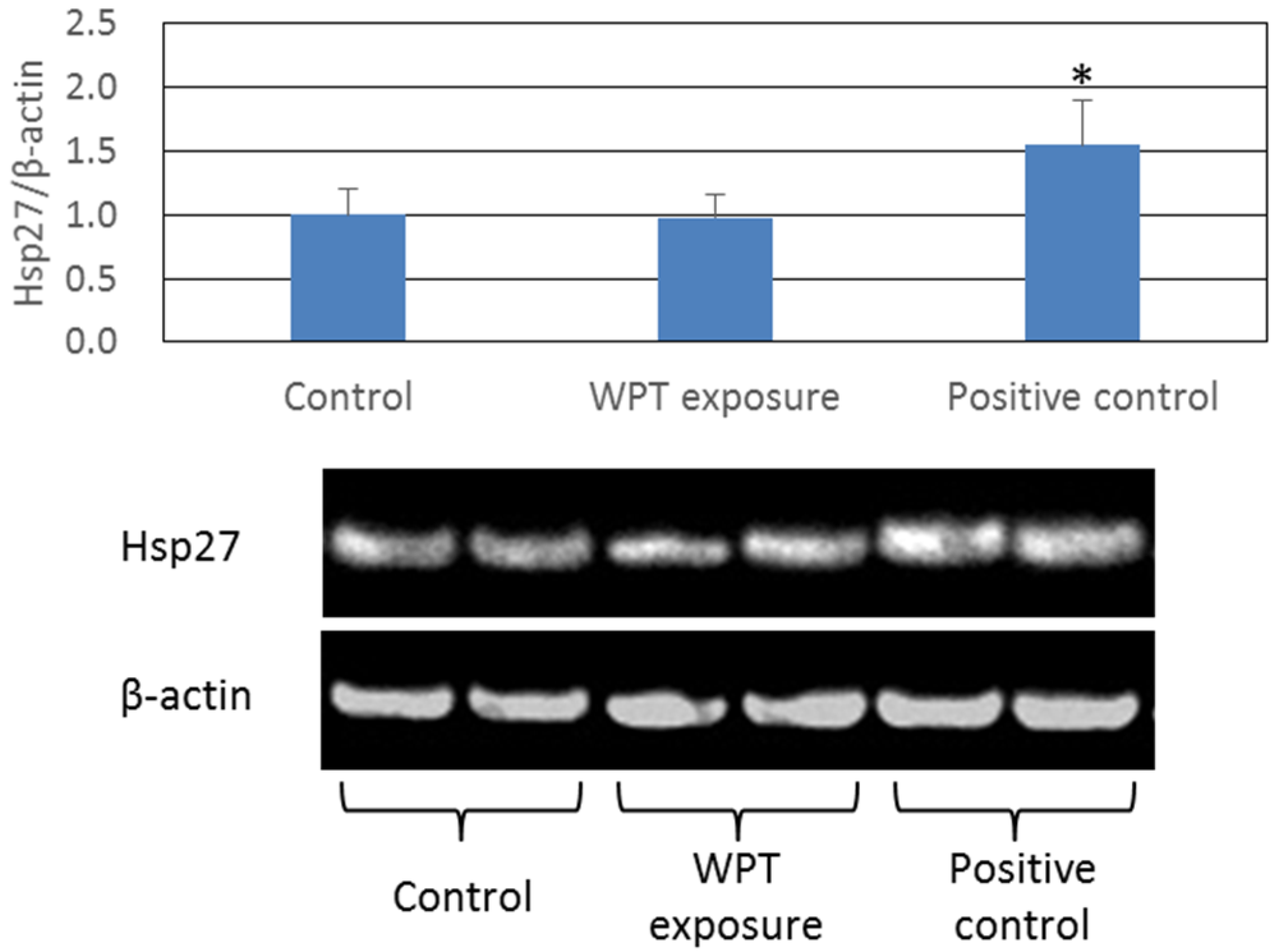
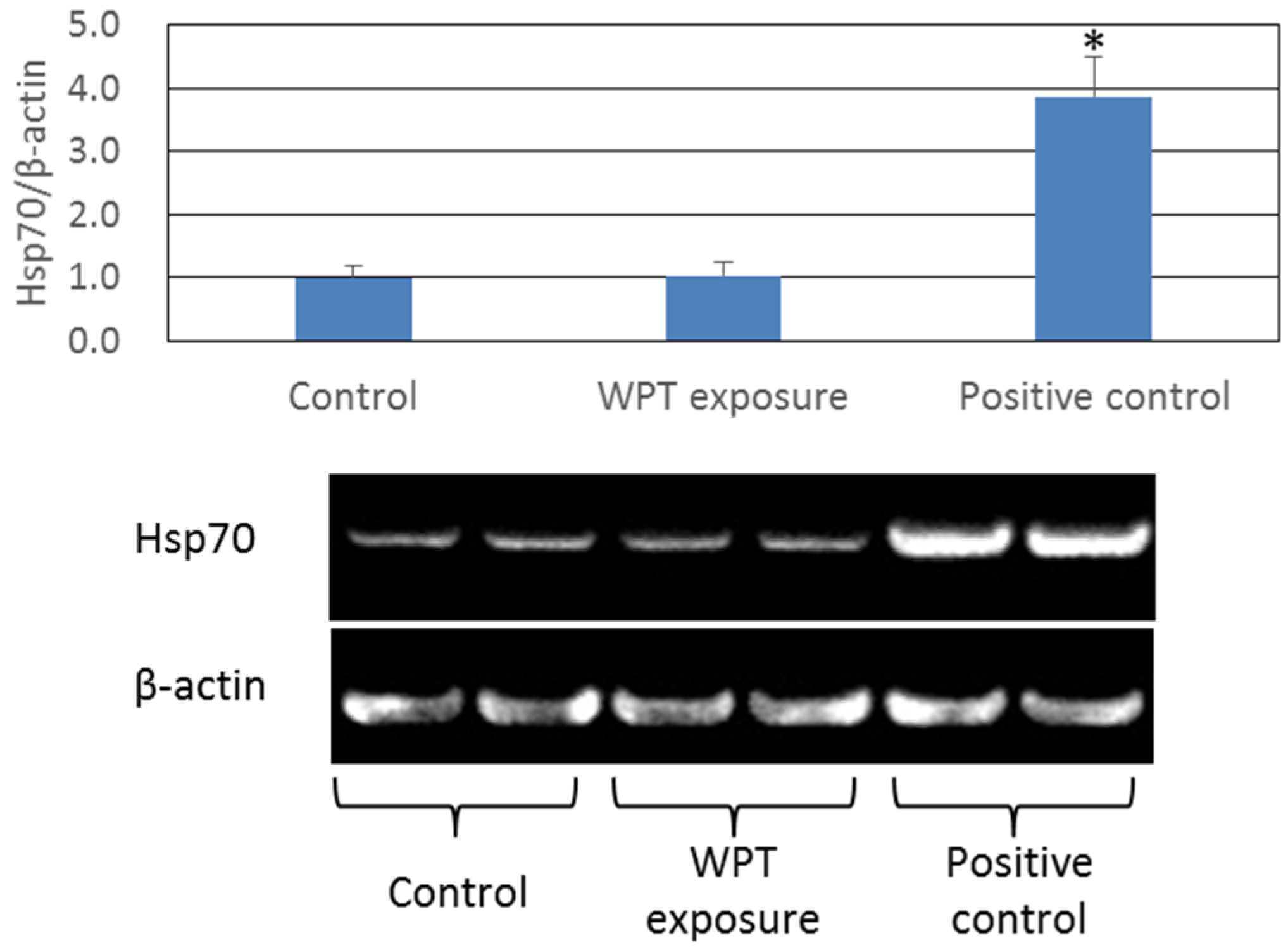
2. Results and Discussion

Expression of Heat Shock Proteins

In this study, we evaluated the expression of Hsps 27, 70 and 90 in WI38VA13 subcloned 2RA human fibroblast cells by western blotting, and the results are shown in Figure 1, Figure 2 and Figure 3, respectively. WI38 fibroblast cells (obtained from the Japanese Cancer Research Resources Bank, Osaka, Japan) have been used to evaluate the biological effects of various environmental stresses such as radiation, ultraviolet irradiation and EMFs [22,23,24,25,26]. It is also known that WI38 fibroblast cells express Hsps in response to heat stress conditions [27,28].
Figure 1. The expression of Hsp 27 in WI38VA13 subcloned 2RA human fibroblast cells exposed to Wireless power transfer (WPT) and control cells for 24 h, or heated at 43 °C as positive control. The expression of Hsp 27 was standardized to that of β-actin. Data are presented as means ± standard deviation (SD) from three separate experiments. Photograph shows the typical results of Western blotting. * p < 0.05 compared with control.
Energies 08 12020 g001
The increase in expression of Hsps is related to the activation of the heat shock transcription factor (HSF), which regulates the expression of Hsps, and binds to the heat shock element (HSE) on heat shock genes. In the absence of heat stress, HSF is inhibited by Hsps, and, therefore, exists in a monomeric form that has no DNA-binding activity. When proteins unfold under heat stress conditions, Hsps bind to the unfolded proteins as a molecular chaperon to facilitate refolding, and release HSF. HSF assembles into a trimeric form that is exhibits DNA-binding activity, phosphorylates and then translocates into the nucleus. In the nucleus, HSF binds to HSEs and activates transcription of Hsp genes, e.g., Hsp 27, 70 and 90, to synthesize new Hsps [29,30,31]. In our results, the cells under heat stress conditions showed a statistically significant increase in expression of Hsps 27, 70 and 90 compared with control cells. In contrast, we found no statistically significant differences in expression of Hsps 27, 70 and 90 in cells exposed to WPT for 24 h compared with control cells. These results suggested that WPT exposure did not induce detectable cell damage or disrupt Hsp expression.
Figure 2. The expression of Hsp 70 in WI38VA13 subcloned 2RA human fibroblast cells exposed to WPT and control cells for 24 h, or heated at 43 °C as positive control. The expression of Hsp 70 was standardized to that of β-actin. Data are presented as means ± SD from three separate experiments. Photograph shows the typical results of Western blotting. * p < 0.05 compared with control.
Energies 08 12020 g002
Figure 3. The expression of Hsp 90 in WI38VA13 subcloned 2RA human fibroblast cells exposed to WPT and control cells for 24 h, or heated at 43 °C as positive control. The expression of Hsp 90 was standardized to that of β-actin. Data are presented as means ± SD from three separate experiments. Photograph shows the typical results of Western blotting. * p < 0.05 compared with control.
Energies 08 12020 g003

3. Experimental Section

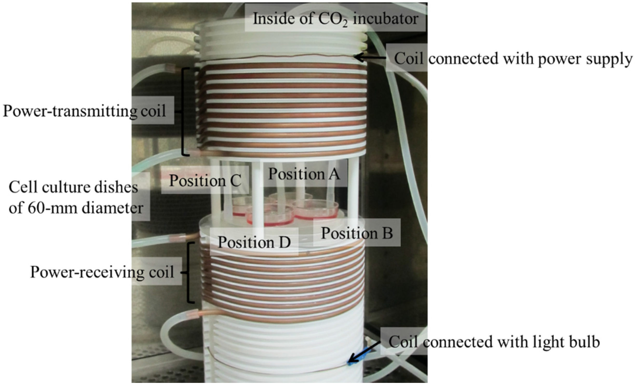
3.1. Exposure System

In this study, WPT exposure was carried out using a magnetic resonant coupling WPT exposure system, as shown in Figure 4. The details of the exposure system have been described in our previous reports [17,32]. In brief, the exposure system, built in a conventional CO2 incubator, has two self-resonant helical coils, a power-transmitting coil and a power-receiving coil, that function at a 12.5-MHz resonant frequency, and transfer power with 85.4% efficiency at 200-W input power. The cells are cultured between the power-transmitting coil and power-receiving coil using 60-mm-diameter cell culture dishes, and are maintained at 37 ± 0.2 °C with humidified 95% air and 5% CO2 during WPT exposure. The magnetic field level and specific absorption rate (SAR) level at each cell culture position are summarized in Table 1. These magnetic field and SAR levels are exceed the ICNIRP guidelines [33,34] of 80 A/m and 20 W/kg (localized SAR (limbs)) at 10 MHz for workers.
In contrast, the control condition was compared with WPT exposure using a conventional CO2 incubator with settings of 37 °C and humidified 95% air and 5% CO2. The cells under the control condition were exposed to ambient magnetic fields less than 0.55 μT (40 Hz–1 kHz), 0.13 μT (9 kHz–1 MHz), and 0.02 μT (1–30 MHz) measured using magnetic field meters (TMM-2, Denryoku Techno Systems, Kawasaki, Japan; EHP-200A, Narda STS, Pfullingen, Germany).
Figure 4. Photograph of the exposure system designed for magnetic resonant coupling WPT and cell culture dishes of 60-mm diameter inside of the conventional CO2 incubator.
Energies 08 12020 g004
Table 1. Magnetic fields and specific absorption rate (SAR) at each cell culture position calculated using the finite element method with a mesh size of 0.1 mm × 0.1 mm. The data presented are expressed as mean ± standard deviation (SD). Data from [17,32].
TypePosition APosition BPosition CPosition D
Magnetic Field169.2 ± 2.5 A/m (±1.5%)170.7 ± 2.2 A/m (±1.3%)167.6 ± 1.7 A/m (±1.0%)171.5 ± 2.2 A/m (±1.3%)
SAR21.8 ± 9.5 W/kg 21.3 ± 10.0 W/kg21.6 ± 12.1 W/kg 20.7 ± 9.7 W/kg

3.2. Cells and Culture Conditions

Human embryo lung-derived SV40 virus transformed WI38VA13 subcloned 2RA cells (obtained from the Japanese Cancer Research Resources Bank, Osaka, Japan) were cultured in Eagle’s Minimum Essential Medium with L-glutamine and phenol red (Wako Pure Chemical Industries, Osaka, Japan) and 10% fetal bovine serum (Bovogen Biologicals, East Keilor, VIC, Australia) at 37 °C in 95% air and 5% CO2 in a conventional CO2 incubator.

3.3. Western Blotting

The WI38VA13 subcloned 2RA human fibroblast cells were seeded at a density of 1 × 10⁶ cells per dish. After incubation for 24 h in the conventional CO2 incubator, the cells were WPT exposed for 24 h. Positive control cells were first incubated at 37 °C for 14 h, then heated at 43 °C for 1 h, and finally incubated at 37 °C for 9 h (total 24 h).
Immediately after exposure, the cells were washed once with cold phosphate-buffered saline (PBS) (T900, Takara Bio, Shiga, Japan) and treated with 250 μL CelLytic™ M (C2978, Sigma-Aldrich, St. Louis, MO, USA) supplemented with 20 μL protease inhibitor cocktail (P8340, Sigma-Aldrich), and then were collected by cell scraper into pre-cooled 1.5-mL centrifuge tubes. The tubes were then incubated for 15 min at 4 °C on a shaker and centrifuged at 1200 rpm (130 g) and 4 °C for 15 min to pellet the cellular debris. The protein-containing supernatant was removed to new pre-cooled 1.5-mL centrifuge tubes and stored at –80 °C for further analysis. Protein concentrations were measured using the Pierce™ BCA Protein Assay Kit (23227, Thermo Fisher Scientific, Waltham, MA, USA).
For electrophoresis, protein was added to the sample buffer (39000, Thermo Fisher Scientific), which was then boiled at 95 °C for 4 min. A 10-μL sample, equivalent to 10 μg of protein, was loaded onto 10% sodium dodecyl sulfate (SDS)-polyacrylamide gel (SuperSep™ Ace 10%, Wako Pure Chemical Industries, Osaka, Japan) and separated by electrophoresis at 20 mA for 70 min with an electrophoresis system (058-07681, Wako Pure Chemical Industries), and then transferred to a nitrocellulose membrane (0.2-μm pore size, IB23001, Life Technologies, Carlsbad, CA, USA) using a iBlot 2 Dry Blotting System (IB21001, Life Technologies).
The nitrocellulose membranes containing blotted proteins were incubated with primary antibody (goat anti-Hsp 27 (polyclonal, AF15801, R & D Systems, Minneapolis, MN, USA), mouse anti-Hsp 70 (monoclonal, SMC-100, Stress Marq, Victoria, BC, Canada), mouse anti-Hsp 90 (monoclonal, SMC-149, Stress Marq) and rabbit anti-β-actin (polyclonal, GTX109639, Gene Tex, Irvine, CA, USA)), followed by secondary antibody [horseradish peroxidase-conjugated anti-goat-Immunoglobulin G (IgG; HAF109, R & D Systems), horseradish peroxidase-conjugated anti-mouse-IgG (NA931, GE Healthcare UK Ltd., Amersham Place, Buckinghamshire, UK), or horseradish peroxidase-conjugated anti-rabbit-IgG (HAF008, R & D Systems)] using a iBind Solution Kit (SLF1020, Life Technologies) and iBind Western System (SLF1000, Life Technologies) for 2.5 h at room temperature.
Protein expression was visualized using ECL Western Blotting Detection Reagents (RPN2209, GE Healthcare UK Ltd., Amersham Place, Buckinghamshire, UK), and scanned by a Light Capture System (AE-6955, ATTO, Tokyo, Japan). Quantification was performed using image analysis software (CS Analyzer version 3, Tokyo, Japan).

3.4. Statistical Analysis

All experiments were repeated three times on separate days, and the data are expressed as the mean ± standard deviation (SD). Statistical analysis was conducted using Tukey’s test for multiple comparisons. P values less than 0.05 were considered statistically significant.

4. Conclusions

In this study, to evaluate whether magnetic resonant coupling WPT induces cellular stress, we determined the expression level of Hsps in WI38VA13 subcloned 2RA human fibroblast cells using a western blotting method. The expression level of Hsps 27, 70 and 90 when cells were exposed to heat stress conditions significantly increased, but the expression level of Hsps under conditions of magnetic resonant coupling WPT for 24 h was not significantly different compared with control cells. These results suggested that exposure to magnetic resonant coupling WPT under conditions stipulated in the ICNIRP guidelines does not cause detectable cellular stress in human fibroblast cells.

Acknowledgments

This work was supported in part by a Grant-in-Aid for Scientific Research (B) (No. 26281032) from the Japan Society for the Promotion of Science (JSPS) and a Development of New Research Area in Sustainable Humanosphere.

Author Contributions

Kohei Mizuno helped run the experiments and analyze the data, and write the paper. Naoki Shinohara and Junji Miyakoshi helped write the paper.

Conflicts of Interest

The authors declare no conflict of interest.

References

  1. Shinohara, N. Power without wire. IEEE Microw. Mag. 2011, 12, S64–S73. [Google Scholar] [CrossRef]
  2. Kurs, A.; Karalis, A.; Moffatt, R.; Joannpoilos, J.D.; Fisher, P.; Soljacic, M. Wireless power transfer via strongly coupled magnetic resonances. Sci. Mag. 2007, 317, 83–86. [Google Scholar] [CrossRef] [PubMed]
  3. Yokoi, Y.; Taniya, A.; Horiuchi, M.; Kobayashi, S. Development of kW Class Wireless Power Transmission System for EV Using Magnetic Resonant Method. In Proceedings of the 1st International Electric Vehicle Technology Conference, Yokohama, Japan, 17–19 May 2011.
  4. Chun, Y.; Park, S.; Kim, J.; Kim, J.; Kim, H.; Kim, J.; Kim, N.; Ahn, S. Electromagnetic compatibility of resonance coupling wireless power transfer in on-line electric vehicle system. IEICE Trans. Commun. 2014, 97, 416–423. [Google Scholar] [CrossRef]
  5. Sony develops highly efficient wireless power transfer system based on magnetic resonance. Available online: http://www.sony.net/SonyInfo/News/Press/200910/09-119E/ (accessed on 18 September 2015).
  6. Miyakoshi, J.; Horiuchi, E.; Nakahara, T.; Sakurai, T. Magnetic fields generated by an induction heating (IH) cook top do not cause genotoxicity in vitro. Bioelectromagnetics 2007, 28, 529–537. [Google Scholar] [CrossRef] [PubMed]
  7. Nakasono, S.; Ikehata, M.; Dateki, M.; Yoshie, S.; Shigemitsu, T.; Negishi, T. Intermediate frequency magnetic fields do not have mutagenic, co-mutagenic or gene conversion potentials in microbial genotoxicity tests. Mutat. Res. 2008, 649, 187–200. [Google Scholar] [CrossRef] [PubMed]
  8. Suzuki, Y.; Taki, M. Measurement of magnetic field from an induction heating hob and estimation of induced current density in human body. IEEJ Trans. Fundam. Mater. 2005, 125, 427–433. [Google Scholar] [CrossRef]
  9. Lin, J.C. Dosimetric comparison between different quantities for limiting exposure in the RF band: Rationale and implications for guidelines. Health Phys. 2007, 92, 547–553. [Google Scholar] [CrossRef] [PubMed]
  10. Verschaeve, L.; Juutilainen, J.; Lagroye, I.; Miyakoshi, J.; Saunders, R.; de Seze, R.; Tenforde, T.; van Rongen, E.; Veyret, B.; Xu, Z. In vitro and in vivo genotoxicity of radiofrequency fields. Mutat. Res. 2010, 705, 252–268. [Google Scholar] [CrossRef] [PubMed]
  11. Vijayalaxmia; Cao, Y.; Scarfic, M.R. Adaptive response in mammalian cells exposed to non-ionizing radiofrequency fields: A review and gaps in knowledge. Mutat. Res. 2014, 760, 36–45. [Google Scholar] [CrossRef] [PubMed]
  12. Laakso, I.; Hirata, A. Evaluation of induced electric field and compliance procedure for wireless power transfer system in an electrical vehicle. Phys. Med. Biol. 2013, 58, 7583–7593. [Google Scholar] [CrossRef] [PubMed]
  13. Park, S.W.; Wake, K.; Watanabe, S. Incident electric field effect and numerical dosimetry for a wireless power transfer system using magnetically coupled resonances. IEEE Trans. Microw. Theory Tech. 2013, 61, 3461–3469. [Google Scholar] [CrossRef]
  14. Christ, A.; Douglas, M.G.; Cooper, E.B.; Sample, A.P.; Waters, B.H.; Smith, J.R.; Kuster, N. Evaluation of wireless resonant power transfer systems with human electromagnetic exposure limits. IEEE Trans. Electromagn. Compat. 2013, 55, 265–274. [Google Scholar] [CrossRef]
  15. Sunohara, T.; Laakso, I.; Hirata, A.; Onishi, T. Analysis of in-situ electric field and specific absorption rate in human models for wireless power transfer with induction coupling. Phys. Med. Biol. 2014, 59, 3721–3735. [Google Scholar] [CrossRef] [PubMed]
  16. Shi, D.; Zhu, C.; Lu, R.; Mao, S.; Qi, Y. Intermediate frequency magnetic field generated by a wireless power transmission device does not cause genotoxicity in vitro. Bioelectromagnetics 2014, 35, 512–518. [Google Scholar] [CrossRef] [PubMed]
  17. Mizuno, K.; Shinohara, N.; Miyakoshi, J. In Vitro Evaluation of Genotoxic Effects under Magnetic Resonant Coupling Wireless Power Transfer. Int. J. Environ. Res. Public Health 2015, 12, 3853–3863. [Google Scholar] [CrossRef] [PubMed] [Green Version]
  18. World Health Organization (WHO). Extremely Low Frequency Fields, Environmental Health Criteria Monograph No. 238; WHO Press: Geneva, Switzerland, 2007. [Google Scholar]
  19. Miyakoshi, J.; Mori, Y.; Yaguchi, H.; Ding, G.; Fujimori, A. Suppression of heat-induced HSP-70 by simultaneous exposure to 50 mT magnetic field. Life Sci. 2000, 66, 1187–1196. [Google Scholar] [CrossRef]
  20. Sakurai, T.; Kiyokawa, T.; Kikuchi, K.; Miyakoshi, J. Intermediate frequency magnetic fields generated by an induction heating (IH) cooktop do not affect genotoxicities and expression of heat shock proteins. Int. J. Radiat. Biol. 2009, 85, 883–890. [Google Scholar] [CrossRef] [PubMed]
  21. Wang, J.; Koyama, S.; Komatsubara, Y.; Suzuki, Y.; Taki, M.; Miyakoshi, J. Effects of a 2450 MHz high-frequency electromagnetic field with a wide range of SARs on the induction of heat-shock proteins in A172 cells. Bioelectromagnetics 2006, 27, 479–486. [Google Scholar] [CrossRef] [PubMed]
  22. Fujimichi, Y.; Hamada, N. Ionizing irradiation not only inactivates clonogenic potential in primary normal human diploid lens epithelial cells but also stimulates cell proliferation in a subset of this population. PLoS ONE 2014, 9. [Google Scholar] [CrossRef] [PubMed]
  23. Tomita, M.; Maeda, M.; Maezawa, H.; Usami, N.; Kobayashi, K. Bystander cell killing in normal human fibroblasts is induced by synchrotron X-ray microbeams. Radiat. Res. 2010, 173, 380–385. [Google Scholar] [CrossRef] [PubMed]
  24. Straface, E.; Vona, R.; Ascione, B.; Matarrese, P.; Strudthoff, T.; Franconi, F.; Malorni, W. Single exposure of human fibroblasts (WI-38) to a sub-cytotoxic dose of UVB induces premature senescence. FEBS Lett. 2007, 581, 4342–4348. [Google Scholar] [CrossRef] [PubMed]
  25. Jin, Y.B.; Kang, G.Y.; Lee, J.S.; Choi, J.I.; Lee, J.W.; Hong, S.C.; Myung, S.H.; Lee, Y.S. Effects on micronuclei formation of 60-Hz electromagnetic field exposure with ionizing radiation, hydrogen peroxide, or c-Myc overexpression. Int. J. Radiat. Biol. 2012, 88, 374–380. [Google Scholar] [CrossRef] [PubMed]
  26. Wolf, F.I.; Torsello, A.; Tedesco, B.; Fasanella, S.; Boninsegna, A.; D’Ascenzo, M.; Grassi, C.; Azzena, G.B.; Cittadini, A. 50-Hz extremely low frequency electromagnetic fields enhance cell proliferation and DNA damage: Possible involvement of a redox mechanism. Biochim. Biophys. Acta 2005, 1743, 120–129. [Google Scholar] [CrossRef] [PubMed]
  27. Bonelli, M.A.; Alfieri, R.R.; Petronini, P.G.; Brigotti, M.; Campanini, C.; Borghetti, A.F. Attenuated expression of 70-kDa heat shock protein in WI-38 human fibroblasts during aging in vitro. Exp. Cell Res. 1999, 252, 20–32. [Google Scholar] [CrossRef] [PubMed]
  28. Bonelli, M.A.; Alfieri, R.R.; Desenzani, S.; Petronini, P.G.; Borghetti, A.F. Proteasome inhibition increases HuR level, restores heat-inducible HSP72 expression and thermotolerance in WI-38 senescent human fibroblasts. Exp. Gerontol. 2004, 39, 423–432. [Google Scholar] [CrossRef] [PubMed]
  29. Morimoto, R.I. Cells in stress: Transcriptional activation of heat shock genes. Science 1993, 259, 1409–1410. [Google Scholar] [CrossRef] [PubMed]
  30. Anckar, J.; Sistonen, L. Regulation of HSF1 function in the heat stress response: Implications in aging and disease. Annu. Rev. Biochem. 2011, 80, 1089–1115. [Google Scholar] [CrossRef] [PubMed]
  31. Wang, X.; Chen, M.; Zhou, J.; Zhang, X. HSPs 27, 70 and 90, anti-apoptotic proteins, in clinical cancer therapy (Review). Int. J. Oncol. 2014, 45, 18–30. [Google Scholar] [PubMed]
  32. Mizuno, K.; Miyakoshi, J.; Shinohara, N. In vitro exposure system using magnetic resonant coupling wireless power transfer. Wirel. Power Transf. 2014, 1, 97–107. [Google Scholar] [CrossRef]
  33. International Commission on Non-Ionizing Radiation Protection (ICNIRP). Guidelines for limiting exposure to time-varying electric and magnetic fields (1 Hz–100 kHz). Health Phys. 2010, 99, 818–836. [Google Scholar]
  34. Ahlbom, A.; Bergqvist, U.; Bernhardt, J.H.; Cesarini, J.; Court, L.A.; Grandolfo, M.; Hietanen, M.; Mckinlay, A.F.; Repacholi, M.H.; Sliney, D.H.; et al. Guidelines for Limiting Exposure to Time-varying electric, magnetic and electromagnetic fields (up to 300 GHz). Health Phys. 1998, 74, 494–522. [Google Scholar]

