Cytogenetic Effects of 1,1-Dichloroethane in Mice Bone Marrow Cells

1
Department of Biological Sciences, Alcorn State University, Lorman, MS, USA
2
Molecular Toxicology Laboratory, Jackson State University, Jackson, MS, USA
*
Author to whom correspondence should be addressed.
Int. J. Environ. Res. Public Health 2005, 2(1), 101-106; https://doi.org/10.3390/ijerph2005010101
Submission received: 15 November 2004
/
Accepted: 6 February 2005
/
Published: 30 April 2005
Abstract
:The major concern for the halogenated compounds is their widespread distribution, in addition to occupational exposures. Several chlorinated alkanes and alkenes were found to induce toxic effects. In this study, we investigated the genotoxic potential of 1,1-dichloroethane in the bone marrow cells obtained from Swiss-Webster mice, using chromosomal aberrations (CA), mitotic index (MI), and micronuclei (MN) formation as toxicological endpoints. Five groups of three male mice each, weighing an average of 24 ± 2 g, were injected intraperitoneally, once with doses of 100, 200, 300, 400, 500 mg/kg body weight (BW) of 1,1-dichloroethane dissolved in ethanol. A control group was also made of three animals injected with ethanol (1%) without the chemical. All animals were sacrificed 24 hours after the treatment. Chromosome and micronuclei preparations were obtained from bone marrow cells following standard protocols. Chromatid and chromosome aberrations were investigated in 100 metaphase cells per animal and percent micronuclei frequencies were investigated in 1,000 metaphase cells per animal. 1,1-dichloroethane exposures significantly increased the number of chromosomal aberrations and the frequency of micronucleated cells in the bone marrow cells of Swiss-Webster mice. Percent chromosomal aberrations of 2.67 ± 0.577, 7.66 ± 2.89, 8.33 ± 2.08, 14.67 ± 2.51, 20.3 ± 3.21, 28 ± 3.61; mitotic index of 9.4%, 7.9%, 6.2%, 4.3%, 3.0%, 2.6% and micronuclei frequencies of 3.33 ± 0.7, 7.33 ± 0.9, 8.00 ± 1.0, 11.67 ± 1.2, 15.33 ± 0.7, 18.00 ± 1.7 were recorded for the control, 100, 200, 300, 400, and 500 mg/kg BW respectively; indicating a gradual increase in number of chromosomal aberrations and micronuclei formation, with increasing dose of 1,1,-dichloroethane. Our results indicate that 1,1-dichloroethane has a genotoxic potential as measured by the bone marrow CA and MN tests in Swiss-Webster mice.
Introduction
1,1-Dichloroethane (DCE) is a short–chain, chlorinated aliphatic hydrocarbon (i.e., halocarbon). It is used primarily as an intermediate in the synthesis of other halocarbons and high vaccum rubber. DCE is utilized to a limited extent as a degreaser, cleaning agent, and finish remover [1]. Environmental releases occur primarily by volatilization, but 1,1-dichloroethane can also be discharged into surface waters and soils. DCE is among the widely used chlorinated hydrocarbon and is currently assigned the classification of C (Possible Human Carcinogen) by the U.S. Environmental Protection Agency [2]. The major concerns for this group of compounds are their widespread distribution, in addition to occupational exposure [3]. Several chlorinated alkanes and alkenes have been found to induce genotoxic effects [4, 5], and also caused miscarriages in women [6]. Judging from the very limited information available it appears that the central nervous system (CNS), kidneys, and liver are the most likely target organs for DCE, causing depression, nausea, dizziness etc [7, 8]. The main objective of this study was to determine the genotoxic effect of 1,1-dichloroethane in the bone marrow cells of Swiss-Webster mice using chromosomal aberrations, mitotic index and micronuclei formation as the toxicological endpoints. These endpoints were selected based on the fact that the bone marrow assay for detecting chromosomal aberrations and micronuclei formation is very sensitive, faster, inexpensive and easier to run. It can also be used to evaluate genotoxic hazards [9].
Materials and Methods
Chemicals
1,1-dichloroethane Cat No: 48512, Potassium chloride solution (0.075 M ) Lot No: 72K2447, Giemsa stain stock solution (0.4%) Lot No: 22k8416, May-Grunwald stain Cat No: MG-500 were purchased from Sigma-Aldrich (St. Louis, MO, USA). Methanol Cat No: A 452-4, Glacial acetic acid Cat No: A38-500, Super frost microscope slides Cat No:12-544-15 was purchased from Fischer-Scientific (Houston, TX, USA). Hanks Balanced Salt Solution Cat No: 14025-092 and Fetal Bovine Serum (FBS) Cat No: 16000-044 was obtained from GIBCO (Grand Island, NY, USA).
Animal Maintenance
Healthy adult male Swiss-Webster mice (6–8 weeks of age, with average bodyweight of 24 ± 2 g were used in this study. They were purchased from Simonsen Breeding Laboratories in Gilrey, California. The animals were randomly selected and housed in polycarbonate cages (three mice per cage) with steel wire tops and corn-cob bedding. They were maintained in a controlled atmosphere with a 12h: 12h dark/light cycle, a temperature of 22 ± 2° C and 50–70% humidity with free access to pelleted feed and fresh tap water. The animals were supplied with dry food pellets commercially available from PMI Feeds Inc (St. Louis, Missouri). They were allowed to acclimate for 2 weeks prior to the treatment.
Dose
Groups of three mice each were treated with five different 1,1-dichloroethane dose levels 100, 200, 300, 400, 500 mg/kg BW. 1,1-dichloroethane was dissolved in ethanol (as required) and intraperitoneally administered to animals at the doses of 0, 100, 200, 300, 400, 500 mg/kg BW. Ethanol (1%) was used as the solvent control.
Chromosome Aberration Assay
Cytogenetic analysis was performed on bone marrow cells according to the recommendations of Preston et al (9) with slight modifications. Experimental animals were injected (i.p.) with colchicine (4mg/kg) 1.5 h prior to sacrifice. Animals were sacrificed by cervical dislocation 24 hours after the exposure; both femora from each animal were removed and freed of muscle. The muscle free femora bone was cut at the proximal end and a syringe needle with about 2 ml of mixture of fetal calf serum and Hanks balanced salt solution (3:1 ratio), was inserted at the distal end and bone marrow cells were flushed in to a centrifuge tube, and gently aspirated for uniform spread [10]. The cells were centrifuged at 800 rpm for 5min and the supernatant was discarded. The collected bone-marrow cells were incubated in KCL [0.075M] at 37° C for 25 min. The cells were then centrifuged at 2000-x g for 10 min, fixed in aceto-methanol (acetic acid: methanol, 1:3 v/v). Centrifugation and fixation were repeated five times at an interval of 20 min. The cells were re-suspended in a small volume of the fixative, dropped onto chilled slides, flame-dried and stained with freshly prepared 2% Giemsa stain for 3–5 min and were washed in distilled water to remove excess stain. 300 metaphase plates containing 40 ± 2 chromosomes were examined per animal to score different types of aberrations.
Mitotic Index Determination
The mitotic index was used to determine the rate of cell division. The slides prepared for the assessment of chromosomal aberrations were also used for calculating the mitotic index. Randomly selected views on the slides were monitored to determine the number of dividing cells (metaphase stage) and the total number of cells. At least 1000 cells were examined in each preparation. The mitotic index was calculated as the ratio of the number of dividing cells to the total number of cells, multiplied by 100.
Micronucleus Test
Mice were sacrificed by cervical dislocation 24h after the treatment. The frequency of micro-nucleated cells in femoral bone marrow was evaluated according to the procedure of Schmid [11], with slight modifications as reported by Agarwal and Chauhan [12]. The bone marrow was flushed out from both femora using 2 ml of Fetal Calf Serum and Hanks Balanced Salt Solution (3:1) and centrifuged at 2000-x g for 10 min. The supernatant was discarded. Evenly spread bone marrow smears were first stained in 5% May-Grumwald solution for 2.5 minutes, and then immersed in 20% Giemsa solution for 20 minutes. Subsequently the slides were placed in Sorensen’s buffer (pH 6.7 and pH 6.8) for 10 seconds, rinsed in distilled water, and then dried overnight. The following day, slides were sequentially immersed in acetone and xylene for 5 seconds. After the slides have dried, the frequency of cells with micronuclei was determined using a microscope. A total of 3000 cells were counted from each treatment group (1000 cells per animal).
Statistical Analysis
Data obtained in this study were analyzed using two-way ANOVA and Tukey test. All values were reported as Means ± SEMs. For all the experiments, the significance level was set at p≤ 0.05.
Results
Induction of Chromosomal Aberrations in Mice Bone Marrow Cells by 1,1-Dichloroethane
1,1-Dichloroethane (DCE) induced a statistically significant increase in chromosome aberrations in metaphase bone marrow cells. Table. 1 shows the mean frequency of cells with aberrations (as percentages) calculated for each dose with 3 animals/dose. The most frequent types of aberrations were gaps, breaks and fragments. Chromatid-type aberrations were detected at high frequencies. Relatively higher frequencies of gaps were observed for all the doses tested. A quantitative assessment of the distribution of breaks and gaps revealed that the distal regions of the chromosomes were more vulnerable to the effects of 1,1-Dichloroethane. The frequency of chromosomal aberrations also increased with increasing doses of 1,1-dichloroethane (Figure 1). The mean percentages of the induced chromosomal aberrations were 2.67 ± 0.577 %, 7.66 ± 2.89 %, 8.33 ± 2.08 %, 14.67 ± 2.51%, 20.3 ± 3.21%, 28 ± 3.61 at 1,1-dichloroethane doses of 0, 100, 200, 300, 400 and 500 mg/kg BW respectively.
Mitotic Index
The mitotic index was used to determine the rate of cell division. The slides prepared for the assessment of chromosomal aberrations were used for calculating the mitotic index. Mitotic Index depression was observed in the bone marrow cells of mice as compared with the control, it was found to be dose-dependent (Figure 2). Mean percentages of mitotic index were 9.4%, 7.9%, 6.2%, 4.3%, 3.0%, 2.6% for the doses 0, 100, 200, 300, 400 and 500 mg/Kg BW of 1,1-Dichloroethane respectively.
Induction of Micronuclei in Mice Bone Marrow Cells Exposed to 1,1-Dichloroethane
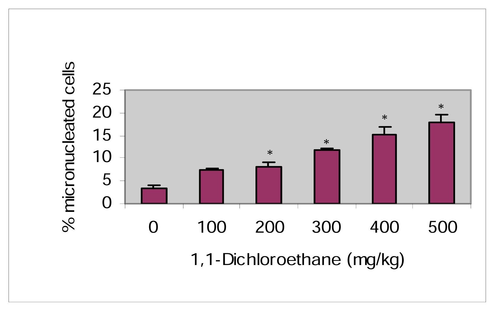
The micronuclei frequencies in bone marrow cells after intra-peritoneal treatment with 1,1-dichloroethane are summarized in Table 2. 1,1-dichloroethane induced a dose-dependent increase in micronuclei frequency (Figure 3) and significant (p>0.05) differences from the control were observed. The mean frequency of micro-nucleated cells were 3.33 ± 07, 7.33 ± 0.9, 8.00 ± 1.0, 11.67 ± 1.2, 15.33 ± 0.7, 18.00 ± 1.7 at 1,1-Dichloroethane doses of 0, 100, 200, 300, 400, and 500 mg/kg BW respectively.
Discussion
Rodent animal bioassays are valuable tools for investigating the pharmacokinetics, mechanisms of action, and differential toxicity of various chemicals [3]. The data obtained from this study clearly shows that 1,1-DCE significantly increased the number of chromosomal aberrations and the formation of micronuclei as compared to the control. These results support earlier studies with halogenated compounds [13–15]. The increase in chromosome aberrations and micronuclei formation was in a dose-dependent manner. The different type of aberrations produced by the test chemical suggests their clastogenic potential. The increased chromosomal aberrations could be attributed to either an increase in induced DNA lesions or interference with their repair [16]. The chromosome gap, which represents only the loss of chromatin material [17], may be due to the damage to the protein part of the chromosome rather than the whole chromosome. The chromatid breaks represent the DNA double strand breaks that have not undergone G2 repair [18]. However, a number of factors can influence the time of appearance of chemically induced aberrations such as compound solubility, rate and distribution of biotransport, availability at the target site as influenced by time and cell permeability [19].
Mitotic index depression was observed in the bone-marrow cells of mice as compared with the control. The decrease was in a dose-dependent manner. These results were in agreement with Rank & Nielsen [15] who observed a decrease in mitotic index in the Allium cepa root tips. Duma et al [20] found decreased mitotic index by the monocrotophos in rodents. Significant decrease in the mitotic index was observed with the Roundup herbicide [21].
Chromosomal breaks or interference in the mitotic process results in lagging of the chromosomal material and during cell division, this leads to the formation of micronuclei [22]. Treatments with 1,1-DCE have shown a dose-dependent increase in the number of micronuclei. These results were in conjunction with the studies of Tafazoli & Kirsch-Volders [23] with chlorinated hydrocarbons in human lymphocytes; Jagetia & Jyothi [24] with Vinesine (desacetyl vinblastinamide sulphate) in mouse bone marrow cells; and Gutierrez et al [25] with 131I sodium iodide exposure in human. Several other studies have shown an increased in micronulei formation in treated cells [22, 26–29]. The induction of micronuclei by 1,1-DCE may be due to the ability of these compounds to affect DNA synthesis, which may lead to DNA strand breaks and as a result, breakage of chromosome and or loss of whole chromosome owing to the spindle failure.
Conclusions
Our data indicate that 1,1-dichloroethane induces chromosome aberrations and the formation of micronuclei in the bone marrow cells of Swiss-Webster mice. The repression in mitotic index indicates the potential for 1,1-dichloroethane to induce growth arrest or to inhibit cell growth. These findings demonstrate that 1,1-dichloroethane has a strong clastogenic/genotoxic potential. The chromosome aberration assay and micronuclei test appear to be promising technique to assess the clastogenic/genotoxic potential of 1,1-dichloroethane and its compounds.
Figure 1.
Effect of 1,1-Dichloroethane on the frequency of chromosomal aberrations.
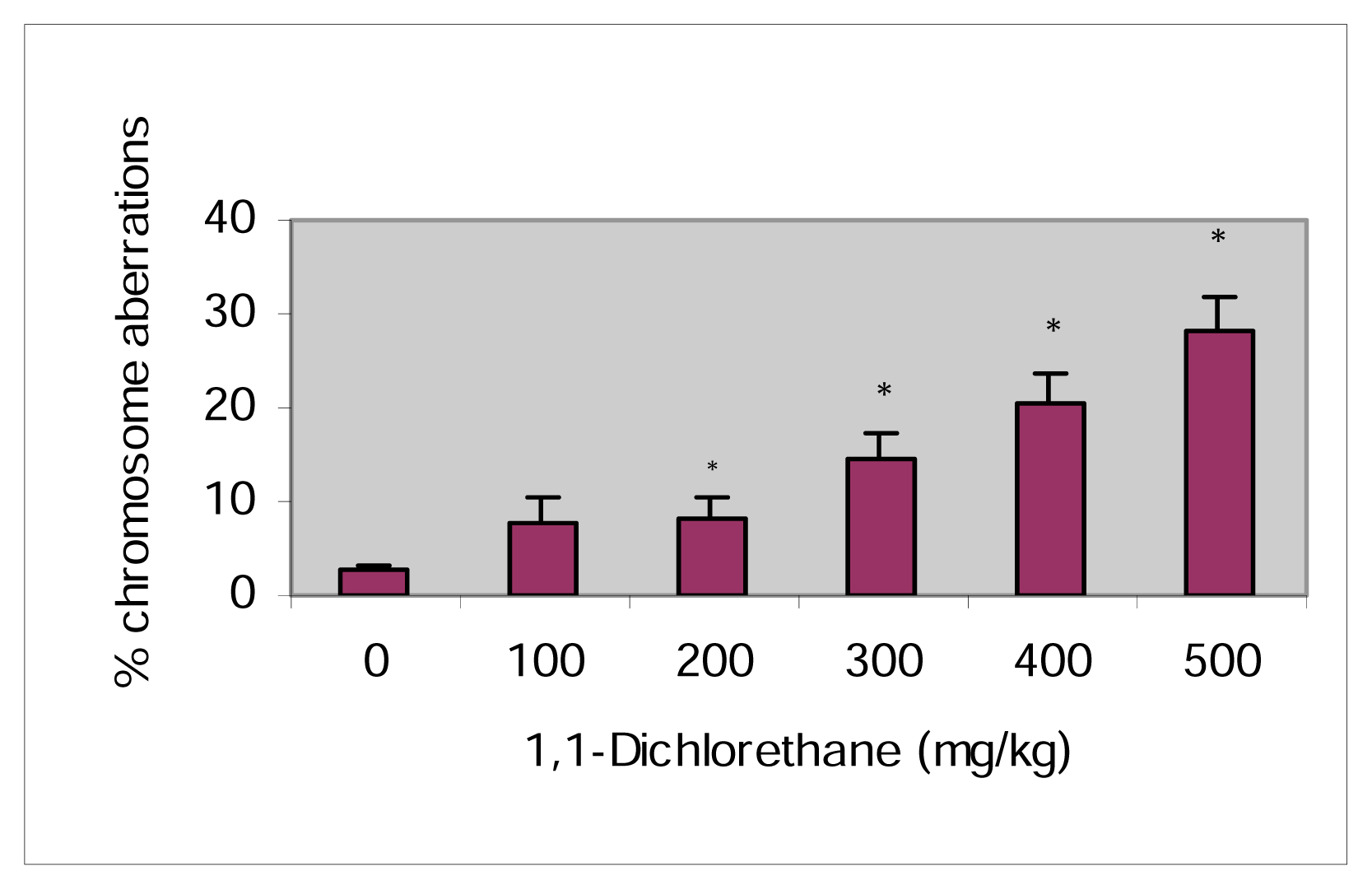
Figure 2.
Effect of 1,1-Dichloroethane on the percent Mitotic Index.
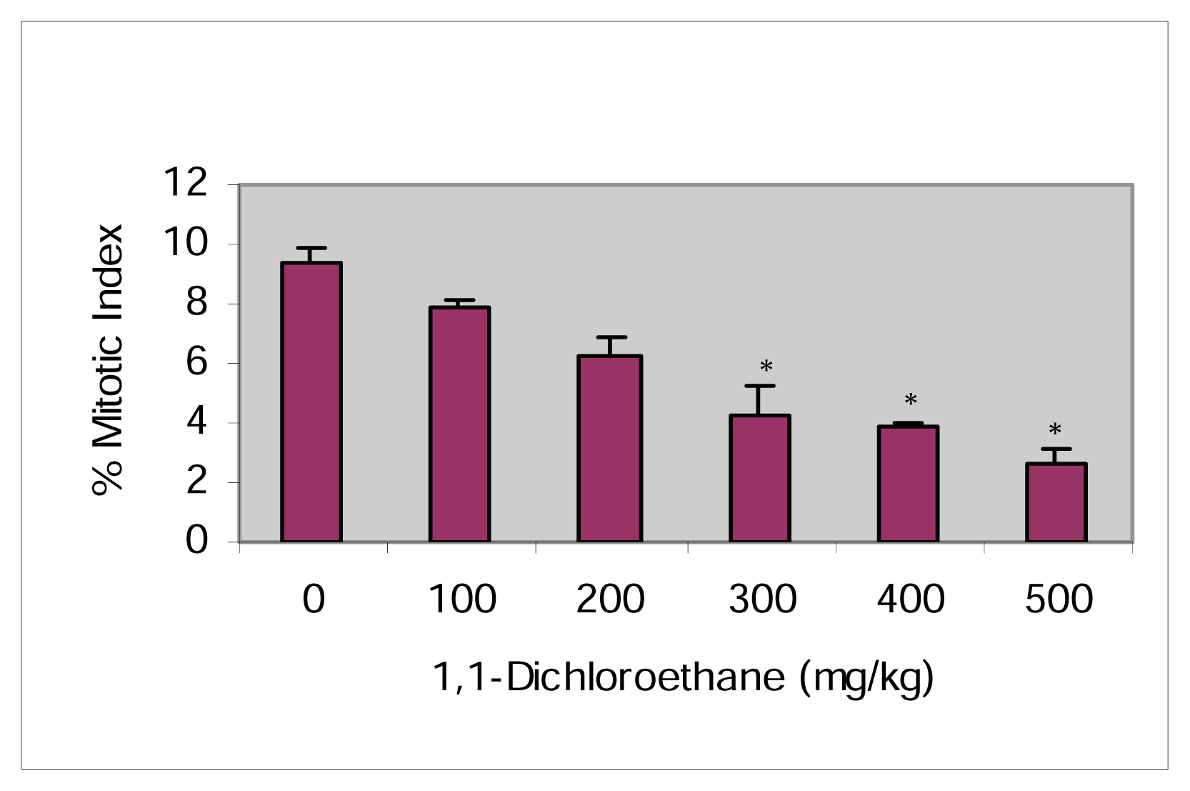
Figure 3.
Effect of 1,1-Dichloroethane on the percent Micronuclei induction.

Table 1.
Frequency of chromosome aberrations in bone marrow cells of Swiss-Webster mice induced by 1,1-Dichloroethane.
| Dose (mg/kg) | Total Metaphases Examined (n) | MI | Chromatid | Chromosome | Total SCA | Mean % SCA ±SEM | ||
|---|---|---|---|---|---|---|---|---|
| Gap | Break | Gap | Break | |||||
| Ethanol | 300/3 | 9.4 | 2 | 1 | 3 | 2 | 8 | 2.67 ± 0.58 |
| 100 | 300/3 | 7.9 | 4 | 6 | 7 | 6 | 23 | 7.67 ± 2.88 |
| 200 | 300/3 | 6.2 | 7 | 3 | 8 | 7 | 25 | 8.33 ± 2.08 |
| 300 | 300/3 | 4.3 | 6 | 5 | 16 | 17 | 44 | 14.67 ± 2.51 |
| 400 | 300/3 | 3.0 | 15 | 10 | 19 | 17 | 61 | 20.3 ± 3.21 |
| 500 | 300/3 | 2.6 | 20 | 19 | 24 | 21 | 84 | 28.0 ± 3.61 |
n: number of animals; SCA: Structural chromosomal aberrations; MI: mitotic index p< 0.05 compared to control
Table 2.
Frequency of micronucleated cells in mice bone marrow. Means followed by a common letter are not significantly different from each other at p≤ 0.05 (Tukey test).
| Dose | Rat Number | MN*per 1000 Cells | Fixation Time (h) |
|---|---|---|---|
| Ethanol (1%) | 1 | 3 | |
| 2 | 4 | ||
| 3 | 3 | ||
| Mean ± S.E. | 3.33 ± 0.58a | 30 | |
| 1.1 DCE (mg/kg BW, i.p) | |||
| 100 | 1 | 7 | |
| 2 | 7 | ||
| 3 | 8 | ||
| Mean ± S.E. | 7.33 ± 0.57b | 30 | |
| 200 | 1 | 9 | |
| 2 | 7 | ||
| 3 | 8 | ||
| Mean ± S.E. | 8.0 ± 1.0b | 30 | |
| 300 | 1 | 11 | |
| 2 | 12 | ||
| 3 | 12 | ||
| Mean ± S.E. | 11.67 ± 0.58c | 30 | |
| 400 | 1 | 15 | |
| 2 | 14 | ||
| 3 | 17 | ||
| Mean ± S.E. | 15.3 ± 1.52d | 30 | |
| 500 | 1 | 17 | |
| 2 | 17 | ||
| 3 | 20 | ||
| Mean ± S.E. | 18.0 ± 1.73d | 30 |
*Micronuclei; DCE: 1,1-dichloroethane
References
- ATSDR, Toxicological Profile for 1,1-dichloroethane. In Agency for Toxic Substances and Disease Registry; U.S. Department of Health and Human Services: Atlanta, GA, 1990.
- U.S. EPA. Integrated Risk Information System; Environmental Protection Agency, 1990. Available at: http://www.epa.gov/ir Accessed January 18, 2001.
- Roldan-Arjona, T.; Garcia-Pedrajas, D.; Luque-Romero, F.; Hera, C.; Pueyo, C. An association between mutagenicity of the Ara test of Salmonella typhimurium and carcinogenicity in rodents for 16 halogenated aliphatic hydrocarbons. Mutagenesis 1991, 6(3), 199–205. [Google Scholar]
- Tafazoli, M.; Baeten, A.; Geerlings, P.; Kirsch-Volders, M. In vitro mutagenicity and genotoxicity study of a number of short-chain chlorinated hydrocarbons using the micronucleus test and the alkaline single cell gel electrophoresis technique (comet assay) in human lymphocytes: a structure-activity relationship (QSAR) analysis of the genotoxic and cytotoxic potential. Mutagenesis 1998, 13(2), 115–126. [Google Scholar]
- Rosenkranz, H. S.; Klopman, G. A study of the structural basis of the ability of chlorinated alkanes and alkenes to induce aneuploidy and toxicity in the mold. Aspergillus nidulans. Mutat. Res 1996, 354, 183–193. [Google Scholar]
- Gerhard, I.; Daniel, V.; Link, S.; Monga, B.; Runnebaum, B. Chlorinated hydrocarbons in women with repeated miscarriages. Environ. Health. Perspect 1998, 106, 675–681. [Google Scholar]
- Evans, E. B.; Blaster, R. L. CNS depressant effects of volatile organic solvents. Neurosci. Biobehav. Rev 1991, 15, 233–241. [Google Scholar]
- Wilson, C. Chemical exposure and human health: A reference to 314 chemicals with a guide to symptoms and a directory of organizations; McFarland & Company: North Carolina, 1993. [Google Scholar]
- Preston, R. J.; Dean, B. J.; Galloway, S.; Holden, H.; McFee, A. F.; Shelby, M. Mammalian in vivo cytogenetic assays: Analysis of chromosome aberrations in bone marrow cells. Mutat.Res 1987, 189, 157–165. [Google Scholar]
- Romanga, F.; Staniforth, C. D. The automated bone marrow micronucleus test. Mutat. Res 1989, 213, 91–104. [Google Scholar]
- Schmid, W. the micronuclei test for cytogenetic analysis. Hollaender, A., Ed.; In Chemical mutagens, Principles and Methods for their detection; Volume 4, Plenum Press: New York, 1976; pp. 31–53. [Google Scholar]
- Agarwal, D. K.; Chauhan, L. K. S. An Improved chemical substitute for fetal calf serum for the micronucleus test. Biotechnol. Histochem 1993, 68, 187–188. [Google Scholar]
- Matsuoka, A.; Yamada, K.; Kusakable, H.; Wakuri, S.; et al. Re-evaluation of chromosomal aberration induction on nine mouse lymphoma assay “unique positive” NTP carcinogens. Mutat.Res 1996, 369, 243–252. [Google Scholar]
- Whittaker, S. G.; Zimmermann, F. K.; Dicus, B.; Piegorsch, W. W.; Resnick, M. A.; Fogel, S. Detection of induced mitotic chromosome loss in Saccharomyces cerevisiae – an interlaboratory assessment of 12 chemicals. Mutat. Res. 1990, 241(3), 225–42. [Google Scholar]
- Rank, J.; Nielsen, M. H. Evaluation of the Allium anaphase-telophase test in relation to genotoxicity screening of industrial wastewater. Mutat. Res 1994, 312(1), 17–24. [Google Scholar]
- Sasaki, F.Y.; Yamada, H.; Sugiyama, C.; Kinae, N. Increasing effect of tri-n-butyltins and triphenyltins on the frequency of chemically induced aberrations in cultured Chinese hamster cells. Mutat. Res 1993, 300, 5–14. [Google Scholar]
- Topaktas, M.; Rencuzogullar, E. Chromosomal aberrations in cultured human lymphocytes treated with Marshal and its effective ingredient Carbosulfan. Mutat. Res. 1993, 319(2), 103–11. [Google Scholar]
- Kihlman, B. A.; Hanssan, K.; Andersson, H. C. The effects of post-treatments with caffeine during S and G2 on the frequencies of chromosomal aberrations induced by thiotepa in root-tips of vicia faba and in human lymphocytes in vitro. Mutat. Res 1982, 104(4–5), 323–330. [Google Scholar]
- McFee, A. F.; Tice, R. R. Influence of treatment to sacrifice time and the presence of BrdUrd on chemically-induced aberration rates in mouse marrow cells. Mutat. Res 1990, 241(1), 95–108. [Google Scholar]
- Duma, D.; Raicu, P.; Hamar, M.; Tuta, A. Cytogenetic effects of some pesticides on rodents. Rev. Roum. Biol 1977, 22, 93–96. [Google Scholar]
- Rank, J.; Jensen, A. G.; Skov, B.; Pedersen, L. H.; Jensen, K. Genotoxicity testing of the herbicide Roundup and its active ingredient glyphosate isopropylamine using the mouse bone marrow micronucleus test, Salmonella mutagenicity test, allium anaphase-telophase test. Mutat. Res 1993, 300, 29–36. [Google Scholar]
- Gudi, R. D.; Sandhu, S. S.; Athwal, S. R. Kinetochore identification in micronuclei in mouse bone marrow erythrocytes: An assay for the detection of aneuploidy-inducing agents. Mutat. Res 1990, 234, 263–268. [Google Scholar]
- Tafazoli, M.; Kirsch-Volders, M. In vitro mutagenicity and genotoxicity study of 1,2-dichloroethylene, 1,1,2-trichloroethane, 1,3-dichloropropane, 1,2,3-trichloropropane and 1,1,3-trichloropropene, using the micronucleus test and the alkaline single cell gel electrophoresis technique (comet assay) in human lymphocytes. Mutat. Res 1996, 371(3–4), 185–202. [Google Scholar]
- Jagetia, G. C.; Jyothi, P. Enhancement of micronuclei frequency by vindesine in mouse bone marrow. Mutat. Res 1997, 388, 1–5. [Google Scholar]
- Gutierrez, S.; Carbonell, E.; Galofre, P.; Creus, A.; Marcos, R. Micronuclei induction by 131I exposure: Study in hyperthyroidism patients. Mutat. Res 1997, 373, 39–45. [Google Scholar]
- Anwar, W.; Khalil, M. M.; Wild, C. P. Micronuclei, chromosomal aberrations and aflatoxin-albumin adducts in experimental animals after exposure to aflatoxinB1. Mutat. Res 1994, 322, 61–67. [Google Scholar]
- Jayashree, I. V.; Vijayalaxmi, K. K.; Abdul Rahiman, M. The genotoxicity of Hinosan, an organophosphorus pesticide in the vivo mouse. Mutat. Res 1994, 322(2), 77–85. [Google Scholar]
- Grover, I. S.; Malhi, P. K. Genotoxic effects of some organophosphorous pesticides. I. Induction of micronuclei in bone marrow cells in rat. Mutat. Res 1985, 155(3), 131–4. [Google Scholar]
- Sun, J. T.; Armstrong, M. J.; Galloway, S. M. Rapid method for improving slide quality in the bone marrow micronucleus assay; an adapted cellulose column procedure. Mutat. Res 1999, 439, 121–126. [Google Scholar]
© 2005 MDPI. All rights reserved.
Share and Cite
MDPI and ACS Style
Patlolla, B.P.; Patlolla, A.K.; Tchounwou, P.B. Cytogenetic Effects of 1,1-Dichloroethane in Mice Bone Marrow Cells. Int. J. Environ. Res. Public Health 2005, 2, 101-106. https://doi.org/10.3390/ijerph2005010101
AMA Style
Patlolla BP, Patlolla AK, Tchounwou PB. Cytogenetic Effects of 1,1-Dichloroethane in Mice Bone Marrow Cells. International Journal of Environmental Research and Public Health. 2005; 2(1):101-106. https://doi.org/10.3390/ijerph2005010101
Chicago/Turabian StylePatlolla, Babu P., Anita K. Patlolla, and Paul B. Tchounwou. 2005. "Cytogenetic Effects of 1,1-Dichloroethane in Mice Bone Marrow Cells" International Journal of Environmental Research and Public Health 2, no. 1: 101-106. https://doi.org/10.3390/ijerph2005010101