Factors Influencing Microbiological Biodiversity of Human Foot Skin

 , and
, and 
Abstract
1. Introduction
2. Materials and Methods
2.1. Characteristics of the Study Participants
2.2. Sampling
2.3. Culture Method
2.4. DNA Extraction and High-Throughput Sequencing.
2.5. Mathematical and Statistical Analyses
3. Results and Discussion
3.1. The Influence of the Studied Factors on the Number of Bacteria and Fungi on Foot Skin
3.2. Determining the Influence of the Studied Factors on the Microbiological Biodiversity of Foot Skin Using Culture Method
3.3. Microbiological Biodiversity of Foot Skin on the Basis of High-Throughput Sequencing
3.4. Comparison of Culture and High-Throughput Sequencing Methods
4. Conclusions
Supplementary Materials
Author Contributions
Funding
Conflicts of Interest
References
- Rosenthal, M.; Goldberg, D.; Aiello, A.; Larson, E.; Foxman, B. Skin microbiota: Microbial community structure and its potential association with health and disease. Infect. Genet. Evol. 2011, 11, 839–848. [Google Scholar] [CrossRef] [PubMed]
- Kong, H.H.; Segre, J.A. Skin Microbiome: Looking back to move forward. J. Investig. Dermatol. 2012, 132, 933–939. [Google Scholar] [CrossRef] [PubMed]
- Scharschmidt, T.C.; Fischbach, M.A. What lives on our skin: Ecology, genomics and therapeutic. Opportunities of the skin microbiome. Drug Discov. Today Dis. Mech. 2013, 10, 1083–1089. [Google Scholar] [CrossRef] [PubMed]
- Grice, E.A.; Kong, H.H.; Renaud, G.; Young, A.C.; NISC Comparative Sequencing Program; Bouffard, G.G.; Blakesley, R.W.; Wolfsberg, T.G.; Turner, M.L.; Segre, J.A. A diversity profile of the human skin microbiota. Genome Res. 2008, 18, 1043–1050. [Google Scholar] [CrossRef] [PubMed]
- Grice, E.A.; Kong, H.H.; Conlan, S.; Deming, C.B.; Davis, J.; Young, A.C.; NISC Comparative Sequencing Program; Bouffard, G.G.; Blakesley, R.W.; Murray, P.R.; et al. Topographical and temporal diversity of the human skin microbiome. Science 2009, 5931, 1190–1192. [Google Scholar] [CrossRef] [PubMed]
- Gao, Z.; Tseng, C.; Pei, Z.; Blaser, M.J. Molecular analysis of human forearm superficial skin bacterial biota. Proc. Natl. Acad. Sci. USA 2007, 104, 2927–2932. [Google Scholar] [CrossRef] [PubMed]
- Fierer, N.; Hamady, M.; Lauber, C.L.; Knight, R. The influence of sex, handedness, and washing on the diversity of hand surface bacteria. Proc. Natl. Acad. Sci. USA 2008, 105, 17994–17999. [Google Scholar] [CrossRef] [PubMed]
- Costello, E.K.; Lauber, C.L.; Hamady, M.; Fierer, N.; Gordon, J.I.; Knight, R. Bacterial Community Variation in Human Body Habitats Across Space and Time. Science 2009, 5960, 1694–1697. [Google Scholar] [CrossRef] [PubMed]
- Paulino, L.C.; Tseng, C.-H.; Strober, B.E.; Blaser, M.J. Molecular analysis of fungal microbiota in samples from healthy human skin and psoriatic lesions. J. Clin. Microbiol. 2006, 44, 2933–2941. [Google Scholar] [CrossRef] [PubMed]
- Findley, K.; Oh, J.; Yang, J.; Conlan, S.; Deming, C.; Meyer, J.A.; Schoenfeld, D.; Nomicos, E.; Park, M.; NISC Comparative Sequencing Program; et al. Human skin fungal diversity. Nature 2013, 7454, 367–370. [Google Scholar] [CrossRef]
- Cogen, A.L.; Nizet, V.; Gallo, R.L. Skin microbiota: A source of disease or defence? Br. J. Dermatol. 2008, 158, 442–455. [Google Scholar] [PubMed]
- Dominguez-Bello, M.G.; Costello, E.K.; Contreras, M.; Magris, M.; Hidalgo, G.; Fierer, N.; Knight, R. Delivery mode shapes the acquisition and structure of the initial microbiota across multiple body habitats in newborns. Proc. Natl. Acad. Sci. USA 2010, 107, 11971–11975. [Google Scholar] [CrossRef] [PubMed]
- Capone, K.A.; Dowd, S.E.; Stamatas, G.N.; Nikolovski, J. Diversity of the human skin microbiome early in life. J. Investig. Dermatol. 2011, 131, 2026–2032. [Google Scholar] [CrossRef] [PubMed]
- Kong, H.H. Skin microbiome: Genomics-based insights into the diversity and role of skin microbes. Trends Mol. Med. 2011, 17, 320–328. [Google Scholar] [CrossRef] [PubMed]
- Grice, E.A.; Segre, J.A. The skin microbiome. Nat. Rev. Microbiol. 2011, 9, 244–253. [Google Scholar] [PubMed]
- Larson, E. Hygiene of the skin: When is clean too clean? Emerg. Infect. Dis. 2001, 7, 225–230. [Google Scholar] [CrossRef] [PubMed]
- Muyzer, G.; de Wall, E.C.; Uitterlinden, A.G. Profiling of complex microbial populations by denaturing gradient gel electrophoresis analysis of polymerase chain reaction-amplified genes coding for 16S rRNA. Appl. Environ. Microbiol. 1993, 59, 695–700. [Google Scholar]
- White, T.J.; Bruns, T.; Lee, S.; Taylor, J. Amplification and direct sequencing of fungal ribosomal RNA genes for phylogenetics. In PCR Protocols: A Guide to Methods and Applications; Innis, M.A., Gelfand, D.H., Shinsky, J.J., White, T.J., Eds.; Academic Press: San Diego, CA, USA, 1990; pp. 315–322. [Google Scholar]
- Zhang, Z.; Schwartz, S.; Wagner, L.; Miller, W. A greedy algorithm for aligning DNA sequences. J. Comput. Biol. 2000, 7, 203–214. [Google Scholar] [CrossRef]
- Fassatiová, O. Microscopic Fungi in Technical Microbiology; Wydawnictwo Naukowo-Techniczne: Warsaw, Poland, 1983. (In Polish) [Google Scholar]
- Samson, R.A.; Hoekstra, E.S.; Frisvad, J.C.; Filtenborg, O. Introduction to Food and Air Born Fungi; Centraalbureau von Schimmelcultures: Utrecht, The Netherlands, 2000. [Google Scholar]
- Quast, C.; Klindworth, A.; Pruesse, E.; Schweer, T.; Horn, M.; Glo, F.O. Evaluation of general 16S ribosomal RNA gene PCR primers for classical and next-generation sequencing-based diversity studies. Nucleic Acids Res. 2013, 41, e1. [Google Scholar]
- Caporaso, J.G.; Kuczynski, J.; Stombaugh, J.; Bittinger, K.; Bushman, F.D.; Costello, E.K.; Fierer, N.; Peña, A.G.; Goodrich, J.K.; Gordon, J.I.; et al. QIIME allows analysis of high—Throughput community sequencing data. Nat. Methods 2010, 7, 335–336. [Google Scholar] [CrossRef]
- Greengenes Database. Available online: http://greengenes.lbl.gov (accessed on 27 March 2019).
- UNITE. Available online: http://unite.ut.ee/repository.php (accessed on 27 March 2019).
- Oh, J.; Byrd, A.L.; Deming, C.; Conlan, S.; NISC Comparative Sequencing Program; Kong, H.H.; Segre, J.A. Biogeography and individuality shape function in the human skin metagenome. Nature 2014, 514, 59–64. [Google Scholar] [CrossRef] [PubMed]
- Jo, J.-H.; Deming, C.; Kennedy, E.A.; Conlan, S.; Polley, E.C.; Ng, W.-I.; NISC Comparative Sequencing Program; Kong, H.H. Diverse human skin fungal communities in children converge in adulthood. J. Investig. Dermatol. 2016, 136, 2356–2363. [Google Scholar] [CrossRef] [PubMed]
- Directive 2000/54/EC—Biological Agents at Work. Available online: https://osha.europa.eu/en/legislation/directives/exposure-to-biological-agents/77 (accessed on 4 November 2016).
- Falkiewicz-Dulik, M. Footwear materials sanitization. In Proceedings of the II Konferencja Naukowa. Rozkład i korozja Mikrobiologiczna Materiałów Technicznych, Lodz, Poland, 30–31 May 2001; Lodz University of Technology: Łódź, Poland, 2001. (In Polish). [Google Scholar]
- Ghanem, N.B. Study on the antimicrobial activity of honey products and some Saudi Folkloric substances. Res. J. Biotechnol. 2011, 6, 38–43. [Google Scholar]
- Kacániová, M.; Vuković, N.; Chlebo, R.; HaŠČík, P.; Rovná, K.; Cubon, J.; Dżugan, M.; Pasternakiewicz, A. The antimicrobial activity of honey, bee pollen loads and beeswax from Slovakia. Arch. Biol. Sci. 2012, 64, 927–934. [Google Scholar] [CrossRef]
- Dhiman, G.; Chakraborty, J.N. Antimicrobial performance of cotton finished with triclosan, silver and chitosan. Fash. Text. 2015, 2, 13. [Google Scholar] [CrossRef]
- Fratini, F.; Cilia, G.; Turchi, B.; Felicioli, A. Beeswax: A minireview of its antimicrobial activity and its application in medicine. Asian Pac. J. Trop. Med. 2016, 9, 839–843. [Google Scholar] [CrossRef] [PubMed]
- Haase, H.; Jordan, L.; Keitel, L.; Keil, C.; Mahltig, B. Comparison of methods for determining the effectiveness of antibacterial functionalized textiles. PLoS ONE 2017, 12, e0188304. [Google Scholar] [CrossRef]


| Microorganisms | Total Microorganisms Number (CFU/cm2) | |||||||
|---|---|---|---|---|---|---|---|---|
| Women | Man | |||||||
| 0–10 Years Old | 11–17 Years Old | 18–50 Years Old | >60 Years Old | 0–10 Years Old | 11–17 Years Old | 18–50 Years Old | >60 Years Old | |
| Bacteria | X: 3.9 × 10⁶ A | X: 6.0 × 10⁴ AB | X: 1.2 × 10⁴ B | X: 1.1 × 10⁴ B | X: 3.6 × 10⁵ A | X: 1.0 × 10⁵ AB | X: 1.1 × 10⁴ B | X: 7.6 × 10³ B |
| SD: 5.9 × 10⁶ | SD: 7.7 × 10⁴ | SD: 6.4 × 10³ | SD: 8.5 × 10³ | SD: 7.5 × 10⁵ | SD: 2.1 × 10⁵ | SD: 1.3 × 10⁴ | SD: 8.8 × 10³ | |
| Total number of bacteria | X: 1.0 × 10⁶ a | X: 1.2 × 10⁵ a | ||||||
| SD: 3.2 × 10⁶ | SD: 3.9 × 10⁵ | |||||||
| Fungi | X: 2.4 × 101 A | X: 6.8 × 101 A | X: 2.5 × 101 A | X: 2.1 × 10² A | X: 3.2 × 101 A | X: 1.7 × 101 A | X: <1 | X: 1.5 × 101 A |
| SD: 3.5 × 101 | SD: 1.1 × 10² | SD: 3.3 × 101 | SD: 3.0 × 10² | SD: 2.9 × 101 | SD: 3.4 × 101 | SD: <1 | SD: 3.1 × 101 | |
| Total number of fungi | 2 X: 8.1 × 101 | X: 1.6 × 101 b | ||||||
| SD: 8.6 × 101 | SD: 2.8 × 101 | |||||||
| Microorganisms | Total Microorganisms Number (CFU/cm2) | |||||
|---|---|---|---|---|---|---|
| Washing Feet Frequency | Physical Activity Frequency | |||||
| Twice a Day | Once a Day | Every Other Day | 3 Times per Week and Above | 1–2 Times per Week | Lack of Physical Activity | |
| Bacteria | X: 8.8 × 10³ A | X: 5.6 × 10⁵ A | X: 1.1 × 10⁶ B | X: 2.2 × 10⁴ A | X: 6.4 × 10⁵ B | X: 6.9 × 10⁵ A |
| SD: 6.1 × 10³ | SD: 2.5 × 10⁶ | SD: 2.5 × 10⁶ | SD: 3.4 × 10⁴ | SD: 1.9 × 10⁶ | SD: 2.8 × 10⁶ | |
| Fungi | X: 5.4 × 101 A | X: 5.6 × 101 A | X: 5.5 × 101 A | X: 1.2 × 101 A | X: 5.7 × 101 B | X: 5.6 × 101 AB |
| SD: 1.1 × 10² | SD: 1.4 × 10² | SD: 5.1 × 101 | SD: 2.5 × 101 | SD: 7.7 × 101 | SD: 1.6 × 10² | |
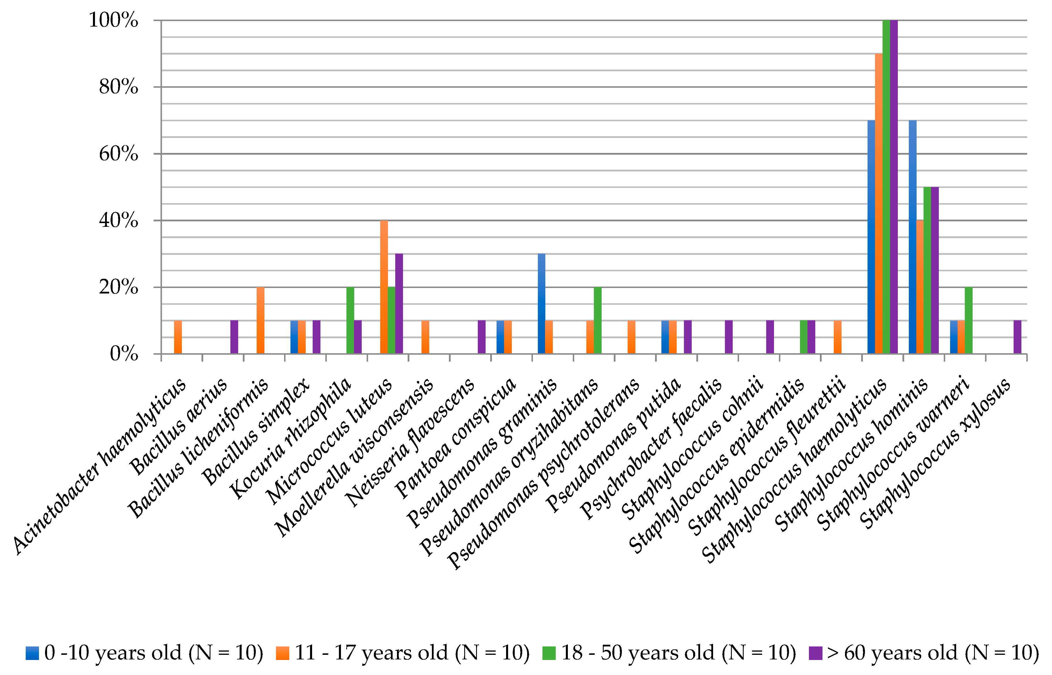
| Microorganisms Isolation Frequency Based on Samples Acquired from Examined People (N = 40) | |||
|---|---|---|---|
| Bacteria | (%) | Fungi | (%) |
| Acinetobacter haemolyticus | 2.5 | Acremonium sp. | 2.5 |
| Bacillus aerius | 2.5 | Alternaria consortiale | 5.0 |
| Bacillus licheniformis | 5.0 | Aspergillus candidus | 12.5 |
| Bacillus simplex | 7.5 | Aspergillus flavus | 5.0 |
| Kocuria rhizophila | 7.5 | Aspergillus fumigatus | 25.0 |
| Micrococcus luteus | 22.5 | Aspergillus niger | 12.5 |
| Moellerella wisconsensis | 2.5 | Cryptococcus adeliensis * | 2.5 |
| Neisseria flavescens | 2.5 | Cryptococcus magnus * | 5.0 |
| Pantoea conspicua | 5.0 | Geotrichum candidum | 5.0 |
| Pseudomonas graminis | 10.0 | Meyerozyma quilliermondii * | 12.5 |
| Pseudomonas oryzihabitans | 7.5 | Mucor circinelloides | 2.5 |
| Pseudomonas psychrotolerans | 2.5 | Naganishia diffluens * | 17.5 |
| Pseudomonas putida | 7.5 | Penicillium citrinum | 7.5 |
| Psychrobacter faecalis | 2.5 | Penicillium corylophilum | 5.0 |
| Staphylococcus cohnii | 2.5 | Penicillium funiculosum | 2.5 |
| Staphylococcus epidermidis | 5.0 | Penicillium glabrum | 17.5 |
| Staphylococcus fleurettii | 2.5 | Penicillium sclerotiorum | 2.5 |
| Staphylococcus haemolyticus | 90.0 | Penicillium sp. (1) | 2.5 |
| Staphylococcus hominis | 52.5 | Penicillium sp. (2) | 10.0 |
| Staphylococcus warneri | 10.0 | Pichia kudriavzevii * | 5.0 |
| Staphylococcus xylosus | 2.5 | Rhodotorula mucilaginosa * | 12.5 |
| Wickerhamomyces anomalus * | 15.0 | ||
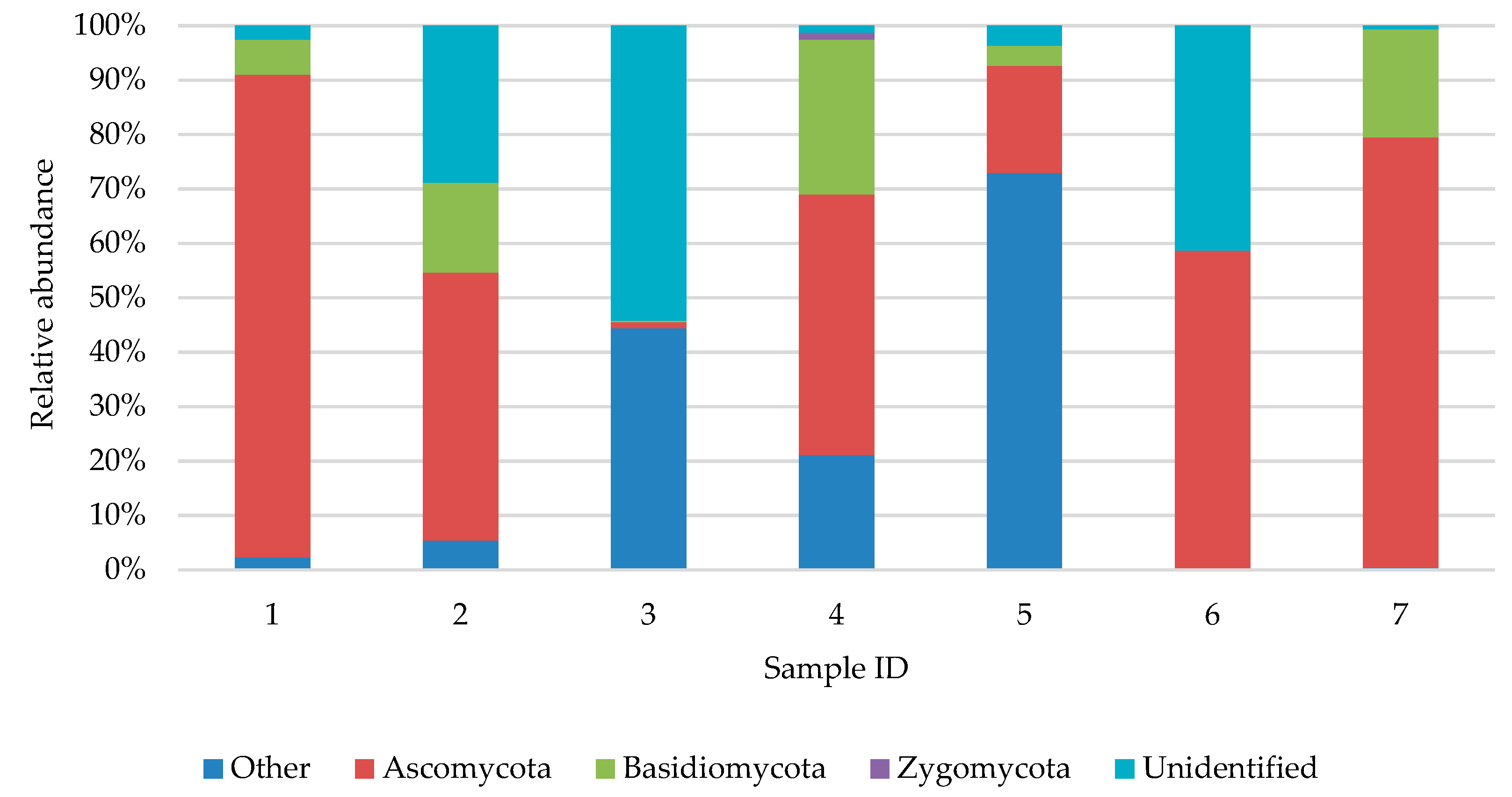
| Sample ID | Bacteria | Fungi |
|---|---|---|
| 1 | Actinobacteria: Brevibacterium, Oerskovia, Corynebacterium, Brachybacterium, Dietzia, Curtobacterium, Leucobacter, Microbacterium, Pseudoclavibacter, Salinibacterium, Yonghaparkia, Arthrobacter, Micrococcus, Sanguibacter, Rhodococcus, Streptomyces;Bacteroidetes: Chryseobacterium;Firmicutes: Bacillus, Brochothrix, Paenibacillus, Sporosarcina, Paenisporosarcina, Exiguobacterium, Aerococcus, Helcococcus, Staphylococcus *, Facklamia, Carnobacterium;Proteobacteria:Devosia, Agrobacterium, Paracoccus, Achromobacter, Acidovorax, Psychrobacter, Pseudomonas, Phenylobacterium, Paracoccus, Pigmentiphaga, Ralstonia, Acinetobacter | Ascomycota:Mycosphaerella, Arachnomyces, Penicillium *, Sagenomella;Basidiomycota:Laetiporus, Goffeauzyma, Malassezia |
| 2 | Actinobacteria: Brevibacterium, Oerskovia, Corynebacterium, Brachybacterium, Dietzia, Rhodococcus, Streptomyces, Actinomyces, Curtobacterium, Leucobacter, Microbacterium, Pseudoclavibacter, Yonghaparkia, Arthrobacter, Micrococcus, Mycobacterium, Nocardioides, Nocardiopsis, Propionibacterium;Bacteroidetes: Myroides, Chryseobacterium, Gelidibacter, Weeksella, Pedobacter, Sphingobacterium;Firmicutes: Bacillus, Paenibacillus, Sporosarcina, Staphylococcus*, Facklamia, Carnobacterium, Jeotgalicoccus, Aerococcus, Desemzia, Trichococcus, Lactococcus, Streptococcus, Dialister, Anaerococcus, Finegoldia, Peptoniphilus;Proteobacteria:Devosia, Agrobacterium, Paracoccus, Achromobacter, Acidovorax, Psychrobacter, Pseudomonas, Brevundimonas, Caulobacter, Mycoplana, Phenylobacterium, Ochrobactrum, Methylobacterium, Paracoccus, Rhodobacter, Kaistobacter, Sphingomonas, Comamonas, Neisseria, Erwinia, Acinetobacter, Enhydrobacter, Luteimonas | Ascomycota:Aspergillus (A. fumigatus) #, Mycosphaerella, Epicoccum, Preussia Cladophialophora, Penicillium *, Thelebolus, Debaryomyces, Wickerhamomyces, Scopulariopsis, Alternaria#;Basidiomycota:Sporobolomyces, Filobasidium, Malassezia |
| 3 | Actinobacteria: Brevibacterium, Oerskovia, Corynebacterium, Brachybacterium, Dietzia, Rhodococcus, Streptomyces, Micrococcus *, Curtobacterium, Leucobacter, Pseudoclavibacter, Salinibacterium, Yonghaparkia, Arthrobacter, Aeromicrobium;Bacteroidetes: Myroides, Chryseobacterium, Wautersiella, Weeksella, Pedobacter;Firmicutes: Bacillus*, Paenibacillus, Sporosarcina, Staphylococcus *, Facklamia, Carnobacterium, Planomicrobium, Jeotgalicoccus, Aerococcus, Desemzia, Trichococcus, Anaerococcus, Finegoldia, Peptoniphilus;Proteobacteria:Devosia, Agrobacterium, Paracoccus, Achromobacter, Tetrathiobacter, Acidovorax, Psychrobacter, Pseudomonas*, Brevundimonas, Caulobacter, Mycoplana, Phenylobacterium, Ochrobactrum, Paracoccus, Sphingomonas, Pigmentiphaga, Tetrathiobacter, Comamonas, Erwinia, Acinetobacter, Luteimonas | Ascomycota:Mycosphaerella, Penicillium, Thelebolus, Candida, Debaryomyces, Aspergillus (A. fumigatus) #, Geotrichum #;Basidiomycota:Malassezia |
| 4 | Actinobacteria: Brevibacterium, Oerskovia, Corynebacterium, Brachybacterium, Dietzia, Rhodococcus, Curtobacterium, Leucobacter, Microbacterium, Pseudoclavibacter, Salinibacterium, Yonghaparkia, Arthrobacter, Aeromicrobium, Xylanimicrobium, Sanguibacter;Bacteroidetes: Myroides, Chryseobacterium, Wautersiella, Pedobacter;Firmicutes: Bacillus, Brochothrix, Paenibacillus, Sporosarcina, Staphylococcus*, Facklamia, Carnobacterium, Planomicrobium, Jeotgalicoccus, Aerococcus, Desemzia, Trichococcus, Lactococcus;Proteobacteria:Devosia, Agrobacterium, Paracoccus, Achromobacter, Tetrathiobacter, Psychrobacter, Pseudomonas, Acinetobacter*, Moellerella#, Brevundimonas, Mycoplana, Phenylobacterium, Ochrobactrum, Pleomorphomonas, Mesorhizobium, Paracoccus, Rhodobacter, Achromobacter, Pigmentiphaga, Erwinia, Acinetobacter, Enhydrobacter, Luteimonas | Ascomycota:Mycosphaerella, Epicoccum, Penicillium *, Pichia #, Candida, Debaryomyces, Wickerhamomyces *, Scopulariopsis, Aspergillus (A. fumigatus #), Aureobasidium, Peltigera, Saccharomyces, Microascus Basidiomycota:Leucosporidium, Malassezia, Rhodotorula#;Zygomycota:Mucor |
| 5 | Actinobacteria: Brevibacterium, Kocuria #, Micrococcus #, Oerskovia, Corynebacterium, Brachybacterium, Dietzia, Rhodococcus, Curtobacterium, Leucobacter, Microbacterium, Salinibacterium, Yonghaparkia, Arthrobacter, Sanguibacter;Bacteroidetes: Chryseobacterium, Sphingobacterium, Wautersiella;Firmicutes: Bacillus, Paenibacillus, Sporosarcina, Staphylococcus *, Facklamia, Jeotgalicoccus, Aerococcus, Lactococcus, Helcococcus;Proteobacteria:Devosia, Agrobacterium, Paracoccus, Achromobacter, Tetrathiobacter, Acidovorax, Pseudomonas, Brevundimonas, Caulobacter, Mycoplana, Ochrobactrum, Aminobacter, Rhodobacter, Sphingomonas, Achromobacter, Pigmentiphaga, Janthinobacterium, Ralstonia, Acinetobacter, Enhydrobacter, Luteimonas | Ascomycota:Penicillium *, Aspergillus #, Alternaria#;Basidiomycota:Sampaiozyma |
| 6 | Actinobacteria: Oerskovia, Kocuria #, Micrococcus #, Corynebacterium, Streptomyces, Agromyces, Curtobacterium, Leucobacter, Microbacterium, Pseudoclavibacter, Salinibacterium, Yonghaparkia, Nocardia, Xylanimicrobium, Propionibacterium, Sanguibacter;Firmicutes: Bacillus, Paenibacillus, Sporosarcina, Staphylococcus *, Facklamia, Carnobacterium, Paenisporosarcina, Lactococcus;Proteobacteria:Achromobacter, Acidovorax, Psychrobacter, Pseudomonas, Sphingomonas, Sphingopyxis | Ascomycota:Mycosphaerella, Epicoccum, Alternaria, Penicillium *, Debaryomyces, Scopulariopsis;Basidiomycota:Filobasidium. Malassezia |
| 7 | Actinobacteria: Brevibacterium, Oerskovia, Corynebacterium, Brachybacterium, Dietzia, Rhodococcus, Streptomyces, Curtobacterium, Leucobacter, Microbacterium, Yonghaparkia, Arthrobacter, Micrococcus, Nocardiopsis, Propionibacterium, Sanguibacter;Firmicutes: Bacillus, Paenibacillus, Sporosarcina, Staphylococcus *, Carnobacterium, Paenisporosarcina;Proteobacteria:Devosia, Agrobacterium, Psychrobacter *, Pseudomonas, Brevundimonas, Caulobacter, Mycoplana, Ochrobactrum, Paracoccus, Sphingomonas, Ralstonia | Ascomycota:Mycosphaerella, Arachnomyces, Penicillium, Aureobasidium, Alternaria, Meyerozyma, Thelebolus, Scopulariopsis, Candida, Acrostalagmus, Microascus;Basidiomycota:Rhodotorula, Cutaneotrichosporon, Guehomyces, Malassezia, Cryptococcus#, Naganishia # |
| 8 | Actinobacteria: Brevibacterium, Micrococcus #, Oerskovia, Corynebacterium, Brachybacterium, Dietzia, Rhodococcus, Streptomyces, Curtobacterium, Leucobacter, Microbacterium, Yonghaparkia, Arthrobacter, Xylanimicrobium, Propionibacterium, Sanguibacter;Bacteroidetes: Myroides, Chryseobacterium, Wautersiella, Gelidibacterm Weeksella;Firmicutes: Bacillus, Paenibacillus, Sporosarcina, Staphylococcus *, Facklamia, Cohnella, Paenisporosarcina, Planomicrobium, Jeotgalicoccus, Desemzia, Trichococcus, Lactococcus;Proteobacteria:Devosia, Agrobacterium, Paracoccus, Achromobacter, Tetrathiobacter, Psychrobacter, Pseudomonas, Neisseria (N. flavescens) #, Brevundimonas, Caulobacter, Mycoplana, Balneimonas, Ochrobactrum, Nitratireductor, Paracoccus, Sphingomonas, Sphingopyxis, Methylocaldum, Acinetobacter, Enhydrobacter, Luteimonas | ns |
| Bacteria | Fungi |
|---|---|
| Arthrobacter psychrolactophilus, Bacillus licheniformis, Brachybacterium conglomeratum, Corynebacterium jeikeium, Jeotgalicoccus psychrophilus, Kocuria rhizophila, Micrococcus luteus, Neisseria flavescens, Pseudoclavibacter bifida, Pseudomonas graminis, Pseudomonas oryzihabitans, Psychrobacter marincola, Rhodococcus fascians, Staphylococcus equorum, Staphylococcus haemolyticus, Staphylococcus hominis, Staphylococcus warneri | Molds: Aspergillus candidus, Aspergillus flavus, Aspergillus fumigatus, Aspergillus niger, Geotrichum candidum, Penicillium citrinum, Penicillium glabrum Yeast: Cryptococcus magnus, Malassezia restricta, Meyerozyma quilermondii, Naganishia diffluens, Pichia kudriavzevii, Rhodotorula mucilaginosa, Wickerhamomyces anomalus |
© 2019 by the authors. Licensee MDPI, Basel, Switzerland. This article is an open access article distributed under the terms and conditions of the Creative Commons Attribution (CC BY) license (http://creativecommons.org/licenses/by/4.0/).
Share and Cite
Steglińska, A.; Jachowicz, A.; Szulc, J.; Adamiak, J.; Otlewska, A.; Pielech-Przybylska, K.; Gutarowska, B. Factors Influencing Microbiological Biodiversity of Human Foot Skin. Int. J. Environ. Res. Public Health 2019, 16, 3503. https://doi.org/10.3390/ijerph16183503
Steglińska A, Jachowicz A, Szulc J, Adamiak J, Otlewska A, Pielech-Przybylska K, Gutarowska B. Factors Influencing Microbiological Biodiversity of Human Foot Skin. International Journal of Environmental Research and Public Health. 2019; 16(18):3503. https://doi.org/10.3390/ijerph16183503
Chicago/Turabian StyleSteglińska, Aleksandra, Anita Jachowicz, Justyna Szulc, Justyna Adamiak, Anna Otlewska, Katarzyna Pielech-Przybylska, and Beata Gutarowska. 2019. "Factors Influencing Microbiological Biodiversity of Human Foot Skin" International Journal of Environmental Research and Public Health 16, no. 18: 3503. https://doi.org/10.3390/ijerph16183503
APA StyleSteglińska, A., Jachowicz, A., Szulc, J., Adamiak, J., Otlewska, A., Pielech-Przybylska, K., & Gutarowska, B. (2019). Factors Influencing Microbiological Biodiversity of Human Foot Skin. International Journal of Environmental Research and Public Health, 16(18), 3503. https://doi.org/10.3390/ijerph16183503