A Case of Incomplete and Atypical Kawasaki Disease Presenting with Retropharyngeal Involvement


Abstract
1. Introduction
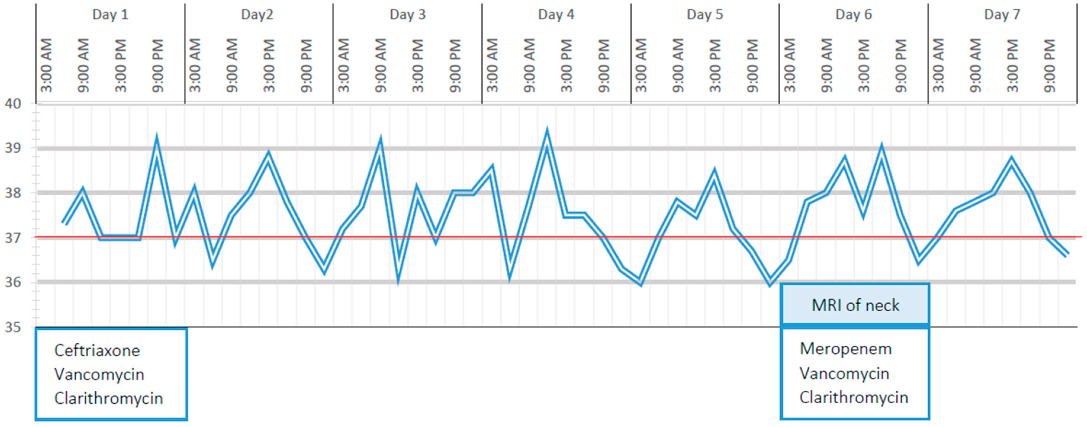
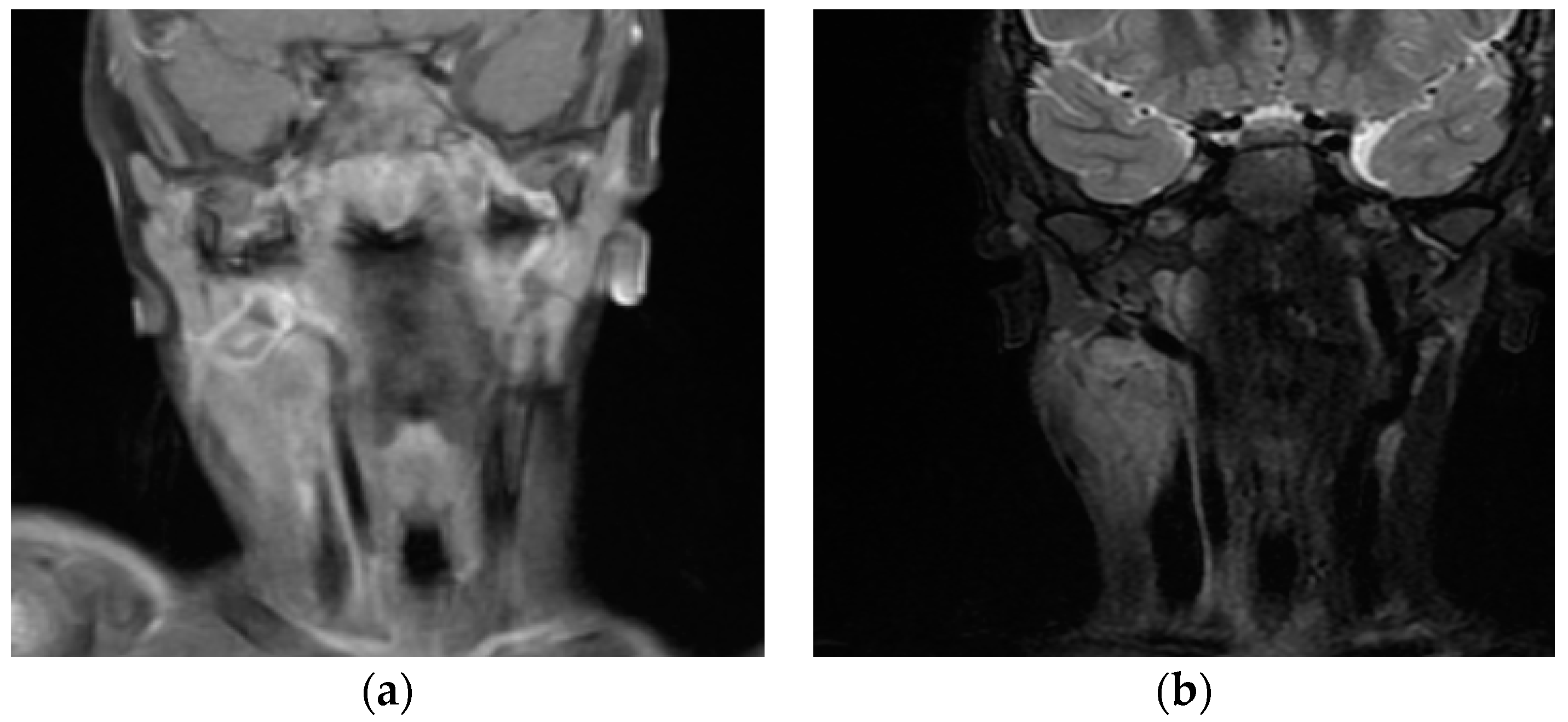
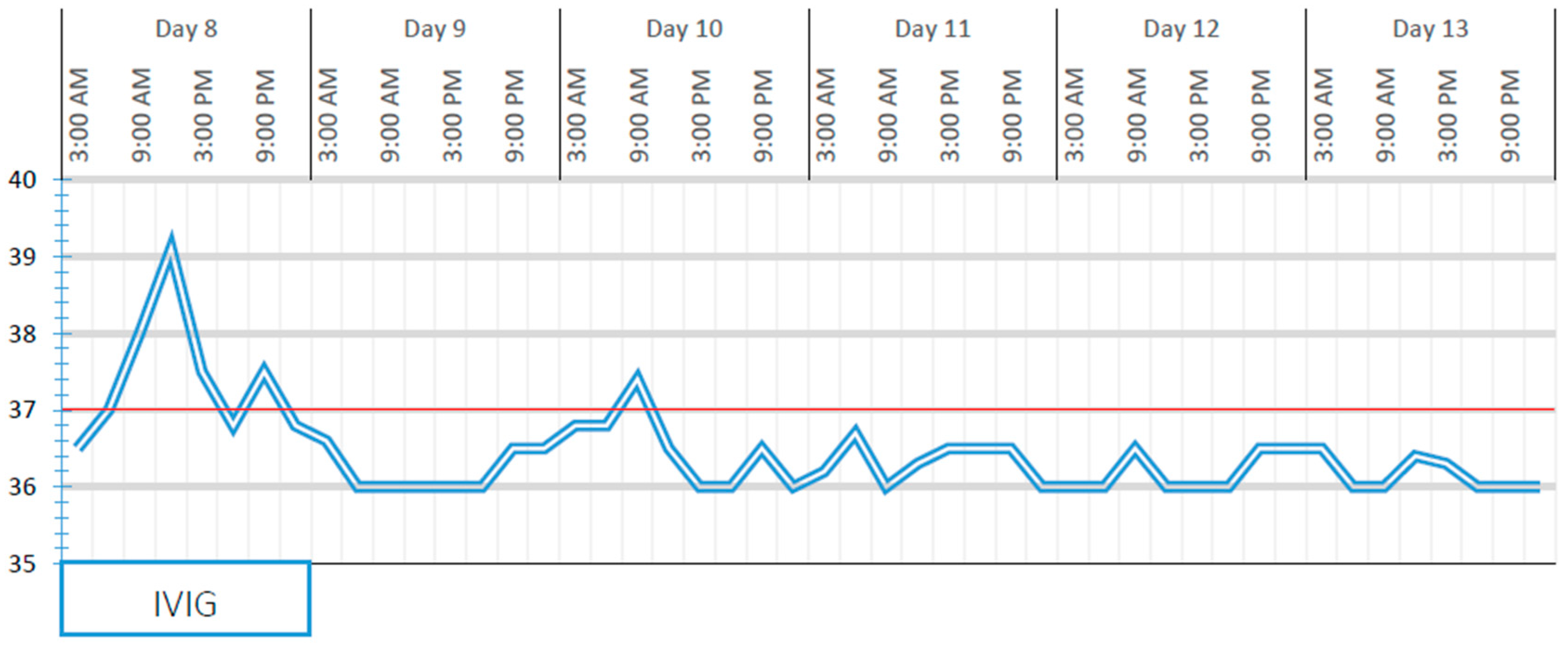
2. Case Report
3. Discussion
4. Conclusions
Author Contributions
Funding
Conflicts of Interest
References
- McCrindle, B.W.; Rowley, A.H.; Newburger, J.W.; Burns, J.C.; Bolger, A.F.; Gewitz, M.; Baker, A.L.; Jackson, M.A.; Takahashi, M.; Shah, P.B.; et al. Diagnosis, Treatment, and Long-Term Management of Kawasaki Disease: A Scientific Statement for Health Professionals from the American Heart Association. Circulation 2017, 135, e927–e999. [Google Scholar] [CrossRef]
- Holman, R.C.; Belay, E.D.; Christensen, K.Y.; Folkema, A.M.; Steiner, C.A.; Schonberger, L.B. Hospitalizations for Kawasaki syndrome among children in the United States, 1997–2007. Pediatr. Infect. Dis. 2010, 29, 483–488. [Google Scholar] [CrossRef]
- Ghimire, L.V.; Chou, F.S.; Mahotra, N.B.; Sharma, S.P. An update on the epidemiology, length of stay, and cost of Kawasaki disease hospitalisation in the United States. Cardiol. Young 2019, 29, 828–832. [Google Scholar] [CrossRef]
- Inagaki, K.; Blackshear, C.; Hobbs, C.V. Deep Neck Space Involvement of Kawasaki Disease in the US: A Population-Based Study. J. Pediatr. 2019, in press. [Google Scholar]
- Marchesi, A.; de Jacobis, I.T.; Rigante, D.; Rimini, A.; Malorni, W.; Corsello, G.; Bossi, G.; Buonuomo, S.; Cardinale, F.; Cortis, E.; et al. Kawasaki disease: Guidelines of the Italian Society of Pediatrics, part I-definition, epidemiology, etiopathogenesis, clinical expression and management of the acute phase. Ital. J. Pediatr. 2018, 44, 102. [Google Scholar] [CrossRef]
- Marchesi, A.; de Jacobis, I.T.; Rigante, D.; Rimini, A.; Malorni, W.; Corsello, G.; Bossi, G.; Buonuomo, S.; Cardinale, F.; Cortis, E.; et al. Kawasaki disease: Guidelines of Italian Society of Pediatrics, part II-treatment of resistant forms and cardiovascular complications, follow-up, lifestyle and prevention of cardiovascular risks. Ital. J. Pediatr. 2018, 44, 103. [Google Scholar] [CrossRef]
- Son, M.B.; Newburger, J.W. Kawasaki disease. Pediatr. Rev. 2013, 34, 151–162. [Google Scholar] [CrossRef]
- Principi, N.; Rigante, D.; Esposito, S. The role of infection in Kawasaki syndrome. J. Infect. 2013, 67, 1–10. [Google Scholar] [CrossRef]
- Esposito, S.; Polinori, I.; Rigante, D. The gut microbiota-host partnership as a potential driver of Kawasaki syndrome. Front. Pediatr. 2019, 7, 124. [Google Scholar] [CrossRef]
- Watanabe, T. Kawasaki disease with retropharyngeal edema following a blackfly bite. Case Rep. Pediatr. 2014, 2014, 296456. [Google Scholar] [CrossRef]
- Kim, J.S.; Kwon, S.H. Atypical Kawasaki disease presenting as a retropharyngeal abscess. Braz. J. Otorhinolaryngol. 2016, 82, 484–486. [Google Scholar] [CrossRef]
- Chiappini, E.; Camaioni, A.; Benazzo, M.; Biondi, A.; Bottero, S.; De Masi, S.; Di Mauro, G.; Doria, M.; Esposito, S.; Felisati, G.; et al. Development of an algorithm for the management of cervical lymphadenopathy in children: Consensus of the Italian Society of Preventive and Social Pediatrics, jointly with the Italian Society of Pediatric Infectious Diseases and the Italian Society of Pediatric Otorhinolaryngology. Expert Rev. Anti Infect. Ther. 2015, 13, 1557–1567. [Google Scholar]
- Yoskovitch, A.; Tewfik, T.L.; Duffy, C.M.; Moroz, B. Head and neck manifestations of Kawasaki disease. Int. J. Pediatr. Otorhinolaryngol. 2000, 52, 123–129. [Google Scholar] [CrossRef]
- Kanegaye, J.T.; Van Cott, E.; Tremoulet, A.H.; Salgado, A.; Shimizu, C.; Kruk, P.; Hauschildt, J.; Sun, X.; Jain, S.; Burns, J.C. Lymphnode-first presentation of Kawasaki disease compared with bacterial cervical adenitis and typical Kawasaki disease. J. Pediatr. 2013, 162, 1259–1263.e2. [Google Scholar] [CrossRef]
- Nozaki, T.; Morita, Y.; Hasegawa, D.; Makidono, A.; Yoshimoto, Y.; Starkey, J.; Kusakawa, I.; Manabe, A.; Saida, Y. Cervical ultrasound and computed tomography of Kawasaki disease: Comparison with lymphadenitis. Pediatr. Int. 2016, 58, 1146–1152. [Google Scholar] [CrossRef]
- Tashiro, N.; Matsubara, T.; Uchida, M.; Katayama, K.; Ichiyama, T.; Furukawa, S. Ultrasonographic evaluation of cervical lymph nodes in Kawasaki disease. Pediatrics 2002, 109, E77–E79. [Google Scholar] [CrossRef]
- Kato, H.; Kanematsu, M.; Kato, Z.; Teramoto, T.; Kondo, N.; Hoshi, H. Computed tomographic findings of Kawasaki disease with cervical lymphadenopathy. J. Comput. Assist. Tomogr. 2012, 36, 138–142. [Google Scholar] [CrossRef]
- Kritsaneepaiboon, S.; Tanaanantarak, P.; Roymanee, S.; Lee, E.Y. Atypical presentation of Kawasaki disease in young infants mimicking a retropharyngeal abscess. Emerg. Radiol. 2012, 19, 159–163. [Google Scholar] [CrossRef]
- NomVura, O.; Hashimoto, N.; Ishiguro, A.; Miyasaka, M.; Nosaka, S.; Oana, S.; Sakai, H.; Takayama, J.I. Comparison of patients with Kawasaki disease with retropharyngeal edema and patients with retropharyngeal abscess. Eur. J. Pediatr. 2014, 173, 381–386. [Google Scholar] [CrossRef]
- Roh, K.; Lee, S.W.; Yoo, J. CT analysis of retropharyngeal abnormality in Kawasaki disease. Korean J. Radiol. 2011, 12, 700–707. [Google Scholar] [CrossRef]
- Vural, C.; Gungor, A.; Comerci, S. Accuracy of computerised tomography in deep neck infections in the pediatric population. Am. J. Otolaryngol. 2003, 24, 143–144. [Google Scholar] [CrossRef]
- Puhakka, L.; Saat, R.; Klockars, T.; Kajosaari, L.; Salo, E.; Nieminen, T. Retropharyngeal involvement in Kawasaki disease—A report of four patients with retropharyngeal edema verified by magnetic resonance imaging. Int. J. Pediatr. Otorhinolaryngol. 2014, 78, 1774–1778. [Google Scholar] [CrossRef]


© 2019 by the authors. Licensee MDPI, Basel, Switzerland. This article is an open access article distributed under the terms and conditions of the Creative Commons Attribution (CC BY) license (http://creativecommons.org/licenses/by/4.0/).
Share and Cite
Isidori, C.; Sebastiani, L.; Esposito, S. A Case of Incomplete and Atypical Kawasaki Disease Presenting with Retropharyngeal Involvement. Int. J. Environ. Res. Public Health 2019, 16, 3262. https://doi.org/10.3390/ijerph16183262
Isidori C, Sebastiani L, Esposito S. A Case of Incomplete and Atypical Kawasaki Disease Presenting with Retropharyngeal Involvement. International Journal of Environmental Research and Public Health. 2019; 16(18):3262. https://doi.org/10.3390/ijerph16183262
Chicago/Turabian StyleIsidori, Chiara, Lisa Sebastiani, and Susanna Esposito. 2019. "A Case of Incomplete and Atypical Kawasaki Disease Presenting with Retropharyngeal Involvement" International Journal of Environmental Research and Public Health 16, no. 18: 3262. https://doi.org/10.3390/ijerph16183262
APA StyleIsidori, C., Sebastiani, L., & Esposito, S. (2019). A Case of Incomplete and Atypical Kawasaki Disease Presenting with Retropharyngeal Involvement. International Journal of Environmental Research and Public Health, 16(18), 3262. https://doi.org/10.3390/ijerph16183262