Article

Cyanobacterial Xenobiotics as Evaluated by a Caenorhabditis elegans Neurotoxicity Screening Test

1
Key Laboratory of Environmental Medicine Engineering, Ministry of Education, School of Public Health, Southeast University, Nanjing 210009, China
2
Department of Biology, Freshwater and Stress Ecology, Humboldt-Universität zu Berlin, Späthstr. 80/81, Berlin 12437, Germany
3
Chair of Water Quality Control, Technische Universität Berlin, Straße des 17. Juni 135, Berlin 10623, Germany
*
Authors to whom correspondence should be addressed.
Int. J. Environ. Res. Public Health 2014, 11(5), 4589-4606; https://doi.org/10.3390/ijerph110504589
Submission received: 7 February 2014 / Revised: 8 April 2014 / Accepted: 14 April 2014 / Published: 25 April 2014

Abstract

:
In fresh waters cyanobacterial blooms can produce a variety of toxins, such as microcystin variants (MCs) and anatoxin-a (ANA). ANA is a well-known neurotoxin, whereas MCs are hepatotoxic and, to a lesser degree, also neurotoxic. Neurotoxicity applies especially to invertebrates lacking livers. Current standardized neurotoxicity screening methods use rats or mice. However, in order to minimize vertebrate animal experiments as well as experimental time and effort, many investigators have proposed the nematode Caenorhabditis elegans as an appropriate invertebrate model. Therefore, four known neurotoxic compounds (positive compounds: chlorpyrifos, abamectin, atropine, and acrylamide) were chosen to verify the expected impacts on autonomic (locomotion, feeding, defecation) and sensory (thermal, chemical, and mechanical sensory perception) functions in C. elegans. This study is another step towards successfully establishing C. elegans as an alternative neurotoxicity model. By using this protocol, anatoxin-a adversely affected locomotive behavior and pharyngeal pumping frequency and, most strongly, chemotactic and thermotactic behavior, whereas MC-LR impacted locomotion, pumping, and mechanical behavior, but not chemical sensory behavior. Environmental samples can also be screened in this simple and fast way for neurotoxic characteristics. The filtrate of a Microcystis aeruginosa culture, known for its hepatotoxicity, also displayed mild neurotoxicity (modulated short-term thermotaxis). These results show the suitability of this assay for environmental cyanotoxin-containing samples.

1. Introduction

The worldwide proliferation of cyanobacterial blooms, most often dominated by Microcystis sp., is caused by eutrophication of water bodies and increases in temperature due to global climate change [1]. The Microcystis sp. is well known for producing a great variety of toxic peptides, such as aeruginosins, microginins, anabaenopeptins, cyanopeptolins, microviridins, and particularly microcystins [2]. For microcystins (MCs) the liver is the main target and they are therefore classified as hepatotoxins [3], however neurotoxic side-effects have also been observed for a long time. For instance, during the classical case of MC-intoxication in a Brazilian hemodialysis unit, even neurotoxic symptoms were observed [4]. Furthermore, it has to be taken into consideration that MC-producing cyanobacterial blooms are frequently contaminated or mixed with cyanobacteria producing additional toxins, particularly with those producing neurotoxic compounds, such as anatoxin-a (ANA) [5]. Probably even more important than these contaminations, strains of the dominant bloom-forming cyanobacterial genus, Microcystis, are capable of producing ANA themselves, even in relatively high concentrations [6]. All of these studies indicate that in blooms of Microcystis, not only hepatotoxic but even neurotoxic potentials should be expected. Recent hole-genomic DNA microarray studies demonstrate that in liver-lacking invertebrates neurotoxicity appears to be the major intoxication pathway even for MC-LR [7]. So far, however, this potentially important toxicity has gained little attention.
Neurotoxic effects serve as important endpoints in environmental toxicity analyses in general. Currently established guidelines are available only for mammalian models [8]. However, these tests are complex, expensive, and time consuming. Additionally, ethical issues limit the use of these tests and underline the need for alternative and/or additional, simple, but robust methods. The nematode C. elegans, as an emerging model in environmental toxicology [9], offers several advantages: it is easy and inexpensive to culture in the laboratory and its short life cycle allows for completion of the procedure within a short time span. However, the most interesting characteristic of C. elegans is that increasing evidence shows its similarity with mammals, both on the genetic and the physiological level [10]. Thus, the results from C. elegans have the potential to predict possible effects in higher animals.
Alterations in the behavior of C. elegans may reflect changes at the cellular or functional level. Therefore, behavioral assays offer a simple, sensitive, and powerful tool to explore the neurotoxicity of environmental substances. C. elegans has served as a successful model for assessing selected neurotoxic chemicals by using various behavioral endpoints [11,12,13,14,15,16,17,18]. The behavioral endpoints of C. elegans could even be used to distinguish neurotoxic mammalian chemicals from non-neurotoxic chemicals, such as organophosphates, metals and organic solvents [12]. In general, exposure of C. elegans to chemicals including pesticides and metals resulted in a reduction of behavioral frequency at exposure concentrations below lethality levels [19,20]. Using these behavioral endpoints of C. elegans to assess the toxicity of chemicals not only yields high sensitivity but also consistency with prior results in mammalian models [13,20,21]. Therefore, we tried to validate the C. elegans model with well-known neurotoxic substances by using systematic behavioral endpoints, which were selected according to the established mammalian guidelines. In addition, one Microcystis aeruginosa culture filtrate (Macf) containing a mixture of cyanotoxins [22] as well as the neurotoxin anatoxin-a (ANA) and the microcystin-LR (MC-LR) were then assessed by this model. Although the major mode of toxic action of MCs is hepatotoxicity, MCs were also shown to be generally toxic [23] and even neurotoxic for the liver-lacking C. elegans. These neurotoxic effects of MCs were shown with some scattered phenotypic endpoints [24,25,26] and on the biomolecular level [7], but not yet in a systematic fashion with cyanotoxins and easy-to-handle endpoints. Overall, neurotoxic responses of C. elegans to the positive compounds, and to Macf as well as the two cyanotoxins, would prove the general suitability of the test system for environmental samples.

2. Experimental Section

2.1. Strains

The wild-type C. elegans strain N2 and the Escherichia coli strain OP50 were obtained from the Caenorhabditis Genetics Center (Minneapolis, MN USA). L4 larvae were used as the starting exposure stage. Nematodes were maintained on nematode growth medium (NGM) plates seeded with OP50 at 20 °C as described by Brenner et al. [27]. All quantitative measurements were carried out using a digital microscope (Keyence VHX 600D, Osaka, Japan) or a stereo microscope (Nikon, Tokyo, Japan).

2.2. Preparation of Plates and Exposure Conditions

MC-LR and ANA (Enzo Life Sciences, Lörrach, Germany) were dissolved by DMSO and then added to the NGM agar and the OP50 bacteria with final concentrations ranging from 0.1 to 100 µg·L‑1. The final concentration of the solvent DMSO was 0.3% (v/v).
The positive neurotoxic compounds which include chlorpyrifos (Sigma-Aldrich, Steinheim, Germany) for the locomotion assay, abamectin (Sigma-Aldrich) for the pharyngeal pumping assay, atropine (Sigma-Aldrich) for the defecation assay, and acrylamide (Sigma-Aldrich) for sensory function were prepared as mentioned above.
To check the applicability of the neurotoxicity assay with an aquatic sample, a filtrate was taken from a M. aeruginosa (Culture Collections of Algae at the University of Göttingen, SAG, Göttingen, Germany) batch culture grown in 20 L pure Z-medium amended with 200 µg·L−1 bromide. The OP50 bacteria as well as the NGM medium were spiked with 40% of this filtrate, resulting in a final total MC concentration of about 300 µg·L−1 consisting of three variants as given in Table 1.
Table 1. Contents of microcystins from the culture filtrate of Microcystis aeruginosa.
The Culture FiltrateMicrocystins, µg·L−1
Microcystis aeruginosa batch culture MC-RR 244 ± 7%
in Z-medium + 200 µg·L−1·BrMC-YR 180 ± 11%
MC-LR 332 ± 3%
Each neurotoxic test was displayed after exposing the nematodes for 24 and 72 h. At least two replicates and 8–30 nematodes per replicate were used in each assay. Locomotion, pharyngeal pumping and the defecation assay were taken as an assay of autonomic function, while sensory function was assessed by chemotaxis, thermotaxis and mechanical assay.

2.3. Analysis of Microcystins in the Culture Filtrate

According to DIN 20179 solid phase extraction (SPE) was used to concentrate dissolved Microcystins from water samples prior to HPLC. Five hundred mL of water was passed through Lichrolute RP 18 (300 mg, 3 cc, Merck Millipore, Darmstadt, Germany) SPE cartridges, conditioned with 4 mL 80% methanol and eluted with 6.5 mL methanol that was acidified with 0.1% formic acid (v/v). The organic solvent was removed with a gentle nitrogen stream. Extracts were dissolved in 0.5 mL ultrapure water and analyzed by an LC system with Triple-Quad MS TSQ Vantage (Thermo Fisher Scientific, Waltham, MA, USA) using an electrospray (ESI) interface. Separation was achieved on a Zorbax Eclipse XDB C18-UPLC column at 40 °C with a flow rate of 0.4 mL·min−1. For LC/MS-MS gradient elution both water (A) and methanol (B) contained 0.006% acetic acid (v/v) and 5 mM HCOONH4. The gradient for microcystin separation was 0%–2% B, 0–1.5 min; 2%–50% B, 1.5–5 min; 50%–70% B, 5–8 min; 70% B, 8–13 min; 0% B for re-equilibration. The ESI+ multiple reaction monitoring transitions were m/z 519.8 → 135.0 and 239.1 (Microcystin-RR), m/z 995.6 → 135.0 and 861.0 (Microcystin-LR) and 1045.5 → 135 and 174.0 (Microcystin-YR). The limit of quantification was 10 ng·L−1 for each microcystin. Further details are given in Ju et al. [28].
MC-RR and MC-YR standards were obtained from Enzo Life Science (solid substances, purity 95%). All solvents used were HPLC grade. The ultrapure water was produced through deionization with a water purification system (maxima ultrapure water; Elga, Upstadt-Weiher, Germany). The cyanotoxins were dissolved in methanol to reach a concentration of 9.5 mg·L−1 as a stock solution.

2.4. Locomotion Behavior

After exposing C. elegans for 24 or 72 h, body bend frequency and relative move length were determined to monitor the locomotive behavior. A body bend was counted as a change in the direction of C. elegans movement. To determine the relative move length, nematodes were transferred to fresh plates for 20 s. After removal, the crawler lanes in the OP50 lawn were measured. The body length was used for normalization. Twenty worms per treatment were examined for each replicate using a digital microscope.

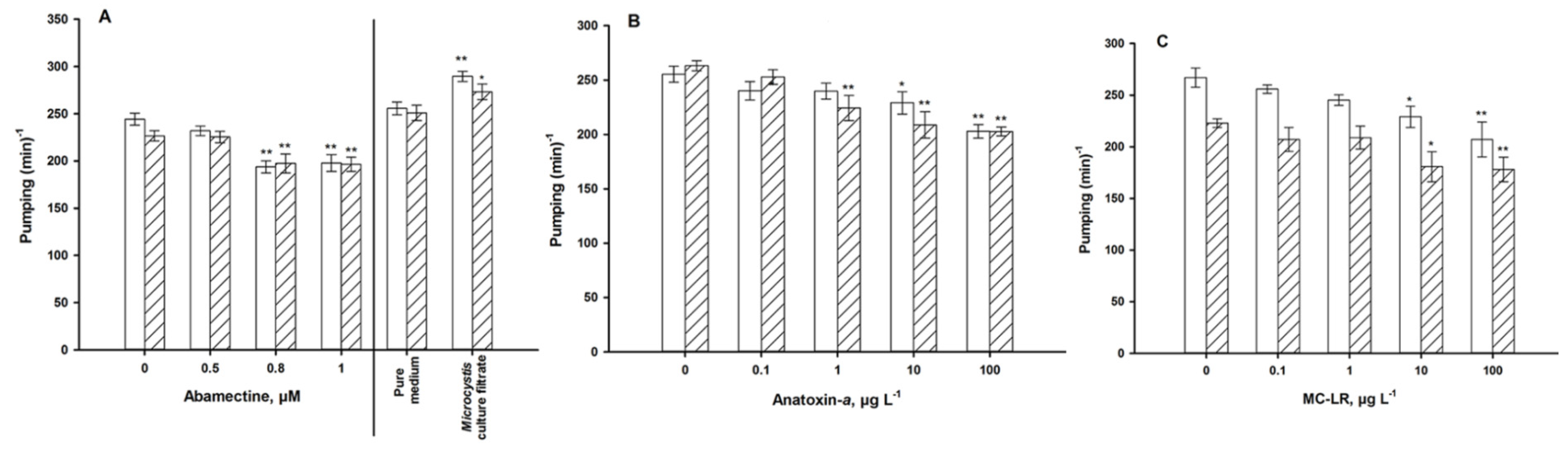
2.5. Pharyngeal Pumping

The pharyngeal pumping rate represents food intake. It was quantified after exposing the nematodes to the positive chemicals as well as cyanobacterial material for 24 or 72 h. Nematodes of the different exposure groups were randomly selected and their pumping frequency was determined three times over a 60 s timespan. Twenty worms per treatment were examined for each replicate using a digital microscope.

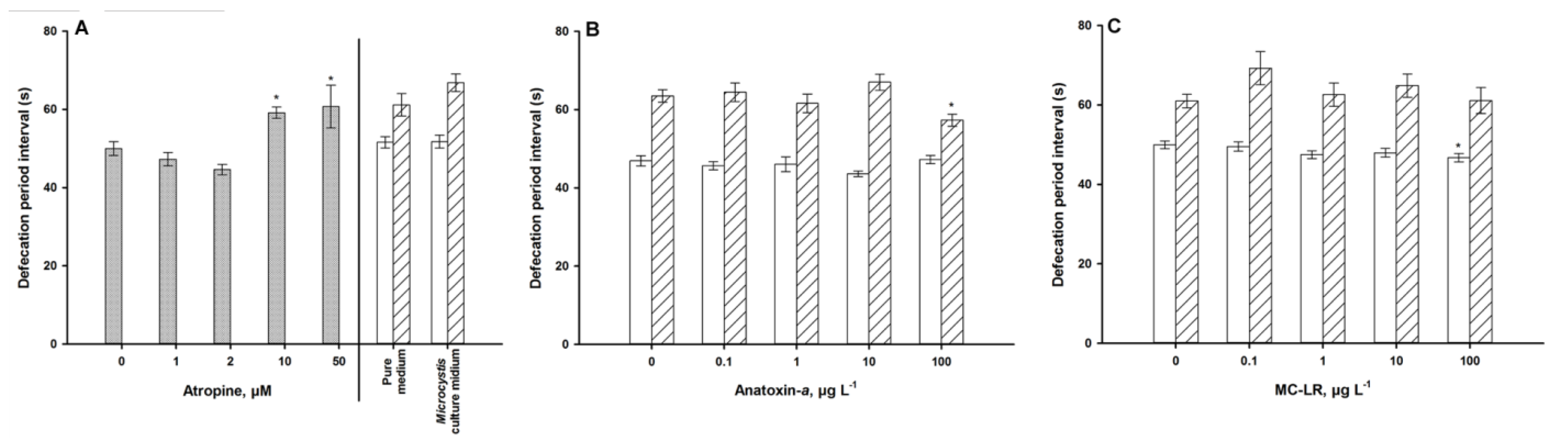
2.6. Defecation Assay

Defecation was assayed as previously described [29]. To a high degree of accuracy, the posterior body-wall muscle contraction of defecation can be clearly recognized. The whole defecation process includes four steps (Figure 1) and we measured the interval between the two posterior body-wall muscle contractions. Twenty worms per treatment were examined for each of the replicates using a digital microscope.
Figure 1. Defecation steps in C. elegans.
Ijerph 11 04589 g001
Notes: pBoc = posterior body-wall muscle contraction; aBoc = anterior body-wall muscle contraction; Exp = Expulsion.

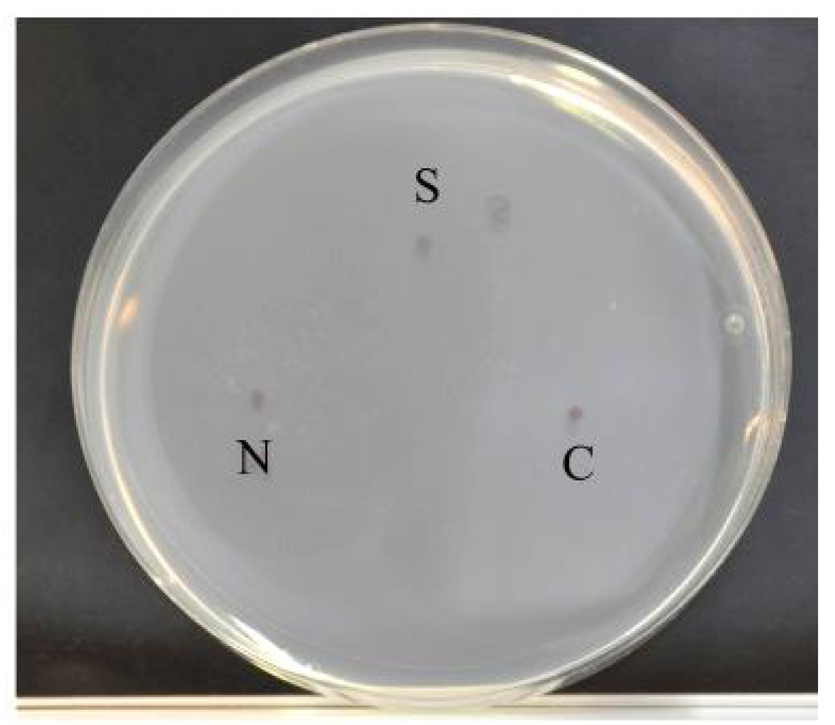
2.7. Chemotaxis Assay

Chemotaxis assays were conducted following the protocol described by Saeki, Yamamoto, and Iino [30]. This assay is based on the C. elegans’ attraction to NaCl. If impaired, the chemotaxis of the nematodes towards NaCl decreases dramatically. Assay plates (5 mM potassium phosphate, pH 6.0, 1 mM CaCl2, 1 mM MgSO4 and 20 g·L−1 agar) were prepared in advance.
An agar plug, which was prepared as described above, but with the addition of 100 mM NaCl, was excised from a NaCl plate and placed on the off-center spot (N, Figure 2) of the assay plate. This plate was then left overnight (14–24 h). Immediately prior to the assay, the NaCl plug was removed and 1 μL sodium azide (0.5 M) was spotted onto the same position to anaesthetize the animals. As a control, the same volume of sodium azide was also spotted onto a position (C, Figure 2) about 4 cm from the NaCl spot. Thirty worms were placed on the starting spot (S, Figure 2), which was about 3 cm away from the C and N spots, and were left to move freely on the assay plate for 1 h at 20 °C. After this amount of time, the number of worms around each spot was counted. The chemotaxis index (CI) was calculated as CI = (NN−NC)/30, where NN is the number of animals within 1.5 cm of the center of the NaCl gradient and NC is the number within 1.5 cm of the control spot. Assays were performed in triplicate, and the CI value was averaged. Thirty worms per treatment were assayed for each of the replicates using a stereo microscope.
Figure 2. Assay plates for chemotaxis
Ijerph 11 04589 g002
Notes: S: Start point; N: NaCl + NaN3; C: NaN3.

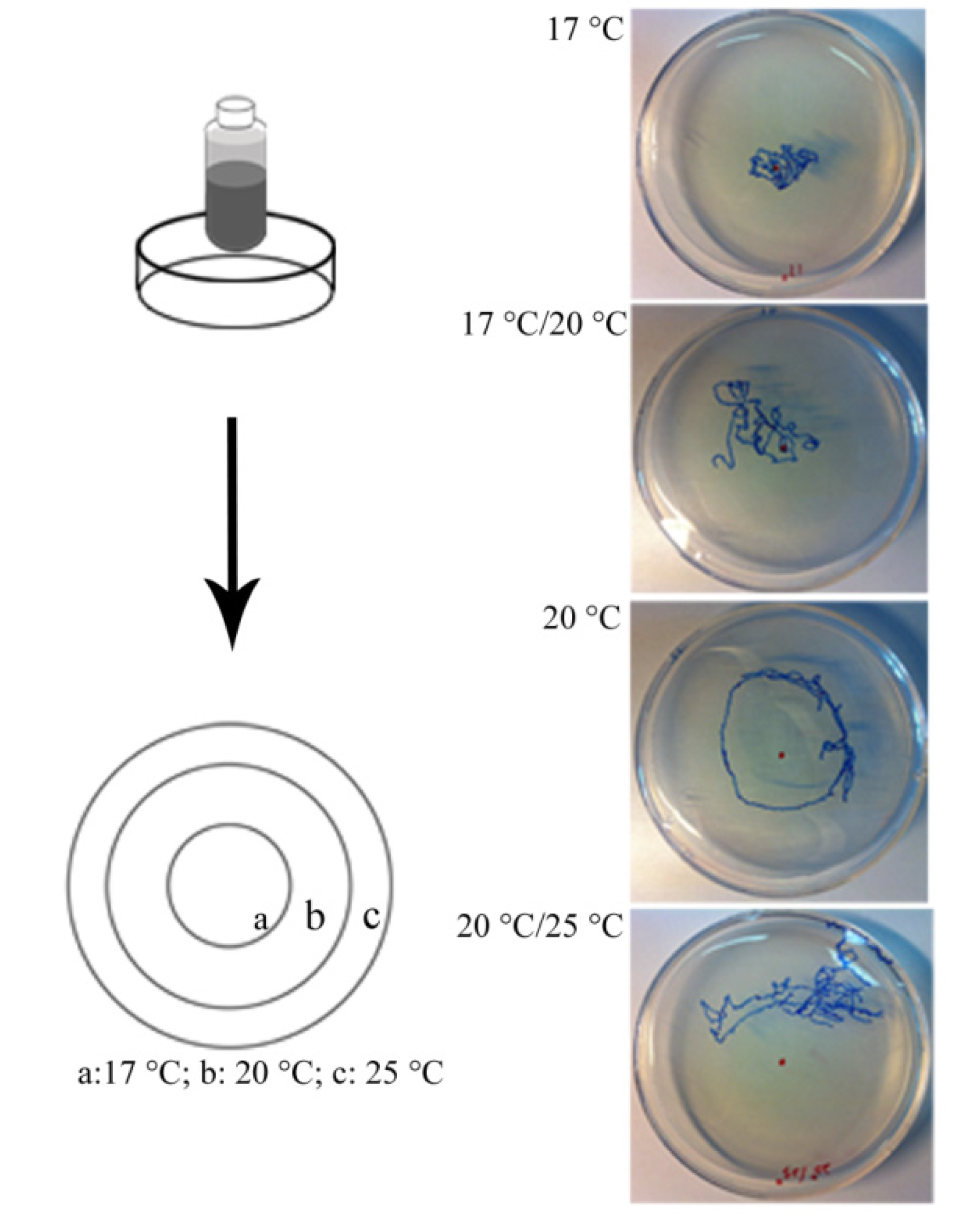
2.8. Thermotaxis Assay

The thermotaxis assay was performed as described by Kuhara et al. [31]. Individual C. elegans “memorize” temperatures, and this stored information modifies their subsequent migration along a temperature gradient. Control and exposed nematodes were kept at 20 °C for 24 h or 72 h on a fresh OP50 lawn. Individual nematodes were then transferred onto a fresh plate (9 cm diameter) without bacteria and cultivated at 25 °C. These plates are characterized by a pre-existing radial temperature gradient (Figure 3). The nematodes were placed on the 20 °C area and allowed to move freely on the plates. After 1 h, the trace was analyzed. If the animals moved within distinct temperature areas, their tracks were classified as “17 °C”, “20 °C”, or “25 °C”. Animals that moved between two distinct areas were classified as “17 °C/20 °C” or “20 °C/25 °C”. Between eight and 15 worms per treatment and replicate were assayed using a stereo microscope.

2.9. Mechanical Sensory Stimulus

Nose touch response, which is an expression of mechanical sensory perception, was conducted as previously described [32]. The tip of the nose of a forward-moving animal was touched with a fine hair, and reversal was scored as a response. Twenty worms per treatment were assayed for each of the replicates using a stereo microscope.
Figure 3. The thermal-gradient assay was performed using a 9 cm plate and a vial containing frozen acetic acid (Left); Traces of individuals of each category after 1 h (Right).
Ijerph 11 04589 g003

3. Data Analysis

All data are displayed as the mean ± Standard Error of the Mean (SEM). A student’s t-tests or ANOVA were used to determine the statistical significance between control and exposed groups. This was followed by the Fisher’s Least Significant Difference (LSD) test or the Dunnett’s t-test to determine the significance of differences between the groups. Probability levels of 0.05 and 0.01 were considered statistically significant.

4. Results

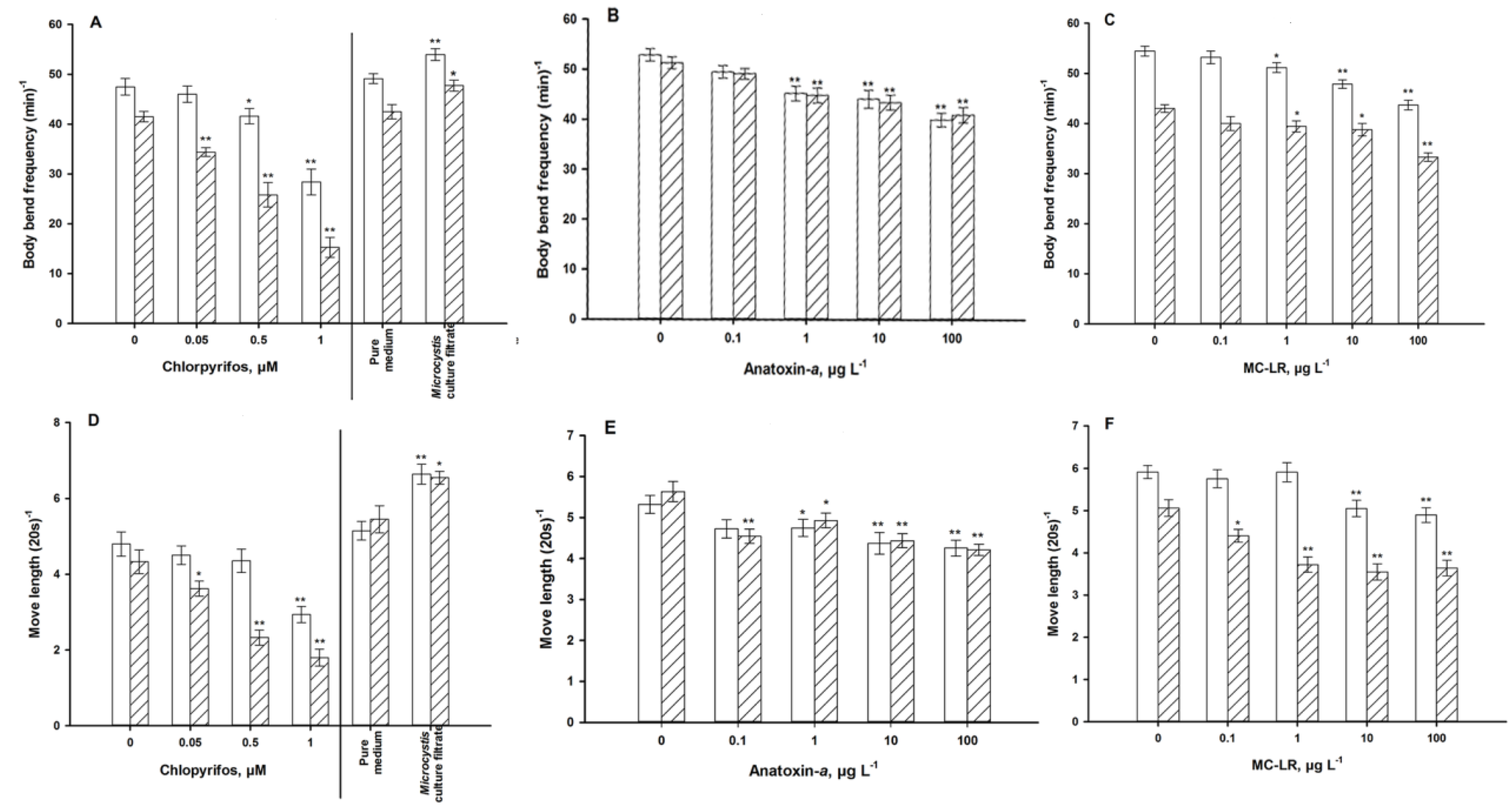
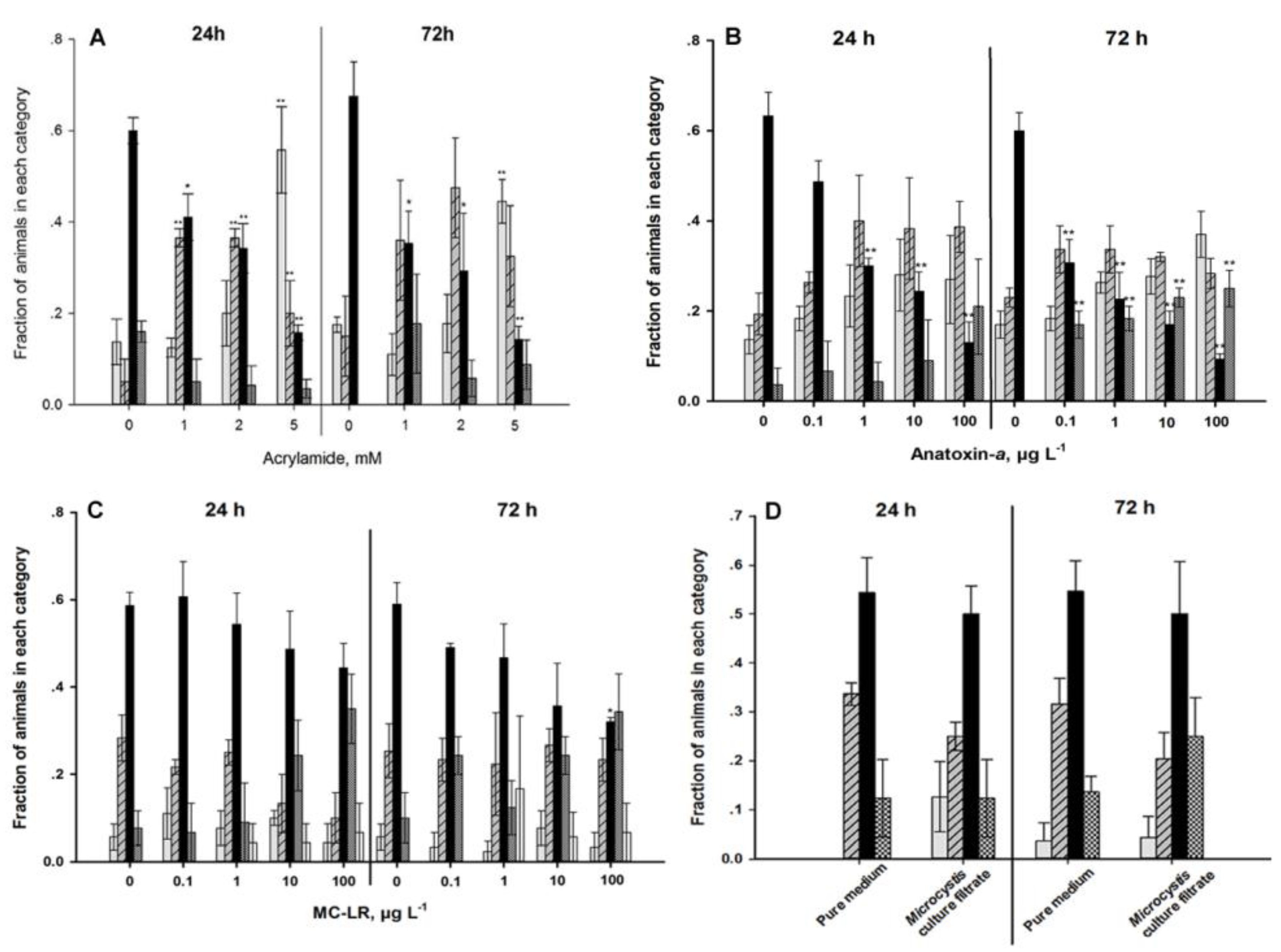
4.1. Autonomic Function

A gradual and time-related decrease in body bend frequency and move length was evident as chlorpyrifos-exposure increased (Figure 4A,D). After 72 h, even 0.05 μM caused a decrease in body bend frequency. The movement speed, measured as body-length–normalized move length in 20 second, showed a noticeable decrease upon exposure to 1.0 μM chlorpyrifos after 24 h and to 0.5 and 1.0 μM after 72 h.
Exposure to abamectin for 24 h and 72 h resulted in a decline of the mean pharyngeal pumping rate; especially for the exposure concentrations of 0.8 and 1 μM which showed a significant decrease in pumping compared with the control (Figure 5A). Due to its low persistence [33], the atropine exposure trials were terminated after 1 h. The whole defecation process, which is important for measuring atropine response, includes four steps (Figure 1). After exposure to more than 10 µM of atropine for 1 h, the defecation periodicity was significantly prolonged (Figure 6A). However, no worm constipation was observed.
As a well-known neurotoxin, ANA not surprisingly adversely affected both of the locomotive behavior variables (Figure 4B,E) and the pharyngeal pumping frequency (Figure 5B), with a defecation activity of only 100 µg·L−1 after 72 h of exposure (Figure 6B). Older worms need longer defecation periods, however, this activity was not modulated by ANA.
The hepatotoxin MC-LR, too, reduced body bends, move length (Figure 4C,F) and pharyngeal pumping frequency (Figure 5C), but not defecation activity (Figure 6C). Exposure to Macf significantly increased the motile activity (Figure 4A,D), pumping activity (Figure 5A), and after long-term exposure tended to increase the defecation period (Figure 6A).

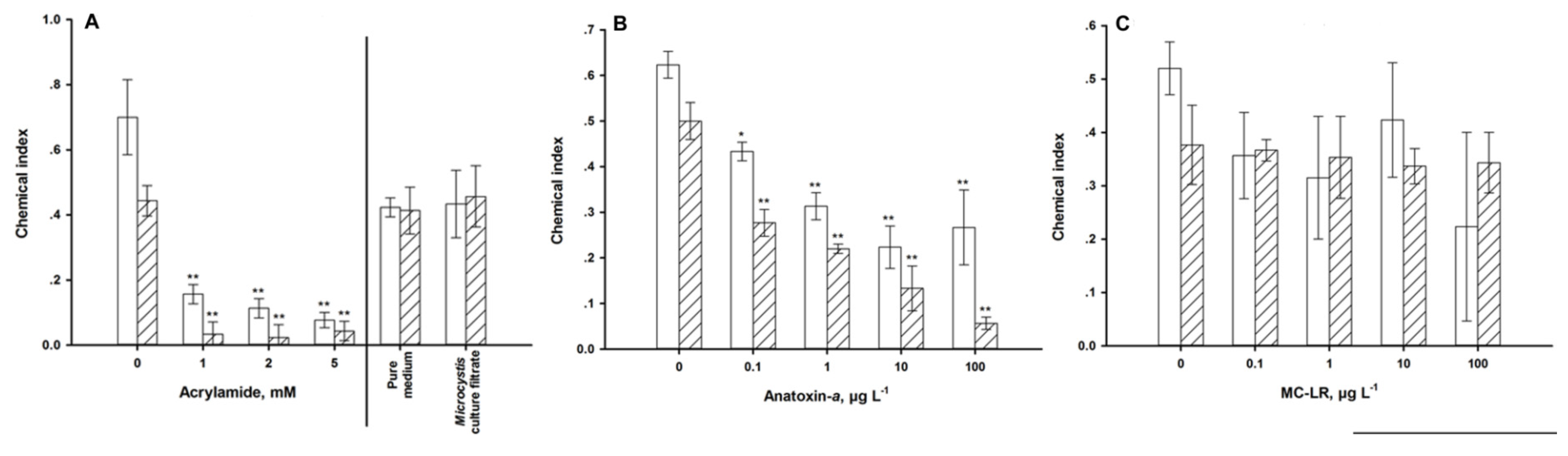
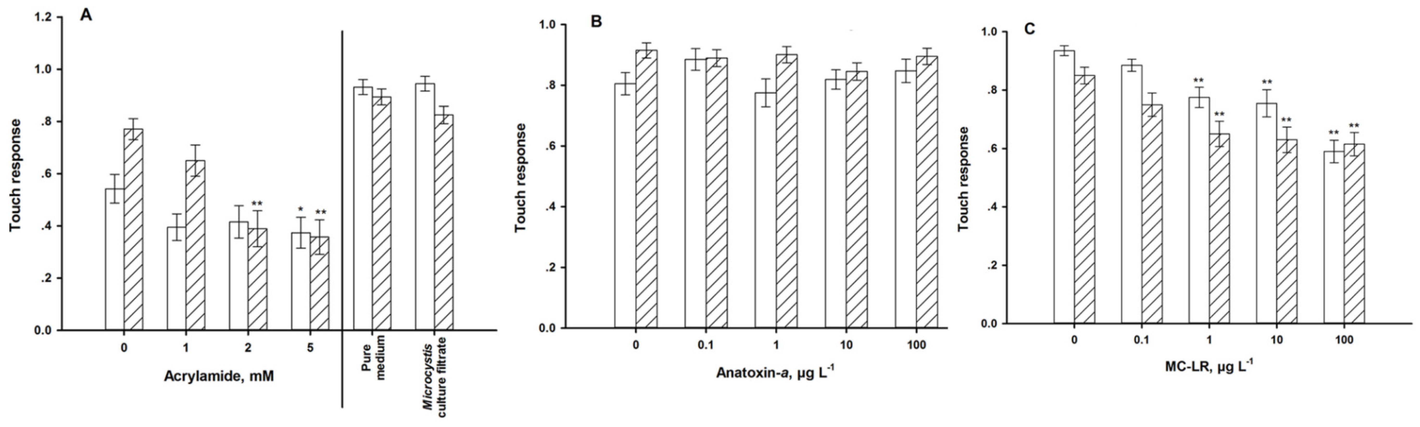
4.2. Sensory Function

When C. elegans was fed on NGM (with NaCl and food), its chemotaxis index was around 0.5 after 24 h and 0.4 after 72 h of exposure. The chemotaxis towards NaCl fell dramatically if C. elegans was exposed to acrylamide (Figure 7A).
The thermotaxis was also significantly changed. As the exposure concentration of acrylamide increased, the fractions of worms which migrated towards 20 °C decreased, and the worms became more attracted by lower temperatures (Figure 8A).
The nose touch avoidance behavior was affected by exposure to acrylamide (Figure 9A). Worms exposed to 5 mM of acrylamide for 24 h exhibited a significant decrease in their nose touch avoidance rate. When the exposure duration was increased to 72 h, the worms displayed a significant decrease, even at 2 mM.
The strongest impacts of ANA on sensory function were found in the chemotactic (Figure 7B) and the thermotactic behaviors (Figure 8B). Exposed worms were increasingly not attracted to NaCl and preferred colder and warmer temperatures. Long-term exposure aggravated these effects. The mechanical sensory behavior remained unaffected by ANA exposure (Figure 9B).
In contrast to ANA, MC-LR impacted the mechanical (Figure 9C), but not the chemical sensory behavior (Figure 7C). MC-LR-exposure also changed the thermotactic behavior; but again unlike the effects of ANA, it caused the nematodes to prefer warmer environments (Figure 8C). Exposure to ≥1 µg·L−1 MC-LR forced between 5 and 18% of the individuals to migrate to the 25 °C environment.
Macf-exposure did not modulate the chemical or mechanical sensory functions (Figure 7A and Figure 9A), but temporarily impacted the thermotactic behavior (Figure 8D). Untreated C. elegans migrated to its growth temperature (20 °C) and then moved isothermally [34]. Exposure to Macf for 24 h reduced the fractions of worms which tended towards 20 °C, the worms were instead more attracted by lower temperatures (Figure 8D). This behavior was no longer observed after 72 h of exposure. Macf contained three MC variants (Table 1).
Figure 4. Locomotive behavior of young adult nematodes for 24 h (white) and 72 h (hatched). Move length is normalized by the mean of body length per group. (A,D): exposure to chlorpyrifos and a Microcystis culture filtrate. (B,E): exposure to anatoxin-a. (C,F): exposure to microcystin-LR. * = p < 0.05, ** = p < 0.01.
Ijerph 11 04589 g004
Figure 5. Pharyngeal pumping of young adults for 24 h (white) and 72 h (hatched). (A): exposure to abamectine, and a Microcystis culture filtrate. (B): exposure to anatoxin-a. (C): exposure to microcystin-LR. * = p < 0.05, ** = p < 0.01.
Ijerph 11 04589 g005
Figure 6. Defecation of young adults. (A) exposure to atropine only for 1 h (grey) and a Microcystis culture filtrate for 24 h (white) and 72 h (hatched). (B): exposure to anatoxin-afor 24 h (white) and 72 h (hatched). (C): exposure to microcystin-LR for 24 h (white) and 72 h (hatched). * = p < 0.05, ** = p < 0.01.
Ijerph 11 04589 g006
Figure 7. Chemical sensory response of young adult nematodes after exposure for 24 h (white) and 72 h (hatched). The smaller the chemical index, the worse the chemotactic behavior. (A): exposure to acrylamide and a Microcystis culture filtrate. (B): exposure to anatoxin-a. (C): exposure to microcystin-LR. * = p < 0.05, ** = p < 0.01.
Ijerph 11 04589 g007
Figure 8. Thermotaxis of young adult nematodes after exposure for 24 h and 72 h. (A) exposure to acrylamide and a Microcystis culture filtrate. (B): exposure to anatoxin-a. (C): exposure to microcystin-LR. (D) exposure to a Microcystis culture filtrate. * = p < 0.05, ** = p < 0.01. (grey: 17 °C, hatched: 17/20 °C, black: 20 °C, dark grey: 20/25 °C, white: 25 °C).
Ijerph 11 04589 g008
Figure 9. Mechnical sensory function of young adults for 24 h (white) and 72 h (hatched). (A): exposure to acrylamide and a Microcystis culture filtrate. (B): exposure to anatoxin-a. (C): exposure to microcystin-LR. * = p < 0.05, ** = p < 0.01.
Ijerph 11 04589 g009

5. Discussion

5.1. Applicability of C. elegans as a Model for Neurotoxicity

The changes in the autonomic and sensory functions of C. elegans took place after exposure to chemicals well identified as neurotoxic compounds by tests in mammals. The autonomic function (locomotion behavior, pharyngeal pumping, and defecation) was significantly affected by chlorpyrifos, abamectin, and atropine. The organophosphate insecticide chlorpyrifos acts on the nervous system by inhibiting acetylcholinesterases; it decreases the locomotor activity of mammalians and other species as well as C. elegans [35]. The pesticide abamectin was reported to elicit a potent and persistent paralysis of the nematode’s pharynx [36]. Atropine is known to reduce secretions and is used as a drug for functional gastrointestinal disorders [37] and, indeed, we observed prolonged defecation periods upon exposure. These three chemicals showed the expected effect in C. elegans.
Acrylamide acts primarily on nerve terminals and causes ataxia, skeletal muscle weakness and numbness in the hands and feet of animals and humans [38]. It also affects the life span and the expression of genes encoding detoxification enzymes in C. elegans [39] and showed the expected effects on sensory function in the assays. Hence, C. elegans responded to all positive neurotoxic compounds.
Consequently, a chemical or a mixture of chemicals may be classified as neurotoxic, if one life trait of exposed C. elegans is responsive. Obviously, C. elegans can only be a valuable toxicity model if its results are predictive in higher organisms. In addition to several existing comparative studies on the ranking of chemicals between C. elegans and mammals, which mainly concentrated on lethality in AchE inhibition and movement [13,20,21], our work supported the consistency of several neurotoxic compounds on various behavioral endpoints. The similarity of the mechanisms should be observed in further study. Another next step in establishing C. elegans as a suitable invertebrate replacement for neurotoxic evaluation will be to perform a cross-check with the neurotoxic compounds. For instance, it is of interest whether or not chlorpyrifos and atropine impair sensory function or if acrylamide impairs autonomic or cognitive functions in mammals. After the eventual successful establishment of these effects, this model could save resources and improve efficiency by serving as a prescreening test before mammalian tests.

5.2. Cyanotoxins and M. aeruginosa Culture Filtrate

Nowadays, we have quantitative ELISA, HPLC methods and mass spectroscopic methods which allow for the detection and quantification of all known classes of cyanobacterial toxins. However, these tests can only prove the existence of the toxins they cannot show their effects since toxicity is not an intrinsic property of the xenobiotic chemical itself, but rather is displayed only by its interaction with exposed/administered individuals. Consequently, biotests can fill this gap.
The concentrations of the cyanotoxins applied in this study lie below the maximal values found in eutrophicated freshwater bodies [5] and cover well the range of values reported from German lakes and reservoirs [40,41]. The major toxic mode of action of MC-LR is hepatotoxicity and of ANA neurotoxicity [42]. In the C. elegans assay which was applied, the neurotoxicity of ANA was well reflected by five out of six test variables indicating the robustness of the applied assays. Particularly, chemotaxis was strongly impaired, deteriorating with exposure time. This indicates that C. elegans loses its ability to avoid adverse environments. Translating this finding to natural environments, it reflects the risk of extinction of the population, because a persistence of the population in chemically hostile environments cannot be warranted, if the nematode can no longer avoid them.
For a long time, MCs were considered only hepatotoxic [3,43]. A classic case of severe MC-intoxication, however, which happened in a hemodialysis unit in Caruaru, Brazil in 1996, had already suggested that MCs could pass the blood–brain barrier and induce neurotoxic symptoms [4]. Quantitative records of the behavior of two fish species, Danio rerio Hamilton and Leucaspius delineatus Heckel, showed that even exposure to low (0.5 µg·L−1) concentrations of MC-LR significantly modulated the swimming performance of these fish suggesting that there could possibly be a neurotoxic action [44]. This assumption gets support from biochemical and histological studies. For instance, Fischer et al. [45] showed that members of the organic anion transporting polypeptides were involved in the uptake of MC-LR and are expressed in both the liver and brain of the clawed frog (Xenopus laevis), which implies that the brain may be one of the potential targets of MC-LR. Analyzing three South American fish species for their tissue distribution of MC-RR, Cazenave, et al. [46] found traces of MC in the brains of Jenynsia multidentata Jenyns, indicating that MC has the ability to cross the blood-brain barrier. These examples clearly illustrate the need for routine neurotoxic evaluations of Microcystis and most likely other cyanobacteria blooms by a cost-effective neurotoxicity assay.
Very recently, L1 larvae of C. elegans were proven to respond to MC-LR at environmentally realistic concentrations [24]. MC-LR in this case impaired a central neurotransmitter. In particular, γ-aminobutyric acid (GABA)ergic neurons were lost in a concentration-dependent manner at concentrations ≥0.1 μg·L−1. GABA is the most abundant inhibitory neurotransmitter in vertebrates and invertebrates and functions in C. elegans as an excitatory as well as an inhibitory neurotransmitter [47].
Again, the demonstrated C. elegans assay used showed strong modifications that indicate neurotoxicity in five out of six variables, namely both locomotive variables (Figure 4C,F), pharyngeal pumping activity (Figure 5C), thermotaxis (Figure 8C) and mechanical sensory ability (Figure 9C). This outcome continues to confirm that the C. elegans assay is sensitive and robust enough to identify the neurotoxicity of novel untested chemicals. Similar to the ANA check, the implications for C. elegans in the environment based on the overall outcome of these assays may be equally adverse, although different traits were found to respond to the toxin. For instance, the MC-induced preference of environments with elevated temperatures may be risky, because C. elegans is thermo sensitive and stops reproduction at temperatures >25 °C [27].
The filtrate of a M. aeruginosa culture showed mild neurotoxic effects for some of the sensory functions in C. elegans indicating the general suitability of the presented assay. Even the Microcystis culture medium appears to contain neuroirritant compounds. However, autonomic functions were surprisingly stimulated during Macf exposure. This effect might be caused by Microcystis metabolites other than microcystin, such as brominated organic compounds [22] and will be addressed in a separate study. Nevertheless, even neurostimulation shows the applicability of the test described, since it indicates that the exposed chemicals target neurons.

6. Conclusions

Neurobehavioral testing is traditionally done using mammals, however there is an upward spiraling of costs in terms of time, resources, and number of animals. This inefficiency and the high costs associated with mammalian testing reduces the possibility to evaluate the neurotoxicity of various environmental chemicals and pollutants. C. elegans, as an emerging model in environmental and toxicological sciences, was chosen for establishing a neurobehavioral screening system according to the guideline used for mammalians. The presented data support the possible establishment of this neurobehavioral model. Every positive control achieved the expected effect in this model organism. Furthermore, the well-known neurotoxin ANA as well as the hepatotoxin MC-LR revealed adverse effects in five out of six life traits. Overall, C. elegans appears to have the potential to replace rodents as neurotoxicity model animals.

Acknowledgments

This work was supported by a scholarship received by J.J. from the China Scholarship Council (CSC) and grants from the National Natural Science Foundation of China (No. 81273123), Natural Science Foundation of Jiangsu Province (No. 2010402), the Research and Innovation Project for College Graduates of Jiangsu Province (CXLX_0156), the Fundamental Research Funds for the Central Universities and by grants received by Nadine Saul (STE673/18-1) and Cindy Kochan (PU199/6-1) from the Deutsche Forschungsgemeinschaft (DFG). Furthermore, we thank the Caenorhabditis Genetics Centre, which is funded by the National Institutes of Health National Centre for Research Resources, for supplying the Caenorhabditis elegans strains. Last but not least we want to thank two anonymous reviewers for their very supportive comments on an earlier version of this manuscript.

Author Contributions

Lihong Yin, Jingjuan Ju, Christian E. W. Steinberg, and Yuepu Pu conceived of and designed the experiments.; Jingjuan Ju performed the experiments about C. elegans; Cindy Kochan and Anke Putschew contributed to the analysis of microcystins in the culture filtrate; Jingjuan Ju and Nadine Saul analyzed data; Jingjuan Ju and Christian E. W. Steinberg, wrote the manuscript; Nadine Saul, Jingjuan Ju and Christian E. W. Steinberg revised the manuscript.

Conflicts of Interest

The authors declare no conflict of interest.

References

  1. Paerl, H.W.; Huisman, J. Climate: Blooms like it hot. Science 2008, 320, 57–58. [Google Scholar] [CrossRef]
  2. Welker, M.; von Döhren, H. Cyanobacterial peptides—Nature’s own combinatorial biosynthesis. FEMS Microbiol. Rev. 2006, 30, 530–563. [Google Scholar] [CrossRef]
  3. Mackintosh, C.; Beattie, K.A.; Klumpp, S.; Cohen, P.; Codd, G.A. Cyanobacterial microcystin-LR is a potent and specific inhibitor of protein phosphatases 1 and 2A from both mammals and higher plants. FEBS Lett. 1990, 264, 187–192. [Google Scholar] [CrossRef]
  4. Pouria, S.; de Andrade, A.; Barbosa, J.; Cavalcanti, R.L.; Barreto, V.T.S.; Ward, C.J.; Preiser, W.; Poon, G.K.; Neild, G.H.; Codd, G.A. Fatal microcystin intoxication in haemodialysis unit in Caruaru, Brazil. Lancet 1998, 352, 21–26. [Google Scholar] [CrossRef]
  5. Dittmann, E.; Wiegand, C. Cyanobacterial toxins—Occurrence, biosynthesis and impact on human affairs. Mol. Nutr. Food Res. 2006, 50, 7–17. [Google Scholar] [CrossRef]
  6. Park, H.-D.; Watanabe, M.F.; Harada, K.-I.; Nagai, H.; Suzuki, M.; Watanabe, M.; Hayashi, H. Hepatotoxin (microcystin) and neurotoxin (anatoxin-a) contained in natural blooms and strains of cyanobacteria from Japanese freshwaters. Nat. Toxins 1993, 1, 353–360. [Google Scholar] [CrossRef]
  7. Saul, N.; Chakrabarti, S.; Stürzenbaum, S.R.; Menzel, R.; Steinberg, C.E.W. Molecular insights into the toxicity mechanisms of microcystin-LR in Caenorhabditis elegans. Chem. Biol. Interact. 2014. accepted. [Google Scholar]
  8. Guidelines for Neurotoxicity Risk Assessment; Environmental Protection Agency: Washington, DC, USA, 1998.
  9. Leung, M.C.K.; Williams, P.L.; Benedetto, A.; Au, C.; Helmcke, K.J.; Aschner, M.; Meyer, J.N. Caenorhabditis elegans: An emerging model in biomedical and environmental toxicology. Toxicol. Sci. 2008, 106, 5–28. [Google Scholar]
  10. Kaletta, T.; Hengartner, M.O. Finding function in novel targets: C. elegans as a model organism. Nat. Rev. Drug Discov. 2006, 5, 387–398. [Google Scholar] [CrossRef]
  11. Anderson, G.L.; Cole, R.D.; Williams, P.L. Assessing behavioral toxicity with Caenorhabditis elegans. Environ. Toxicol. Chem. 2004, 23, 1235–1240. [Google Scholar]
  12. Anderson, G.L.; Boyd, W.A.; Williams, P.L. Assessment of sublethal endpoints for toxicity testing with the nematode Caenorhabditis elegans. Environ. Toxicol. Chem. 2001, 20, 833–838. [Google Scholar]
  13. Rajini, P.S.; Melstrom, P.; Williams, P.L. A comparative study on the relationship between various toxicological endpoints in Caenorhabditis elegans exposed to organophosphorus insecticides. J. Toxicol. Environ. Health Pt. A 2008, 71, 1043–1050. [Google Scholar]
  14. Dhawan, R.; Dusenbery, D.B.; Williams, P.L. Comparison of lethality, reproduction, and behavior as toxicological endpoints in the nematode Caenorhabditis elegans. J. Toxicol. Environ. Health Pt. A 1999, 58, 451–462. [Google Scholar]
  15. Boyd, W.A.; Anderson, G.L.; Dusenbery, D.B.; Williams, P.L. Computer Tracking Method for Assessing Behavioral Changes in the Nematode Caenorhabditis elegans. ASTM Spec. Tech. Publ. 2000, 9, 225–238. [Google Scholar]
  16. Melstrom, P.C.; Williams, P.L. Measuring movement to determine physiological roles of acetylcholinesterase classes in Caenorhabditis elegans. J. Nematol. 2007, 39, 317–320. [Google Scholar]
  17. Boyd, W.A.; Cole, R.D.; Anderson, G.L.; Williams, P.L. The effects of metals and food availability on the behavior of Caenorhabditis elegans. Environ. Toxicol. Chem. 2003, 22, 3049–3055. [Google Scholar]
  18. Jones, D.; Candido, E.P.M. Feeding is inhibited by sublethal concentrations of toxicants and by heat stress in the nematode Caenorhabditis elegans: Relationship to the cellular stress response. J. Exp. Zool. 1999, 284, 147–157. [Google Scholar]
  19. Dhawan, R.; Dusenbery, D.B.; Williams, P.L. A comparison of metal-induced lethality and behavioral responses in the nematode Caenorhabditis elegans. Environ. Toxicol. Chem. 2000, 19, 3061–3067. [Google Scholar]
  20. Cole, R.D.; Anderson, G.L.; Williams, P.L. The nematode Caenorhabditis elegans as a model of organophosphate-induced mammalian neurotoxicity. Toxicol. Appl. Pharmacol. 2004, 194, 248–256. [Google Scholar]
  21. Hunt, P.R.; Olejnik, N.; Sprando, R.L. Toxicity ranking of heavy metals with screening method using adult Caenorhabditis elegans and propidium iodide replicates toxicity ranking in rat. Food Chem. Toxicol. 2012, 50, 3280–3290. [Google Scholar]
  22. Hütteroth, A.; Putschew, A.; Jekel, M. Natural production of organic bromine compounds in Berlin lakes. Environ. Sci. Technol. 2007, 41, 3607–3612. [Google Scholar] [CrossRef]
  23. Li, Y.; Wang, Y.; Yin, L.; Pu, Y.; Wang, D. Using the nematode Caenorhabditis elegans as a model animal for assessing the toxicity induced by microcystin-LR. J. Environ. Sci. 2009, 21, 395–401. [Google Scholar] [CrossRef]
  24. Ju, J.; Ruan, Q.; Li, X.; Liu, R.; Li, Y.; Pu, Y.; Yin, L.; Wang, D. Neurotoxicological evaluation of microcystin-LR exposure at environmental relevant concentrations on nematode Caenorhabditis elegans. Envir. Sci. Pollut. Res. 2013, 20, 1823–1830. [Google Scholar] [CrossRef]
  25. Li, Y.; Ye, H.; Du, M.; Zhang, Y.; Ye, B.; Pu, Y.; Wang, D. Induction of chemotaxis to sodium chloride and diacetyl and thermotaxis defects by microcystin-LR exposure in nematode Caenorhabditis elegan. (in Chinese). J. Environ. Sci.-China 2009, 21, 971–979. [Google Scholar]
  26. Li, Y.H.; Wang, Y.; Yin, L.H.; Pu, Y.P.; Wang, D.Y. Using the nematode Caenorhabditis elegans as a model animal for assessing the toxicity induced by microcystin-LR. (in Chinese). J. Environ. Sci.-China 2009, 21, 395–401. [Google Scholar]
  27. Brenner, S. The genetics of Caenorhabditis elegans. Genetics 1974, 77, 71–94. [Google Scholar]
  28. Ju, J.; Lieke, T.; Saul, N.; Pu, Y.; Yin, L.; Cindy, K.; Anke, P.; Baberschke, N.; Steinberg, C.E.W. Neurotoxic evaluation of two organobromine model compounds and natural AOBr-containing surface water samples by a Caenorhabditis elegans test. Ecotoxicol. Environ. Safety 2014, 104, 194–201. [Google Scholar] [CrossRef]
  29. Mahoney, T.R.; Luo, S.; Round, E.K.; Brauner, M.; Gottschalk, A.; Thomas, J.H.; Nonet, M.L. Intestinal signaling to GABAergic neurons regulates a rhythmic behavior in Caenorhabditis elegans. Proc. Nat. Acad. Sci. USA 2008, 105, 16350–16355. [Google Scholar]
  30. Saeki, S.; Yamamoto, M.; Iino, Y. Plasticity of chemotaxis revealed by paired presentation of a chemoattractant and starvation in the nematode Caenorhabditis elegans. J. Exp. Biol. 2001, 204, 1757–1764. [Google Scholar]
  31. Kuhara, A.; Okumura, M.; Kimata, T.; Tanizawa, Y.; Takano, R.; Kimura, K.D.; Inada, H.; Matsumoto, K.; Mori, I. Temperature sensing by an olfactory neuron in a circuit controlling behavior of C. elegans. Science 2008, 320, 803–807. [Google Scholar] [CrossRef]
  32. Liedtke, W.; Tobin, D.M.; Bargmann, C.I.; Friedman, J.M. Mammalian TRPV4 (VR-OAC) directs behavioral responses to osmotic and mechanical stimuli in Caenorhabditis elegans. Proc. Nat. Acad. Sci. USA 2003, 100, 14531–14536. [Google Scholar] [CrossRef]
  33. Mirakhur, R.K. Comparative study of effects of oral and i.m. atropine and hyoscine in volunteers. Brit. J. Anaesth. 1978, 50, 591–598. [Google Scholar] [CrossRef]
  34. Hedgecock, E.M.; Russell, R.L. Normal and mutant thermotaxis in the nematode Caenorhabditis elegans. Proc. Nat. Acad. Sci. USA 1975, 72, 4061–4065. [Google Scholar] [CrossRef]
  35. Moser, V.C.; Phillips, P.M.; Levine, A.B.; Mcdaniel, K.L.; Sills, R.C.; Jortner, B.S.; Butt, M.T. Neurotoxicity produced by dibromoacetic acid in drinking water of rats. Toxicol. Sci. 2004, 79, 112–122. [Google Scholar] [CrossRef]
  36. Geary, T.G.; Sims, S.M.; Thomas, E.M.; Vanover, L.; Davis, J.P.; Winterrowd, C.A.; Klein, R.D.; Ho, N.F.H.; Thompson, D.P. Haemonchus contortus: Ivermectin-induced paralysis of the pharynx. Exp. Parasitol. 1993, 77, 88–96. [Google Scholar] [CrossRef]
  37. Sun, E.A.; Snape, W.J., Jr.; Cohen, S.; Renny, A. The role of opiate receptors and cholinergic neurons in the gastrocolonic response. Gastroenterology 1982, 82, 689–693. [Google Scholar]
  38. Lopachin, R.M. Acrylamide Neurotoxicity: Neurological, Morhological and Molecular Endpoints in Animal Models. In Advances in Experimental Medicine and Biology: Chemistry and Safety of Acrylamide in Food; Friedman, M., Mottram, D., Eds.; Springer: New York, NY, USA, 2005; Volume 561, pp. 21–37. [Google Scholar]
  39. Hasegawa, K.; Miwa, S.; Isomura, K.; Tsutsumiuchi, K.; Taniguchi, H.; Miwa, J. Acrylamide-responsive genes in the nematode Caenorhabditis elegans. Toxicol. Sci. 2008, 101, 215–225. [Google Scholar] [CrossRef]
  40. Bumke-Vogt, C.; Mailahn, W.; Chorus, I. Anatoxin-a and neurotoxic cyanobacteria in German lakes and reservoirs. Environ. Toxicol. 1999, 14, 117–125. [Google Scholar] [CrossRef]
  41. Welker, M.; von Döhren, H.; Täuscher, H.; Steinberg, C.E.W.; Erhard, M. Toxic Microcystis in shallow lake Müggelsee (Germany)—Dynamics, distribution, diversity. Arch. Hydrobiol. 2003, 157, 227–248. [Google Scholar] [CrossRef]
  42. Bell, S.G.; Codd, G.A. Cyanobacterial toxins and human health. Rev. Med. Microbiol. 1994, 5, 256–264. [Google Scholar]
  43. Barford, D. Molecular mechanisms of the protein serine/threonine phosphatases. Trends Biochem. Sci. 1996, 21, 407–412. [Google Scholar] [CrossRef]
  44. Baganz, D.; Staaks, G.; Steinberg, C. Impact of the cyanobacteria toxin, microcystin-LR on behaviour of zebrafish, Danio rerio. Water Res. 1998, 32, 948–952. [Google Scholar] [CrossRef]
  45. Fischer, W.J.; Altheimer, S.; Cattori, V.; Meier, P.J.; Dietrich, D.R.; Hagenbuch, B. Organic anion transporting polypeptides expressed in liver and brain mediate uptake of microcystin. Toxicol. Appl. Pharmacol. 2005, 203, 257–263. [Google Scholar]
  46. Cazenave, J.; Wunderlin, D.A.; Bistoni, M.D.L.A.; Amé, M.V.; Krause, E.; Pflugmacher, S.; Wiegand, C. Uptake, tissue distribution and accumulation of microcystin-RR in Corydoras paleatus, Jenynsia multidentata and Odontesthes bonariensis: A field and laboratory study. Aquat. Toxicol. 2005, 75, 178–190. [Google Scholar] [CrossRef]
  47. Mcintire, S.L.; Jorgensen, E.; Kaplan, J.; Horvitz, H.R. The GABAergic nervous system of Caenorhabditis elegans. Nature 1993, 364, 337–341. [Google Scholar] [CrossRef]

