Eutypellenoids A–C, New Pimarane Diterpenes from the Arctic Fungus Eutypella sp. D-1
by
 ,
,
Hao-Bing Yu
1,2,† ,
,
Xiao-Li Wang
1,†,
Wei-Heng Xu
3,†,
Yi-Xin Zhang
1,
Yi-Sen Qian
4,
Jian-Peng Zhang
1,
Xiao-Ling Lu
1 and
Xiao-Yu Liu
1,* 1
Department of Biochemistry and Molecular Biology, College of Basic Medical Sciences, and Marine Biopharmaceutical Institute, Second Military Medical University, Shanghai 200433, China
2
Center for Marine Biotechnology and Biomedicine, Scripps Institution of Oceanography, University of California, San Diego, CA 92093, USA
3
School of Pharmacy, Second Military Medical University, Shanghai 200433, China
4
School of Physical Sciences, University of California, Irvine, CA 92697, USA
*
Author to whom correspondence should be addressed.
†
These authors contributed equally to this work.
Mar. Drugs 2018, 16(8), 284; https://doi.org/10.3390/md16080284
Submission received: 29 July 2018
/
Revised: 12 August 2018
/
Accepted: 14 August 2018
/
Published: 16 August 2018
(This article belongs to the Special Issue Terpenoids from Marine Organisms)
Abstract
:Three new pimarane diterpenes, eutypellenoids A–C (1–3), together with a known compound, eutypenoid C (4), were isolated from the culture extract of Eutypella sp. D-1 derived from the Arctic region. Compounds 1–3 possessed an uncommon tetrahydrofuran-fused pimarane diterpene skeleton. The structures of all compounds were determined by detailed spectroscopic analysis, electronic circular dichroism (ECD) analysis, as well as a comparison with the literature data. Antibacterial, antifungal, and cytotoxic activities of these compounds were evaluated. Compound 2 displayed antibacterial activity against Staphylococcus aureus and Escherichia coli with MIC values of 8 and 8 μg/mL, respectively. Additionally, compound 2 showed antifungal activity against Candida parapsilosis, Candida albicans, Candida glabrata, and Candida tropicalis with MIC values of 8, 8, 16, and 32 μg/mL, respectively. Furthermore, compound 2 exhibited moderate cytotoxic activity against HCT-116 cell line with IC50 value of 3.7 μM.
1. Introduction
Marine-derived fungi continue to embody an important spring of novel chemical structures with potentially useful applications as drugs [1]. Secondary metabolites from fungi of polar regions, including the Arctic, Antarctic, and their subregions, are rarely studied because of the challenging living environment [2]. The Eutypella species, found not only from the Arctic, but also from tropical forests and marine sources [2,3], produces a rich source of varied secondary metabolites, including pimarane diterpenoids, sesquiterpenoids, γ-lactones, cytochalasin derivatives, cyclic dipeptides, and cytosporin-related compounds [4,5,6,7,8,9,10]. Among them, pimarane diterpenes have attracted considerable interest because of their unique chemical structures and varied biological activities, such as antitumor, antimicrobial, and immunosuppressive activities [10,11].
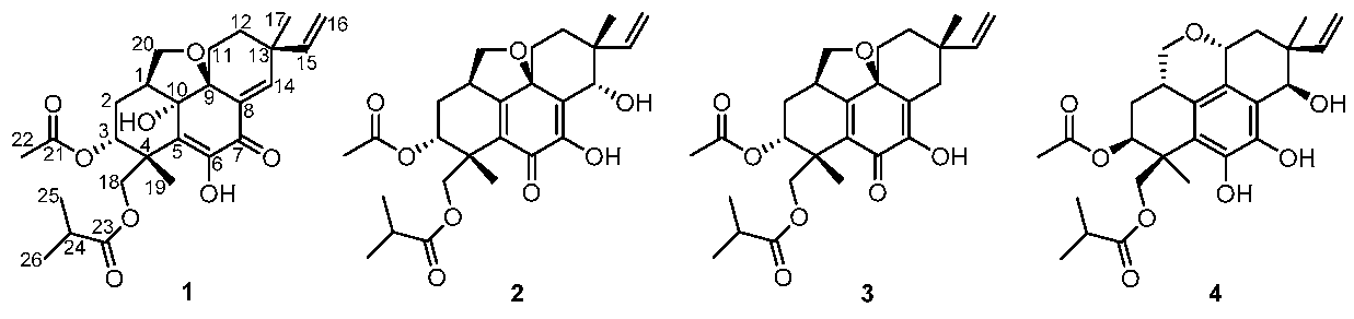
As part of our continuous commitment to discovering structurally intriguing and biologically active natural products from polar fungi, we investigated the Eutypella sp. D-1, collected from the Arctic region, which led to the isolation of a series of cyclopropyl-fused and cyclobutyl-fused pimarane diterpenes with potent cytotoxic activity [9,10,11]. Subsequently, in an attempt to investigate the structurally diverse metabolites from this fungi, Eutypella sp. D-1 was further investigated by adding ethanol as an elicitor to the same liquid culture media as previously reported [11]. This work led to the isolation of three new pimarane diterpenes (1–3) with an uncommon tetrahydrofuran-fused skeleton (Figure 1).
2. Results
Eutypellenoid A (1) was obtained as a yellow oil and possessed a molecular formula of C26H34O8 by HRESIMS data (m/z 473.2187 [M − H]−), implying ten degrees of unsaturation. The UV spectrum exhibited absorptions at λmax 202 and 301 nm. The IR absorptions at 3532, 3353, 1736, and 1659 cm–1 revealed the presence of hydroxyl, ester, and α,β-unsaturated carbonyl groups [9]. These spectroscopic characteristics and the initial inspections of the 1H and 13C NMR spectra indicated that 1 seemed to share common structural features with the libertellenone class [9]. The 1H NMR spectrum (Table 1) demonstrated signals assigned to five methyl groups at δH 1.13 (3H, d, 7.0 Hz), 1.14 (3H, d, 7.0 Hz), 1.23 (3H, s), 1.61 (3H, s), and 2.08 (3H, s), one olefinic proton at δH (7.11, s), and a terminal vinyl group at δH 5.71 (1H, dd, 17.5, 10.5 Hz), 5.04 (1H, d, 10.5 Hz), and 4.86 (1H, d, 17.5 Hz). Additionally, two hydroxyl protons were observed at δH 4.41 (s) and 7.17(s) from 1H NMR and HSQC spectra, respectively. The 13C NMR and DEPT spectra (Table 1) indicated the presence of 26 carbon resonances, including ten quaternary carbons (three carbonyl, three sp2, two sp3, and two oxygenated sp3 carbons), five methines (two sp2, one oxygenated sp3, and two sp3 carbons), six methylenes (one sp2, two oxygenated sp3, and three sp3 carbons), and five methyls. Apart from the three carbonyl groups (δC 181.3, 176.6, and 168.6) and three double bonds (δC 114.2, 124.7, 129.5, 142.7, 145.9, and 152.7), the remaining four degrees of unsaturation implied that 1 was likely to be a tetracyclic pimarane diterpenoid.
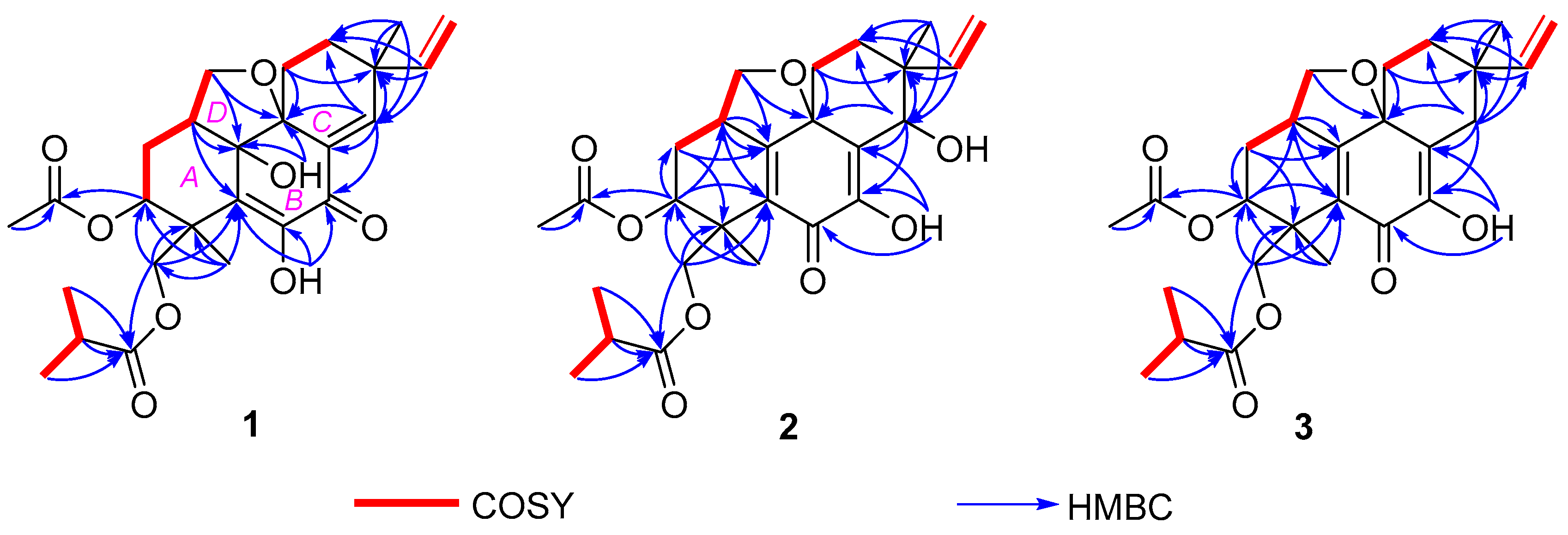
The basic libertellenone carbon skeleton of 1 was confirmed by the COSY correlations from H-1/H-2β, H-2β/H-3, H-11β/H-12β, H-15/H2-16, and H3-25/H-24/H3-26, as well as the key HMBC correlations from H-1 to C-5 and C-10, from H-3 to C-21, from 6-OH to C-5, C-6, and C-7, from 10-OH to C-9 and C-10, from H-11β to C-9 and C-13, from H-14 to C-7, C-8, C-9, C-12, and C-13, from H-15 to C-12, C-13, and C-14, from H3-17 to C-12, C-13, and C-14, from H-18α to C-3, C-4, C-5, and C-23, from H3-19 to C-3, C-4, C-5, and C-18, from H3-22 to C-21, and from H-24, H3-25 and H3-26 to C-23 (Figure 2). Additionally, the remaining unsigned signals H-20α and H-20β showed COSY correlations with H-1 and HMBC correlations with C-1, C-2, C-9, and C-10 (Figure 2). These, taken together with the downfield shift of C-20 (δC 72.5) and the molecular formula, linked C-20 to C-9 and C-10 via an O-atom and C-1, respectively. Thus, eutypellenoid A (1) was elucidated as a new pimarane diterpene derivative.
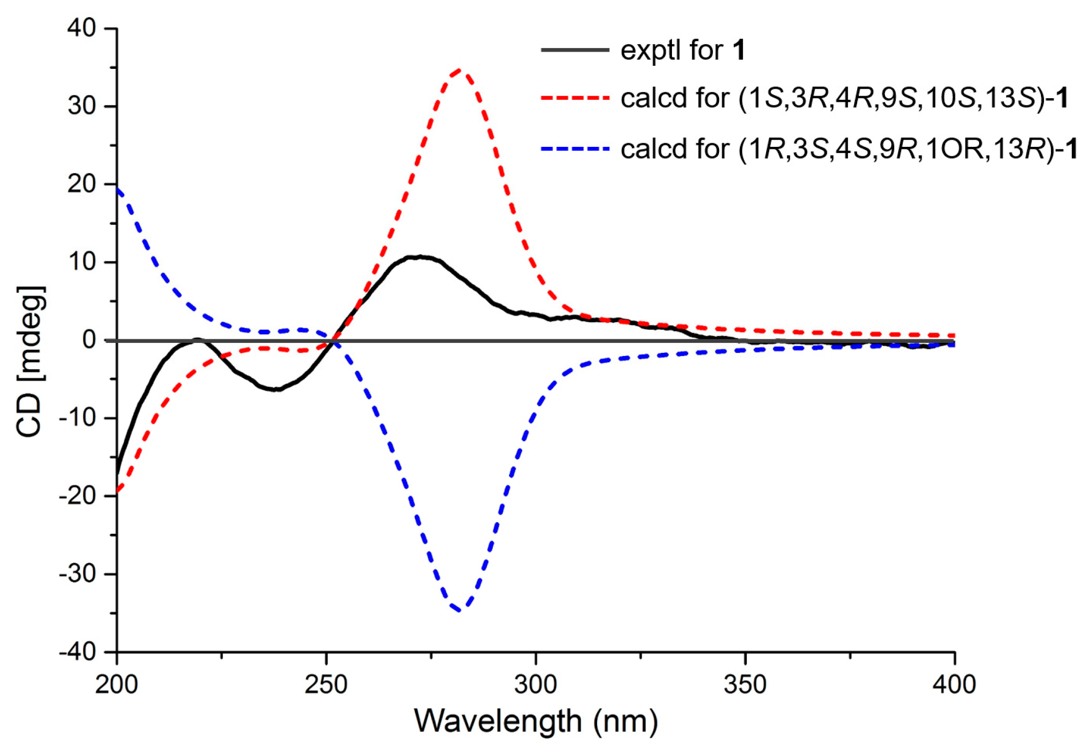
The relative configuration of compound 1 was established by a NOESY experiment (Figure 3). The NOESY correlations of H-1/10-OH, 10-OH/H-11α, H-11α/H3-17, and H-12α/H3-17 indicated that they were cofacial and assigned randomly as α-oriented. In addition, the NOESY correlations of H-2β/H3-19, H-2β/H-20β, H-3/H3-19, H-11β/H-20β, and H-15 /H-12β suggested the β-orientation for these protons (Figure 3). The absolute configuration of 1 was established by ECD experiments (Figure 4). The theoretical calculation of ECD was conducted in MeOH using time-dependent density functional theory (TD-DFT). The calculated ECD spectrum of 1S,3R,4R,9S,10S,13S was well matched with the experimental spectrum of 1, thus determining the absolute configuration of 1 as 1S,3R,4R,9S,10S,13S.
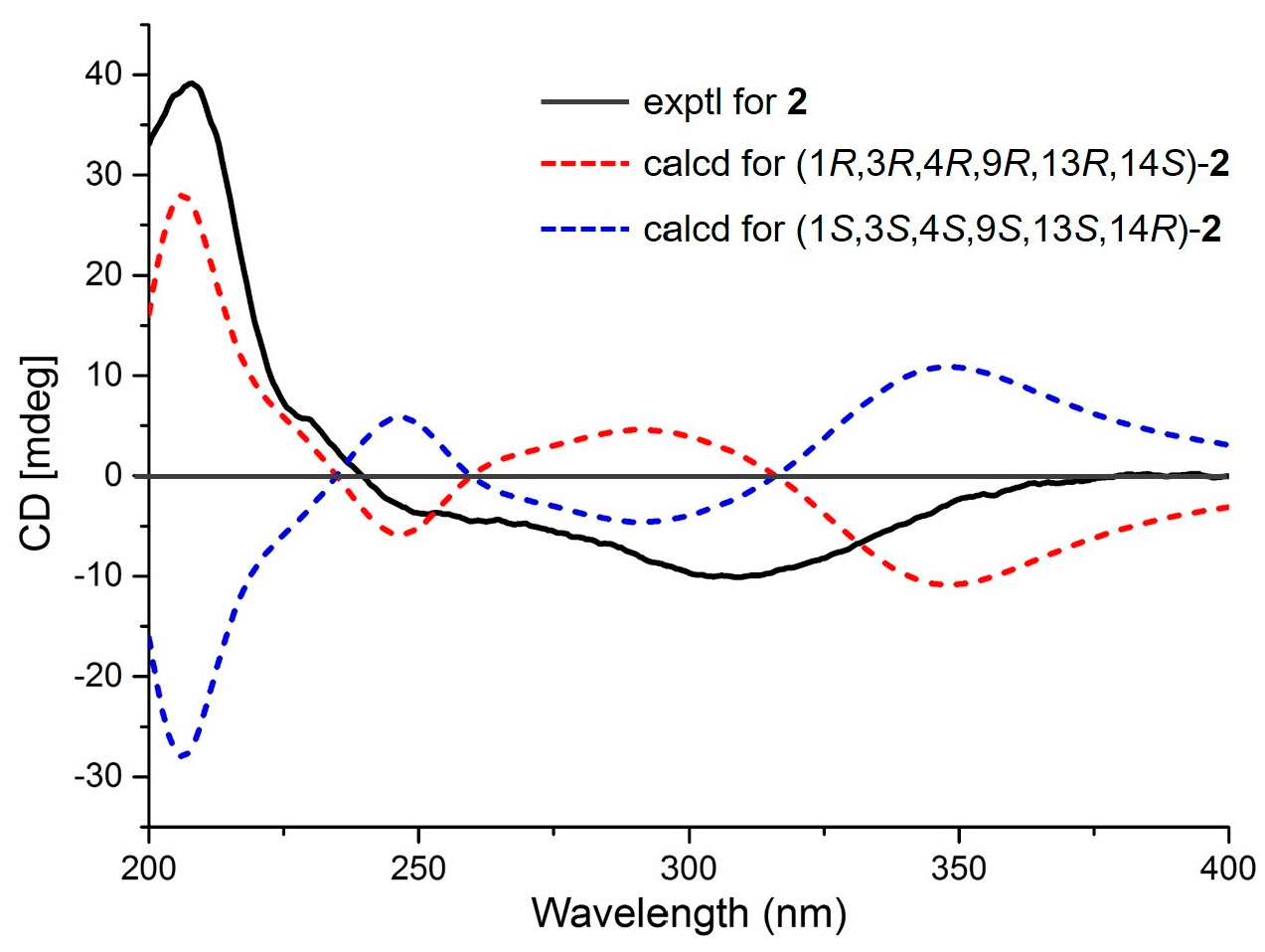
Eutypellenoid B (2) was also isolated as a yellow oil, with a the molecular formula C26H34O8 with 10 degrees of unsaturation based on HRESIMS data (m/z 492.2590 [M + NH4]+). The UV spectrum showed absorptions at λmax 211 and 250 nm. The IR absorptions exhibited the presence of hydroxyl (3353 cm−1) and carbonyl carbons (1734 and 1640 cm−1) [9]. The 1H NMR spectrum (Table 1) also showed the characteristic pattern for a vinyl group at δH 5.72 (1H, dd, 17.6, 10.8 Hz), 5.09 (1H, d, 17.6 Hz), and 5.03 (1H, d, 10.8 Hz). A comparison of the 1H and 13C NMR data of 2 with those of compound 1 showed that they shared the same tetrahydrofuran-fused pimarane diterpene skeleton, except for the replacement of sp2 methine with a hydroxyl group at C-14 (δC 72.2), and the presence of a benzoquinone subunit in ring B of 2. These was confirmed by the HMBC correlations (Figure 2) from H-3 to C-1, C-2, and C-5, from 7-OH to C-6, C-7, and C-8, from H-14 to C-7, C-8, C-9, C-12, and C-13, from H3-19 to C-3, C-4, and C-5, from H-20β to C-1, C-9, and C-10. The NOESY correlations of H-1/H-11α, H-12α/H-15, H-2β/H3-19, H-2β/H-20β, H-3/H3-19, H-11β/H3-17, H-11β/H-20β, H-12β/H3-17, and H-14/H3-17 established the relative configuration of 2 (Figure 3). Likewise, the experimental ECD spectrum of 2 was in good agreement with the calculated spectrum for 1R,3R,4R,9R,13R,14S, indicating that the absolute configuration of 2 is 1R,3R,4R,9R,13R,14S (Figure 5).
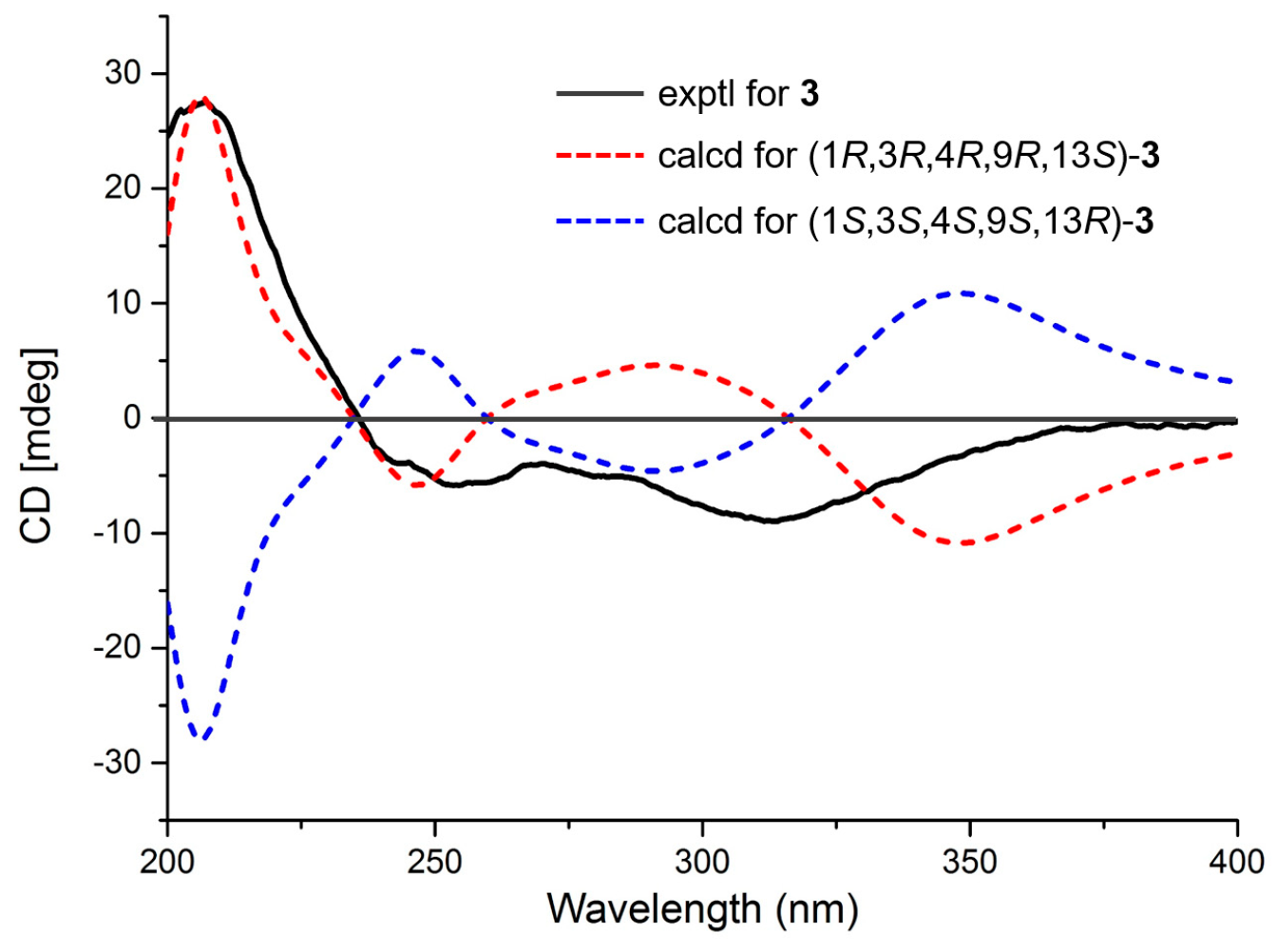
Eutypellenoid C (3) was isolated as a yellow oil. The molecular formula, C26H34O7, consistent with nine degrees of unsaturation, was determined based on HRESIMS data (m/z 457.2236 [M − H]−). The NMR spectra of 2 displayed close structure similarities to that of 3, except for the absence of a hydroxyl group substituted at C-14 (δC 35.0). Therefore, C-8 was attached to C-13 via the methylene carbon C-14, which was supported by the HMBC correlations from H-14α and H-14β to C-7, C-8, C-9, C-12, C-13, C-15, and C-17. The NOESY cross-peaks (Figure 3) of H-1/H-11α, H-11α/H-15, H-12α/H-15, and H-14α/H-15 indicated that these protons were α-oriented, while the NOESY cross-peaks of H-2β/H3-19, H-2β/H-20β, H-3/H3-19, H-12β/H3-17, H-14β/H3-17, and H-12β/H-14β indicated that they were β-oriented. A comparison of the calculated and experimental ECD spectra of 3 determined its absolute configuration as 1R,3R,4R,9R,13S (Figure 6).
In addition to the three new compounds 1−3, one known compound, eutypenoid C (4), was also obtained and elucidated by comparing the spectroscopic data with those reported in the literature [10].
All the isolated compounds were tested for antibacterial activity against Staphylococcus aureus (ATCC 27217), Escherichia coli (ATCC 25922), Bacillus subtilis (ATCC 21951), Vibrio alginolyticus (ATCC 33787), Vibrio vulnificus (ATCC 27562), Streptococcus agalactiae (ATCC 12386), and Aeromonas hydrophila (ATCC 35654) (Table 2), antifungal activity against Candida parapsilosis (ATCC 22019), Candida albicans (SC5314), Candida glabrata (537), Cryptococcus neoformans (32609), Microsporum gypseum (Cmccfmza), and Candida tropicalis (Table 3), and cytotoxic activities against HeLa (human cervical cancer cell line), MCF-7 (human breast adenocarcinoma cell line), HCT-116 (human colon carcinoma cancer cell line), K562 (human chronic myelogenous leukemia cell line), and SW1990 (human pancreatic cancer cell line) (Table 4). Compound 2 displayed antibacterial activity against S. aureus and E. coli with MIC values of 8 and 8 μg /mL, respectively. Compound 2 also showed antifungal activity against C. parapsilosis, C. albicans, C. glabrata, and C. tropicalis with MIC values of 8, 8, 16, and 32 μg/mL, respectively. Moreover, compound 2 exhibited moderate cytotoxic activity against HCT-116 cell line with IC50 value of 3.7 μM. The biological evaluation indicated that the hydroxylation at C-14, as in the case of comparison between 2 and 3, correlated with a positive effect on the inhibitory activity. However, compared with the pimaranes with the cyclopropyl-fused or cyclobutyl-fused system [11], the tetrahydrofuran-fused system decreased the inhibitory activity.
3. Experimental Section
3.1. General Experimental Procedures
Optical rotations were measured on a Perkin-Elmer model 341 polarimeter (Perkin-Elmer Inc., Waltham, MA, USA). UV spectra were obtained on a UV-8000 spectrophotometer (Shanghai Metash instruments Co., Shanghai, China). IR (KBr) spectra of all compounds were performed on a Jasco FTIR-400 spectrometer (Jasco Inc., Tokyo, Japan). 1D and 2D NMR spectra were recorded on Bruker AMX-500 or Bruker AMX-400 instruments (Bruker Biospin Corp., Billerica, MA, USA) at room temperature (rt). HRESIMS data were obtained on an Agilent 6210 LC/MSD TOF mass spectrometer (Agilent Technologies Inc. Lake Forest, CA, USA). Semi-preparative HPLC chromatography was performed on a Waters 1525 separation module (Waters Corp., Milford, MA, USA) equipped with a Waters 2998 photodiode array (PDA) detector (Waters Corp., Milford, MA, USA) by using YMC-Pack Pro C18 RS (5 μm) columns (YMC Co. Ltd., Kyoto, Japan. Silica gel (200–300 mesh, Qingdao Ocean Chemical Co., Qingdao, China), ODS (50 μm, YMC Co. Ltd., Kyoto, Japan), and Sephadex LH-20 (18–110 μm, Pharmacia Co., Piscataway, NJ, USA) were used for column chromatography.
3.2. Fungal Strain
The strain Eutypella sp. D-1 was isolated from the soil of London Island of Kongsfjorden of Ny-Ålesund District (altitude of 100 m) in the Arctic. It was incubated at 20 °C by using potato dextrose agar (PDA) medium. The fungus was identified as Eutypella sp. by 18S rDNA gene sequence analysis (GenBank accession number FJ430580). The strain was stored in PDA medium at the Second Military Medical University, Shanghai, China.
3.3. Fermentation, Extraction, and Isolation
The strain Eutypella sp. D-1 was maintained on PDA medium at 28 °C for seven days, and then three pieces (1 × 1 cm) of mycelial agar plugs were inoculated into 250 mL Erlenmeyer flasks, each containing 100 mL of seed medium (glucose 125 g/L, NaNO3 3.3 g/L, MgSO4·7H2O 0.4 g/L, K2HPO4·3H2O 0.07 g/L, KCl 0.625 g/L, yeast extract 0.7 g/L, FeSO4·7H2O 18.75 mg/L, CoCl2·6H2O 3.125 mg/L, CaCl2 6.5 mg/L, and L-ornithine hydrochloride 15 g/L, pH 5.8). After three days of incubation at 20 °C on a rotary shaker at 180 r/min, 20 mL seed cultures were transferred into a total of 150 flasks (2 L) containing 400 mL of fermentation medium (sucrose 51.4 g/L, NaNO3 3.3 g/L, MgSO4·7H2O 0.4 g/L, K2HPO4·3H2O 0.07 g/L, KCl 0.625 g/L, yeast extract 0.7 g/L, FeSO4·7H2O 18.75 mg/L, CoCl2·6H2O 3.125 mg/L, and CaCl2 6.5 mg/L, pH 5.8). The liquid cultivation was incubated for 10 days at 20 °C and 180 rpm on a rotary shaker. Meanwhile, 5 mL ethanol was added to the liquid cultivation three times, at 72 h, 96 h, and 120 h, respectively.
The whole culture (30 L) was filtered to give the broth and mycelia. The former was extracted with EtOAc three times, while the latter was extracted with a mixture of CH2Cl2/CH3OH (1:1, v/v) three times. The CH2Cl2/CH3OH solution was combined and evaporated under reduced pressure to obtain an aqueous solution and then extracted with EtOAc three times. These two EtOAc layers were almost the same by TLC and HPLC analysis, so they were combined and evaporated under reduced pressure to yield a dark brown gum (20.0 g).
The crude extract was subjected to vacuum liquid chromatography (VLC) on silica gel eluting with a step gradient of a mixture of petroleum ether (PE) and EtOAc (from 60:1 to 0:1) to afford seven fractions (A−G). Fraction C was separated on an ODS (50 μm) column followed by stepwise gradient elution with MeOH/H2O (3:5, 2:3, 4:5, 1:0) to obtain six subfractions (C1−C6). Fraction C4 was then further purified by HPLC (55% CH3CN/H2O, 2.0 mL/min) detected at the wavelength of 300 nm to yield compound 1 (tR = 48.3 min, 2.0 mg) and 4 (tR = 59.1 min, 3.8 mg). Fraction D was separated by CC on Sephadex LH-20 eluting with CH2Cl2/MeOH (1:1) to obtain three subfractions (D1−D3), and then fraction D2 was isolated by HPLC with an elution of 60% CH3CN detected at the wavelength of 252 nm to afford compound 2 (tR = 37.6 min, 2.0 mg). Fraction E was separated on an ODS (50 μm) column followed by stepwise gradient elution with MeOH/H2O (50%→100%) to obtain five subfractions (E1−E5). Fraction E3 was then further purified by HPLC (65% CH3CN/H2O, 2.0 mL/min) to yield compound 3 (252 nm, tR = 49.7 min, 2.0 mg).
Eutypellenoid A (1): yellow oil, [α] −12.0 (c 0.1, MeOH); UV (MeOH) λmax (log ε) 202 (3.63), 301 (3.34) nm; IR (KBr) vmax 3532, 3353, 2962, 2928, 2874, 1736, 1659, 1620, 1464, 1371, 1342, 1224, 1159, 1076, 1034, 997, 955, 920 cm−1; CD (MeOH) (Δε) 219 (+0.1), 237 (−4.6), 272 (+7.6); 1H and 13C NMR data, see Table 1; HRESIMS m/z 473.2187 [M − H]− (calcd for C26H33O8, 473.2193, Δ +1.24 ppm).
Eutypellenoid B (2): yellow oil, [α] −105.0 (c 0.1, MeOH); UV (MeOH) λmax (log ε) 211 (3.62), 250 (3.23) nm; IR (KBr) vmax 3353, 2972, 2928, 1734, 1640, 1463, 1370, 1298, 1242, 1200, 1159, 1081, 1031, 987, 947, 921 cm−1; CD (MeOH) (Δε) 208 (+28.1), 229 (−7.2); 1H and 13C NMR data, see Table 1; HRESIMS m/z 492.2590 [M + NH4]+ (calcd for C26H38NO8, 492.2592, Δ +0.40 ppm).
Eutypellenoid C (3): yellow oil, [α] −100.0 (c 0.1, MeOH); UV (MeOH) λmax (log ε) 208 (3.99), 249 (3.73), 320 (3.36) nm; IR (KBr) vmax 3404, 2961, 2929, 2879, 1736, 1658, 1629, 1464, 1371, 1346, 1299, 1241, 1195, 1156, 1076, 1032, 990, 948, 924, 890, 844, 803, 754 cm−1; CD (MeOH) (Δε) 207 (+19.1), 313 (−6.2); 1H and 13C NMR data, see Table 1; HRESIMS 457.2236 [M − H]− (calcd for C26H33O7, 457.2232, Δ −0.99 ppm).
3.4. ECD Calculations
Conformational searches for compounds 1–3 were carried out via Spartan’s 10 software (Wave-function, Inc., Irvine, CA, USA) in the MMFF94 force field. Subsequently, the conformers with a Boltzmann population of over 5% were re-optimized at the B3LYP/6-31+G(d,p) level by employing the conductor-like polarizable continuum model (CPCM) in MeOH. The theoretical calculation of ECD for 1–3 were calculated using the time-dependent density functional theory (TDDFT) methodology at the B3LYP/6-311++G (2d, 2p) level in MeOH, respectively. The ECD spectra were generated by the program SpecDis 1.6 using a Gaussian function (σ = 0.3 eV, half the bandwidth at 1/e peak height) [12].
3.5. Biological Assays
The antimicrobial activities of compounds 1–4 against E. coli, S. aureus, B. subtilis, V. vulnificus, V. alginolyticus, A. hydrophila, and S. agalactiae were evaluated by the broth dilution method [13,14], and chloromycetin was used as a positive control. The antifungal activities of compounds 1–4 against C. parapsilosis, C. albicans, C. glabrata, C. neoformans, M. gypseum, and C. tropicalis were determined using the National Center for Clinical Laboratory Standards (NCCLS) methods [15,16,17]. Fluconazole, posaconazole, and voriconazole were used as the positive control. The cytotoxic activity of compounds 1–4 against HeLa, MCF-7, HCT-116, K562, and SW1990 cell lines was performed by the Cell Counting Kit-8 (CCK-8) assay, as described before [11,13]. Each cancer cell line was treated with the indicated test compound at various concentrations, in triplicate, and cisplatin was used as a positive control.
4. Conclusions
Investigation on the secondary metabolites from fungus Eutypella sp. D-1 isolated from the Arctic led to the isolation and structure elucidation of three new pimarane diterpenes (1–3), together with one known compound 4. Structurally, compounds 1–3 possess an uncommon tetrahydrofuran-fused pimarane diterpene skeleton. These compounds were evaluated in antibacterial, antifungal, and cytotoxic activities. Only compound 2 displayed weak antibacterial activity against S. aureus and E. coli with MIC values of 8 and 8 μg/mL, respectively. Additionally, compound 2 showed antifungal activity against C. parapsilosis, C. albicans, C. glabrata, and C. tropicalis with MIC values of 8, 8, 16, and 32 μg/mL, respectively. Moreover, compound 2 exhibited moderate cytotoxic activity against HCT-116 cell line with IC50 value of 3.7 μM.
Supplementary Materials
1D and 2D NMR, UV, IR, HRESMS data, and detailed ECD calculations of 1–3 are available online at https://www.mdpi.com/1660-3397/16/8/284/s1.
Author Contributions
The contributions of the respective authors are listed as follows: H.-B.Y. and X.-L.W. drafted the work. H.-B.Y., X.-L.W., and Y.-X.Z. performed the fermentation, extraction, isolation, and structure elucidation. Y.-S.Q. contributed to the antimicrobial and antifungal activities evaluation. W.-H.X. performed the cytotoxicity evaluation. W.-H.X., J.-P.Z., X.-L.L., and X.-Y.L. contributed to checking and confirming all of the procedures of the isolation and the structure elucidation. H.-B.Y. and X.-Y.L. designed the study, supervised the laboratory work, and contributed to the critical reading and revision of the manuscript. All the authors have read the final manuscript and approved the submission.
Funding
The research was funded by National Natural Science Foundation of China (81741152, 41606173, and 41306197), the National High Technology Research and Development Program of China (863 Projects, SS2012AA09160703), and the Guangdong Innovative Development of Marine Economy Regional Demonstration Project (GD2012-D01-001).
Acknowledgments
The authors thank Prof. Bo Chen of Polar Research Institute of China for supplying the Arctic fungus Eutypella sp. D-1.
Conflicts of Interest
The authors declare no conflict of interest.
References
- Ivanets, E.V.; Yurchenko, A.N.; Smetanina, O.F.; Rasin, A.B.; Zhuravleva, O.I.; Pivkin, M.V.; Popov, R.S.; Amsberg, G.; Afiyatullov, S.S.; Dyshlovoy, S.A. Asperindoles A–D and a p-Terphenyl Derivative from the Ascidian-Derived Fungus Aspergillus sp. KMM 4676. Mar. Drugs 2018, 16, 232. [Google Scholar] [CrossRef] [PubMed]
- Tian, Y.; Li, Y.-L.; Zhao, F.-C. Secondary Metabolites from Polar Organisms. Mar. Drugs 2017, 15, 28. [Google Scholar] [CrossRef] [PubMed]
- Sun, L.; Li, D.; Tao, M.; Chen, Y.; Dan, F.; Zhang, W. Scopararanes C‒G: New oxygenated pimarane diterpenes from the marine sediment-derived fungus Eutypella scoparia FS26. Mar. Drugs 2012, 10, 539–550. [Google Scholar] [CrossRef] [PubMed]
- Tan, J.; Zhang, J.; Li, Y. Secondary Metabolites from Eutypella species and their Bioactivities. Mycosystema 2017, 36, 1181–1191. [Google Scholar]
- Niu, S.; Liu, D.; Shao, Z.; Proksch, P.; Lin, W. Eutypellazines A‒M, Thiodiketopiperazine-type Alkaloids from Deep Ssea Derived Fungus Eutypella sp. MCCC 3A00281. RSC Adv. 2017, 7, 33580–33590. [Google Scholar] [CrossRef]
- Pongcharoen, W.; Rukachaisirikul, V.; Phongpaichit, S.; Rungjindamai, N.; Sakayaroj, J. Pimarane Diterpene and Cytochalasin Derivatives from the Endophytic Fungus Eutypella scoparia PSU-D44. J. Nat. Prod. 2006, 69, 856–858. [Google Scholar] [CrossRef] [PubMed]
- Ciavatta, M.L.; Lopez-Gresa, M.P.; Gavagnin, M.; Nicoletti, R.; Manzo, E.; Mollo, E.; Guo, Y.-W.; Cimino, G. Cytosporin-related Compounds from the Marine-derived Fungus Eutypella scoparia. Tetrahedron 2008, 64, 5365–5369. [Google Scholar] [CrossRef]
- Isaka, M.; Palasarn, S.; Lapanun, S.; Chanthaket, R.; Boonyuen, N.; Lumyong, S. γ-Lactones and ent-Eudesmane Sesquiterpenes from the Endophytic Fungus Eutypella sp. BCC 13199. J. Nat. Prod. 2009, 72, 1720–1722. [Google Scholar] [CrossRef] [PubMed]
- Lu, X.-L.; Liu, J.-T.; Liu, X.-Y.; Gao, Y.; Zhang, J.; Jiao, B.-H.; Zheng, H. Pimarane Diterpenes from the Arctic Fungus Eutypella sp. D-1. J. Antibiot. 2014, 67, 171–174. [Google Scholar] [CrossRef] [PubMed]
- Zhang, L.-Q.; Chen, X.-C.; Zhu, S.-G.; Yang, Y.-F.; Chen, K.-X.; Li, Y.-M.; Chen, Z.-Q.; Wang, G.-M.; Chen, K.-X.; Liu, X.-Y. Eutypenoids A‒C: Novel Pimarane Diterpenoids from the Arctic Fungus Eutypella sp. D-1. Mar. Drugs 2016, 14, 44. [Google Scholar] [CrossRef] [PubMed]
- Yu, H.-B.; Wang, X.-L.; Zhang, Y.-X.; Xu, W.-H.; Zhang, J.-P.; Zhou, X.-Y.; Lu, X.-L.; Liu, X.-Y.; Jiao, B.-H. Libertellenones O−S and Eutypellenones A and B, Pimarane Diterpene Derivatives from the Arctic Fungus Eutypella sp. D-1. J. Nat. Prod. 2018, 81, 1553–1560. [Google Scholar] [CrossRef] [PubMed]
- Li, J.; Gu, B.B.; Sun, F.; Xu, J.R.; Jiao, W.H.; Yu, H.B.; Han, B.N.; Yang, F.; Zhang, X.C.; Lin, H.W. Sesquiterpene Quinones/Hydroquinones from the Marine Sponge Spongia pertusa Esper. J. Nat. Prod. 2017, 80, 1436–1445. [Google Scholar] [CrossRef] [PubMed]
- Yu, H.-B.; Jiao, H.; Zhu, Y.-P.; Zhang, J.-P.; Lu, X.-L.; Liu, X.-Y. Bioactive metabolites from the Arctic fungus Nectria sp. B-13. J. Asian Nat. Prod. Res. 2018. [Google Scholar] [CrossRef] [PubMed]
- Klink, J.W.; Larsen, L.; Perry, N.B.; Weavers, R.T.; Cook, G.M.; Bremer, P.J.; MacKenzie, A.D.; Kirikae, T. Triketones Active Against Antibiotic-resistant Bacteria: Synthesis, Structure-activity Relationships, and Mode of Action. Bioorg. Med. Chem. 2006, 13, 6651–6662. [Google Scholar] [CrossRef] [PubMed]
- Yu, H.-B.; Yang, F.; Sun, F.; Li, J.; Jiao, W.-H.; Gan, J.-H.; Hu, W.-Z.; Lin, H.-W. Aaptamine Derivatives with Antifungal and Anti-HIV-1 Activities from the South China Sea Sponge Aaptos aaptos. Mar. Drugs 2014, 12, 6003–6013. [Google Scholar] [CrossRef] [PubMed]
- Clinical and Laboratory Standards Institute. Reference Method for Broth Dilution Antifungal Susceptibility Testing of Yeast; Approved Standard, 3rd ed.; Document M27 A3; Clinical and Laboratory Standards Institute: Wayne, PA, USA, 2009. [Google Scholar]
- Clinical and Laboratory Standards Institute. Reference Method for Broth Dilution Antifungal Susceptibility Testing of Filamentous Fungi; Approved Standard, 2nd ed.; Document M38 A2; Clinical and Laboratory Standards Institute: Wayne, PA, USA, 2008. [Google Scholar]
Figure 1.
Chemical structures of compounds 1–4.

Figure 2.
COSY and key HMBC correlations of compounds 1–3.

Figure 3.
Key NOESY correlations of compounds 1–3.
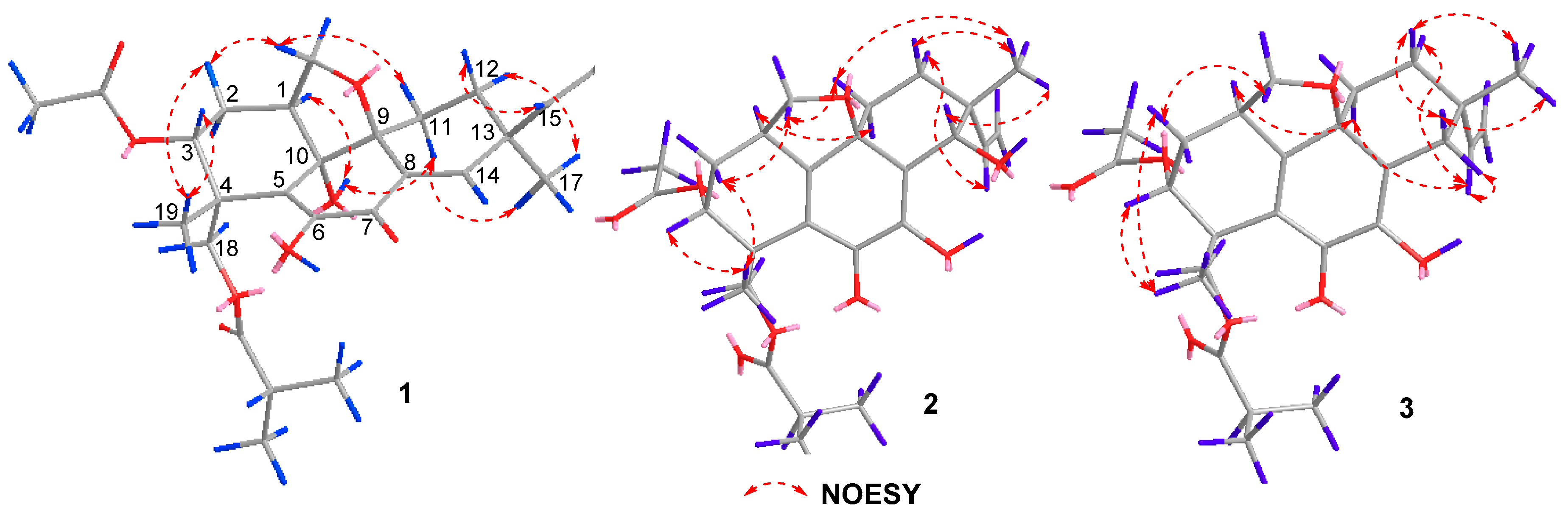
Figure 4.
Experimental and calculated ECD spectra of compound 1.

Figure 5.
Experimental and calculated ECD spectra of compound 2.

Figure 6.
Experimental and calculated ECD spectra of compound 3.

Table 1.
1H and 13C NMR data of compounds 1−3 in CDCl3.
| Position | 1 a | 2 b | 3 b | |||
|---|---|---|---|---|---|---|
| δC | δH, mult. (J in Hz) | δC | δH, mult. (J in Hz) | δC | δH, mult. (J in Hz) | |
| 1 | 40.6, CH | 2.93, m | 32.3, CH | 3.27, m | 33.0, CH | 3.32, m |
| 2α | 26.0, CH2 | 2.32, m | 26.5, CH2 | 2.35, m | 26.8, CH2 | 2.33, m |
| 2β | 1.70, m | 1.46, m | 1.46, m | |||
| 3 | 74.1, CH | 5.11, t (7.0, 3.5) | 72.7, CH | 5.12, d (1.2) | 72.9, CH | 5.11, d (0.8) |
| 4 | 42.8, C | 40.5, C | 40.5, C | |||
| 5 | 124.7, C | 127.3, C | 126.9, C | |||
| 6 | 145.9, C | 180.3, C | 179.9, C | |||
| 6-OH | 7.17, s | |||||
| 7 | 181.3, C | 142.3, C | 142.4, C | |||
| 7-OH | 6.66, s | 6.52, s | ||||
| 8 | 129.5, C | 122.7, C | 125.8, C | |||
| 9 | 80.9, C | 79.2, C | 77.7, C | |||
| 10 | 76.2, C | 164.8, C | 164.8, C | |||
| 10-OH | 4.41, s | |||||
| 11α | 24.4, CH2 | 1.72, m | 35.8, CH2 | 2.04, m | 35.4, CH2 | 2.04, m |
| 11β | 2.09, m | 1.54, m | 1.41, m | |||
| 12α | 29.7, CH2 | 1.82, m | 26.9, CH2 | 1.57, m | 32.6, CH2 | 1.60, m |
| 12β | 1.64, m | 2.08, m | 1.93, m | |||
| 13 | 39.8, C | 44.9, C | 41.1, C | |||
| 14α | 152.7, CH | 7.11, s | 72.2, CH | 4.68, s | 35.0, CH2 | 2.96, dd (12.8, 2.0) |
| 14β | 2.15, d (12.8) | |||||
| 15 | 142.7, CH | 5.71, dd (17.5, 10.5) | 142.4, CH | 5.72, dd (17.6, 10.8) | 144.0, CH | 5.68, dd (17.6, 10.8) |
| 16a | 114.2, CH2 | 4.86, d (17.5) | 113.7, CH2 | 5.03, d (10.8) | 112.9, CH2 | 4.96, d (10.8) |
| 16b | 5.04, d (10.5) | 5.09, d (17.6) | 5.12, d (17.6) | |||
| 17 | 27.4, CH3 | 1.23, s | 25.2, CH3 | 1.20, s | 30.1, CH3 | 1.15, s |
| 18α | 64.9, CH2 | 4.42, d (14.0) | 65.2, CH2 | 4.35, d (10.8) | 65.3, CH2 | 4.35, d, (10.4) |
| 18β | 4.57, d (14.0) | 4.79, d (10.8) | 4.81, d (10.4) | |||
| 19 | 20.6, CH3 | 1.61, s | 20.9, CH3 | 1.31, s | 20.9, CH3 | 1.31, s |
| 20α | 72.5, CH2 | 4.22, t (18.5, 9.5) | 72.9, CH2 | 4.42, t (8.8) | 71.9, CH2 | 4.36, t (8.0) |
| 20β | 3.47, t (18.0, 9.0) | 3.77, t (8.8) | 3.70, t (8.0) | |||
| 21 | 168.6, C | 169.9, C | 170.0, C | |||
| 22 | 21.1, CH3 | 2.08, s | 21.0, CH3 | 1.99, s | 21.0, CH3 | 1.99, s |
| 23 | 176.6, C | 176.4, C | 176.4, C | |||
| 24 | 34.1, CH | 2.51, m | 34.1, CH | 2.52, m | 34.1, CH | 2.52, m |
| 25 | 18.8, CH3 | 1.13, d (7.0) | 18.8, CH3 | 1.13, d (6.8) | 18.8, CH3 | 1.13, d (6.8) |
| 26 | 18.9, CH3 | 1.14, d (7.0) | 18.9, CH3 | 1.14, d (6.8) | 18.9, CH3 | 1.13, d (6.8) |
a 500 MHz for 1H NMR and 125 MHz for 13C NMR. b 400 MHz for 1H NMR and 100 MHz for 13C NMR.
Table 2.
Antibacterial activities of compounds 1–4.
| Compound | MIC (μg/mL) | ||||||
|---|---|---|---|---|---|---|---|
| S. aureus | E. coli | B. subtilis | V. alginolyticus | V. vulnificus | S. agalactiae | A. hydrophila | |
| 1 | 32 | 32 | 64 | 64 | 32 | 64 | 64 |
| 2 | 8 | 8 | 32 | 32 | 32 | 32 | 32 |
| 3 | 32 | 32 | 32 | 64 | 64 | 64 | 64 |
| 4 | 32 | 64 | 64 | 64 | 64 | 64 | 64 |
| Chloromycetin | 4 | 4 | 2 | 1 | 1 | 1 | 1 |
Table 3.
Antifungal activities of compounds 1–4.
| Compound | MIC (μg/mL) | |||||
|---|---|---|---|---|---|---|
| C. parapsilosis | C. albicans | C. glabrata | C. neoformans | M. gypseum | C. tropicalis | |
| 1 | >64 | >64 | >64 | >64 | >64 | >64 |
| 2 | 8 | 8 | 16 | >64 | >64 | 32 |
| 3 | >64 | >64 | 64 | >64 | >64 | >64 |
| 4 | >64 | >64 | 64 | >64 | >64 | >64 |
| Fluconazole | 0.50 | 2 | 0.50 | 1 | 2 | 0.25 |
| Posaconazole | 0.50 | 0.12 | 0.50 | 0.02 | 1 | 0.02 |
| Voriconazole | 0.02 | 0.03 | 0.02 | 0.02 | 0.06 | 0.02 |
Table 4.
Cytotoxic activities of compounds 1–4.
| Compound | IC50 (μM) | ||||
|---|---|---|---|---|---|
| HeLa | MCF-7 | HCT-116 | K562 | SW1990 | |
| 1 | 24.4 | 26.2 | 20.7 | 30.9 | 23.6 |
| 2 | 15.1 | 20.3 | 3.7 | 23.3 | 33.6 |
| 3 | 41.5 | 36.5 | 31.6 | >50 | >50 |
| 4 | 46.5 | 40.1 | 27.1 | 32.1 | >50 |
| Cisplatin | 0.5 | 4.5 | 2.7 | 2.7 | 1.0 |
© 2018 by the authors. Licensee MDPI, Basel, Switzerland. This article is an open access article distributed under the terms and conditions of the Creative Commons Attribution (CC BY) license (http://creativecommons.org/licenses/by/4.0/).
Share and Cite
MDPI and ACS Style
Yu, H.-B.; Wang, X.-L.; Xu, W.-H.; Zhang, Y.-X.; Qian, Y.-S.; Zhang, J.-P.; Lu, X.-L.; Liu, X.-Y. Eutypellenoids A–C, New Pimarane Diterpenes from the Arctic Fungus Eutypella sp. D-1. Mar. Drugs 2018, 16, 284. https://doi.org/10.3390/md16080284
AMA Style
Yu H-B, Wang X-L, Xu W-H, Zhang Y-X, Qian Y-S, Zhang J-P, Lu X-L, Liu X-Y. Eutypellenoids A–C, New Pimarane Diterpenes from the Arctic Fungus Eutypella sp. D-1. Marine Drugs. 2018; 16(8):284. https://doi.org/10.3390/md16080284
Chicago/Turabian StyleYu, Hao-Bing, Xiao-Li Wang, Wei-Heng Xu, Yi-Xin Zhang, Yi-Sen Qian, Jian-Peng Zhang, Xiao-Ling Lu, and Xiao-Yu Liu. 2018. "Eutypellenoids A–C, New Pimarane Diterpenes from the Arctic Fungus Eutypella sp. D-1" Marine Drugs 16, no. 8: 284. https://doi.org/10.3390/md16080284
Note that from the first issue of 2016, this journal uses article numbers instead of page numbers. See further details here.