Article

Cytotoxic Polyketides from the Deep-Sea-Derived Fungus Engyodontium album DFFSCS021

1
Key Laboratory of Tropical Marine Bio-resources and Ecology, Guangdong Key Laboratory of Marine Materia Medica, RNAM Center for Marine Microbiology, South China Sea Institute of Oceanology, Chinese Academy of Sciences, 164 West Xingang Road, Guangzhou 510301, Guangdong, China
2
University of Chinese Academy of Sciences, Beijing 100049, China
*
Author to whom correspondence should be addressed.
Mar. Drugs 2014, 12(12), 5902-5915; https://doi.org/10.3390/md12125902
Submission received: 20 October 2014 / Revised: 25 November 2014 / Accepted: 25 November 2014 / Published: 9 December 2014

Abstract

:
Eight new chromones, engyodontiumones A–H (18), and three new phenol derivatives (911) together with eight known polyketides (1219) were isolated from the deep-sea-derived fungus Engyodontium album DFFSCS021. Their structures were identified by extensive spectroscopic analysis. Compounds 8 and 16 showed significant selective cytotoxicity against human histiocytic lymphoma U937 cell line with IC50 values of 4.9 and 8.8 μM, respectively. In addition, this is the first time to report that 8, 15 and 16 had mild antibacterial activity against Escherichia coli and Bacillus subtilis, and 15 showed potent antilarval activity against barnacle Balanus amphitrite larval settlement.

1. Introduction

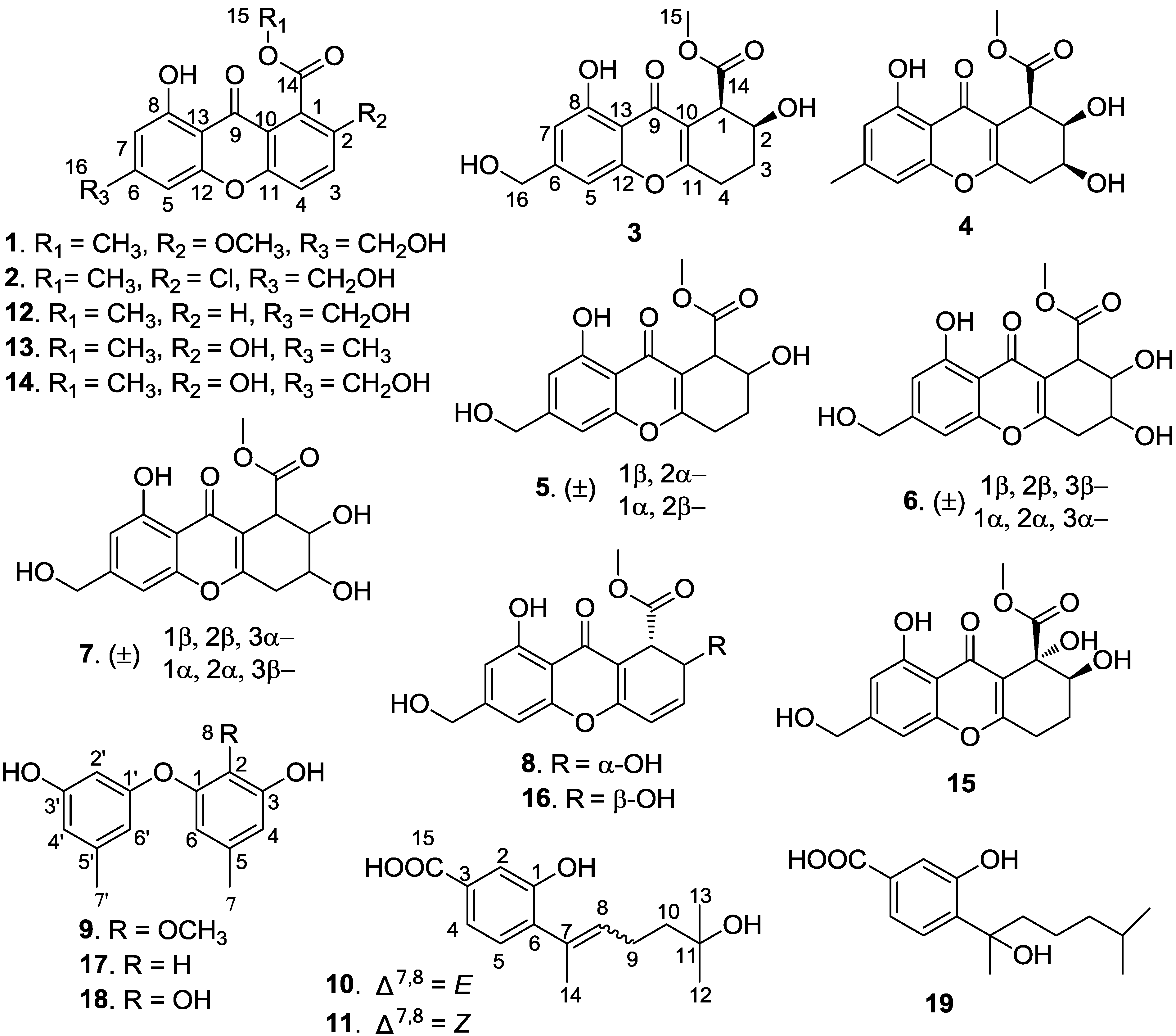
Recently, with the development of methods for sample collection and culturing technologies, an increasing number of deep-sea-derived fungi have been reported to produce novel bioactive secondary metabolites [1,2]. In our ongoing search for structurally unique and biologically active compounds from deep-sea-derived microorganisms [3], a fungal strain, identified as Engyodontium album, isolated from a marine sediment sample collected in the South China Sea (19°00′368″N, 117°58′223″E, 3739 m depth), attracted our attention. Preliminary experiments demonstrated that the extract of the culture medium of the strain showed toxicity towards brine shrimp and antibacterial activity. Further investigation on the chemical constituents of the extract resulted in the isolation of eight new chromones (18) and three new phenol derivatives (911) together with eight known compounds: sydowinin A (12) [4], pinselin (13) [5], sydowinin B (14) [5,6], aspergillusone B (15) [7], AGI-B4 (16) [8], diorcinol (17) [9], cordyol C (18) [10], and hydroxysydonic acid (19) [11] (Figure 1). The cytotoxicity, antibacterial, and antilarval activities of these compounds were individually evaluated. This paper describes the isolation, structure elucidation, and bioactivity of these compounds.
Figure 1. Structures of compounds 119.
Marinedrugs 12 05902 g001

2. Results and Discussion

Engyodontiumone A (1) had a molecular formula of C17H14O7 as established by HRESIMS (m/z 331.0801 [M + H]+). Its NMR data (Table 1 and Table 2) was similar to that of sydowinin B [5,6] with the only difference of one methoxyl (δC 56.8) and the absence of a hydroxyl proton signal in 1, which indicated that 1 was a chromone skeleton. In the HMBC spectrum (see Supporting Information Figure S3), correlations of δH 12.11 with C-7/C-8/C-13 suggested a hydroxyl group located at C-8, correlations of δH 3.88 (3H, s) with C-2 suggested a methoxy group located at C-2, and correlations of δH 4.60 (2H, s) with C-5/C-6/C-7 suggested a hydroxymethene group attached at C-6. In addition, HMBC correlations of δH 3.87 (3H, s) with C-14 combined with the chemical shift of C-1 (δC 120.3, C), suggested a methoxycarbonyl group located at C-1. Therefore, the structure of 1 was determined as shown.
Engyodontiumone B (2) had a molecular formula of C16H11ClO6 as inferred from its HRESIMS (m/z 335.0309 [M + H]+) showing peaks at m/z 335.0309 and 337.0264 with the height ratio of 3:1. Its NMR data (Table 1 and Table 2) showed great similarity to those of sydowinin B [5,6] with the only obvious difference of the high-field shift of the chemical shift of C-2 (from δC 148.9 in sydowinin B to δC 125.5 in 2) suggesting the loss of the hydroxyl functionality. Combined with the molecular formula and HMBC spectral data (see Supporting Information Figure S8), these data suggested a chlorine atom attached at C-2. Therefore, the structure of 2 was determined as shown.
The molecular formula C16H16O7 of engyodontiumone C (3) was deduced from its HRESIMS (m/z 321.0972 [M + H]+). Its NMR data (Table 1 and Table 2) showed great similarity to those of aspergillusone B7 with the only difference is the presence of one extra methine (δH 3.84 and δC 43.5) and the absence of one oxygenated quaternary carbon (δC 74.3 (C-1) in aspergillusone B), which suggested that 3 had the same skeleton as aspergillusone B with the only difference being the substitution at C-1. This was proved by the HMBC spectrum (Figure 2) showing correlation of H-1 (δH 3.84) with C-3/C-9/C-11. The coupling constant values of JH-1,H-2 (6.0 Hz) and JH-2,H-3 (3.5, 9.5 Hz) indicated that H-1 and H-2 were equatorial–axial relationship and required the placement of H-1 and H-2 in pseudo equatorial and axial orientations [12], respectively (Table 3). We failed to determine the absolute structure of 3 by Mosher ester methods, because the OH- at C-16 was easier to be esterified than OH- at C-2. In the experiment, the main product of the Mosher ester reaction of 3 was the C-16 esterification, and the trace product was the ester esterified at C-2, C-8, and C-16.
Table 1. 1H NMR Data for 18 (500 MHz, in DMSO-d6, δ ppm, J in Hz).
Position12345678
δH (J)δH (J)δH (J)δH (J)δH (J)δH (J)δH (J)δH (J)
1 3.84, d (6.0)3.78, d (5.5)3.62, d (3.5)3.60, d (4.5)3.63, d (6.0)4.03, d (10.0)
2 4.11, ddd (3.5, 6.0, 9.5)3.99, br t (5.5)4.14, ddd (2.5, 3.5, 6.0)4.15, br t (4.5)3.99, dd (1.5, 6.0)5.01, br d (10.0)
37.79, d (9.0)8.06, d (9.0)1.84, m
2.01, m
4.04, ddd (4.5, 5.0, 5.5)1.80, m
1.90, m
3.96, ddd (3.5, 4.5, 4.5)3.92, ddd (1.5, 4.5, 6.5)6.49, d (2.0)
47.82, d (8.5)7.84, d (9.0)2.73–2.89, m3.10, dd (4.0, 18.5)2.73–2.87, m3.03, dd (4.5, 18.5)3.07, dd (4.5, 18.5)6.36, d (2.0)
2.60, dd (5.0, 18.5) 2.70, dd (3.5, 18.5)2.79, dd (6.5, 18.5)
57.02, s7.05, s6.96, s6.89, s6.98, s6.98, s6.98, s6.98, s
76.77, s6.81, s6.73, s6.65, s6.74, s6.74, s6.74, s6.75, s
153.87, s3.95, s3.60, s3.60, s3.64, s3.65, s3.65, s3.58, s
164.60, s4.62, d (4.5)4.56, s2.37, s4.58, s4.56, s4.56, s4.56, s
2-OH 5.49, brs 5.90, brs
8-OH12.11, s11.88, brs12.29, s 12.29, s12.41, brs12.25, brs12.39, s
16-OH 5.58, t (4.5) 5.50, brs 5.50, brs
8-OCH33.88, s
Table 2. 13C NMR Data of 18 (125 MHz, in DMSO-d6, δ ppm).
Position12345678
δCδCδCδCδCδCδCδC
1120.3, C131.0, C43.5, CH42.3, CH46.2, CH43.5, CH44.6, CH41.7, CH
2154.4, C125.5, C65.1, CH68.1, CH65.4, CH66.3, CH65.8, CH65.9, CH
3121.3, CH136.4, CH26.1, CH266.1, CH26.1,CH269.8, CH69.9, CH145.0, CH
4120.8, CH121.3, CH25.7, CH232.8, CH223.8, CH232.9, CH233.4, CH2119.7, CH
5103.9, CH104.1, CH103.8, CH107.2, CH103.9, CH103.9, CH103.9, CH104.1, CH
6155.5, C154.8, C155.5, C147.2, C152.3, C152.1, C152.3, C152.2, C
7107.2, CH107.8, CH107.7, CH111.4, CH107.8, CH107.9, CH107.9, CH108.3, C
8160.2, C160.4, C159.3, C159.2, C159.3, C159.4, C159.3, C159.3, C
9180.2, C179.2, C181.1, C180.7, C181.2, C180.1, C180.8, C180.0, C
10117.1, C118.0, C113.9, C113.1, C113.1, C112.7, C113.0, C111.0, C
11152.0, C154.3, C166.9, C164.1, C166.7, C164.3, C164.8, C160.4, C
12156.2, C155.3, C152.2, C155.6, C155.5, C155.7, C155.7, C154.9, C
13107.0, C106.9, C107.9, C107.2, C107.9, C107.8, C107.9, C108.3, C
14166.2, C165.7, C171.4, C170.0, C172.0, C170.7, C172.3, C169.7, C
1552.4, CH352.9, CH351.5, CH351.5, CH352.0, CH351.5, CH351.9, CH351.5, CH3
1662.3, CH262.2, CH262.1, CH221.7, CH362.2, CH262.2, CH262.1, CH262.1, CH2
2-OCH358.6, OCH3
Figure 2. Key HMBC and 1H–1H COSY correlations of compounds 3, 4 and 10.
Marinedrugs 12 05902 g002
Table 3. 1H NMR (DMSO-d6) J values (Hz) for compounds 37. Marinedrugs 12 05902 i001
CompoundJH-1,H-2JH-2,H-3JH-3,H-4Orientations of H-1, H-2, H-3
36.03.5, 9.5 H-1: equatorial, H-2: axial
45.55.54.5, 5.0H-1: equatorial, H-2: axial, H-3: equatorial
53.52.5, 6.0 H-1: equatorial, H-2: equatorial
64.54.53.5, 4.5H-1: equatorial, H-2: axial, H-3: equatorial
76.01.54.5, 6.5H-1: axial, H-2: equatorial, H-3: equatorial
The molecular formula C16H16O8 of engyodontiumone D (4) was inferred from its HRESIMS (m/z 321.0974 [M + H]+). Its NMR data (Table 1 and Table 2) were very similar to those of 3, except for the presence of one methyl group (δH 2.37, δC 21.7) and one oxygenated methine (δH 4.06, δC 66.1) and absence of two methylenes. The HMBC spectrum (Figure 2) showing correlations of H-1 with C-2/C-10/δC 66.1, H-2 with C-1/δC 66.1, and 1H–1H COSY spectrum (Figure 2) showing correlations of H-2 with H-1/δH 4.06, suggested the oxygenation of C-3 (δH 4.06, δC 66.1). In addition, correlations of δH 2.37 with C-5/C-6/C-7 suggested that a methyl group CH3-16 was attached at C-6. The coupling constant values of JH-1,H-2 (5.5 Hz), JH-2,H-3 (5.5 Hz) and JH-3,H-4 (4.5, 5.0 Hz) indicated the equatorial–axial relationship for H-1/H-2 and H-2/H-3 placing H-1, H-2 and H-3 in pseudo equatorial, axial and equatorial orientations [12], respectively (Table 3). Therefore, the structure of 4 was determined as shown.
Engyodontiumone E (5) had the same molecular formula of C16H16O7 as 3, which was deduced from its HRESIMS (m/z 321.0968 [M + H]+). Its NMR data (Table 1 and Table 2) were very similar to those of 3 except for the obvious changes of the chemical shifts of H-1 and C-1. Further detailed analysis of HSQC and HMBC spectra (see Supporting Information Figure S27) suggested that 3 and 5 had the same planar structure, and 5 that was an isomer of 3. The coupling constant values of JH-1,H-2 (3.5 Hz) and JH-2,H-3 (2.5, 6.0 Hz) indicated the equatorial–equatorial relationship for H-1 and H-2 placing of H-1 and H-2 in pseudo equatorial orientation (Table 3). Compound 5 was inferred to be racemic by its zero value of specific optical rotation and HPLC analysis using a chiral column (see Supporting Information Figure S68).
Engyodontiumone F (6) had a molecular formula of C16H16O8 as deduced from its HRESIMS (m/z 337.0922 [M + H]+). Its NMR data (Table 1 and Table 2) showed great similarity to those of 4 with the only difference of one methyl group (CH3-16) oxygenated to be methylene (δH 5.50, δC 62.2). This was supported by the HMBC spectrum (see Supporting Information Figure S34) showing correlation of H-16 (δH 5.50) with C-5/C-6/C-7. The coupling constant values of JH-1, H-2 (4.5 Hz), JH-2,H-3 (4.5 Hz) and JH-3,H-4 (3.5, 4.5 Hz) indicated the axial–equatorial relationship for H-1/H-2 and H-2/H-3 placing H-1, H-2 and H-3 in pseudo equatorial, axial and equatorial orientations, respectively (Table 3). The relative configuration of H-2 and H-3 was supported by the NOESY spectrum showing correlations of OH-2/OH-3 with OH-8. Compound 6 was also inferred to be racemic by its zero value of specific optical rotation and HPLC analysis using a chiral column (see Supporting Information Figure S69).
Engyodontiumone G (7) had the same molecular formula of C16H16O8 as 6, which was deduced from its HRESIMS (m/z 337.0922 [M + H]+). Its NMR data (Table 1 and Table 2) was similar to those of 6. Further detailed analysis of HSQC and HMBC spectra (see Supporting Information Figures S41 and S42) suggested that 7 and 6 shared the same planar structure suggesting that 7 is an isomer of 6. The coupling constant values of JH-1,H-2 (4.5 Hz), JH-2,H-3 (1.5 Hz), and JH-3,H-4 (4.5, 6.5 Hz) indicated that axial–equatorial relationship for H-1 and H-2 and equatorial–equatorial relationship for H-2 and H-3 placing H-1, H-2 and H-3 in pseudo axial, equatorial and equatorial orientations, respectively (Table 3). Compound 7 was also inferred to be racemic by its zero value of specific optical rotation and HPLC analysis using a chiral column (see Supporting Information Figure S70).
Engyodontiumone H (8) had the same molecular formula of C16H14O7 as AGI-B4 [8] as deduced from its HRESIMS (m/z 321.0496 [M + H]+). Its NMR data (Table 1 and Table 2) also showed great similarity to those of AGI-B4 [7] except for a change in the chemical shift of H-2 (from δH 4.60 in AGI-B4 to δH 5.01 in 8) and the coupling constant value JH-1,H-2 (from 3.5 Hz in AGI-B4 to 10.0 Hz in 8), suggesting 8 is an epimer of AGI-B4 at C-2. Further detailed analysis of HMBC spectrum (see Supporting Information Figure S48) proved that 8 and AGI-B4 shared the same planar structure, and the coupling constant value of JH-1,H-2 (10.0 Hz) indicated that H-1 and H-2 were in a cis-configuration. We also failed to determine the absolute structure of 8 by Mosher ester methods as compound 3.
The molecular formula of 9 was established as C15H16O4 by its HRESIMS (m/z 261.1122 [M + H]+). Its NMR data showed close similarity to those of cordyol C [10] with the exception of one methoxyl group (δC 60.1, δH 3.60), which indicated that 9 had the same skeleton as cordyol C. The HMBC spectrum showed correlations of δH 3.60 (H-8) with C-2 (δC137.3), suggesting the methoxyl group (δC 60.1, δH 3.60) attached at C-2. Therefore, the structure of 9 was determined as shown, and named 2-methoxyl cordyol C.
Engyodontiumone I (10) had a molecular formula of C15H20O4 as inferred from its HRESIMS data (m/z 265.1426 [M + H]+). Its 1H NMR spectrum exhibited characteristic signals for a trisubstituted benzene ring (δH 7.40 (1H, d, J = 1.5 Hz), 7.33 (1H, dd, J = 1.5, 7.5 Hz), 7.09 (1H, d, J = 7.5 Hz)) and a trisubstituted double-bond olefinic proton (δH 5.49 (1H, m)). The 13C NMR and DEPT spectral data revealed 15 carbon signals including six quaternary carbons, four methines, two methylenes, and three methyls. These data showed similarity to those of hydroxysydonic acid [11] with the obvious difference of the additional appearance of one trisubstituted double-bond (δC 129.9 (C), 130.8 (CH)) and disappearance of one methine and one methene. In the HMBC spectrum (Figure 2), correlations of H-8 with C-6/C-7/C-10/C-14 suggested one double bond between C-7 and C-8, and correlations of H-12/H-13 with δC 68.5 (C, C-11) suggested the oxygenation of C-11. The chemical shift of C-14 (δC 16.4) indicated that the double-bond between C-7 and C-8 was in an E-configuration [13]. Therefore, the structure of 10 was determined as shown.
Engyodontiumone J (11) had the same molecular formula C15H20O4 as 10, which was deduced from its HRESIMS (m/z 265.1428 [M + H]+). Its NMR data were very similar to those of 10 except for the chemical shift changes of H-9, C-3 and C-14. Further detailed analysis of the HMBC spectrum suggested that 11 and 10 had the same planar structure. The chemical shift of C-14 (δC 23.8) indicated that the double bond between C-7 and C-8 was in a Z-configuration [13].
Antibacterial activities of compounds 119 were tested against Escherichia coli and Bacillus subtilis. Preliminary antibacterial assay results (Table 4) showed that at a concentration of 25 μg/disc compounds 8, 10, 15, 16 and 19 inhibiting the growth of E. coli and B. subtilis, while the remaining compounds had no obvious antibacterial activity. The MIC values of 8, 10, 15, 16 and 19 were further tested using micro-dilution method. The results (Table 4) showed that 8, 15 and 16 had mild antibacterial activity against E. coli and B. subtilis with MIC ≤ 64 μg/mL.
Chromones (28, 1214, 16) were also tested for cytotoxicity against human helacyton gartleri Hela, breast cancer MCF-7, liver hepatocellular carcinoma HepG2 and Huh7 cell lines (Table 4) by MTT method, and against human histiocytic lymphoma U937 cell line by CCK-8 (Cell Counting Kit-8) method. Compounds 8 and 16 showed significant selective cytotoxicity against U937 cell line with IC50 values of 4.9 and 8.8 μM, respectively. According to the literature, 16 was an inhibitor of VEGF signaling with the ability of inhibiting the proliferation of HUVECs induced by VEGF, bFGF or ECGS with IC50 values of 1.4, 2.8, and 6.2 μM [8], respectively, and showed weak cytotoxicity against human cancer cell lines SGC-7901 and BEL-7404 [14]. By comparison of the structures and cytotoxicities of these compounds, it seems that the substituent group at C-2 and the retainment of double bond between C-3 and C-4 are important for the cytotoxicity of this kind of chromone.
Table 4. Cytotoxicity and Antibacterial Activity of Compounds 219.
CompoundZone of Inhibition (mm) aMIC (μg/mL)Cytotoxicity (IC50 μM)
E. coliB. subtilisE. coliB. subtilisU937HelaMCF-7HepG2Huh7
2- b---55.596.1172.373.8 >300
3----218.4>300>300>300>300
4----208.6>300>300>300>300
5----15.9205.2>300>300>300
6----192.7>300>300>300>300
7----287.2 >300>300>300>300
813.816.564.032.04.924.838.560.553.3
10-10.0-256.0c NTNTNTNTNT
12----75.6>300>300>300>300
14----127.0>300>300>300>300
1511.014.464.064.0NTNTNTNTNT
1615.817.564.064.08.860.0102.252.7133.3
1911.413.664.0128.0NTNTNTNTNT
d Dox 0.060.823.13.31.2
d PG31.843.32.02.0
a Zones of inhibition of compounds 8, 10, 15, 16, and 19 compounds at concentration of 25 μg/disc and penicillin at concentration of 10 μg/disc; b “-”: trace or no effect was observed; c “NT”: not tested; d Doxorubicin (Dox) and penicillin (PG) were used as positive control.
In addition, antilarval activities of the known compounds 1218 were evaluated in settlement inhibition assays with laboratory-reared Balanus amphitrite larvae. Larval settlement bioassays revealed that 15 showed potent antilarval activity with EC50 value of 19.1 μg/mL and low toxicity with LC50/EC50 value of 70, and at the primal tested concentration of 25 μg/mL, 16 and 18 exhibited strong toxicity towards B. amphitrite larvae with lethality rates of 87.5% and 81.3% respectively, while other compounds showed a weak effect on larvae.

3. Experimental Section

3.1. General Experimental Procedure

Optical rotations were measured with an Anton Paar MCP 500 polarimeter. UV spectra were obtained using a Shimadzu UV-2600 UV-Vis spectrophotometer. CD spectra were measured with a Chirascan circular dichroism spectrometer (Applied Photophysics). IR spectra were measured with a Shimadzu IR Affinity-1 Fourier transform infrared spectrophotometer. 1H, 13C NMR and 2D NMR spectra were recorded on a Bruker AV-500 MHz NMR spectrometer with TMS as reference. MS spectroscopic data were obtained on a LCQDECA XP HPLC/MSn spectrometer for ESIMS. High-resolution electrospray-ionization (HRESIMS) was performed on a UPLC/Q-TOF Micro MS spectrometer under 70 eV. Semi-preparative MPLC was performed on a CHEETAHTM MP 200. Semi-preparative HPLC was performed on a Shimadzu LC-20A preparative liquid chromatography with an YMC-Pack ODS column, 250 × 20 mm i.d., S-5 μm. Sephadex LH-20 (GE Healthcare) was used for CC. Silica gel (200–300 mesh) for columm CC and GF254 for TLC were obtained from the Qindao Marine Chemical Factory, Qindao, China.

3.2. Fungal Material

The fungal strain DFFSCS021 (GenBank access number JX156368) was isolated from a marine sediment sample collected in the South China Sea (19°00′368″N, 117°58′223″E; 3739 m depth), and identified as Engydontium album by ITS rDNA sequence homolgy (99% similarity with E. album isolate AHF (Genbank accession No. KC3111469)). The fungal strain was inoculated into a 7 mL centrifuge tube containing 1 mL potato glucose medium and cultured at 28 °C, shaking at 150 rpm for 7 days. Total genomic DNA was extracted from the strain, as described by Lai et al. [15]. From the genomic DNA (gDNA), the ITS region of rDNA was amplified by PCR using primers ITS1 (5′-TCCGTAGGT GAACCTGCGG-3′) and ITS4 (5′-TCCT CCGCT TATTG AT ATGC-3′). The primers were synthesized by SBS Genetech (China). The PCR mixtures consisted of 12.5 μL Taq premix (TakaRa, China), 0.25 μL (10 μM) of each primer (Taka Ra, China), 0.75 μL DMSO, 10.25 μL water, and 1 μL template gDNA. After denaturation at 94 °C for 6 min, amplification was performed with 30 cycles of 45 s at 94 °C, 45 s at 53 °C, 2 min at 72 °C and a final extension at 72 °C for 10 min. In addition, the macro- and micro-morphological characteristics of strain DFFSCS021 were examined (see Supporting Information Figure S71). The strain was deposited in the RNAM Center, South China Sea Institute of Oceanology, Chinese Academy of Sciences.

3.3. Fermentation and Extraction

The fungal strain was inoculated in soluble starch medium (containing 10 g/L glucose, 10 g/L starch soluble, 1 g/L KH2PO4, 1 g/L MgSO4, 1 g/L bacterial peptone and 30 g/L sea salt) in 500 mL shake flask loading 120 mL as seed culture and incubated on a rotary shaker (200 rpm) at 28 °C for 2 days. Then, each of the seed cultures (100 mL) was transferred into autoclaved 5000 mL Erlenmeyer flasks that contained solid rice medium (each flask contained 500 g of commercially available rice, 2.5 g of yeast extract, 2.5 g of glucose, 22.5 g of sea salt, and 750 mL of water). Ultimately, the fungal stain was statically cultured in 5 kg rice medium at 26 °C for 28 days.
After incubation, the 5 kg rice culture was crushed and extracted with 80% acetone four times. The acetone extract was evaporated under reduced pressure to afford an aqueous solution, and then the aqueous solution was extracted with EtOAc to afford a 121 g brown crude gum.

3.4. Purification

The EtOAc extract (121 g) was chromatographed on a silica gel column eluting with a CHCl3/MeOH step gradient system at the ratios of 100:0, 99:1, 98:2, 95:5, 90:10, 80:20 and 0:100 (v/v) to yield nine fractions. Fraction 4 (5 g) was subjected to MPLC with an ODS column, eluting with MeOH/H2O (from 10:90 to 0:100, 120 min, 20 mL/min) to give five sub-fractions (Fr.4-1−Fr.4-5). Fr.4-1 was purified by semi-preparative reversed-phase (SP-RP) HPLC (YMC-Pack, ODS-A S-5 μm × 12 nm 250 × 20 mm i.d., 5 mL/min) eluting with C2H3N-H2O-TFA (46:54:0.03) to afford compounds 1 (10.3 mg, 0.085%) and 12 (20.9 mg, 0.173%). Fr.4-2 was purified by (SP-RP) HPLC eluting with C2H3N-H2O (38:62) to afford compound 9 (4.5 mg, 0.037%). Fr.4-3 was purified by (SP-RP) HPLC eluting with C2H3N-H2O-TFA (50:50:0.03) to afford compound 2 (7.9 mg, 0.065%). Fr.4-4 was purified by (SP-RP) HPLC eluting with C2H3N-H2O-TFA (45:55:0.03) to give compound 13 (16.7 mg, 0.138%). Fraction 6 (2.3 g) was isolated by MPLC with an ODS column, eluting with MeOH/H2O (from 20:80 to 0:100, 90 min, 20 mL/min) to give three sub-fractions (Fr.6-1−Fr.6-3) and compound 16 (18.5 mg, 0.153%). Fr.6-1 was purified by (SP-RP) HPLC eluting with MeOH-H2O (30:70) to give compound 15 (25.5 mg, 0.211%). Fr.6-2 was purified by (SP-RP) HPLC eluting with MeOH-H2O (60:40) to give compound 14 (15.5 mg, 0.128%). Fr.6-3 was purified by preparative TLC with a mobile phase of CHCl3-MeOH (9:1) to give compound 18 (14.3 mg, 0.118%). Fraction 7 (4.3 g) was subjected to silica gel CC eluting with a CHCl3/MeOH step gradient system at the ratios of 100:0, 98:2, 95:5, 90:10, 80:20, 50:50, and 0:100 (v/v) to give nine sub-fractions (Fr.7-1−Fr.7-9). Fr.7-2 was purified by (SP-RP) HPLC eluting with MeOH-H2O-TFA (38:62:0.03) to give compounds 6 (3.2 mg, 0.026%) and 7 (2.2 mg, 0.018%). Fr.7-5 was purified by (SP-RP) HPLC eluting with MeOH-H2O-TFA (52:48:0.03) to give compounds 10 (45.4 mg, 0.375%) and 11 (2.5 mg, 0.021%). Fr.7-8 was purified by (SP-RP) HPLC eluting with C2H3N-H2O-TFA (52:48:0.03) to give compound 19 (55.4 mg, 0.458%). Fraction 8 (3.9 g) was separated by MPLC with an ODS column, eluting with MeOH/H2O (from 10:90 to 0:100, 120 min, 20 mL/min) to give six sub-fractions (Fr.8-1−Fr.8-6). Fr.8-1 was purified by (SP-RP) HPLC eluting with C2H3N-H2O-TFA (12:88:0.03) to offer compound 15 (26.2 mg, 0.217%). Fr.8-2 was purified by (SP-RP) HPLC eluting with C2H3N-H2O-TFA (20:80:0.03) to afford compounds 3 (9.0 mg, 0.074%) and 16 (89.3 mg, 0.738%). Fr.8-3 was further purified by (SP-RP) HPLC eluting with C2H3N-H2O-TFA (35:65:0.03) to give compounds 5 (4.0 mg, 0.033%) and 8 (13.4 mg, 0.111%). Fr.8-4 was purified by (SP-RP) HPLC eluting with MeOH-H2O-TFA (38:62:0.03) to yield compounds 14 (8.0 mg, 0.066%) and 4 (4.6 mg, 0.038%). Fr.8-5 was purified by (SP-RP) HPLC eluting with C2H3N-H2O-TFA (38:62:0.03) to yield compound 17 (123.0 mg, 1.017%).
Engyodontiumone A (1): Pale yellow powder; UV (MeOH) λmax (log ε) 385 (2.91), 293 (3.13), 263 (3.66), 236 (3.63) nm; FT-IR: 3356, 1678, 1647 cm−1; 1H and 13C NMR data, see Table 1 and Table 2; HRESIMS m/ z 331.0801 [M + H]+ (calcd for C17H14O7, 331.0812).
Engyodontiumone B (2): Yellow needles; UV (MeOH) λmax (log ε) 372 (2.75), 287 (3.05), 259 (3.52), 234 (3.55) nm; FT-IR: 3394, 1674 cm−1; 1H and 13C NMR data, see Table 1 and Table 2; HRESIMS m/ z 335.0309 [M + H]+ (calcd for C17H11ClO6, 335.0317).
Engyodontiumone C (3): Brown needles; [ α ] D 25 5 (c 0.5, MeOH); UV (MeOH) λmax (log ε) 328 (3.38), 239 (4.13), 203 (4.06) nm; FT-IR: 3417, 1728, 1697, 1651 cm−1; 1H and 13C NMR data, see Table 1 and Table 2; HRESIMS m/z 321.0972 [M + H]+ (calcd for C16H16O8 , 321.0969).
Engyodontiumone D (4): Brown needles; [ α ] D 25 14 (c 0.3, MeOH); UV (MeOH) λmax (log ε) 325 (3.38), 322 (3.38), 238 (4.07), 230 (4.05), 202 (4.07) nm; FT-IR: 3456,1728,1658, 1627 cm−1; 1H and 13C NMR data, see Table 1 and Table 2; HRESIMS m/z 321.0974 [M + H]+ (calcd for C16H16O7, 321.0969).
Engyodontiumone E (5): Brown needles; [ α ] D 25 0 (c 0.4, MeOH); UV (MeOH) λmax (log ε) 329 (3.41), 258 (3.97), 238 (4.10), 203 (4.07) nm; FT-IR: 3336,1724,1654, 1620 cm−1; 1H and 13C NMR data, see Table 1 and Table 2; HRESIMS m/z 321.0968 [M + H]+ (calcd for C16H16O8, 321.0969).
Engyodontiumone F (6): Brown needles; [ α ] D 25 0 (c 0.3, MeOH); UV (MeOH) λmax (log ε) 328 (3.42), 238 (4.17), 203 (4.06) nm; FT-IR: 3334, 1732, 1651 cm−1; 1H and 13C NMR data, see Table 1 and Table 2; HRESIMS m/z 337.0922 [M + H]+ (calcd for C16H16O8 , 337.0918).
Engyodontiumone G (7): Brown needles; [ α ] D 25 0 (c 0.2, MeOH); UV (MeOH) λmax (log ε) 328 (3.14), 238 (3.94), 202 (3.94) nm; FT-IR: 3336, 1728, 1651 cm−1; 1H and13C NMR data, see Table 1 and Table 2; HRESIMS m/z 337.0922 [M + H]+ (calcd for C16H16O8 , 337.0918).
Engyodontiumone H (8): Brown needles; [ α ] D 25 −56 (c 1, MeOH); UV (MeOH) λmax (log ε) 344 (3.31), 272 (3.98), 213 (3.90), 203 (3.97) nm; FT-IR: 3420, 1735, 1658 cm−1; 1H and13C NMR data, see Table 1 and Table 2; HRESIMS m/z 319.0922 [M + H]+ (calcd for C16H14O7 , 319.0812).
2-methoxyl cordyol C (9): Yellow amorphous solid; UV (MeOH) λmax (log ε) 360 (2.64), 356 (2.64), 274 (3.20) nm; FT-IR: 3271, 1678 cm−1; 1H NMR (500 MHz, DMSO-d6) δH: 9.35 (2H, brs, OH-3/3′), 6.49 (1H, s, H-6), 6.26 (1H, s, H-6′), 6.24 (1H, s, H-4), 6.17 (1H, s, H-4′), 6.06 (1H, s, H-3′) , 3.60 (3H, s, H-8), 2.16 (3H, s, H-7′) and 2.14 (3H, s, H-7); 13C NMR (125 MHz, DMSO-d6) δC: 158.7 (s, C-1′), 158.2 (s, C-3′), 151.0 (s, C-3) , 148.3 (s, C-1), 139.7 (s, C-5′), 137.3 (s, C-2), 133.1 (s, C-5), 113.2 (d, C-6), 112.4 (d, C-4), 110.1 (d, C-6′), 108.0 (d, C-4′), 100.9 (d, C-2′), 60.1 (q, C-8), 21.1 (q, C-7′) and 20.6 (q, C-7). HRESIMS m/z 261.1122 [M + H]+ (calcd for C15H16O4 , 261.1121).
Engyodontiumone I (10): Yellow gum; UV (MeOH) λmax (log ε) 302 (3.40), 255 (3.64), 211 (4.16) nm; FT-IR: 3402, 3132, 1689, 1651, 979, 948 cm−1; 1H NMR (500 MHz, DMSO-d6) δH: 7.40 (1H, d, J = 1.5 Hz, H-2), 7.33 (1H, dd, J = 1.5, 7.5 Hz, H-4), 7.09 (1H, d, J = 7.5 Hz, H-5), 5.49 (1H, m, H-5), 2.18 (2H, m, H-9), 1.93 (3H, s, H-14), 1.48 (2H, m, H-10), 1.11 (6H, s, H-12/13); 13C NMR (125 MHz, DMSO-d6) δC: 167.2 (C, C-15), 154.3 (C, C-1), 136.7 (C, C-3), 133.4 (C, C-6), 130.8 (CH, C-8), 129.9 (CH, C-7), 129.2 (CH, C-5), 120.1 (CH, C-4), 116.1 (CH, C-2), 68.8 (C, C-11), 43.1 (CH2, C-10), 29.2 (CH3, C-12/13), 23.1 (CH2, C-9), 16.4 (CH3, C-14); HRESIMS m/z 265.1426 [M + H]+ (calcd for C15H20O4 , 265.1434).
Engyodontiumone J (11): Pale yellow gum; UV (MeOH) λmax (log ε) 297 (3.26), 244 (3.57), 207 (4.10) nm; FT-IR: 3344, 1689, 1612 cm−1; 1H NMR (500 MHz, DMSO-d6) δH: 7.42 (1H, d, J = 2.0 Hz, H-2), 7.35 (1H, dd, J = 2.0, 6.0 Hz, H-4), 7.03 (1H, d, J = 6.0Hz, H-5), 5.44 (1H, m, H-5), 2.18 (2H, m, H-9), 1.93 (3H, m, H-14), 1.81 (2H, s, H-9), 1.37 (3H, s, H-10), 0.95 (6H, s, H-12/13); 13C NMR (125 MHz, DMSO-d6) δC: 167.2 (C, C-15), 154.2 (C, C-1), 133.3 (C, C-3), 132.5 (C, C-6), 130.1 (CH, C-8), 129.6 (CH, C-7), 128.7 (CH, C-5), 119.7 (CH, C-4), 116.0 (CH, C-2), 68.5 (C, C-11), 43.4 (CH2, C-10), 29.0 (CH3, C-12/13), 24.0 (CH2, C-9), 23.8 (CH3, C-14); HRESIMS m/z 265.1428 [M + H]+ (calcd for C15H20O4 , 265.1434).

3.5. Cytotoxicity

Chromones (28, 1214, 16) were tested for cytotoxicity against Hela, MCF-7, HepG2 and Huh7 human carcinoma cell lines using a MTT (3-(4,5-dimethyl-2-thiazolyl)-2,5-diphenyl-2-H-tetrazolium bromide) method, and against U937 human carcinoma cell line using a CCK-8 (2-(2-methoxy-4-nitrophenyl)-3-(4-nitrophenyl)-5-(2,4-disulfophenyl)-2H-tetrazolium, monosodium salt) method. The media for U937 and the other four cell lines were RPMI1640 and DMEM, respectively. Cells in the logarithmic phase were seeded into 96-well plates in a 100 μL volume of medium, maintained at 37 °C in a 5% CO2 incubator overnight, then treated with the corresponding compounds or vehicle control at the indicated concentration. After 48 h of incubation, 5 μL of MTT or CCK-8 was added into the 96-well plates and incubated for additional 2–3 h, then the absorbance was measured in a microplate reader at 450 nm and 650 nm. Cell toxicity was evaluated as the ratio of the absorbance of the sample to that of the control. The IC50 were further calculated using Graphpad Prism 5 (Graphpad Software, Inc., San Diego, CA, USA).

3.6. Antibacterial Activities

Antibacterial activities of 119 were tested against common bacteria Bacillus subtilis and Escherichia coli using a standard disc diffusion assay [16]. Briefly, the tested bacteria were cultured on nutrient agar slants at 37 °C for 1−2 days, then a 40 μL cell suspension (108 CFU/mL) was mixed with 20 mL of corresponding nutrient agar and poured in 100 mm sterile petri plates. Sterile 6 mm diameter circular discs of filter paper were loaded with 25 μg of tested compound dissolved in methanol, evaporated to dryness, and then placed onto the seeded bacterial plates. An additional set of discs with 10 μg penicillin was used as the positive controls. The agar plates were incubated for 18 h at 37 °C. The inhibition zones were measured.
The minimum inhibitory concentration (MIC) of active compounds were determined by a microbroth dilution method [17]. Briefly, the tested bacteria were cultured on starch culture-medium including tryptone (10 g/L), yeast extract (5 g/L), NaCl (10 g/L) at 37 °C for 1–2 days on a rotary shaker (200 rpm), then scraped to make a cell suspension (105 CFU/mL) in the starch culture-medium. Tested samples and penicillin (antibiotic control) were dissolved in DMSO and dispensed into 96-well plates to create a dilution series (half fold dilutions ranging from 1024 to 0.5 µg/mL) in a 100 μL volume of starch culture-medium. Following incubation at 37 °C for 12–24 h, the tubes were examined for visible bacterial growth as evidenced by turbidity. The lowest concentration of test compound that prevented 100% growth represented the minimal inhibitory concentration (MIC). The experiment was run in 3 replicates.

3.7. Barnacle Balanus amphitrite Larval Settlement Bioassays

Larval settlement bioassays were performed using sterile 24-well polystyrene plates. Tested samples were dissolved in DMSO to a concentration of 25 μg mL−1 for preliminary bioassay. To define the EC50 values of antilarval compounds found in the preliminary bioassay, different dilutions of the tested compounds were further prepared to the concentrations ranging from 1 to 200 μg mL−1 in autoclaved FSW (filtered seawater). About 20 competent larvae were added to each well in 1 mL of the test solution. Wells containing only FSW with DMSO served as the controls. The plates were incubated at 27 °C for 24 h. The percentage of larval settlement was determined by counting the settled, live individuals under a dissecting microscope and expressing the result as a proportion of the total number of larvae in the well [18].

4. Conclusions

In conclusion, eight new chromones (18) and three new phenol derivatives (911) together with eight known polyketides were isolated from the culture medium of the deep-sea-derived fungus Engyodontium album DFFSCS021. Among then, 8 and 16 showed significant selective cytotoxicity against U937 cell line with IC50 < 10 μM. This is the first time to report that 8, 15 and 16 had mild antibacterial activity against E. coli and B. subtilis with MIC ≤ 64.0 μg/mL, and 15 showed potent antilarval activity against barnacle B. amphitrite larval settlement with EC50 value of 19.1 μg/mL and low toxicity.

Supplementary Files

Supplementary File 1

Acknowledgments

We are grateful for the financial support provided by the National Marine Public Welfare Research Project of China (201305017), Natural Science Foundation of China (41376160), National High Technology Research and Development Program of China (863 Program, 2012AA092104), Strategic Leading Special Science and Technology Program of Chinese Academy of Sciences (XDA100304002), Regional Innovation Demonstration Project of Guangdong Province Marine Economic Development (GD2012-D01-002), Science and Technology Planning Project of Guangdong Province (2013B031100001), and Marine Fishery Science and Technology Promotion Project of Guangdong Province (A201301B05).

Author Contributions

S. Qi conceived and designed the experiments; Q. Yao and J. Wang performed the experiments; S. Qi and X. Zhang analyzed the data; S. Qi, X. Xu and X. Nong contributed reagents/materials/analysis tools; S. Qi and Q. Yao wrote the paper.

Conflicts of Interest

The authors declare no conflict of interest.

References

  1. Blunt, J.W.; Copp, B.R.; Munro, M.H.G.; Northcote, P.T.; Prinsep, M.R. Marine natural products. Nat. Prod. Rep. 2011, 28, 196–268. [Google Scholar] [CrossRef] [PubMed]
  2. Skropeta, D. Deep-sea natural products. Nat. Prod. Rep. 2008, 25, 1131–1166. [Google Scholar] [CrossRef]
  3. Peng, J.; Zhang, X.Y.; Tu, Z.C.; Xu, X.Y.; Qi, S.H. Alkaloids from the deep-sea-derived fungus Aspergillus westerdijkiae DFFSCS013. J. Nat. Prod. 2013, 76, 983–987. [Google Scholar] [CrossRef]
  4. Hamasaki, T.; Sato, Y.; Hatsuda, Y. Structure of sydowinin A, sydowinin B, and sydowinol, metabolites from Aspergillus sydowi. Agric. Biol. Chem. 1975, 39, 1345–2341. [Google Scholar]
  5. Healy, P.C.; Hocking, A.; Tran-Dinh, N.; Pitt, J.I.; Shivas, R.G.; Mitchell, J.K.; Kotiw, M.; Davis, R.A. Xanthones from a microfungus of the genus Xylaria. Phytochemistry 2003, 65, 2373–2378. [Google Scholar] [CrossRef]
  6. Little, A.; Porco, J.J. Total syntheses of graphisin A and sydowinin B. Org. Lett. 2012, 14, 2862–2865. [Google Scholar] [CrossRef] [PubMed]
  7. Trisuwan, K.; Rukachaisirikul, V.; Kaewpet, M.; Phongpaichit, S.; Hutadilok-Towatana, N.; Preedanon, S.; Sakayaroj, J.; Rukachaisirikul, V. Sesquiterpene and xanthone derivatives from the sea fan-derived fungus Aspergillus sydowii PSU-F154. J. Nat. Prod. 2011, 74, 1663–1667. [Google Scholar] [CrossRef] [PubMed]
  8. Kim, H.S.; Park, I.Y.; Park, Y.J.; Lee, J.H.; Hong, Y.S.; Lee, J.J. A novel dihydroxanthenone, AGI-B4 with inhibition of VEGF-induced endothelial cell growth. J. Antibiot 2002, 55, 669–672. [Google Scholar] [CrossRef] [PubMed]
  9. Philip, M.H.; Bijay, K.S.; Iwona, T.S.; Jeffrey, H.B. Synthesis and herbicidal activity of cyperin. J. Agric. Food Chem. 1995, 43, 804–808. [Google Scholar]
  10. Bunyapaiboonsri, T.; Yoiprommarat, S.; Intereya, K.; Kocharin, K. New diphenyl ethers from the insect pathogenic fungus Cordyceps sp. BCC 1861. Chem. Pharm. Bull. 2007, 55, 304–307. [Google Scholar] [CrossRef] [PubMed]
  11. Takashi, H.; Kouzou, N.; Yuichi, H. Two new metabolites, sydonic acid and hydroxysydonic acid, from Aspergillus sydowi. Agric. Biol. Chem. 1978, 42, 37–40. [Google Scholar] [CrossRef]
  12. Ratnayake, R.; Lace, E.; Tennant, S.; Gill, J.H.; Capon, R.J. Kibdelones: Novel anticancer polyketides from a rare Australian actinomycete. Chem. Eur. J. 2007, 13, 1610–1619. [Google Scholar] [CrossRef] [PubMed]
  13. Lange, G.L.; Lee, M. 13C NMR determination of the configuration of methyl substituted double bonds in medium- and large-ring terpenoids. Mag. Res. Chem. 1986, 24, 656–658. [Google Scholar] [CrossRef]
  14. Tan, Q.W.; Ouyang, M.A.; Shen, S.; Li, W. Bioactive metabolites from a marine-derived strain of the fungus Neosartorya fischeri. Nat. Prod. Res. 2012, 26, 1402–1407. [Google Scholar] [CrossRef]
  15. Lai, X.; Cao, L.; Tan, H.; Fang, S.; Huang, Y.; Zhou, S. Fungal communities from methane hydrate-bearing deep-sea marine sediments in South China Sea. ISME J. 2007, 1, 756–762. [Google Scholar] [CrossRef] [PubMed]
  16. Lorian, V. Antibiotics in Laboratory Medicine: The Disc Susceptibility Test; Williams & Wilkins: Baltimore, MD, USA, 1980; pp. 24–54. [Google Scholar]
  17. Brantner, A.; Grein, E. Antibacterial activity of plant extracts used externally in traditional medicine. J. Ethnopharmacol. 1994, 44, 35–40. [Google Scholar] [CrossRef] [PubMed]
  18. Qi, S.H.; Zhang, S.; Qian, P.Y.; Xiao, Z.H.; Li, M.Y. Ten new antifouling briarane diterpenoids from the South China Sea gorgonian Junceella juncea. Tetrahedron 2006, 62, 9123–9130. [Google Scholar]