1. Introduction
Gorgonian corals have provided an abundance of novel structures with many of these demonstrating potentially useful biological activities. The gorgonian
Briareum asbestinum has shown to be a plentiful source of diterpenoids belonging to the eunicellin, asbestinane, cembrane, and briarane classes and display numerous biological activities (e.g., cytotoxicity, antimicrobial, anti-inflammatory, antiviral, immunomodulatory, antifouling, and ichthotoxicity) [
1]. Briarane-type diterpenoids contain a highly oxidized bicyclo[8.4.0] system of which most contain a γ-lactone ring. The briareolate esters are a small group of briarane diterpenoids isolated from
Briareum asbestinum that contain a C-19 methyl ester instead of the typical γ-lactone ring [
2,
3,
4].
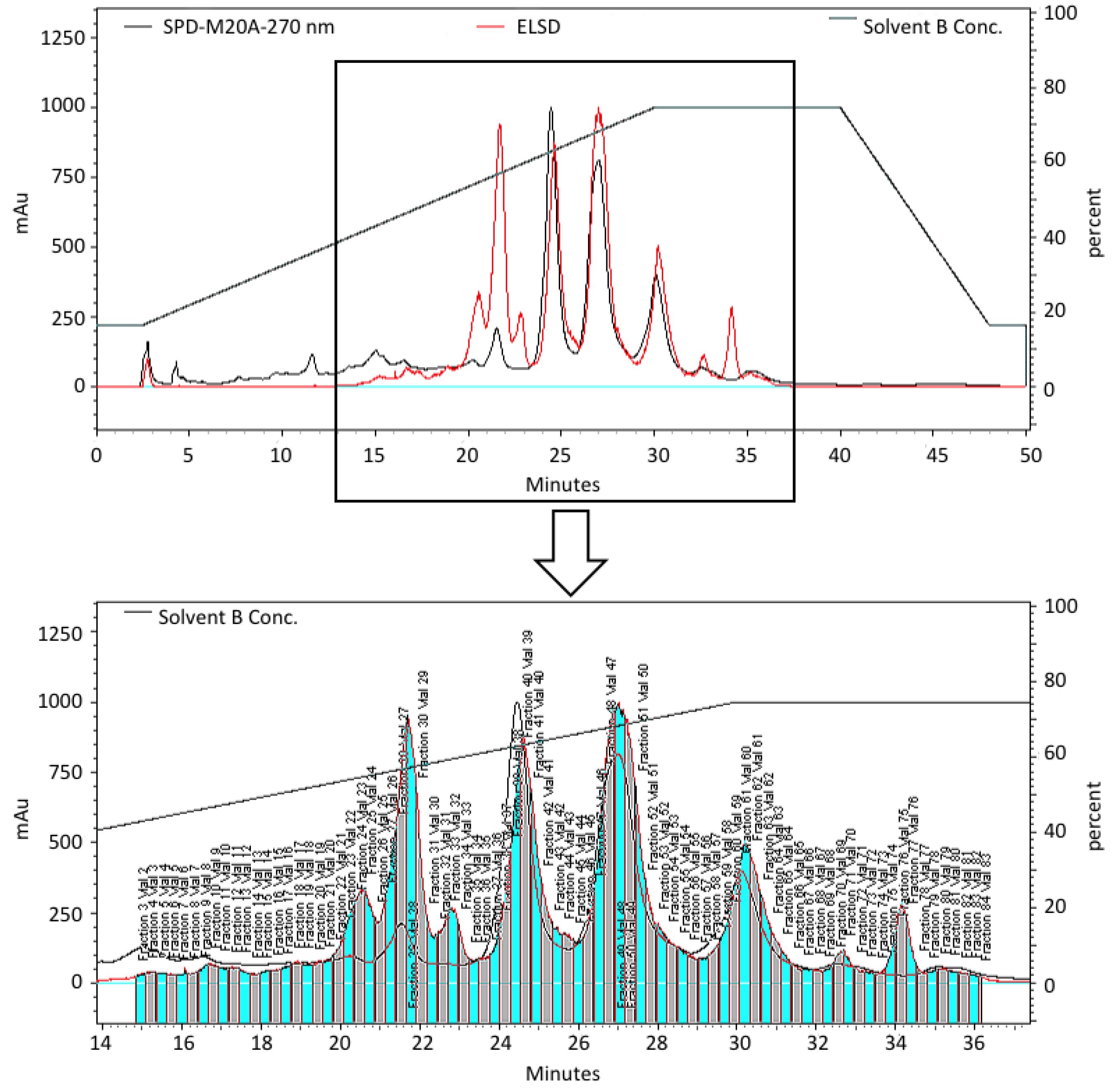
As part of an ongoing study to discover compounds that impact human embryonic stem cell (hESC) growth we have been screening pre-fractionated and semi-purified marine natural product extract libraries generated using a solid phase extraction (SPE) procedure followed by semi-preparative high pressure liquid chromatography (HPLC) using evaporative light scattering detection (ELSD) directed fractionation (
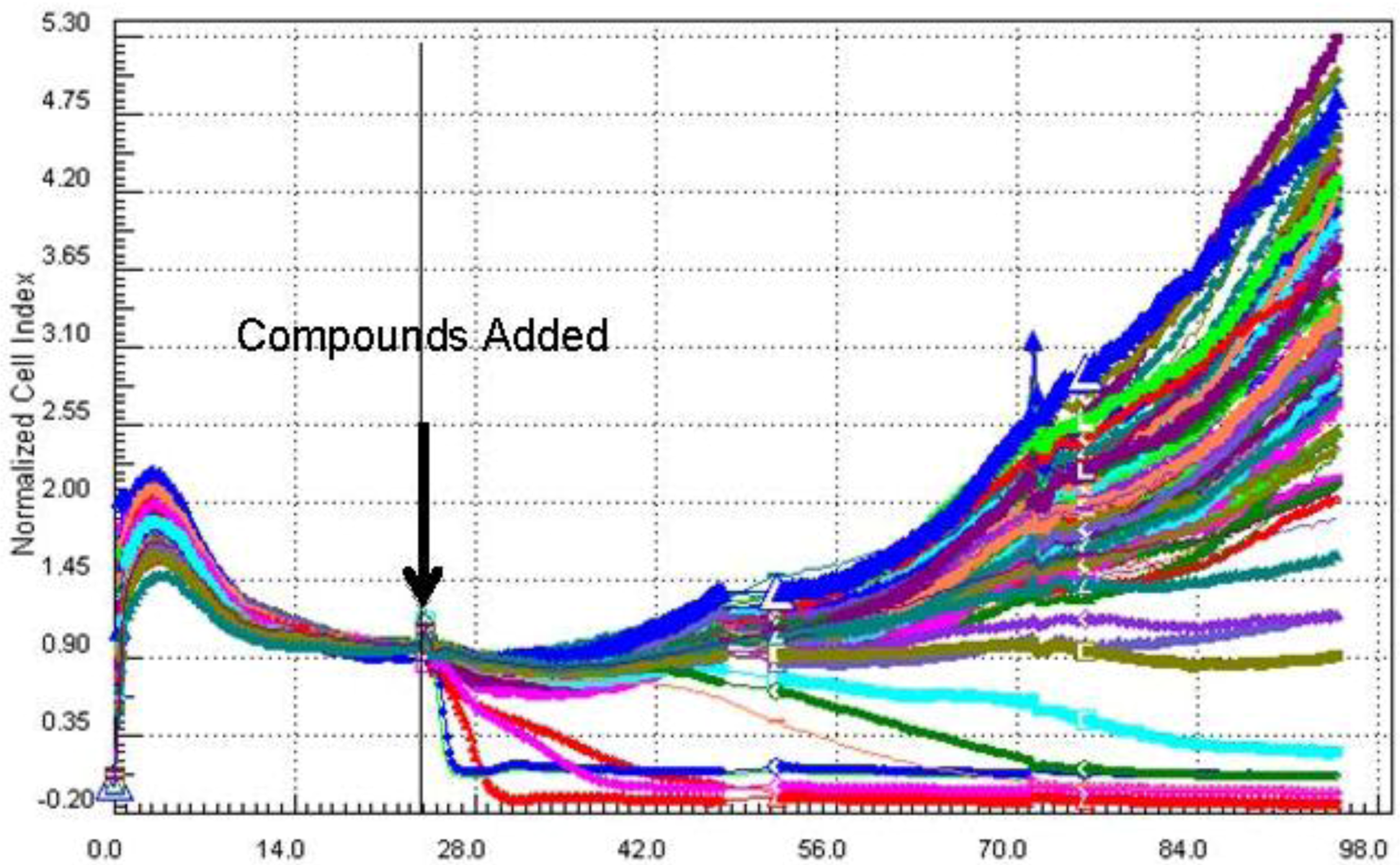
Figure 1). The cell growth inhibitory activities of the pre-fractionated extract libraries are then evaluated against human embryonic stem cells (BG02) using a 96-well plate real-time cell electronic sensing (RT-CES) system to identify compounds that impact self-renewal, differentiation or apoptosis. A plot of cell index (impedance)
versus time is used to indicate relative proliferation, differentiation, or death in real-time (
Figure 2).
Our previous work using this approach on
B. asbestinum resulted in the isolation of three new briareolate esters L–N (
3–
5) [
5]. This included the biologically active compound briareolate ester L (
3) that was found to possess a 10-membered macrocyclic ring with a (
E,
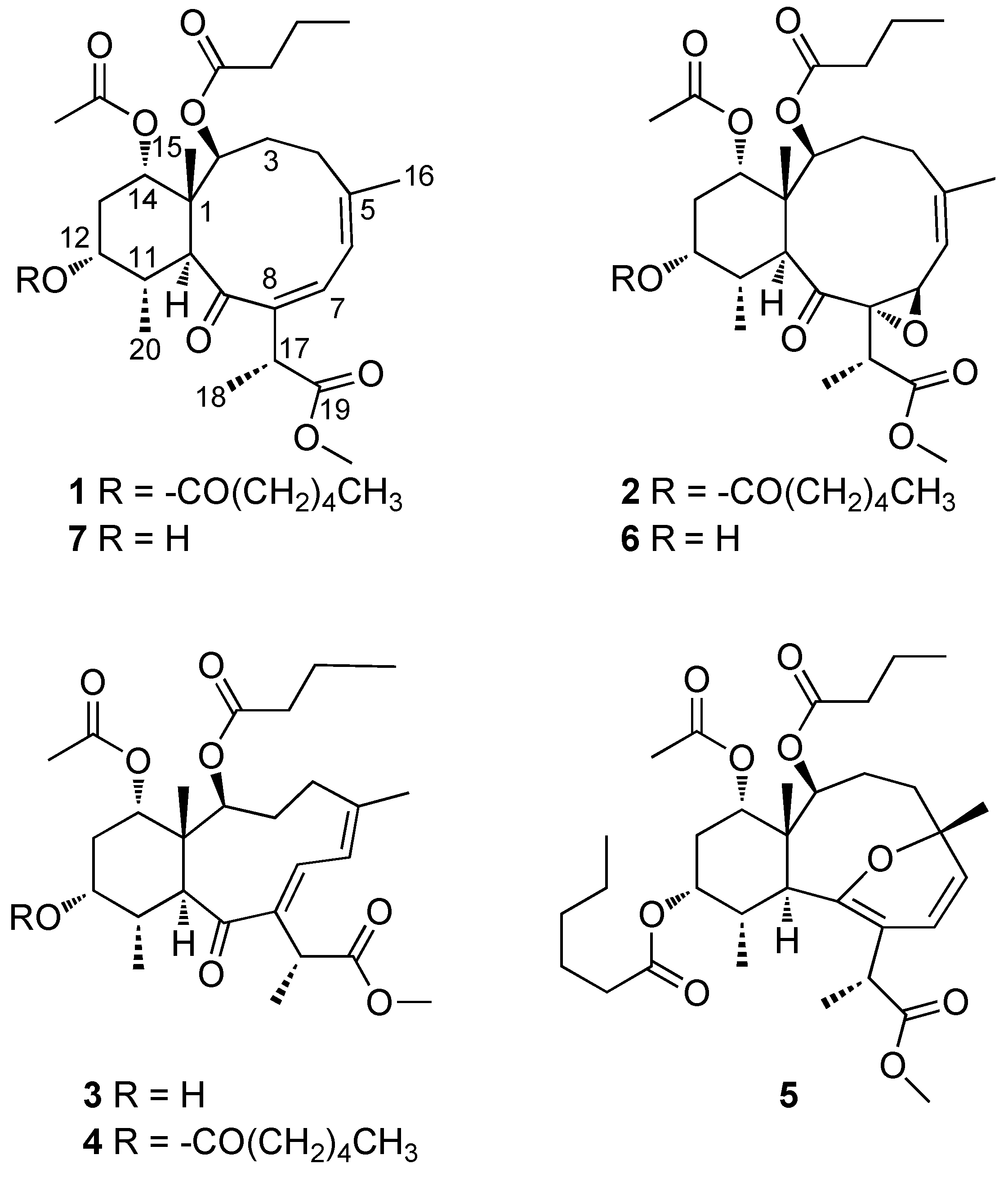
Z)-dieneone and was shown to contain a “spring loaded” Michael acceptor that is capable of forming a reversible covalent bond to model sulfur-based nucleophiles. In the present study we report the isolation and structural elucidation of two additional new briarane diterpenoids briareolate esters J (
1) and K (
2), along with three known compounds from the methanolic extract of
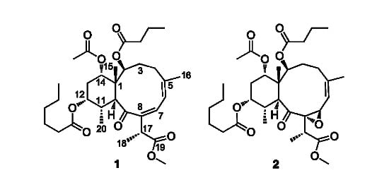
B. asbestinum that was found to exhibit growth inhibition against BG02 cells (
Figure 3).
Figure 1.
HPLC chromatogram of the pre-fractionated extract of B. asbestinum showing the fractions separated using ELSD-directed collection to generate a semi-purified extract library for biological screening.
Figure 1.
HPLC chromatogram of the pre-fractionated extract of B. asbestinum showing the fractions separated using ELSD-directed collection to generate a semi-purified extract library for biological screening.
Figure 2.
A cell index vs. time plot for a pre-fractionated extract library of B. asbestinum against the BG02 cell line. A drop in cell index within the first 24–48 h after addition of the compounds is interpreted as toxicity or induction of apoptosis.
Figure 2.
A cell index vs. time plot for a pre-fractionated extract library of B. asbestinum against the BG02 cell line. A drop in cell index within the first 24–48 h after addition of the compounds is interpreted as toxicity or induction of apoptosis.
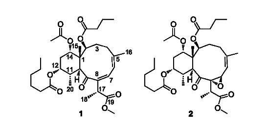
Figure 3.
Structures of compounds 1–7.
Figure 3.
Structures of compounds 1–7.
2. Results and Discussion
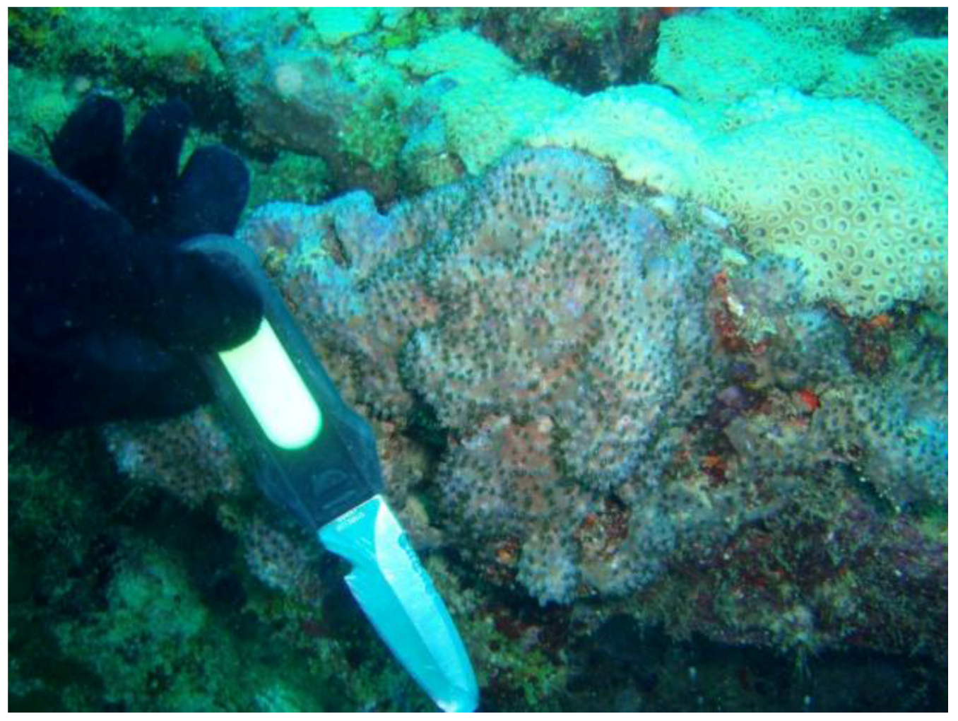
Specimens of
Briareum asbestinum (
Figure 4) were collected at Hillsboro Ledge, Boca Raton Florida and kept frozen until extraction. The methanolic extract was first fractionated on polymeric HP-20 resin using cyclic loading [
6]. The HP-20 column was eluted with 800 mL fractions of (1) H
2O, (2) 40% Me
2CO/H
2O, (3) 75% Me
2CO/H
2O and (4) Me
2CO. The Me
2CO fraction was then subjected to column chromatography on HP-20SS and normal phase HPLC to yield two new briareolate esters J (
1) and K (
2) and three known briareolate esters D (
6), G (
7), and M (
4).
Figure 4.
Briareum asbestinum.
Figure 4.
Briareum asbestinum.
Briareolate ester J (
1) was isolated as a colorless oil. The molecular formula of briareolate ester J (
1), C
33H
50O
9, was determined from the HRESIMS of the [M + Na]
+ ion at
m/
z 613.3346, 98 mass units higher than that of briareolate ester G (
7). The presence of a ketone conjugated with two double bonds (α,β,γ,δ-unsaturated ketone) was indicated from a carbonyl carbon with a chemical shift of δ
C 207 (C-9), and the C-C double bond carbons [δ
C 146.3 (C-5), δ
C 125.3 (C-6), δ
C 140.9 (C-7), δ
C 146.3 (C-8)]. The observation of absorption maxima at λ
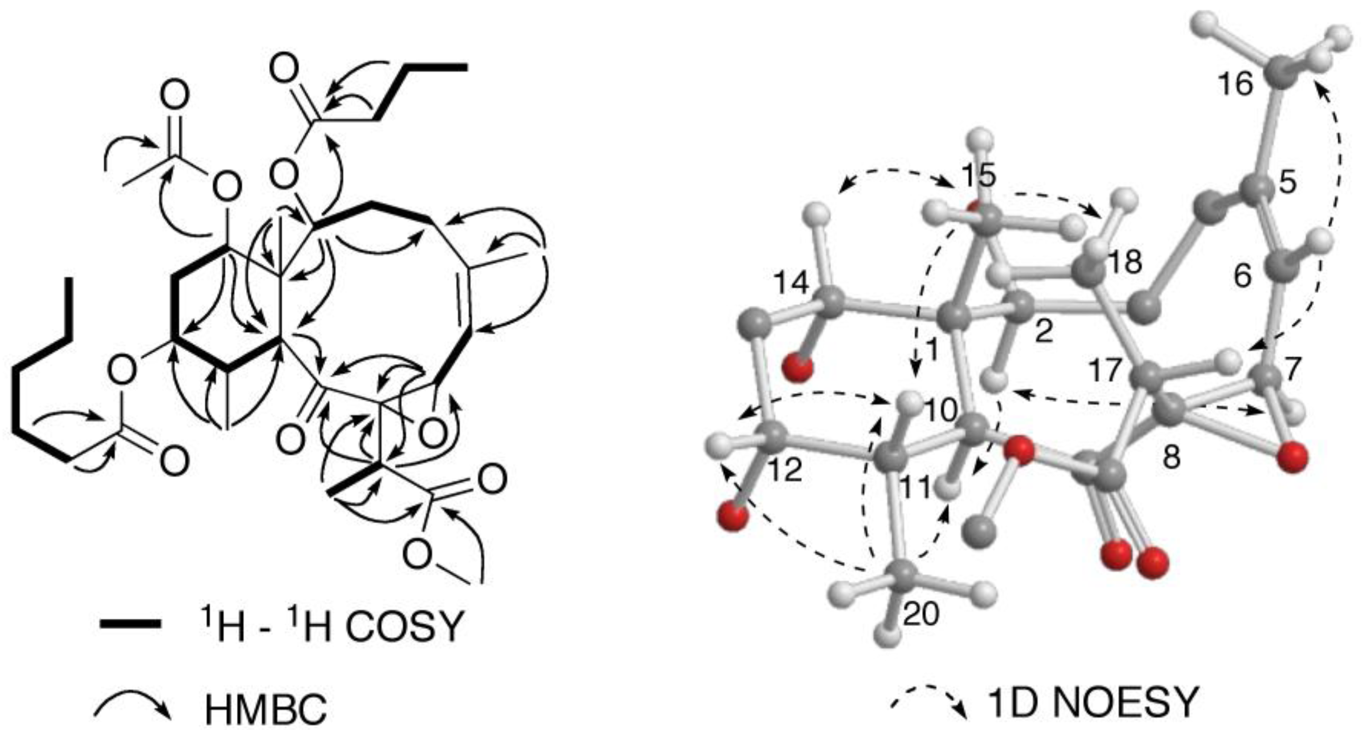
max = 287 and 225 nm in the UV spectrum was consistent with this assignment. NOE correlations observed from the olefinic proton H-6 to H-7 and H
3-16, together with correlations from H-7 to both H-17 and H
3-18, established the (
Z,
Z)-configuration of the dieneone and the
s-
cis conformation of the diene (
Figure 5). Additionally, the upfield chemical shift of the olefinic proton H-7 in
1 at δ
H 6.74 compared toδ
H 7.64 in the (
E,
Z)-dieneone briareolate ester L (
3), with the
s-
trans diene conformation, confirmed this assignment [
5].
A close inspection of the
1H and
13C NMR data (
Table 1) revealed the similarity of
1 to that of briareolate ester G (
7), except that H-12 [δ
H 4.89, br s] was shifted downfield by 1.24 ppm as compared with that of
1. In addition, in the
13C NMR spectrum the resonance of C-12 (δ
C 73.7) was shifted downfield by 2.7 ppm and those of C-11 (δ
C 38.4) and C-13 (δ
C 30.2) were shifted upfield by 0.5 and 1.3 ppm, respectively, in comparison with those of
1 [
4]. This suggested that the 12-hydroxy group of
7, was replaced by a hexanoate group at C-12 in
1, as observed in briareolate esters M (
4) and N (
5). The presence of the hexanoate group was confirmed by the NMR data [δ
H 0.92 (3H, t,
J = 8.0 Hz),
ca. 1.31 (4H, overlapped), 1.63 (2H, overlapped), 2.32 (2H, m), δ
C 17.6 (q), 24.0, 26.3, 33.0, and 36.1 (each t), 175.1 (CO)]. These assignments were confirmed by COSY, HSQC, and HMBC correlations similar to those observed for
4,
5 and
7 (
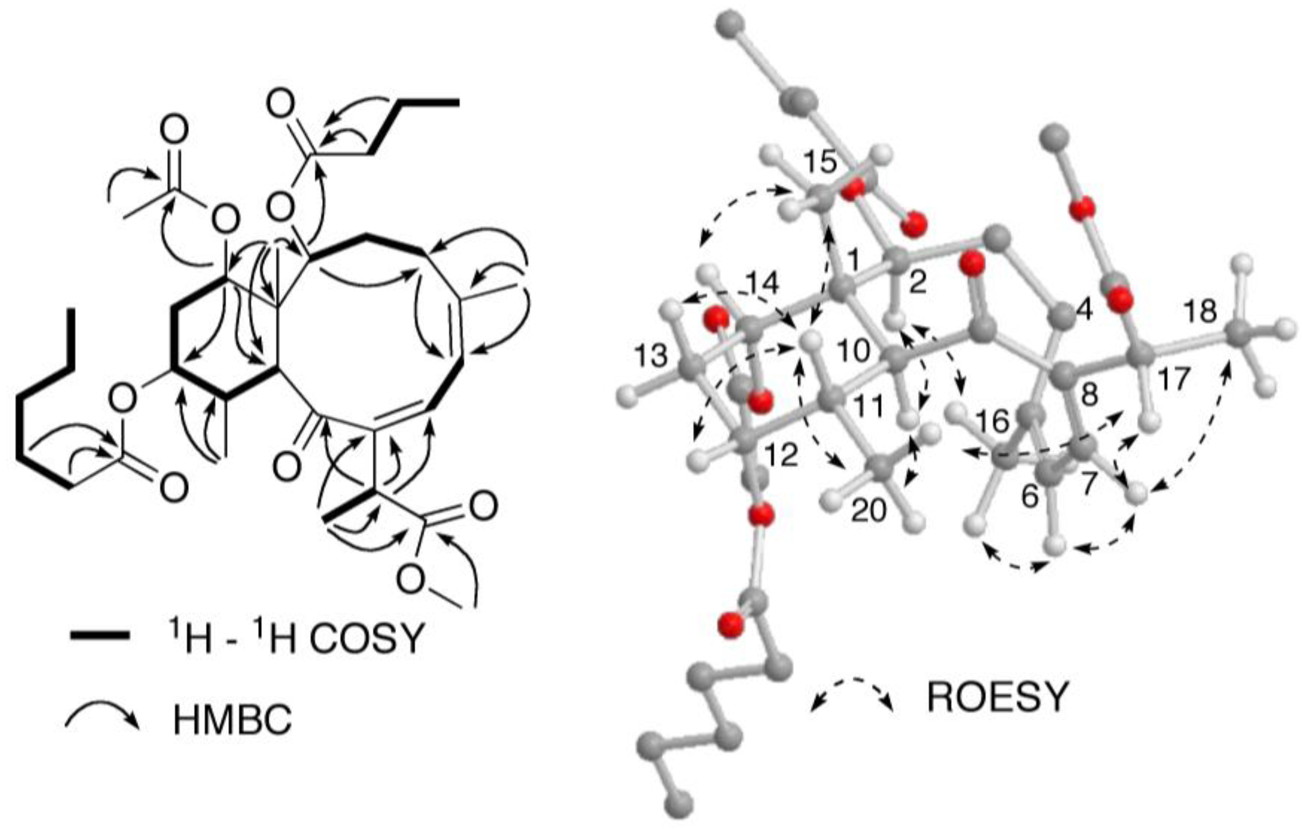
Figure 5). The relative configuration of briareolate ester J (
1) was determined to be the same as that of
7 from the similarity of proton-proton coupling constants and
1H and
13C chemical shifts together with NOE correlations observed in a ROESY experiment (
Figure 5).
Table 1.
NMR Spectroscopic Data for Briareolate Esters J (1) and K (2) a.
Table 1.
NMR Spectroscopic Data for Briareolate Esters J (1) and K (2) a.
| position | 1 | 2 |
|---|
| δC, mult | δH (
J
in Hz) | δC, mult | δH (
J in Hz) |
|---|
| 1 | 46.4, C | | 48.8, C | |
| 2 | 73.7, CH | 5.17, br d (6.0) | 80.5, CH | 5.52, d (8.0) |
| 3α | 31.3, CH2 | 1.89, m | 31.1, CH2 | 2.31, m |
| 3β | | 1.89, m | | 1.58, m |
| 4α | 30.2, CH2 | 2.20, m | 34.1, CH2 | 2.53, dd (18.0, 8.0) |
| 4β | | 2.08, m | | 2.28, m |
| 5 | 146.3, C | | 147.1, C | |
| 6 | 125.3, CH | 6.12, br s | 116.2, CH | 5.47, br d (4.0) |
| 7 | 140.9, CH | 6.74, br s | 64.3, CH | 4.42, br d (4.0) |
| 8 | 146.3, C | | 71.3, C | |
| 9 | 207.0, C | | 211.5, C | |
| 10 | 48.3, CH | 3.79, d (12.0) | 44.7, CH | 3.06, d (12.0) |
| 11 | 38.4, CH | 2.19, m | 36.2, CH | 2.34, m |
| 12 | 73.7, CH | 4.89, br s | 72.6, CH | 4.98, br s |
| 13α | 30.2,CH2 | 2.05, m | 29.9, CH2 | 2.05, br d (16.0) |
| 13β | | 1.86, m | | 1.99, m |
| 14 | 75.6, CH | 4.68, br s | 74.6, CH | 4.69, br s |
| 15 | 14.9, CH3 | 1.19, s | 12.8, CH3 | 0.99, s |
| 16 | 26.3, CH3 | 2.19, s | 26.8, CH3 | 1.75, s |
| 17 | 46.4, CH | 3.45, q (8.0) | 40.8, CH | 2.39, q (8.0) |
| 18 | 19.8, CH3 | 1.32, d (8.0) | 13.5, CH3 | 1.29, d (8.0) |
| 19 | 176.7, C | | 175.3, C | |
| 20 | 17.6, CH3 | 0.79, d (8.0) | 16.8, CH3 | 0.75, d (8.0) |
| OMe | 52.8, CH3 | 3.62, s | 52.5, CH3 | 3.61, s |
| ester at C-2 | 175.1, C | | 174.3, C | |
| | 37.7, CH2 | 2.19, m | 37.9, CH2 | 2.25, m |
| | 19.2, CH2 | 1.57, m | 19.7, CH2 | 1.60, m |
| | 14.5, CH3 | 0.92, t (8.0) | 14.5, CH3 | 0.93, t (8.0) |
| ester at C-12 | 175.1, C | | 175.4, C | |
| | 36.1, CH2 | 2.32, m | 35.9 CH2 | 2.37, m |
| | 26.3, CH2 | 1.63, m | 26.4 CH2 | 1.64, m |
| | 33.0, CH2 | 1.31, m | 33.0 CH2 | 1.33, m |
| | 24.0, CH2 | 1.31, m | 24.0 CH2 | 1.33, m |
| | 17.6, CH3 | 0.92, t (8.0) | 14.8 CH3 | 0.95, t (8.0) |
| ester at C-14 | 172.1, C | | 172.6, C | |
| | 22.1, CH3 | 1.98, s | 22.3 CH3 | 1.99, s |
Figure 5.
Selected 2D NMR correlations for briareolate ester J (1).
Figure 5.
Selected 2D NMR correlations for briareolate ester J (1).
Briareolate ester K (
2) was isolated as a colorless oil. The molecular formula of briareolate ester K (
2), C
33H
50O
10, was determined from the HRESIMS of the [M + Na]
+ ion at
m/
z 629.3291, one more oxygen atom than that of briareolate ester J (
1). A comparison of the
1H and
13C NMR data (
Table 1) revealed that
2 was similar to
1, except that the NMR signals for the C-7–C-8 double bond were missing in the NMR spectra of
2, and instead, resonances for an epoxide were observed in the
1H NMR (δ 4.42, H-6) and
13C NMR [δ
C 64.3 (s); 71.3 (d)] spectra indicating oxidation of the C-7–C-8 double bond to be an epoxide. HMBC correlations from the oxygenated methine signal at δ 4.42 (H-7) to C-5 (δ 147.1) and C-6 (δ 116.2) of the trisubstituted double bond, and to C-8 (δ 71.3) and C-9 (δ 211.5) are consistent with this assignment. Additional HMBC correlations observed from H-17 to C-7, C-8 and C-9 confirmed this assignment (
Figure 6).
Figure 6.
Selected NMR correlations for briareolate ester K (2).
Figure 6.
Selected NMR correlations for briareolate ester K (2).
The relative configuration of briareolate ester K (
2) was determined to be identical to that of
1 on the basis of NOE enhancements revealed in a series of 1D NOESY experiments (
Figure 6). The configuration of the C-7/8 epoxide was determined from NOE enhancements observed between H-7 and H-2 together with NOE enhancements observed from H-6 to H-17 which placed H-7 on the inside of the 10-membered ring and indicated the
trans orientation of the epoxide. This assignment is consistent with that of briareolate ester D (
6) whose structure was assigned on the basis of X-ray crystallography [
4].
Compounds
1 and
2 were evaluated for cell growth inhibitory activities against human embryonic stem cells (BG02) using a 96-well plate real-time cell electronic sensing (RT-CES) system [
7]. Briareolate ester K (
2) showed weak growth inhibition against BG02 cells with an EC
50 value of 40 µM. No inhibitory activity was detected for briareolate ester J (
1) at 40 µM. Previously the (
E,
Z)-dienone containing briareolate esters L (
3) and M (
4) were found to have growth inhibition against both the BG02 and a pancreatic cancer cell line (BxPC-3) cells with EC
50 values of 2.4 and 9.3 µM, respectively for
3, and 8.0 µM against BG02 cells and only cytostatic effects at 13.0 and 17.0 µM against the BxPC-3 cells for
4 [
5]. No growth inhibition was found for the briareolate esters B, C, G (
7), and N (
5). The absence of significant growth inhibition activity for the (
Z,
Z)-dieneone compound
1 and the epoxide containing compound
2 further confirms the importance of the (
E,
Z)-dieneone for biological activity.


