Article

Structural Characterization and Anti-HSV-1 and HSV-2 Activity of Glycolipids from the Marine Algae Osmundaria obtusiloba Isolated from Southeastern Brazilian Coast

1
Department of Biochemistry and Molecular Biology, Federal University of Paraná, Curitiba 81531-990, PR, Brazil
2
Department of Virology , Institute of Microbiology, Federal University of Rio de Janeiro, Rio de Janeiro 21941-902, RJ, Brazil
3
Department of General Microbiology, Federal University of Rio de Janeiro, Rio de Janeiro 21941-902, RJ, Brazil
*
Author to whom correspondence should be addressed.
Mar. Drugs 2012, 10(4), 918-931; https://doi.org/10.3390/md10040918
Submission received: 5 March 2012 / Revised: 11 April 2012 / Accepted: 17 April 2012 / Published: 23 April 2012
(This article belongs to the Special Issue Marine Anti-infective Agents)

Abstract

:
Glycolipids were extracted from the red alga Osmundaria obtusiloba from Southeastern Brazilian coast. The acetone insoluble material was extracted with chloroform/methanol and the lipids, enriched in glycolipids, were fractionated on a silica gel column eluted with chloroform, acetone and then methanol. Three major orcinol-positive bands were found in the acetone and methanol fractions, being detected by thin layer chromatography. The structures of the corresponding glycolipids were elucidated by ESI-MS and 1H/13C NMR analysis, on the basis of their tandem-MS behavior and HSQC, TOCSY fingerprints. For the first time, the structure of sulfoquinovosyldiacylglycerol from the red alga Osmundaria obtusiloba was characterized. This molecule exhibited potent antiviral activity against HSV-1 and HSV-2 with EC50 values of 42 µg/mL to HSV-1 and 12 µg/mL to HSV-2, respectively. Two other glycolipids, mono- and digalactosyldiacylglycerol, were also found in the alga, being characterized by ESI-MS/MS. The structural elucidation of algae glycolipids is a first step for a better understanding of the relation between these structures and their biological activities.

Graphical Abstract

1. Introduction

Glycolipids constitute an important class of membrane lipids that are synthesized by both prokaryotic and eukaryotic organisms. Major glycolipids such as monogalactosyldiacylglycerol (MGDG), digalactosyldiacylglycerol (DGDG) and sulfoquinovosyldiacylglycerol (SQDG), along with phospholipids were isolated from red, green and brown algae, such as, for example, Anfeltia tobuchiensis (Rhodophyta), Ulva fenestrate (Chlorophyta), Laminaria japonica (Chlorophyta), Sargassum pallidum (Phaeophyta) [1], Sargassum thunbergii (Phaeophyta) [2], Exophlyllum wentii (Rhodophyta) [3] and Chondria armata (Rhodophyta) [4], as well as in simbiotic organisms having mycroalgae [5,6]. These lipids are reported to exhibit diverse biological functions [7,8]. MGDGs from the green alga Chlorella vulgaris, exhibit anti-tumor effect [9]. SQDG from the red alga Gigartina tenella inhibits the eukaryotic DNA polymerase and HIV-reverse transcriptase type 1 [10] and SQDG identified in Porphyridium purpureum and other microalgae presents antivirus activity [11]. Most studies show the antiviral activity of sulfated polysaccharides from marine alga [12,13,14,15,16,17,18] but studies using sulfoglycolipids have received less attention [19,20,21,22]. Due to economic importance of algae, as a rich source of bioactive compounds with potential biomedicinal interest, there is an increasing need for improved techniques for isolation pure compounds. In our search for potentially useful bioactive molecules of marine origin, we have isolated and identified the glycolipids from the red alga Osmundaria obtusiloba from Southeastern Brazilian coast using electrospray ionization tandem mass spectrometry (ESI-MS/MS) complemented with NMR. In the present study we also evaluated the inhibitory activity of sulfoquinovosyldiacylglycerol from the red marine alga Osmundaria obtusiloba on the herpes simplex virus type 1 (HSV-1) and type 2 (HSV-2). Herpes simplex viruses are responsible for a broad range of human infectious diseases. Despite acyclovir is the drug of choice for treating the HSV infections, new antiviral agents exhibiting different mechanisms of action is urgently needed, mainly due to the high number of acyclovir-resistant strains [23].

2. Results and Discussion

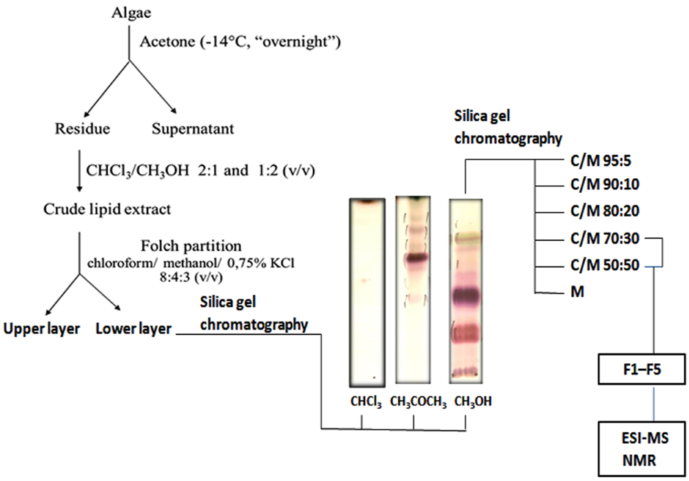
2.1. Lipid Fractionation

Osmundaria obtusiloba was treated with acetone and kept overnight at −14 °C. The acetone insoluble material was successively extracted with chloroform/methanol 2:1 and 1:2 (v/v) at room temperature [22,24]. The combined extracts were concentrated in vacuo and the crude lipid fraction was partitioned according to Folch and coworkers [25]. The lipids recovered from the Folch lower layer, enriched in glycolipids, were fractionated on a silica gel column, eluted with chloroform, acetone and then methanol. Figure 1 shows the steps of purification.
Figure 1. Overview of the strategy used for purification of glycolipids from Osmundaria obtusiloba. Purified or partially purified fractions are usually resolved by TLC and visualized with iodine and orcinol-H2SO4.
Marinedrugs 10 00918 g001
Orcinol reactive bands were detected on TLC from fractions eluted with acetone and methanol. A single main spot orcinol-positive was detected in the acetone fraction (F-Ac), whereas at least three positive spots could be visualized on TLC of methanol fraction (F-MeOH). This enriched glycolipid fraction was then purified by a second silica gel chromatography, eluted by a gradient of CHCl3/MeOH, increasing the solvent polarity, giving rise to fractions F1–F5 (Figure 1). These fractions were analyzed on TLC plates developed with CHCl3:MeOH:2M NH4OH (40:10:1 v/v/v) and visualized through iodine vapor and by spraying the plates with orcinol-sulfuric acid [26]. The partially purified glycolipids were then analyzed by ESI-MS in the positive and negative ionization modes.

2.2. Mass Spectrometry of Neutral Glycolipids

In order to improve the positive ion detection of neutral lipids, Li+ (LiCl) was added in the sample solvent, giving the molecules as lithiated ions. The acetone fraction (F-Ac) gave in MS1 an ion with m/z 777 [M + Li]+. This was fragmented by CID-MS, giving rise to characteristic spectrum of neutral glyceroglycolipids, with a fragment-ion at m/z 615, consistent with a monosaccharide residue loss, those at m/z 521 and 481 were consistent with loss of a palmitc acid (C16:0) and an unusual nonadecenoic (C19:1) acid, respectively. The fragment-ions from glycan moiety appeared at m/z 227, 187 and 169 (Figure 2A), thus being consistent with the fragmentation partner of a monogalactosyldiacylglycerol (MGDG) [27].
Figure 2. (A) Tandem-MS and fragmentation profile obtained from lithiated monogalactosyldiacylglycerol, precursor ion at m/z 777, from acetone Fraction (F-Ac); (B) from lithiated digalactosyldiacylglycerol, precursor ion at m/z 939, from Fraction 5.
Marinedrugs 10 00918 g002
Fraction 5, an orcinol-positive sub-fraction from F-MeOH, was also analyzed in positive ion mode assisted by Li+, giving a main ion at m/z 939. These were fragmented giving rise to fragments at m/z 777 and a small m/z 615, consistent with loss a monosaccharide and a disaccharide, respectively. The fragments at m/z 683 and 643 were consistent with loss of a palmitic and a nonadecenoic acid, respectively. The fragments at m/z 389, 331, 187 and 169 confirmed the presence of a disaccharide, being this glycolipid consistent with a digalactosyldiacylglycerol (DGDG) [27,28]. Other fragments are shown in Figure 2B.

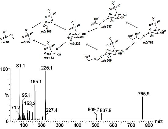
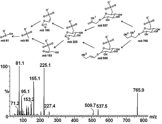
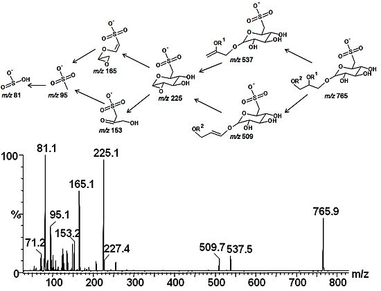
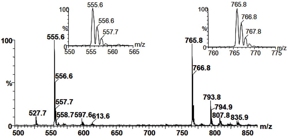
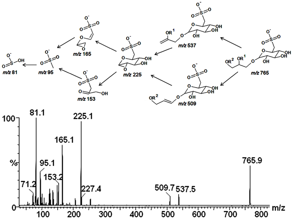
2.3. Mass Spectrometry of Sulfolipids

Fraction 1, a sub-fraction from F-MeOH, gave two positive spots for carbohydrate with lower Rf values. Furthermore, the spectrum obtained in negative MS1 exhibited two main deprotonated ions with m/z 555 and 765 [M − H] (Figure 3).
Figure 3. Spectrum from MS1 obtained in negative ionization mode from Fraction 1. The ion at m/z 555 is compatible with the structure of a sulfoquinovosylmonoacylglycerol, whereas the one at m/z 765, with sulfoquinovosyldiacylglycerol.
Marinedrugs 10 00918 g003
The distribution of isotopologues was 10:4:1.5 (for m/z 555, 556 and 557, respectively) and 10:5:2 (for m/z 765, 766 and 767). The high intensity of the heavy isotopologues at m/z 557 and 767 is an indicative for the presence of a sulfur element in the molecules [29], occurring because of the presence of >4% of the heavy isotope (34S). In lipid fractions from vegetable sources, the presence of sulfur element added to positive result for carbohydrate strongly suggests the presence of sulfolipids, such as sulfoquinovosyldiacylglycerol (SQDG), a lipid associated with phototrophic organisms [30,31].
In order to confirm the structures, the ions at m/z 555 and 765 were fragmented by the second stage tandem-MS. The results were consistent with the structure of the sulfoquinovosylmonoacylglycerol (SQMG-m/z 555) with a palmitic acid (C16:0), as indicated by the presence of fragments at m/z 299, 243, 225, 165, 153, 95 and 81, whereas the ion at m/z 765 gave fragments at m/z 537 (M–C14:0), 509 (M–C16:0), 225, 165, 153, 95 and 81, as indicated in the fragmentation pathway (Figure 4), being consistent with the SQDG structure, esterified by myristic and palmitic acids. Structures of sulfonoglycolipids are frequently studied by tandem-MS, and the fragments at m/z 225, 165, 153, 95 and 81can be considered as diagnostics for 6-deoxy-6-sulfono-hexosyl residue [6,28,32,33].
Figure 4. Tandem-MS fingerprint and a proposed fragmentation pathway of sulfoquinovosyldiacylglycerol, precursor ion at m/z 765, under collision induced dissociation-mass spectrometry.
Marinedrugs 10 00918 g004
The lower abundant ions in the MS1 spectrum occurred due to other fatty acids combination, such as that one at m/z 793, usually associated with sulfoquinovosyl-dipalmitoylglycerol or myristoyl-steaorylglycerol. Although the structures had been well characterized by MS2, the absolute hydroxyl configuration could not be determined by mass spectrometry. Since SQDG (m/z 765) was the predominant glycolipid found in Fraction 2, this fraction was used for NMR experiments, such as COSY, TOCSY and HSQC and biological activity as well.

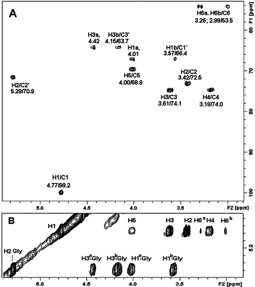
2.4. NMR Spectroscopy of Sulfolipids

The structure of the sulfoglycolipid present in fraction 2, was confirmed by 1H and 13C NMR analysis, based on HSQC and TOCSY fingerprints [5,6]. The chemical shift map was obtained by HSQC and discussed as follows. The HSQC spectrum of glycolipid contained aliphatic signals from δ 22.8 to 34.6 with predominant CH2 signals at δ 29.9 and CH3 at δ 14.1. The spectrum suggests the presence of exclusively saturated fatty acids, since no double bond signals were observed at δ 122.0 to 133.0 [6,33] and confirms the previous ESI-MS/MS results. The signals of 1H/13C-HSQC and 1H/1H-TOCSY from glycerol were observed at δ 4.009, 3.570/66.4; 5.280/70.9; and 4.417, 4.159/63.5, arising from C-1, C-2, and C-3, respectively (Figure 5A,B).
Figure 5. (A) Partial 2D 1H/13C-HSQC fingerprint spectrum of sulfoquinovosyldiacylglycerol in fraction 1, showing the polar region of lipid; (B) Partial 2D 1H/1H-TOCSY spectrum, showing the fingerprint region containing the monosaccharide ring connectivity and glycerol moiety of the sulfonoglycolipid.
Marinedrugs 10 00918 g005
On the 1H/13C-HSQC experiment, the anomeric region contained a single signal at δ 4.770/99.2, consistent with α-quinovopyranosyl group. Also, other key 1H/13C-HSQC signals were found at δ 3.267, 2.990/53.5. The presence of doublets of CH2 signals in a high-field region is characteristic of S-substituted C-6, typical of 6-sulfo-α-quinovopyranosyl unit [5,6]. In addition, the total hydroxyl connectivity observed in 1H/1H-TOCSY experiment (Figure 5B) confirmed the hydroxyl configuration of a 6-deoxy-α-glucopyranoyl (α-quinovosyl) unit. These results and those from mass spectrometry were very similar to previous findings for sulfonoglycolipids [4,5,6,7,34,35,36], confirming the lipid identity as 1,2-di-O-acyl-3-O-(6-deoxy-6-sulfo-α-D-glucopyranosyl)-sn-glycerol (SQDG).

2.5. Antiviral Activity

In a previous study from our group, the antiviral activity of the crude acetone and methanol fractions from a lipid extract of O. obtusiloba was evaluated and the results have demonstrated that both fractions had potent inhibitory activity against HSV-1 (herpes simplex virus type 1) and present low toxicity for cell cultures [22]. A better activity was found in the crude methanol fraction. In order to find out which compounds are responsible for this activity, a purified sulfoglycolipid fraction (Fraction 2) was obtained in the present work and the antiviral activity against HSV-1 and HSV-2 (herpes simplex virus type 2) was measured (Table 1).
Table 1. Cytotoxicity and antiviral activity of fraction 2 from Osmundaria obtusiloba and acyclovir.
HSV-1HSV-2
CompoundsMNTC (µg/mL)CC50 (µg/mL)PIEC50 (µg/mL)SIPIEC50 (µg/mL)SI
Fraction 2 O. obtusiloba507275421.796126.0
Acyclovir200>200990.8>250991.38>145
HSV-1, herpes simplex virus type 1; HSV-2, herpes simplex virus type 2; MNTC, maximum non-toxic concentration; CC50, 50% cytotoxic concentration; PI, percentage of inhibition; EC50, effective concentration to reduce virus titers by 50%; SI, selectivity index. Acyclovir, standard compound.
Our results showed a reduction in the antiviral activity of the purified glycolipid (75%) when compared with the crude methanol fraction (99.5%) [22]. The somewhat higher activity of the crude methanol fraction relative to the purified fraction 2 occurred, presumably, as a result of the absence of other polar lipids in this fraction , such as MGDG and DGDG, which can trigger a synergic effect. These results suggest the importance of both glycolipids (glycoglycerolipids and sulfoglycolipids) in the antiviral activity. The anti-HSV activity of glycolipids extracted from seaweed has been demonstrated [19,20,21,22]. El-Baroty and coworkers showed that glycolipid isolated from the brown alga Dilophys fasciola was able to block the viral infectivity by interacting with HSV-1 glycoprotein [22]. Studies will be conducted to determine the mechanism of action of sulfoglycolipid from O. obtusiloba. DGDG and SQDG from O. obtusiloba identified in this present work have been previously detected in other red algae Hypnea musciformis, Porphyra acanthophora, Pterocladiella capillacea, the brown alga Dictyota menstrualis and D. cervicomis and in the green alga Caulerpa racemosa [22], confirming that these glycolipids are conserved molecules in green, red and brown algae isolated from the Southeastern Brazilian coast [22].

3. Experimental Section

3.1. Biological Material

Osmundaria obtusiloba (C. Agardh) R. E. Norris (Rhodophyta-Fam. Rhodomelaceae) was collected at Praia Rasa, (22°44′3.15″S, 41°57′30.15″O), located at the city of Buzios, Rio de Janeiro, Brazil. The seaweed was washed with local sea water and separated from sediments, epiphytes and other associated organisms.

3.2. Extraction and Fractionation of Lipids

Osmundaria obtusiloba was treated with acetone and kept overnight at −14 °C. The acetone insoluble material was successively extracted at room temperature with chloroform/methanol 2:1 and 1:2 (v/v). Extracts were combined, dried and the crude lipid extract was partitioned according to Folch and coworkers [25]. The lipids recovered from the Folch lower phase were fractionated on a silica gel column eluted with cloroform, acetone and then methanol. The methanol fraction containing the glycolipids was further purified on a silica gel column, which was sequentially eluted with chloroform/methanol with increasing concentrations of methanol (95:5, 90:10, 80:20, 70:30, 50:50, v/v) and finally with 100% methanol. Fractions eluted with chloroform/methanol 70:30 and 50:50 (v/v) yielded partially purified glycolipid fractions F1 to F5. These fractions were analysed by TLC, developed with CHCl3:CH3OH:2M NH4OH (40:10:1 v/v/v) and the spots visualized with iodine and by spraying with orcinol/H2SO4 [26]. Fraction 2, was further purified by preparative TLC, yielding a purified glycolipid fraction.

3.3. Mass Spectrometry

The MS analysis was carried out in an electrospray ionization mass spectrometry (ESI-MS), model Quattro-LC (Waters) with a triple-quadrupole mass analyzer, operating at atmospheric pressure ionization (API), assisted by a syringe pump (KDScientific) for sample infusion. Nitrogen was used as nebulizing and desovation gas and the ionization energies were 50 V on the cone and 2 kV on the capillary, operating in the negative ionization mode or 80 V (cone) and 2.5 kV (capillary) when operating in the positive ionization mode. The second stage tandem-MS was obtained by collision induced dissociation mass spectrometry (CID-MS) using argon as collision gas and collision energies ranging between 35–60 eV. The samples were prepared in MeOH at 1 mg/mL, then diluted to 0.1 mg/mL in MeOH-H2O (7:3, v/v) containing 1 mM LiCl, for the positive ion detection, and direct infused into ESI source, at a flow rate of 10 µL/min.

3.4. Nuclear Magnetic Resonance

The glycolipid component of F2 was deuterium exchanged by repeated dissolution in MeOD–D2O (2:1 v/v) and freeze-drying. The spectra were obtained from solutions of MeOD–CDCl3 (1:1) at 30 °C, using TMS (tetramethylsilane) as reference standard (δ = 0). All spectra were obtained with a Bruker 400 MHz AVANCE III NMR spectrometer with a 5 mm inverse gradient probe. 2D-NMR experiments were carried out using HSQC, COSY and TOCSY. The experiments were recorded for quadrature detection in the indirect dimension, edited-HSQC spectra were acquired using 128 scans per series of 1 K × 256 W data points with zero filling in F1 (4 K) prior to Fourier transformation [37].

3.5. Cells and Viruses

Vero cells (African green monkey kidney) were grown in Eagle’s minimum essential medium (Eagle-MEM) and supplemented with 10% (v/v) fetal bovine serum, glutamine (2 mM), garamycin (50 µg/mL), fungizone (amphotericin B) (2.5 µg/mL), NaHCO3 (0.25%) and HEPES (10 mM). HSV-1 was isolated from a typical lip lesion and HSV-2 from a typical genital lesion in the Virology Department of the Federal University of Rio de Janeiro (UFRJ), Brazil. Viruses were typed by polymerase chain reaction (PCR) using specific primers for identification [38].

3.6. Cytotoxicity Assay

The cytotoxicity of glycolipids was performed by incubating Vero (African green monkey kidney cell) cell line monolayers cultivated in 96-well microplates with two-fold serial dilutions (200–3.1 µg/mL) of the SQDG (F-2) for 48 h at 37 °C in a 5% CO2 atmosphere. Cellular viability was evaluated by the neutral red dye-uptake method [39]. The 50% cytotoxic concentration (CC50) was defined as the SQDG concentration which caused a 50% reduction in the number of viable cells.

3.7. Antiviral Activity Assay

The antiviral activity of SQDG and acyclovir was evaluated by the titer reduction. The virus titers were calculated using the Reed and Muench statistical method [40] and expressed as 50% tissue culture infective dose (TCID50) per mL. Vero cell monolayers were treated with the SQDG and acyclovir at the MNTC and 100 TCID50/mL of HSV-1 or HSV-2 suspensions were added to treated and untreated cell cultures and incubated at 37 °C for 48 h in a 5% CO2 atmosphere. After incubation, the supernatant was collected and virus titers in treated and untreated cells were determined. The antiviral activity was expressed as percentage of inhibition (PI) [41] using antilogarithmic TCID50 values as follows: PI = [1 − (antilogarithmic test value/antilogarithmic control value)] × 100. The dose-response curve was established starting from the MNTC (50–0.78 µg/mL to SQDG and 200–0.2 µg/mL to acyclovir) and the 50% effective concentration (EC50) was defined as the concentration required for 50% protection against virus induced cytopathic effects. The selectivity index (SI) was determined as the ratio of CC50 to EC50.

4. Conclusions

SQDG molecules have been isolated for the first time from the red alga Osmundaria obtusiloba and their structure elucidated by ESI-MS/MS and 1H and 13C NMR analysis, based on HSQC and TOCSY fingerprints. MGDG and DGDG have also been identified on the basis of electrospray ionization tandem MS/MS spectrometry. We also demonstrated that SGDG had potent antiviral activity against HSV-1 and HSV-2 and present low toxicity for cell cultures. Although acyclovir has shown better activity, the inhibitory effect in different steps of viral infection is a desirable feature allowing a more efficient action and preventing the emergence of resistance strains. The identification of the chemical structure of algae glycolipids could contribute for a better understanding of the relation between these structures and biological activities.

Acknowledgments

We thank Yocie Yoneshigue Valentin for the macroalgae and Maria de Fátima F. Soares for technical assistance. This work was supported by grants from the Brazilian agencies Fundação Araucária, FINEP (PRONEX-CARBOIDRATOS, PADCT II/SBIO), CAPES (Coordenação de Aperfeiçoamento de Pessoal de Nível Superior), Conselho Nacional de Desenvolvimento Científico e Tecnológico (CNPq), FAPERJ (Fundação de Amparo a Pesquisa do Estado do Rio de Janeiro) and Universidade Federal do Rio de Janeiro (UFRJ).
  • Samples Availability: Available from the authors.

References

  1. Sanina, N.M.; Goncharova, S.N.; Kostetsky, E.Y. Fatty acid composition of individual polar lipid classes from marine macrophytes. Phytochemistry 2004, 65, 721–730. [Google Scholar]
  2. Kim, Y.H.; Kim, E.H.; Lee, C.; Kim, M.H.; Rho, J.R. Two new monogalactosyl diacylglycerols from brown alga Sargassum thunbergii. Lipids 2007, 42, 395–399. [Google Scholar]
  3. Illijas, M.I.; Indy, J.R.; Yasui, H.; Itabashi, Y. Lipid class and fatty acid composition of a little-known and rarely collected alga Exophyllum wentii Weber-van Bosse from Bali Island, Indonesia. J. Oleo Sci. 2009, 58, 103–110. [Google Scholar]
  4. Al-Fadhli, A.; Wahidulla, S.; D’Souza, L. Glycolipids from the red alga Chondria armata (Kutz.) Okamura. Glycobiology 2006, 16, 902–915. [Google Scholar] [CrossRef]
  5. Sassaki, G.L.; Gorin, P.A.J.; Tischer, C.A.; Iacomini, M. Sulfonoglycolipids from the lichenized basidiomycete Dictyonema glabratum: Isolation, NMR, and ESI-MS approaches. Glycobiology 2001, 11, 345–351. [Google Scholar]
  6. Souza, L.M.; Iacomini, M.; Gorin, P.A.J.; Sari, R.S.; Haddad, M.A.; Sassaki, G.L. Glyco- and sphingophosphonolipids from the medusa Phyllorhiza punctata: NMR and ESI-MS/MS fingerprints. Chem. Phys. Lipids 2007, 145, 85–96. [Google Scholar]
  7. Gustafson, K.R.; Cardellina, J.H.; Fuller, R.W.; Weislow, O.W.; Kiser, R.F.; Snader, K.M.; Patterson, G.M.L.; Boyd, M.R. AIDS-antiviral sulfolipids from cyanobacteria (blue-green algae). J. Nat. Cancer Inst. 1989, 81, 1254–1258. [Google Scholar]
  8. Reshef, V.; Mizrachi, E.; Maretzki, T.; Silberstein, C.; Loya, S.; Hizi, A.; Carmeli, S. New acylated sulfoglycolipids and digalactolipids and related known glycolipids from cyanobacteria with a potential to inhibit the reverse transcriptase of HIV-1. J. Nat. Prod. 1997, 60, 1251–1260. [Google Scholar]
  9. Morimoto, T.; Nagatsu, A.; Murakami, N.; Sakakibara, J.; Tokuda, H.; Nishimo, H.; Iwashima, A. Antitumor promoting glyceroglycolipids from the green alga Chlorella vulgaris. Phytochemistry 1995, 40, 1433–1437. [Google Scholar]
  10. Ohta, K.; Mizushina, Y.; Hirata, N.; Takemura, M.; Sugawara, F.; Matsukage, A.; Yoshida, S.; Sakaguchi, K. Sulfoquinovosyldiacylglycerol, KM043, a new potent inhibitor of eukaryotic DNA polymerases and HIV-reverse transcriptase type 1 from a marine red alga, Gigartina tenella. Chem. Pharm. Bull. (Tokyo) 1998, 46, 684–686. [Google Scholar] [CrossRef]
  11. Naumann, I.; Darsow, K.H.; Walter, C.; Lange, H.A.; Buchholz, R. Identification of sulfoglycolipids from the alga Porphyridium purpureum by matrix-assisted laser desorption/ionisation quadrupole ion trap time of-flight mass spectrometry. Rapid Commun. Mass Spectrom. 2007, 21, 3185–3192. [Google Scholar]
  12. Damonte, E.B.; Matulewicz, M.C.; Cerezo, A.S.; Coto, C.E. Herpes simplex virus-inhibitory sulfated xylogalactans from the red seaweed Nothogenia fastigiata. Exp. Chemoter. 1996, 42, 57–64. [Google Scholar]
  13. Witvrouw, M.; de Clercq, E. Sulfated polysaccharides extracted from sea algae as potential antiviral drugs. Gen. Pharm. 1997, 29, 497–511. [Google Scholar]
  14. Carlucci, M.J.; Ciancia, M.; Matulewicz, M.C.; Cerezo, A.S.; Damonte, E.B. Antiherpetic activity and mode of action of natural carrageenans of diverse structural types. Antiviral Res. 1999, 43, 93–102. [Google Scholar]
  15. Duarte, M.E.R.; Noseda, D.G.; Noseda, M.D.; Tulio, S.; Pujol, C.A.; Damonte, E.B. Inhibitory effect of sulfated galactans from the marine alga Bostrychia montagnei on herpes simplex virus replication in vitro. Phytomedicine 2001, 8, 53–58. [Google Scholar] [CrossRef]
  16. Romanos, M.T.V.; Andrada-Serpa, M.J.; Mourão, P.A.S.; Yoneshigue-Valentin, Y.; Costa, S.S.; Pereira, M.S.; Miranda, M.M.F.S.; Gonçalves, J.L.S.; Wigg, M.D. A sulphated fucan from the Laminaria abyssalis inhibits the human T cell lymphotropic virus type 1-induced syncytium formation in HeLa cells. Antivir. Chem. Chemother. 2002, 13, 219–221. [Google Scholar]
  17. Duarte, M.E.R.; Cauduro, J.P.; Noseda, D.G.; Noseda, M.D.; Gonçalves, A.G.; Pujol, C.A.; Damonte, E.B.; Cerezo, A.S. The structure of the agaran sulfate from Acanthophora spicifera (Rhodomelaceae, Ceramiales) and its antiviral activity. Relation between structure and antiviral activity in agarans. Carbohydr. Res. 2004, 339, 335–347. [Google Scholar] [CrossRef]
  18. Talarico, L.B.; Pujol, C.A.; Zibetti, R.G.; Faria, P.C.; Noseda, M.D.; Duarte, M.E.; Damonte, E.B. The antiviral activity of sulfated polysaccharides against dengue virus is dependent on virus serotype and host cell. Antivir. Res. 2005, 66, 103–110. [Google Scholar]
  19. Wang, H.; Li, Y.L.; Shen, W.Z.; Rui, W.; Ma, X.J.; Cen, Y.Z. Antiviral activity of a sulfoquinovosyldiacylglycerol (SQDG) compound isolated from the green alga Caulerpa racemosa. Bot. Mar. 2007, 50, 185–190. [Google Scholar]
  20. Chirasuwan, N.; Chaiklahan, R.; Kittakoop, P.; Chanasattru, W.; Ruengjitchatchawalya, M.; Tanticharoen, M.; Bunnag, B. Anti HSV-1 activity of sulphoquinovosyl diacylglycerol isolated from Spirulina platensis. Sci. Asia 2009, 35, 137–141. [Google Scholar]
  21. El-Baroty, G.S.; El-Baz, F.K.; Abd-Elmoein, A.; Abd El Baky, H.H.; Ali, M.M.; Ibrahim, A.E. Evaluation of glycolipids of some egyptian marine algae as a source of bioactive substances. EJEAFChe 2011, 10, 2114–2128. [Google Scholar]
  22. Mattos, B.B.; Romanos, M.T.V.; Souza, L.M.; Sassaki, G.L.; Barreto-Bergter, E. Glycolipids from macroalgae: potential biomolecules for marine biotechnology? Braz. J. Pharmacogn. 2011, 21, 244–247. [Google Scholar]
  23. Christophers, J.; Sutton, R.N. Characterisation of acyclovir-resistant and -sensitive clinical herpes simplex virus isolates from an immunocompromised patient. J. Antimicrob. Chemother. 1987, 20, 389–398. [Google Scholar]
  24. Barreto-Bergter, E.; Sassaki, G.L.; Souza, L.M. Structural analysis of fungal cerebrosides. Front. Microbiol. 2011, 2, 1–11. [Google Scholar]
  25. Folch, J.; Lees, M.; Sloane-Stanley, G.H. A simple method for the isolation and purification of total lipids from animal tissues. J. Biol. Chem. 1957, 226, 497–509. [Google Scholar]
  26. Skipski, V.P. Thin layer chromatography of neutral glycolipids. Methods Enzymol. 1975, 35, 396–425. [Google Scholar]
  27. Kim, Y.H.; Gil, J.H.; Hong, J.; Yoo, J.S. Tandem mass spectrometric analysis of fatty acyl groups of galactolipid molecular species from wheat flour. Microchem. J. 2001, 68, 143–155. [Google Scholar]
  28. Scoparo, C.T.; Souza, L.M.; Dartora, N.; Sassaki, G.L.; Gorin, P.A.J.; Iacomini, I. Analysis of Camellia sinensis green and black teas via ultra high performance liquid chromatography assisted by liquid-liquid partition and two-dimensional liquid chromatography (size exclusion × reversed phase). J. Chromatogr. A 2012, 1222, 29–37. [Google Scholar]
  29. Souza, L.M.; Müller-Santos, M.; Iacomini, M.; Gorin, P.A.J.; Sassaki, G.L. Positive- and negative-tandem mass spectrometric fingerprints of lipids from the halophilic archaea Haloarcula marismortui. J. Lipid Res. 2009, 50, 1363–1373. [Google Scholar]
  30. Benson, A.A.; Daniel, H.; Wiser, R.A. A sulfolipid in plants. Proc. Natl. Acad. Sci. USA 1959, 45, 1582–1587. [Google Scholar]
  31. Benson, A.A. The plant sulpholipid. Adv. Lipid Res. 1963, 1, 387–394. [Google Scholar]
  32. Naumann, I.; Klein, B.C.; Bartel, S.J.; Darsow, K.H.; Buchholz, R.; Lange, H.A. Identification of sulfoquinovosyldiacyglycerides from Phaeodactylum tricornutum by matrix-assisted laser desorption/ionization QTrap time-of-flight hybrid mass spectrometry. Rapid Commun. Mass Spectrom. 2011, 25, 2517–2523. [Google Scholar]
  33. Zhang, X.; Fhaner, C.J.; Ferguson-Miller, S.M.; Reid, G.E. Evaluation of ion activation strategies and mechanisms for the gas-phase fragmentation of sulfoquinovosyldiacylglycerol lipids from Rhodobacter sphaeroides. Int. J. Mass Spectrom. 2012, in press.. [Google Scholar]
  34. Sassaki, G.L.; Machado, M.J.; Tischer, C.A.; Gorin, P.A.J.; Iacomini, M. Glycosyldiacylglycerolipids from the lichen Dictyonema glabratum. J. Nat. Prod. 1999, 62, 844–847. [Google Scholar]
  35. Siddantha, A.K.; Ramvat, B.K.; Chauvan, V.D.; Achari, B.; Dutta, P.K.; Pakrashi, S.C. Sulphoglycolipid from the green alga Enteromorpha flexuosa (Wulf). J. Agric. Bot. Mar. 1991, 34, 365–367. [Google Scholar]
  36. Siddhanta, A.K.; Mody, K.H.; Ramavat, B.K.; Chauan, V.D.; Sharma, M.; Garg, S.H. Characterization of Sulphonoglycolipid from the Red Alga Laurencia pedicularoides. Bot. Mar. 1995, 38, 329–331. [Google Scholar]
  37. Sassaki, G.L.; Riter, D.S.; Filho, A.S.P.; Guerrini, M.; Lima, M.A.; Cosentino, C.; Souza, L.M.; Cipriani, T.R.; Rudd, T.R.; Nader, H.B.; et al. A robust method to quantify low molecular weight contaminants in heparin: detection of tris(2-n-butoxyethyl) phosphate. Analyst 2011, 136, 2330–2338. [Google Scholar]
  38. Markoulatos, P.; Georgopoulou, A.; Siafakas, N.; Plakokefalos, E.; Tzanakaki, G.; Kourea-Kremastinou, J. Laboratory diagnosis of common herpesvirus infections of the central nervous system by a multiplex PCR assay. J. Clin. Microbiol. 2001, 39, 4426–4432. [Google Scholar]
  39. Borenfreund, E.; Puerner, J.A. Toxicity determined in vitro by morphological alterations and neutral red absorption. Toxicol. Lett. 1985, 24, 119–124. [Google Scholar]
  40. Reed, L.J.; Muench, H. A simple method of estimating 50 per cent end-points. Am. J. Hyg. 1938, 27, 493–497. [Google Scholar]
  41. Nishimura, T.; Toku, K.; Fukuyasu, H. Antiviral compounds. XII. Antiviral activity of aminohydrazones of alkoxyphenyl substituted carbonyl compounds against influenza virus in eggs and mice. Kitasato Arch. Exp. Med. 1977, 50, 39–46. [Google Scholar]

Supplementary Files

  • Supplementary File 1::

    PDF-Document (PDF, 322 KB)

  • Share and Cite

    MDPI and ACS Style

    De Souza, L.M.; Sassaki, G.L.; Romanos, M.T.V.; Barreto-Bergter, E. Structural Characterization and Anti-HSV-1 and HSV-2 Activity of Glycolipids from the Marine Algae Osmundaria obtusiloba Isolated from Southeastern Brazilian Coast. Mar. Drugs 2012, 10, 918-931. https://doi.org/10.3390/md10040918

    AMA Style

    De Souza LM, Sassaki GL, Romanos MTV, Barreto-Bergter E. Structural Characterization and Anti-HSV-1 and HSV-2 Activity of Glycolipids from the Marine Algae Osmundaria obtusiloba Isolated from Southeastern Brazilian Coast. Marine Drugs. 2012; 10(4):918-931. https://doi.org/10.3390/md10040918

    Chicago/Turabian Style

    De Souza, Lauro M., Guilherme L. Sassaki, Maria Teresa Villela Romanos, and Eliana Barreto-Bergter. 2012. "Structural Characterization and Anti-HSV-1 and HSV-2 Activity of Glycolipids from the Marine Algae Osmundaria obtusiloba Isolated from Southeastern Brazilian Coast" Marine Drugs 10, no. 4: 918-931. https://doi.org/10.3390/md10040918

    Article Metrics

    Back to TopTop