Article

Secondary Metabolites from an Algicolous Aspergillus versicolor Strain

1
Yantai Institute of Coastal Zone Research, Chinese Academy of Sciences, Yantai 264003, China
2
Natural Products Discovery Group, Department of Chemistry and Biochemistry, University of Oklahoma, Norman, OK 73019, USA
*
Author to whom correspondence should be addressed.
Mar. Drugs 2012, 10(1), 131-139; https://doi.org/10.3390/md10010131
Submission received: 29 November 2011 / Revised: 6 January 2012 / Accepted: 6 January 2012 / Published: 16 January 2012

Abstract

:
Two new compounds, asperversin A (1) and 9ξ-O-2(2,3-dimethylbut-3-enyl)brevianamide Q (2), and nine known compounds, brevianamide K (3), brevianamide M (4), aversin (5), 6,8-di-O-methylnidurufin (6), 6,8-di-O-methylaverufin (7), 6-O-methylaverufin (8), 5α,8α-epidioxyergosta-6,22-dien-3β-ol (9), ergosta-7,22-diene-3β,5α,6β-triol (10), and 6β-methoxyergosta-7,22-diene-3β,5α-diol (11), were obtained from the culture of Aspergillus versicolor, an endophytic fungus isolated from the marine brown alga Sargassum thunbergii. The structures of these compounds were established by spectroscopic techniques. Compounds 4, 7 and 8 exhibited antibacterial activities against Escherichia coli and Staphyloccocus aureus, and 7 also showed lethality against brine shrimp (Artemia salina) with an LC50 value of 0.5 μg/mL.

1. Introduction

As primary producers, marine algae are faced with a variety of survival stresses, including predation and diseases caused by microorganisms. Algicolous fungi, which are widespread among marine algae, are proposed to play important ecological adaptations for the host that include providing increased resistance against biotic stresses. These protective effects are thought to be mediated by fungal-derived natural products, which makes algicolous fungi and their secondary metabolites a valuable resource for new bioactive compound discovery [1,2].
Figure 1. Structures of compounds 111.
Marinedrugs 10 00131 g001
The brown alga Sargassum thunbergii is distributed widely throughout the marine environment surrounding eastern China and it is recognized for its ability to generate large quantities of biomass. In our program focused on the identification of new and bioactive compounds from marine algae and their associated fungi, endophytic fungi from S. thunbergii (collected near Pingtan Island) were selected for chemical investigations. In the course of these studies, a strain of Aspergillus versicolor (strain designation pt20) was isolated from the inner tissue of S. thunbergii. Our chemical investigation of this strain resulted in the identification of two new compounds, asperversin A (1) and 9ξ-O-2(2,3-dimethylbut-3-enyl)brevianamide Q (2), as well as nine known compounds that included brevianamide K (3) [3], brevianamide M (4) [3], aversin (5) [4], 6,8-di-O-methylnidurufin (6) [5], 6,8-di-O-methylaverufin (7) [6], 6-O-methylaverufin (8) [7], 5α,8α-epidioxyergosta-6,22-dien-3β-ol (9) [8], ergosta-7,22-diene-3β,5α,6β-triol (10) [9], and 6β-methoxyergosta-7,22-diene-3β,5α-diol (11) [9]. This paper describes the isolation, structure determination, and bioactivity of compounds 111 (Figure 1).

2. Results and Discussion
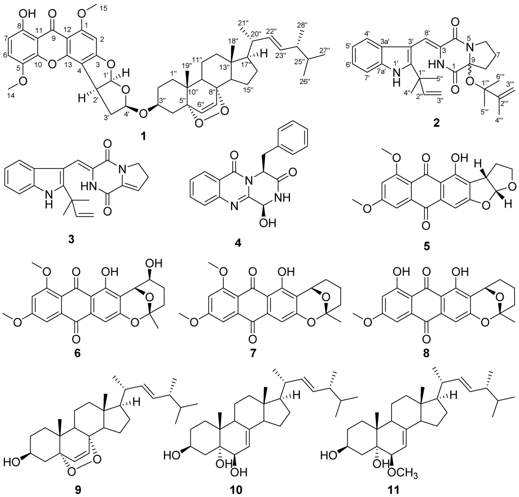
Compound 1 was obtained as yellow crystals from CHCl3. The molecular formula was determined as C47H58O10 on the basis of HREIMS with the [M]+ peak at m/z 782.4024 (calcd. for C47H58O10, 782.4030), indicating nineteen degrees of unsaturation. The 1H-NMR spectrum (Table 1) displayed two tertiary methyl singlets, four secondary methyl doublets, two oxygenated methyl singlets, one multiplet and two doublets assignable to three oxygenated methines, four doublets ascribed to two mutually coupled olefinic protons and two mutually coupled aromatic protons, two double doublets attributed to two mutually coupled olefinic protons, and one singlet characteristic of an aromatic proton. The 13C NMR spectrum (Table 1) along with the DEPT and HSQC experiments revealed the presence of eight methyl groups, eight methylenes, seventeen methines, and fourteen quaternary carbon atoms. Upon further inspection of the NMR data, it was realized that approximately half of the chemical shifts in 1 were superimposeable with 5α,8α-epidioxyergosta-6,22-dien-3β-ol (9), which we also isolated from the same fungal extract [8]. This enabled us to focus on the remaining chemical shifts in 1, which we were able to deduce had remarkable similarities to the natural product 5-methoxysterigmatocystin [4]. However, several key differences remained between our NMR data and those reported for 5-methoxysterigmatocystin. Namely, our metabolite 1 exhibited new resonances for a methylene and an oxygenated methine, but lacked two olefinic methines. Therefore, we deduced that this portion of the molecule represented a 3',4'-dihydro-5-methoxysterigmatocystin residue, which was verified by the 1H–1H COSY correlations between H-6/H-7, H-1'/H-2', H-2'/H-3', H-3'/H-4' and HMBC correlations from H-2 to C-1, C-3, C-4, and C-12, from H-6 to C-5, C-8, and C-10, from H-7 to C-5, C-8, and C-11, from H-14 to C-5, from H-15 to C-1, from H-1' to C-3, C-4, C-2', C-3', and C-4', from H-2' to C-3, C-4, C-13, C-1', C-3', and C-4', and from H-4' to C-1', C-2', and C-3'. The linkage between the two portions of compound 1 was established by HMBC correlations from H-4' to C-3" and from H-3" to C-4'. The relative configuration of 1 was confirmed by analysis of NOESY spectrum. Based on biogenetic considerations, the configurations of steroid moiety should be the same as those of 5α,8α-epidioxyergosta-6,22-dien-3β-ol (9) [8], which were confirmed by the observed NOESY correlations between H-6"/H-19", H-7"/H-18", H-18"/H-20", H-2"a/H-19", H-2"b/H-3". H-3"and H-4' were located on the same side by the NOESY correlation between H-3"/H-4', while H-4', H-1', and H-2' were positioned on the same face based on the NOESY correlations of H-1' with H-2' and H-4'. We have given this new molecule from A. versicolor pt20 the trivial name asperversin A.
Table 1. 1H and 13C NMR data for compound 1 (500 MHz for 1H and 125 MHz for 13C, CDCl3).
PositionδH (J in Hz)δC, mult.PositionδH(J in Hz)δC, mult.
1 163.3, C6"6.11, d (8.5)135.3, CH
26.34, s90.1, CH7"6.44, d (8.5)130.7, CH
3 165.3, C8" 79.4, C
4 108.2, C9"1.44, m51.1, CH
5 139.4, C10" 37.1, C
67.18, d (9.0)120.4, CH11"a1.36, m20.6, CH2
76.68, d (9.0)109.3, CH11"b1.56, m
8 155.3, C12"a1.20, m39.3, CH2
9 181.6, C12"b1.92, m
10 144.9, C13" 44.5, C
11 109.6, C14"1.52, m51.6, CH
12 105.7, C15"a1.16, m23.3, CH2
13 153.9, C15"b1.45, m
143.91, s57.8, CH316"a1.33, m28.6, CH2
153.99, s56.8, CH316"b1.73, m
1'6.51, d (6.0)113.5, CH17"1.20, m56.2, CH
2'4.21, dd (9.2, 6.0)43.0, CH18"0.78, s12.8, CH3
3'a2.34, ddd (13.2, 9.2, 4.9)36.9, CH219"0.72, s18.0, CH3
3'a2.47, d (13.2) 20"2.00, m39.7, CH
4'5.39, d (4.9)104.2, CH21"0.98, d (6.6)20.9, CH3
1"a1.57, m34.6, CH222"5.13, dd (15.3, 8.3)135.2, CH
1"b1.86, m 23"5.21, dd (15.3, 7.6)132.3, CH
2"a1.13, m27.6, CH224"1.84, m42.8, CH
2"b1.73, m 25"1.45, m33.1, CH
3"3.79, m72.1, CH26"0.81, d (6.8)19.6, CH3
4"a1.54, m33.6, CH227"0.83, d (6.8)20.0, CH3
4"b1.91, m 28"0.90, d (6.8)17.6, CH3
5" 81.8, COH12.73, s
Compound 2 was obtained as colorless crystals from CHCl3. The molecular formula was established to be C27H33N3O3 based on HREIMS (m/z 447.2507 [M]+, calcd. for C27H33N3O3, 447.2522), requiring thirteen degrees of unsaturation. The 1H NMR spectrum (Table 2) of 2 exhibited five methyl singlets, two singlets characteristic of terminal olefinic protons, one double doublet and two doublets attributed to a terminal vinyl group, two doublets and two double doublets ascribed to an ortho-substituted phenyl group, and two broad singlets assigned to two presumably exchangeable protons. The 13C NMR spectrum (Table 2) exhibited fifteen resonances, which were identified as five methyls, five methylenes, six methines, and eleven quaternary carbons by the DEPT and HSQC experiments. The HMBC correlations from H-3"' to C-1"', C-2"', and C-4"', from H-4"' to C-1"', C-2"', and C-3"', from H-5"' to C-1"', C-2"', and C-6"', and from H-6"' to C-1"', C-2"', and C-5"' established the presence of structural unit CH2=CH(CH3)–C(CH3)2–. The remaining NMR resonances were similar to those reported for brevianamide Q [10]. The major exception was the significant downfield shift of C-9 (δC 94.3) due to it being the site of an ether linkage to the new CH2=CH(CH3)–C(CH3)2– group. The structure of the remaining portion of the compound was confirmed based on 1H–1H COSY correlations between H-6/H-7, H-7/H-8, H-4'/H-5', H-5'/H-6', H-6'/H-7', H-2"/H-3", as well as HMBC correlations from H-1 to C-4, from H-8 to C-1 and C-9, from H-1' to C-3' and C-3a', from H-4' to C-3', C-6', and C-7a', from H-7' to C-3a' and C-5', from H-8' to C-4 and C-2', and from H-4" and H-5" to C-2', C-1", and C-2". The above evidence established the structure of 2, named 9ξ-O-2(2,3-dimethylbut-3-enyl)brevianamide Q.
Table 2. 1H and 13C NMR data for compound 2 (500 MHz for 1H and 125 MHz for 13C, CDCl3).
PositionδH (J in Hz)δC, mult.PositionδH(J in Hz)δC, mult.
1 162.3, C6'7.19, dd (7.5, 8.0)122.3, CH
27.48 br, s 7'7.35, d (8.0)111.1, CH
3 126.2, C7a' 134.2, C
4 159.4, C8'7.29, s111.6, CH
5 1" 39.3, C
6a3.75, m45.2, CH22"6.08, dd (17.4, 10.6)144.3, CH
6b3.95, m 3"a5.21, d (17.4)113.4, CH2
7a2.00, m19.8, CH23"b5.24, d (10.6)
7b2.10, m 4"1.54, s27.3, CH3
8a2.25, m33.5, CH25"1.54, s27.7, CH3
8b2.37, m 1"' 84.6, C
9 94.3, C2"' 148.1, C
1'8.29 br, s 3"'a4.89, s111.7, CH2
2' 144.1, C3"'b4.96, s
3' 103.5, C4"'1.83, s18.5, CH3
3a' 126.2, C5"'1.40, s24.0, CH3
4'7.47, d (7.5)119.9, CH6"'1.32, s25.0, CH3
5'7.13, dd (7.5, 7.5)120.9, CH
In addition to these two new compounds, we also isolated several known compounds including brevianamide K (3) [3], brevianamide M (4) [3], aversin (5) [4], 6,8-di-O-methylnidurufin (6) [5], 6,8-di-O-methylaverufin (7) [6], 6-O-methylaverufin (8) [7], 5α,8α-epidioxyergosta-6,22-dien-3β-ol (9) [8], ergosta-7,22-diene-3β,5α,6β-triol (10) [9], and 6β-methoxyergosta-7,22-diene-3β,5α-diol (11) [9]. The structures of these metabolites were confirmed by comparisons of their respective spectroscopic data with those reported earlier.
Table 3. Antibacterial activities at 30 μg/disk and toxicities against brine shrimp at 100 μg/mL of 18.
CompoundsInhibition Zone (mm)Lethal Rates (%)
Escherichia coliStaphylococcus aureusArtemia salina
1771.8
27743.2
37730.9
4111047.6
56617.5
67729.1
71010100.0
8101038.5
chloramphenicol3231
Compounds 18 were tested for biological activities against several target organisms including bacteria, fungi, and brine shrimp. Antibacterial activity was assessed by disk diffusion assay against Escherichia coli and Staphylococcus aureus at a concentration of 30 μg/disk. Compounds 4, 7, and 8 were found to exhibit modest inhibitory activity against these bacterial strains (Table 3). None of the compounds inhibited the fungal species Colletotrichum lagenarium or Fusarium oxysporium at 30 μg/disk in the disk diffusion assay [11]. Interestingly, compound 7 exhibited significant toxicity toward brine shrimp with an LC50 value of 0.5 μg/mL [12].

3. Experimental Section

3.1. General

NMR spectra were recorded in CDCl3 at 500 and 125 MHz for 1H and 13C, respectively, on a Bruker Avance III 500 NMR spectrometer using TMS as internal standard. High resolution mass data were acquired on Autospec Premier P776 mass spectrometer with an EI source. IR spectra were obtained on a JASCO FT/IR-4100 Fourier Transform InfraRed spectrometer. UV spectrum was measured on a TU-1810 Spectrophotometer. HPLC separation was carried out on an Elite HPLC system (P270 pump, UV230+ detector, Dalian Elite Analytical Instruments Co., Ltd., Dalian, China) using an Eclipse XDB-C18 (5 μm, 9.4 × 250 mm) column. Column chromatography was performed with silica gel (100–200 and 200–300 mesh, Qingdao Haiyang Chemical Co., Qingdao, China) and Sephadex LH-20 (Pharmacia). Precoated silica gel plates (GF-254, Qingdao Haiyang Chemical Co., Qingdao, China) were used for preparative TLC purification. All solvents were of analytical grade.

3.2. Microorganism and Fermentation

The endophytic fungus A. versicolor pt20 was isolated from a fresh, surface-sterilized tissue sample of the marine brown alga S. thunbergii, which was collected from Pingtan Island, China. The fungus was identified based on morphological and molecular taxonomic methods by one of the authors (F.-P.M.). A voucher sample has been preserved in Bio-Resource Laboratory of Yantai Institute of Coastal Zone Research, Chinese Academy of Sciences. The initial cultures were maintained on the potato dextrose agar plates. Pieces of mycelia were cut into small segments and aseptically inoculated into 1000 mL Erlenmeyer flasks containing 300 mL potato dextrose broth (PDB) culture media. The static fermentation was carried out for 30 days at room temperature (25 °C).

3.3. Extraction and Isolation

The culture broth (15 L) was extracted with EtOAc to yield 3.6 g gum after removal of the solvent by evaporation (40 °C) at reduced pressure. The dried and powdered mycelia (162.2 g) were extracted with a mixture of CHCl3 and MeOH (1:1, v/v), concentrated, and partitioned between H2O and EtOAc to give 29.8 g gum. Since the TLC profiles of the two extracts were nearly identical, they were combined before further separation. The total EtOAc-soluble fraction (33.4 g) was subjected to silica gel column chromatography (CC, petroleum ether (PE)/EtOAc gradient) to afford 16 fractions (Fr. 1–16), monitored by TLC. Fr. 10 eluted with PE/EtOAc (5:1) and was further purified by CC on Sephadex LH-20 (CHCl3/MeOH, 1:1) to yield three sub-fractions, 10-1, 10-2, and 10-3. Sub-fraction 10-1 was further purified by silica gel CC (PE/EtOAc, 5:1) and HPLC (MeOH/H2O, 85%) to give 9 (3.0 mg). Sub-fraction 10-2 was further purified by silica gel CC (PE/EtOAc, 4:1) and preparative TLC (CHCl3/EtOAc, 2:1) to afford 7 (13.0 mg). Sub-fraction 10-3 was further purified by silica gel CC (CHCl3/EtOAc, 4:1) and preparative TLC (CHCl3/EtOAc, 3:2) to produce 1 (8.3 mg). Fr. 11 eluted with PE/EtOAc (2:1) and was further purified by CC on Sephadex LH-20 (CHCl3/MeOH, 1:1) to afford 5 (27.1 mg). Fr. 12 eluted with PE/EtOAc (2:1) and was further purified by CC on Sephadex LH-20 (CHCl3/MeOH, 1:1) and preparative TLC (CHCl3/EtOAc, 3:2) to yield 2 (2.0 mg). Fr. 13 eluted with PE/EtOAc (1:1) and was further purified by CC on Sephadex LH-20 (CHCl3/MeOH, 1:1) to produce two sub-fractions, 13-1 and 13-2. Sub-fraction 13-1 was further purified by silica gel (PE/EtOAc, 2:1) and preparative TLC (CHCl3/EtOAc, 2:1) to give 3 (4.3 mg). Sub-fraction 13-2 was further purified by CC on silica gel (PE/EtOAc, 2:1–1:1) and HPLC (MeOH /H2O, 85%) to afford 11 (4.4 mg). Fr. 14 eluted with PE/EtOAc (1:1) and was further purified by CC on Sephadex LH-20 (CHCl3/MeOH, 1:1) and silica gel (PE/EtOAc, 1:1) and preparative TLC (CHCl3/EtOAc, 1:1) to yield 6 (2.0 mg). Fr. 15 eluted with EtOAc and was further purified by CC on Sephadex LH-20 (CHCl3/MeOH, 1:1) to yield three sub-fractions, 15-1, 15-2, and 15-3. Sub-fraction 15-1 was further purified by CC on silica gel (PE/EtOAc, 1:2) and preparative TLC (PE/EtOAc, 1:2) to afford 8 (10.2 mg). Sub-fraction 15-2 was further purified by CC on silica gel (CHCl3/EtOAc, 2:3) and preparative TLC (CHCl3/EtOAc, 2:3) to produce 4 (29.0 mg). Sub-fraction 15-3 was further purified by HPLC (MeOH /H2O, 85%) and preparative TLC (EtOAc) to give 10 (3.0 mg).
Asperversin A (1): Yellow crystals; m.p. 273–275 °C; [α]25D −309.7 (c 0.12, CHCl3); UV (CHCl3) λmax (log ε) 248 (4.51), 330 (4.14) nm. IR (KBr) νmax 3448, 2927, 2866, 1631, 1581, 1485, 1369, 1234, 972 cm−1. 1H and 13C NMR data, see Table 1. HREIMS m/z 782.4024 [M]+, calcd. for C47H58O10, 782.4030.
9ξ-O-2(2,3-dimethylbut-3-enyl)brevianamide Q (2): Colorless crystals; m.p. 89–92 °C; [α]24D −16.0 (c 0.11, CHCl3); UV (CHCl3) λmax (log ε) 238 (3.61), 334 (3.28) nm. IR (KBr) νmax 3340, 2927, 2858, 1693, 1624, 1427, 1385, 1238, 1149, 910, 748 cm−1. 1H and 13C NMR data, see Table 2. HREIMS m/z 447.2507 [M]+, calcd. for C27H33N3O3, 447.2522.

3.4. Antimicrobial Assay

Antibacterial and antifungal activities were assayed as described previously [11].

3.5. Brine Shrimp Lethality Assay

Brine shrimp (Artemia salina) lethality assay procedure followed the micro-well plate method described by Solis et al with some modifications [12]. Briefly, brine shrimp eggs were left to hatch in sea water for 48 hours at 28 °C under natural light. For brine shrimp lethality testing, compounds were dissolved in DMSO prior to preparing serial dilutions in 200 µL volume of sea water prepared in 96 well microplates. A well containing DMSO without compounds added was used as a positive control. Approximately, 10 brine shrimp were placed in a well with a volume of 200 µL sea water for lethality testing. Brine shrimp lethality was observed after 24 hours of cultivation under continuous light. Dead shrimp were identified with the aid of a handheld magnifying lens.

4. Conclusions

In summary, two new (1 and 2) and nine known (311) secondary metabolites were purified from the algicolous fungus A. versicolor pt20. To the best of our knowledge, compound 1 represented the first described example of a steroid-xanthone heterodimer. Compunds 4, 7, and 8 were more active against E. coli and S. aureus, and 7 also showed strong toxicity against brine shrimp.

Acknowledgments

This work was financially supported by the National Natural Science Foundation of China (41106137, 41106136), Chinese Academy of Sciences for Key Topics in Innovation Engineering (KZCX2-YW-QN209, KSCX2-EW-G-12B), and Foundation of the Chinese Academy of Sciences for President’s Scholarship (awarded to N.-Y. Ji).
  • Samples Availability: Available from the authors.

Supplementary Files

  • Supplementary File 1::

    PDF-Document (PDF, 1791 KB)

  • References

    1. König, G.M.; Kehraus, S.; Seibert, S.F.; Abdel-Lateff, A.; Müller, D. Natural products from marine organisms and their associated microbes. ChemBioChem 2006, 7, 229–238. [Google Scholar]
    2. Osterhage, C.; König, G.M.; Höller, U.; Wright, A.D. Rare sesquiterpenes from the algicolous fungus Drechslera dematioidea. J. Nat. Prod. 2002, 65, 306–313. [Google Scholar]
    3. Li, G.Y.; Yang, T.; Luo, Y.G.; Chen, X.Z.; Fang, D.M.; Zhang, G.L. Brevianamide J, a new indole alkaloid dimer from fungus Aspergillus versicolor. Org. Lett. 2009, 11, 3714–3717. [Google Scholar]
    4. Shao, C.; She, Z.; Guo, Z.; Peng, H.; Cai, X.; Zhou, S.; Gu, Y.; Lin, Y. Spectral assignments and reference data. Magn. Reson. Chem. 2007, 45, 434–438. [Google Scholar]
    5. Kingston, D.G.I.; Chen, P.N.; Vercellotti, J.R. Metabolites of Aspergillus versicolor. 6,8-di-Ο-methylnidurufin, griseofulvin, dechlorogriseofluvin, and 3,8-dihydroxy-6-methoxy-1-methylxanthone. Phytochemistry 1976, 15, 1037–1039. [Google Scholar]
    6. Ren, H.; Gu, Q.Q.; Cui, C.B. Anthraquinone derivatives produced by marine-derived Penicillium flavidorsum SHK1-27 and their antitumor activities. Chin. J. Med. Chem. 2007, 17, 148–154. [Google Scholar]
    7. Steyn, P.S.; Vleggaar, R.; Wessels, P.S.; Cole, R.J.; Scott, D.B. Structure and carbon-13 nuclear magnetic resonance assignments of versiconal acetate, versiconol acetate, and versiconol, metabolites from cultures of Aspergillus parasiticus treated with ditchlorvos. J. Chem. Soc. Perkin Trans. I 1979, 451–459. [Google Scholar]
    8. Greca, M.D.; Mangoni, L.; Malinaro, A.; Monaco, P.; Previtera, L. 5β,8β-Epidioxyergosta-6,22-dien-3β-ol from Typha latifolia. Gazz. Chim. Ital. 1990, 120, 391–392. [Google Scholar]
    9. Kawagishi, H.; Katsumi, R.; Sazawa, T.; Mizuno, T.; Hagiwara, T.; Nakamura, T. Cytotoxic steroids from the mushroom Agaricus blazei. Phytochemistry 1988, 27, 2777–2779. [Google Scholar]
    10. Li, G.Y.; Li, L.M.; Yang, T.; Chen, X.Z.; Fang, D.M.; Zhang, G.L. Four new alkaloids, brevianamides O–R, from the fungus Aspergillus versicolor. Helv. Chim. Acta 2010, 93, 2075–2080. [Google Scholar]
    11. Schulz, B.; Sucker, J.; Aust, H.J.; Krohn, K.; Ludewig, K.; Jones, P.G.; Döring, D. Biologically active secondary metabolites of endophytic Pezicula species. Mycol. Res. 1995, 99, 1007–1015. [Google Scholar]
    12. Solis, P.N.; Wright, C.W.; Anderson, M.M.; Gupta, M.P.; Phillipson, J.D. A microwell cytotoxicity assay using Artemia salina (brine shrimp). Planta Med. 1993, 59, 250–252. [Google Scholar]

    Share and Cite

    MDPI and ACS Style

    Miao, F.-P.; Li, X.-D.; Liu, X.-H.; Cichewicz, R.H.; Ji, N.-Y. Secondary Metabolites from an Algicolous Aspergillus versicolor Strain. Mar. Drugs 2012, 10, 131-139. https://doi.org/10.3390/md10010131

    AMA Style

    Miao F-P, Li X-D, Liu X-H, Cichewicz RH, Ji N-Y. Secondary Metabolites from an Algicolous Aspergillus versicolor Strain. Marine Drugs. 2012; 10(1):131-139. https://doi.org/10.3390/md10010131

    Chicago/Turabian Style

    Miao, Feng-Ping, Xiao-Dong Li, Xiang-Hong Liu, Robert H. Cichewicz, and Nai-Yun Ji. 2012. "Secondary Metabolites from an Algicolous Aspergillus versicolor Strain" Marine Drugs 10, no. 1: 131-139. https://doi.org/10.3390/md10010131

    Article Metrics

    Back to TopTop