Article

The Antistaphylococcal Activity of Citropin 1.1 and Temporin A against Planktonic Cells and Biofilms Formed by Isolates from Patients with Atopic Dermatitis: An Assessment of Their Potential to Induce Microbial Resistance Compared to Conventional Antimicrobials

1
Faculty of Pharmacy, Medical University of Gdansk, Al. Gen. J. Hallera 107, 80-416 Gdansk, Poland
2
Faculty of Medicine, Medical University of Gdansk, Debinki 7, 80-211 Gdansk, Poland
*
Author to whom correspondence should be addressed.
Pharmaceuticals 2016, 9(2), 30; https://doi.org/10.3390/ph9020030
Submission received: 17 February 2016 / Revised: 16 May 2016 / Accepted: 19 May 2016 / Published: 25 May 2016

Abstract

:
Staphylococcus aureus (SA) colonizes the vast majority of patients with atopic dermatitis (AD). Its resistance to antibiotics and ability to form biofilms are the main origins of therapeutic complications. Endogenous antimicrobial peptides (AMPs) exhibit strong activity against SA, including antibiotic resistant strains as well as bacteria existing in biofilm form. The purpose of the present work was to determine the antistaphylococcal activity of two amphibian peptides against SA isolated from patients with AD. The AMPs demonstrated permanent activity towards strains exposed to sublethal concentrations of the compounds and significantly stronger antibiofilm activity in comparison to that of conventional antimicrobials. The results suggest the potential application of amphibian AMPs as promising antistaphylococcal agents for the management of skin infections.

1. Introduction

The skin serves as the first line of defense against infection by reducing microbial adherence and invasion [1]. In atopic dermatitis (AD), a chronic, relapsing inflammatory skin disease, the protective epidermal barrier is significantly compromised [2,3]. Both the etiology and pathogenesis of AD are very complex in nature. The disease is caused by the interplay of various factors, such as genetically related disturbances of both the structure and function of the epidermal barrier, allergic and immunologic abnormalities as well as the surrounding environment and infectious determinants such as Staphylococcus aureus (SA) [4].
SA is the major cause of skin and soft tissue infections which, despite the continuous development of medicines, are still difficult to treat [5,6]. Therapeutic problems are caused by the development of strains with reduced susceptibility to many antibiotics, such as methicillin-resistant SA (MRSA) [7]. SA is also a medically significant biofilm former. In terms of infections, these complex sessile microbial communities can grow on medical devices or host tissues. The process of biofilm formation is controlled by the accessory gene regulator (agr) quorum-sensing system which is one of SA’s virulence factors [8]. Due to their recalcitrance to host defense mechanisms and antimicrobial therapy, the biofilm-related infections move towards chronic disease status and reoccur after the withdrawal of antimicrobials [9,10].
Patients with AD have a predisposition towards frequent cutaneous infection with SA, C. albicans or the herpes simplex virus. Actually, over 90 percent of patients with AD have shown colonization with SA. More than half of the SA strains isolated from AD skin lesions have been shown to secrete superantigenic exotoxins, primarily SEA, SEB, and TSST-1, that stimulate marked activation of T cells and macrophages and presumably contribute to the exacerbation of AD [11,12].
Recently, the mechanisms of increased vulnerability to skin infections have been an area of intensive investigation. It has been postulated that the innate immune defense system of atopic skin based on naturally occurring antimicrobial peptides (AMPs) fails to restrict the growth of the organisms [3,13,14,15]. In view of the increasing microbial resistance to traditional antibiotics, AMPs are considered a source for the development of a novel antistaphylococcal therapy for patients with AD. Peptides secreted by the skin of amphibians constitute one of the best-studied groups of AMPs. Citropin 1.1 is a basic, highly hydrophobic, 16-amino acid peptide, produced by the dorsal and submental glands of the green tree frog Litoria citropa. It is one of the simplest broad-spectrum amphibian AMPs reported to date. Its antimicrobial potential towards a wide range of microorganisms has been established [16]. A broad spectrum of antimicrobial activity has also been displayed by temporin A, a basic, highly hydrophobic, 13-amino acid peptide derived from the frog Rana temporaria [17]. In the present study, we determined the activity of these two amphibian peptides and conventional antistaphylococcal antibiotics towards strains isolated from patients with AD. A characterization of SA isolates according to their variant of agr, their ability to produce superantigens, their development of resistance to antimicrobials and their biofilm formation has been performed.

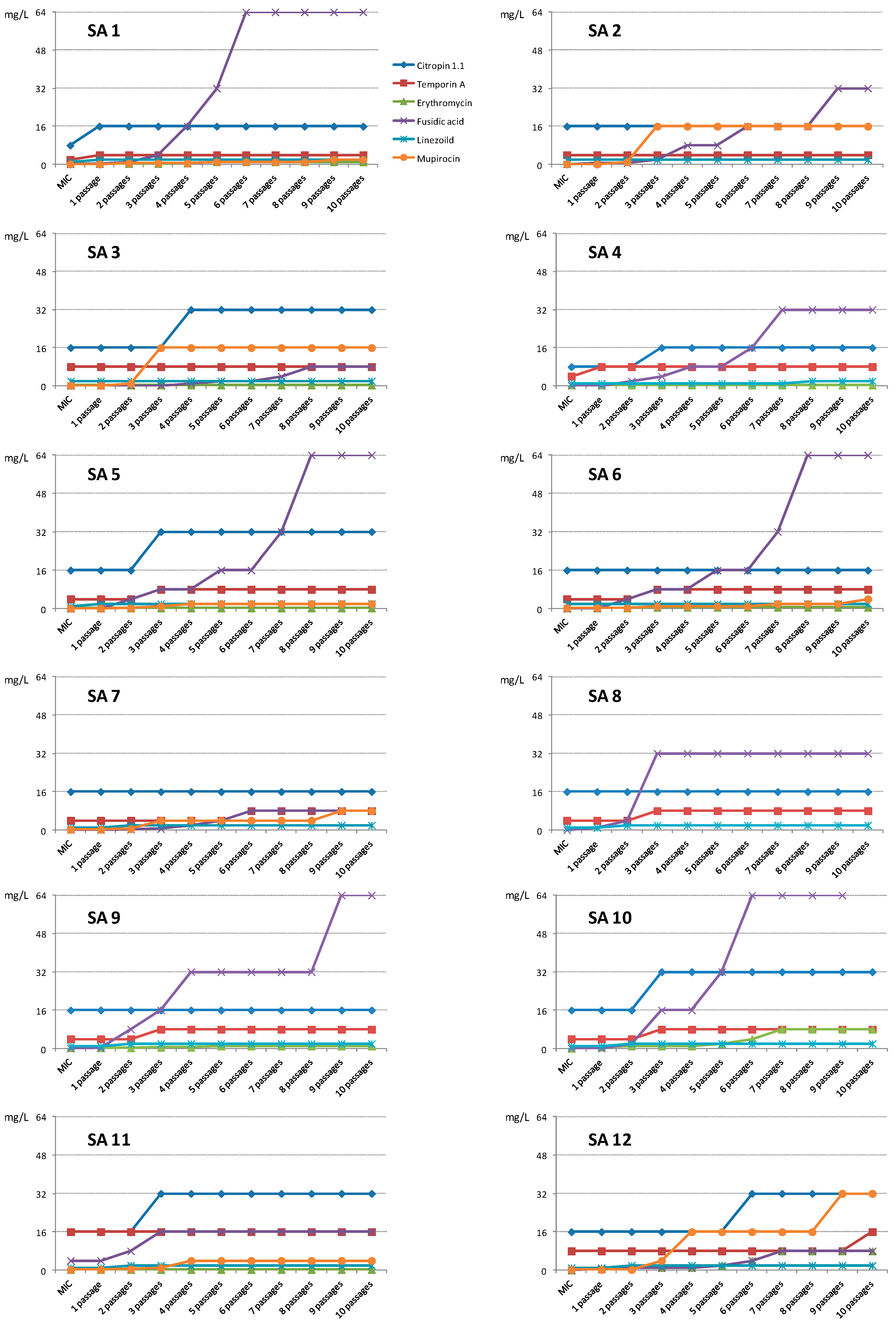
2. Results and Discussion

2.1. Results

2.1.1. SA Isolates

Among 15 examined patients with AD, 12 were colonized by SA. This corresponds to over 80 percent of the tested subjects. Nine strains showed the ability to produce superantigens (Table 1). Five SA isolates secreted staphylococcal enterotoxin A (SEA). C (SEC) and D (SED) were produced by two and three strains respectively, wherein one of these isolates secreted both toxins simultaneously.
None of the SA strains showed the ability to produce staphylococcal enterotoxin B or a toxin of staphylococcal shock syndrome TSST-1. The results obtained using toxin test kits were consistent with those obtained by PCR. All tested isolates carried the accessory regulator gene (agr). Agr types I and III were identified for six and five strains, respectively. Only one strain turned out to be agr II, while agr IV was missing.

2.1.2. Activity against Planktonic Cells

The most potent antimicrobial agent was fusidic acid with MICs of 0.0625–0.125 mg/L against 11 tested strains. One isolate (SA 11) showed a reduced susceptibility to the compound (MIC = 4 mg/L). Erythromycin and mupirocin also inhibited staphylococcal growth in the majority of the strains at low concentrations (MICs below 1 mg/L). However, we have identified three and four isolates resistant to erythromycin (SA 2, 7, 8) and mupirocin (SA 4, 8, 9, 10), respectively. Linezolid was active at concentrations of 1–2 mg/L towards all SA isolates.
The amphibian peptides showed antimicrobial activity following a single application at higher concentrations. The MICs obtained for the majority of strains were 4 mg/L and 16 mg/L for citropin 1.1 and temporin A, respectively (Table 2).
The minimum inhibitory concentration was determined for each strain after serial passages in a medium supplemented with the compounds below their active concentrations (0.5 × MIC). After 10 consecutive passages with the AMPs, the SA strains did not show any significant reduction in their susceptibility to AMPs. For these compounds, the active concentration after 10 passages either remained at the same level or was twice as high as the initial MIC (Figure 1, Table 2).
Linezolid also did not induce resistance in tested SA strains; the MIC10 were equal or twice as high as the initial MICs. No significant reduction in activity was observed also for erythromycin. For SA 3, 4, 5 and 11, the MIC did not change during the study, while for SA 1, 6 and 9 it increased 2–4 times. After 10 passages of isolates SA 10 and 12, their susceptibility was reduced 32 and 64 times in comparison to that of the initial MICs.
During this test with mupirocin, a significant decrease in antistaphylococcal activity was observed. For the strains SA 1, 5, 6, 7 and 11, which were very sensitive to the compound in the first MIC test, the active concentrations increased 16–32 times after the tenth passage. The strains SA 2, 3 and 12 reduced their susceptibility 128 times (Figure 1, Table 2).
An even more significant reduction in the susceptibility of the isolates was demonstrated by strains cultured in a medium supplemented with sublethal concentrations of fusidic acid. In this case, after the exposure of the strains to the compounds following the last passage, SA 11 and 7 were inhibited by the compound at concentrations 4 and 64 times higher than that of the initial MICs, respectively. SA 1, 2, 3, 4, 8 and 12 reduced their susceptibility 128–512 fold, while for the remaining strains MIC10 it was over 1000 times higher than that of MIC0 (Figure 1, Table 2).

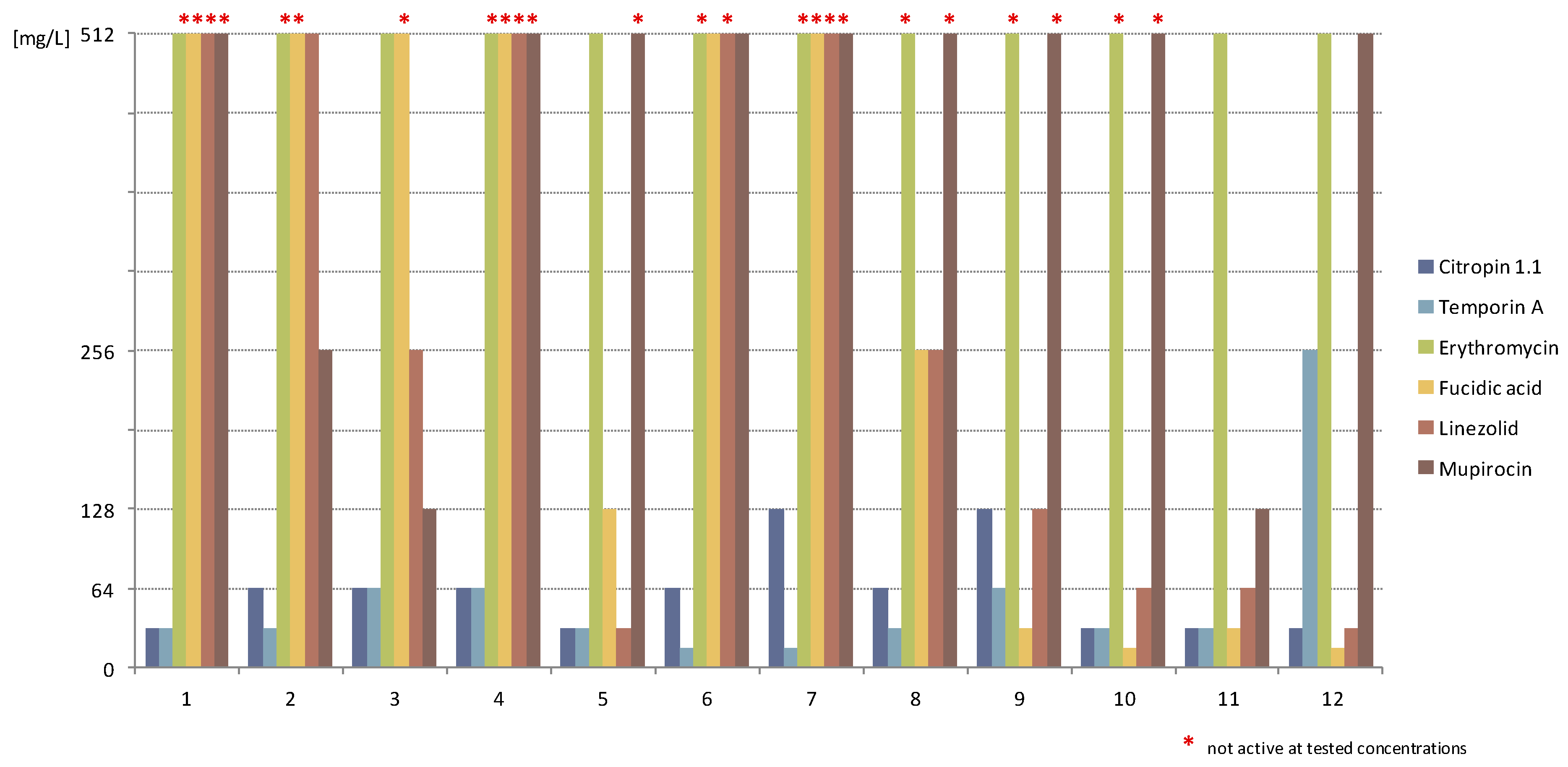
2.1.3. Activity against Staphylococcal Biofilm

Conventional antimicrobials demonstrated rather poor or no antibiofilm activity against the isolates. The least effective agent was erythromycin, which did not exhibit activity against biofilms formed by the majority of the SA isolates at the tested concentrations. Biofilm formed by the remaining strains (SA 3, 5, 10 and 11) was eliminated after exposure to erythromycin at a concentration over 2000 times higher than that of MICs.
Mupirocin was slightly more effective. For the most susceptible strains (SA 3 and 11), the elimination of the structures was noticed after using the antibiotic at a concentration of 128 mg/L, which was equal to (512–1024) times those of the MICs obtained for those strains. Biofilms formed by strains SA 3,6 and 12 were eradicated by mupirocin at 512 mg/L, while the remaining strains were not susceptible to the compound at the applied concentrations (over 2000 times that of the MICs).
Fusidic acid was inactive against the biofilm cultures of SA 1–4 and 7. SA 5, 6 and 8 were eliminated after application of the compound at concentrations of 128–512 mg/L (over (2–8) thousand × MIC). Biofilms formed by SA 9–12 were the most susceptible—the obtained MBEC ranged from 16 to 32 mg/L.
The same strains (SA 9–12) as well as SA 5 were the most susceptible to linezolid (MBEC 32–128 mg/L). SA 2 and 3 were eradicated at MBECs of 256–512 mg/L, whilst the remaining strains formed structures unsusceptible to the compound at the applied concentrations. The amphibian peptides showed markedly higher antibiofilm activity against all tested SA isolates. Temporin A eliminated the majority of the structures at concentrations (2–8) times those of the MICs. For strains SA 1, 4, 9 and 12, the effective concentrations towards biofilms (32–256 mg/L) were (16–32) times higher in comparison to the activity against planktonic cells. With citropin 1.1, the majority of the MBECs (16–32 mg/L) corresponded to (2–4) times those of the MICs, while biofilms formed by the strains SA 4, 7 and 9 were eradicated after application of citropin 1.1 at concentrations 8 times higher than those of the MICs (Table 2, Figure 2).

2.2. Discussion

According to previous studies, SA has been isolated from 75 to 90% of AD patients. This is compatible with our results. SA was found in the skin samples of 80% of the examined patients. Colonization with SA has been well documented as an exacerbating factor in AD [3,18,19,20]. According to the literature, more than half of the SA strains isolated from AD patients possess the ability to produce superantigens, and the prevalence rate is significantly higher than in the control groups (less than 5% of patients). In our study, 75% of the strains produced superantigens, which is consistent with the previous findings. Recent data suggest that SEA, SEB and TSST-1 are the most abundant toxins found in those strains. Four of the nine superantigen positive strains tested in the present work secreted SEA, whereas neither SEB nor TSST-1 were identified.
According to Lee et al., SEA and SEB play a significant role in the initiation of atopic skin inflammation [2,12]. It has been proposed that toxins may stimulate TNF-α and IL-1α release from keratinocytes, macrophages and Langerhans cells. It has also been found that the majority of patients with AD possess circulating IgE specific to SEA, SEB and TSST-1, so the toxins are likely to function as allergens [21]. It has been established that the expression of SEA is not regulated by the accessory gene regulator agr, unlike SEB, SEC and SED [22]. In our study, the SA strains producing SEA belonged to agr group III. For isolates secreting SEC or SED, agr group I was identified. One strain belonged to agr group II and showed the ability to produce two toxins simultaneously (SEC and SED).
The agr locus of SA is described as a quorum-sensing gene cluster and may be a crucial regulator of bacterial cellular metabolism. Numerous studies demonstrate that the vast majority of clinical SA strains belong to agr group I. Among the MRSA isolated around the world, agr group III has often been identified. In our study, we found six, one and five strains belonging to agr groups I, II and III, respectively [23]. Strains representing agr type II showed a reduced susceptibility to β-lactams and glycopeptides [24].
The influence of the ability to produce staphylococcal enterotoxins on the activity of various antibiotics has previously been suggested. Superantigen-positive isolates have demonstrated a reduced susceptibility to chloramphenicol, ciprofloxacin, clindamycin and erythromycin [25]. Our study was conducted on a small population of SA strains and therefore, no statistical analysis in order to determine a relationship between the secretion of enterotoxins or the agr group and antibiotic effectiveness could have been performed. However, all the isolates demonstrating resistance to erythromycin belonged to agr group III and produced SEA. Also, two SA strains showing resistance to mupirocin were of agr type III and secreted SEA. Other mupirocin-resistant isolates were of agr type I; one produced SED, whilst the other did not secrete superantigens.
The type of agr has been reported to influence the ability of SA to form biofilms. Mirani et al. found that among biofilm positive methicillin sensitive SA (MSSA) strains (only 13.7% of all MSSA strains), ca. 50% carry agr type II, while 43.7% and 6.2% belong to group agr type I and 6.2% carry type III, respectively. The vast majority (81%) of biofilm negative MSSA isolates belong to group agr type I; 16% carry agr type III and the remaining 3% carry agr type II [23]. These data suggest that SA isolates from agr group I are not expected to be strong biofilm formers. In our study, SA 3, 5 and 9–12 indeed formed structures more susceptible to the majority of the antimicrobials in comparison to other strains.
The overuse of antibiotics is the main factor responsible for the high prevalence of bacterial strains resistant to antimicrobials. A significant decrease in susceptibility to erythromycin has often been reported over recent years [26]. In our study of three SA isolates, the lack of activity of erythromycin towards planktonic cells was noticed. However, the remaining strains showed a rather low rate of resistance development during the multiple passages of isolates in a medium supplemented with sublethal concentrations of those compounds.
Four of the 12 tested parental strains were not inhibited once exposed to mupirocin at the tested concentrations. After the tenth passage, the remaining isolates reduced their susceptibility significantly in comparison to that of the parental strains. Mupirocin is an approved antibiotic for topical use in the management of MRSA skin infections and is recommended for the eradication of SA carriage [27,28]. The high risk of reduction in its activity against SA has previously been debated. Long-term use of mupirocin induces the development of resistant strains. Some studies have revealed the colonization of patients with mupirocin-resistant strains without its previous widespread use, while others have demonstrated effective use of the antibiotic without a reduction in its activity [29,30,31].
A major decrease in activity was observed for fusidic acid, which proved to be the most active antibiotic in the initial MIC test. The compound has frequently been used in dermatology in the treatment of infected AD, and has a proven track record of efficacy [32]. Fusidic acid resistant SA has been reported all over the world, and its prevalence is higher in dermatology patients in comparison to other disciplines [33].
All the tested parental isolates were also susceptible to linezolid, and only an insignificant loss in its activity after consecutive passages was observed. According to the literature, the resistance rate of SA to this antimicrobial is low and estimated to be less than 1% [34].
Due to their mechanism based on interactions with the microbial cell plasma membrane [35], AMPs are believed not to induce bacterial resistance. This hypothesis was confirmed by several in vitro studies. Lipopeptide Laur-KK-NH2 proved to reduce the ability of E. faecalis strains to develop resistance to daptomycin [36]. The resistance of P. aeruginosa to a panel of eight peptides was increased by no more than fourfold after 30 passages in media supplemented with peptides below their MIC [37]. Our study provided corresponding results—the susceptibility of clinical isolates to amphibian peptides was reduced twofold as compared to that of the initial strains, or remained unchanged.
The unique mechanism of action also determines the activity of AMPs towards microbial biofilms. The compounds have proved to be active in vitro and in vivo against multidrug resistant bacteria in planktonic forms as well as towards biofilms at various stages of their development [38,39,40,41,42,43,44,45]. In the present study, the conventional antimicrobials demonstrated rather low antibiofilm activity, whilst AMPs were effective towards all SA isolates. Bacteria in biofilm form are frequently reported to exhibit tolerance to antimicrobials, while the same cells are sensitive in the planktonic state. This resistance/persistence of biofilm is directly related to low metabolic activity and the slow growth rate of bacteria in such a community. According to the mechanism of action, based on interactions with the microbial cell membrane, AMPs can act on both slow-growing and non-growing bacteria [46].
Human β-defensins HB43, HB55 and HBPM4 have demonstrated activity towards MRSA [38]. Human β-defensin 3 (HBD-3) was also an effective agent against S. aureus in biofilm form [39]. The antistaphylococcal activity of human cathelicidin LL-37 and its analogues has also been extensively investigated. LL-37 disrupted the formation of biofilm and eradicated the pre-grown biofilms of P. aeruginosa. LL-37 also inhibited the attachment and development of the biofilms of S. epidermidis [40,41].
In previous studies, amphibian peptides, aurein 1.2 and uperin 3.6 demonstrated strong antibacterial activity towards Gram-positive nosocomial cocci [42,43]. Citropin 1.1 demonstrated synergic action with rifampin and minocycline against an S. aureus biofilm [44]. Temporin A also proved its effectiveness against biofilms formed by gram-positive strains [45]. The results of the present study confirm the strong antibiofilm potential towards SA in comparison to that of conventional antimicrobials, which exhibited very poor antibiofilm activity. This is compatible with our previous study, where those antimicrobials were ineffective against structures formed by SA isolated from patients suffering from staphylococcal skin infections. The results obtained for conventional antistaphylococcal agents are also in accordance with data in the literature reporting that biofilm-associated bacteria exhibit inherent tolerance to antimicrobial compounds and that the MBECs are over 1000 fold higher in comparison to those of the MICs [47].
Both citropin 1.1 and temporin A have proved not only to be very potent antimicrobials, but also to be non-toxic towards human keratinocytes. In our previous study, the compounds did not exert any toxic effect on HaCaT cells at their microbiologically active concentrations and presented the most promising activity vs. toxicity ratio [48]. Therefore, the AMPs studied could be considered for treatment of skin infections, both on intact and abraded skin.

3. Experimental Section

3.1. Peptides

The compounds included in this study, citropin 1.1 and temporin A (Table 3), were synthesized manually using Fmoc chemistry on polystyrene resin that had been modified by the Rink amide linker [49]. Deprotection of the Fmoc group was carried out in a 20% solution of piperidine (Merck, Darmstadt, Germany) in dimethylformamide (DMF, Honeywell, Seelze, Germany). After washing the resin with DMF and dichloromethane (DCM, Honeywell), the amino acids were coupled using the DMF/DCM (1:1, v/v) mixture in the presence of coupling agents such as 1-hydroxybenzotriazole (HOBt; Orpegen, Heidelberg, Germany) and diisopropylcarbodiimide (DIC; Peptideweb, Zblewo, Poland). The extent of both the deprotection and acylation was monitored by the chloranil test. The peptides were cleaved from the resin with trifluoroacetic acid (Apollo Scientific, Denton, UK), water, triisopropylsilane and phenol (Sigma Aldrich, St. Louise, MO, USA) in the ratio 92.5:2.5:2.5:2.5 v/v, precipitated in cold diethyl ether (Chempur, Piekary Slaskie, Poland) and lyophilized. All crude products were purified by reversed-phase high performance liquid chromatography (RP-HPLC) in a gradient of acetonitrile (Sigma Aldrich)—water. The confirmed purity of both compounds was over 97 percent. The identity of the peptides was confirmed by matrix-assisted laser desorption/ionization time-of-flight-mass spectrometry (MALDI-TOF-MS).

3.2. Bacterial Isolates

Fifteen patients diagnosed with staphylococcal skin infections and hospitalized at the Dermatological Clinic of the Medical University of Gdansk, were enrolled in the study. Skin swabs were taken from each patient. SA was identified using standard previously described microbiological procedures [47].

Superantigen Detection

A Staphylococcal Enterotoxin Test Kit (SET-RPLA KIT TOXIN DETECTION KIT, Oxoid, Basingstoke, UK) was used for the detection of staphylococcal enterotoxins A, B, C and D, and a staphylococcal toxic shock syndrome toxin kit (TST-RPLA TOXIN DETECTION KIT, Oxoid) was employed for the detection of STSS in a culture, using reversed passive latex agglutination.

3.3. Toxin Gene Detection Using the PCR Technique

DNA was isolated in accordance with Barski et al. [50]. DNA amplification was carried out in a Perkin Elmer 2400 Thermocycler (Perkin Elmer Corporation, Norwalk, CT, USA). The primers for enterotoxins (SEA, SEB, SEC and SED) and the TSST-1 toxin were used as described previously [51]. DNA amplification was carried out with the following thermal cycling profile: an initial denaturation at 94 °C for 5 min was followed by 35 cycles of amplification (denaturation at 94 °C for 2 min, annealing at 57 °C for 2 min, and an extension at 72 °C for 1 min) and a final extension at 72 °C for 7 min. The PCR products were analyzed on a 2% agarose gel (Sigma Aldrich, St. Louise, MO, USA) in the presence of ethidium bromide under UV illumination. Control strains for the PCR detection of virulence factor genes were obtained from the National Institute of Public Health (NIPH; Warsaw, Poland).

3.4. Identification of S. Aureus agr Groups I, II, III, IV

DNA was isolated in accordance with Barski et al. [50]. Clinical S. aureus isolates were studied with the use of the PCR, using agr groups I, II, III, and IV-specific primers reported elsewhere [51]. The PCR program consisted of an initial denaturation step at 94 °C for 5 min, 30 cycles of denaturation at 94 °C for 1 min, annealing at 55 °C for 1 min, an extension at 72 °C for 1 min and a final extension step at 72 °C for 0.5 min. The products were analyzed using the standard methods of agarose gel electrophoresis and UV detection.

3.5. Antimicrobial Assay

The minimum inhibitory concentration (MIC) and minimum biofilm elimination concentration (MBEC) of the peptides were determined using a microbroth dilution method with the Mueller Hinton Broth II (MHB II; Becton Dickinson, Heidelberg, Germany). Bacteria at initial inoculums of 5 × 10⁵ CFU/mL were placed on polypropylene 96-well plates and exposed to tested compounds at graded concentrations for 18 h at 37 °C. The MIC was taken as the lowest drug concentration at which a noticeable growth was inhibited. Biofilms formed by clinical SA strains were cultured on polystyrene 96-well plates (Kartell, Noviglio, Italy). Bacteria at initial inoculums of 5 × 10⁸ CFU/mL were added to the plates and incubated at 37 °C. After 72 h, the biofilms were exposed to tested compounds for 24 h. Resazurin (Sigma Aldrich, St. Louise, MO, USA) was added as a cell-viability reagent. MBEC was taken as the lowest drug concentration at which the reading was compatible with the negative control (sterile MHB II with resazurin).
Ten consecutive passages of SA strains in a medium supplemented with tested compounds were performed in order to evaluate the progressive reduction in susceptibility to the tested compounds. After each passage, an MIC determination with the above described microbroth dilution method with MHB II was performed.
Identical tests were performed for conventional antimicrobials: erythromycin, fusidic acid, mupirocin (Sigma Aldrich, St. Louise, MO, USA) and linezolid (kindly provided by Pfizer). The experiments were performed in triplicate.

4. Conclusions

The AMPs exhibited activity against all SA strains isolated from patients with AD, while several strains were resistant to conventional antistaphylococcal agents. In general, the activity of antibiotics against the majority of the isolates in their planktonic form was significantly higher in comparison to that of the AMPs. However, the effectiveness of the most potent conventional antimicrobials, such as mupirocin or fusidic acid, was decreasing gradually during the multiple passages in the medium supplemented with the subinhibitory concentrations of the compounds. A significantly lower ability to induce bacterial resistance was observed for linezolid and the AMPs. Citropin 1.1 and temporin A also proved to be potent antibiofilm agents, unlike conventional antibiotics. The antimicrobials used in the management of staphylococcal infections either exhibited poor activity against structures grown on polystyrene surfaces or turned out to be inactive at the tested concentrations.
Taking under consideration the role played by S. aureus in etiopathogenesis in AD and its growing resistance to conventional antibiotics, the development of new therapeutic agents is crucial for modern medicine. Based on the literature data and the obtained results, AMPs seem to be highly promising candidates for the management of staphylococcal skin infections.

Acknowledgments

This study was supported by a grant from the Polish National Science Centre (Project No. 2011/03/B/NZ7/00548) and by the statutory found from the Medical University of Gdansk (Project No. 02-0087/07/508). We are grateful to the Pfizer Company for supplying linezolid.

Author Contributions

M.D. designed the experiments, performed the microbiological assays and wrote the paper, W.B.-R. designed the experiments, examined the patients and provided the clinical strains, L.P. performed the toxin gene detection and identification of SA agr groups; M.B. assisted in preparation of manuscript for publication, D.N. performed the synthesis and analysis of peptides, R.N. consulted the patients and provided professional support in the field of dermatology; W.K. supervised the synthesis of peptides and microbiological assays and contributed reagents, materials and analysis tools.

Conflicts of Interest

The authors declare no conflicts of interest.

References

  1. Percival, S.L.; Emanuel, C.; Cutting, K.F.; Williams, D.W. Microbiology of the skin and the role of biofilms in infection. Int. Wound J. 2012, 9, 14–32. [Google Scholar] [CrossRef] [PubMed]
  2. So-Young, N.; Joo-Young, R.; Jeung-Min, K.; Migma, D.T.; Jong-Rok, L. Analysis of Colonization and Genotyping of the Exotoxins of Staphylococcus aureus in Patients with Atopic dermatitis. Ann. Dermatol. 2012, 24, 413–419. [Google Scholar]
  3. Pastuszka, M.; Matych, M.; Kaszuba, A.; Poznańska-Kurowska, K. Microorganisms in the etiopathogenesis of atopic dermatitis. Postep. Derm. Alergol. 2012, 3, 215–221. [Google Scholar]
  4. Williams, H.C. Clinical practice. Atopic dermatitis. N. Engl. J. Med. 2005, 352, 2314–2324. [Google Scholar] [CrossRef] [PubMed]
  5. Kim, H.K.; Thammavongsa, V.; Schneewind, O.; Missiakas, D. Recurrent infections and immune evasion strategies of Staphylococcus aureus. Curr. Opin. Microbiol. 2012, 15, 92–99. [Google Scholar] [CrossRef] [PubMed]
  6. Kurlenda, J.; Grinholc, M. Alternative therapies in Staphylococcus aureus diseases. Act. Pol. Bichim. 2012, 59, 215–221. [Google Scholar]
  7. Wertheim, H.F.; Melles, D.C.; Vos, M.C.; van Leeuwen, W.; van Belkum, A.; Verbrugh, H.A.; Nouwen, J.L. The role of nasal carriage in Staphylococcus aureus infections. Lancet Infect. Dis. 2005, 5, 751–762. [Google Scholar] [CrossRef]
  8. Antunes, L.C.M.; Ferreira, R.B.R.; Buckner, M.M.C.; Finlay, B.R. Quorum sensing in bacterial virulence. Microbiology 2010, 156, 2271–2282. [Google Scholar] [CrossRef] [PubMed]
  9. Boles, B.R.; Horswill, A.R. Staphylococcal biofilm disassembly. Trends Microbiol. 2011, 19, 449–455. [Google Scholar] [CrossRef] [PubMed]
  10. Speziale, P.; Visai, L.; Rindi, S.; Pietrocola, G.; Provenza, G.; Provenzano, M. Prevention and treatment of staphylococcal biofilms. Curr. Med. Chem. 2008, 15, 3185–3195. [Google Scholar] [CrossRef] [PubMed]
  11. Mahgoub, D.M.; El-Eishi, N.H.; Rashed, L.A. A quantitative study of the antimicrobial peptide granulysin and its possible role in atopic dermatitis. J. Egypt. Women Dermatol. 2010, 7, 177–122. [Google Scholar]
  12. Lee, H.; Kim, S.M.; Kim, J.M.; Oh, B.M.; Kim, J.Y.; Jung, H.J.; Lim, H.J.; Kim, B.S.; Lee, W.J.; Lee, S.; et al. Potential immunoinflammatory role of staphylococcal enterotoxin A in atopic dermatitis: Immunohistopathological analysis and in vitro assay. Ann. Dermatol. 2013, 25, 173–180. [Google Scholar] [CrossRef] [PubMed]
  13. Ong, P.Y.; Ohtake, T.; Brandt, C.; Strickland, I.; Boguniewicz, M.; Ganz, T.; Gallo, R.L.; Leung, D.Y. Endogenous antimicrobial peptides and skin infections in atopic dermatitis. N. Engl. J. Med. 2002, 347, 1151–1160. [Google Scholar] [CrossRef] [PubMed]
  14. Nakatsuji, T.; Gallo, R.L. Antimicrobial peptides: Old molecules with new ideas. J. Investig. Dermatol. 2012, 132, 887–895. [Google Scholar] [CrossRef] [PubMed]
  15. Harder, J.; Schroeder, J.M.; Glaeser, R. The skin surface as antimicrobial barrier: Present concepts and future outlooks. Exp. Dermatol. 2013, 22, 1–5. [Google Scholar] [CrossRef] [PubMed]
  16. Sikorska, E.; Greber, K.; Motowidło-Rodziewicz, S.; Szultka, Ł.; Łukasiak, J.; Kamysz, W. Synthesis and antimicrobial activity of truncated fragments and analogs of citropin 1.1: The solution structure of the SDS micelle-bound citropin-like peptides. J. Struct. Biol. 2009, 168, 250–258. [Google Scholar] [CrossRef] [PubMed]
  17. Wade, D.; Silberring, J.; Soliymani, R.; Heikkinen, S.; Kilpeläinen, I.; Lankinen, H.; Kuusela, P. Antimicrobial activities of temporin A analogs. FEBS Lett. 2000, 479, 6–9. [Google Scholar] [CrossRef]
  18. Ou, L.S.; Leing, D.Y. Advances in atopic dermatitis. Chang Gung Med. J. 2005, 28, 1–8. [Google Scholar] [PubMed]
  19. Sung, H.C.; Jung, H.D.; Park, K.D.; Lee, W.J.; Lee, S.J.; Kim, D.W. A quantitative culture study of Staphylococcus aureus in adolescent and adult patients with atopic dermatitis using the contact-plate sampling technique. Korean J. Dermatol. 2007, 45, 673–679. [Google Scholar]
  20. Lebon, A.; Labout, J.A.; Verbrugh, H.A.; Jaddoe, V.W.; Hofman, A.; van Wamel, W.J.; van Belkum, A.; Moll, H.A. Role of Staphylococcus aureus nasal colonization in atopic dermatitis in infants: The Generation R Study. Arch. Pediatr. Adolesc. Med. 2009, 163, 745–749. [Google Scholar] [CrossRef] [PubMed]
  21. Manders, S.M. Toxin-mediated streptococcal and staphylococcal disease. J. Am. Acad. Dermatol. 1998, 39, 383–398. [Google Scholar] [CrossRef]
  22. Balaban, N.; Rasooly, A. Staphylococcal enterotoxins. Int. J. Food Microbiol. 2000, 61, 1–10. [Google Scholar] [CrossRef]
  23. Mirani, Z.A.; Aziz, M.; Khan, M.N.; Lal, I.; Hassan ul, N.; Khan, S.I. Biofilm formation and dispersal of Staphylococcus aureus under the influence of oxacillin. Microb Pathog. 2013, 61–61, 66–72. [Google Scholar] [CrossRef] [PubMed]
  24. Sakoulas, G.; Eliopoulos, G.M.; Moellering, R.C., Jr.; Novick, R.P.; Venkataraman, L.; Wennersten, C.; DeGirolami, P.C.; Schwaber, M.J.; Gold, H.S. Staphylococcus aureus accesory gene regulator (agr) group II: Is there a relationship to the development of intermediate-level glycopeptides resistance? J. Invest. Dermatol. 2003, 187, 929–938. [Google Scholar]
  25. Baranska-Rybak, W.; Cirioni, O.; Dawgul, M.; Sokolowska-Wojdylo, M.; Naumiuk, L.; Szczerkowska-Dobosz, A.; Nowicki, R.; Roszkiewicz, J.; Kamysz, W. Activity of antimicrobial peptides and conventional antibiotics against superantigen positive Staphylococcus aureus isolated from the patients with neoplastic and inflammatory erythrodermia. Chemother. Res. Pract. 2011, 2011, 270932. [Google Scholar] [PubMed]
  26. Gherardi, G.; De Florio, L.; Lorino, J.; Fico, L.; Dicuonzo, G. Macrolide resistance genotypes and phenotypesamong erythromycin-resistant clinical isolates of Staphylococcus aureus and coagulase-negative staphylococci. FEMS Immunol. Med. Microbiol. 2009, 55, 62–67. [Google Scholar] [CrossRef] [PubMed]
  27. Gadepalli, R.; Dhawan, B.; Mohanty, S.; Kapil, A.; Das, B.K.; Chaudhry, R.; Samantaray, J.C. Mupirocin resistance in Staphylococcus aureus in an Indian hospital. Diagn. Microbiol. Infect. Dis. 2007, 58, 125–127. [Google Scholar] [CrossRef] [PubMed]
  28. Jones, J.C.; Rogers, T.J.; Brookmeyer, P.; Dunne, W.M., Jr.; Storch, G.A.; Coopersmith, C.M.; Fraser, V.J.; Warren, D.K. Mupirocin resistance in patients colonized with methicillin-resistant Staphylococcus aureus in a surgical intensive care unit. Clin. Infect. Dis. 2007, 45, 541–547. [Google Scholar] [CrossRef] [PubMed]
  29. Wertheim, H.F.L.; Verveer, J.; Boelens, H.A.M.; van Belkum, A.; Verbrugh, H.A.; Vos, M.C. Effect of Mupirocin Treatment on Nasal, Pharyngeal, and Perineal Carriage of Staphylococcus aureus in Healthy Adults. Antimicrob. Agents. Chemother. 2005, 49, 1465–1467. [Google Scholar] [CrossRef] [PubMed]
  30. Abbasi-Montazeri, E.; Khosravi, A.D.; Feizabadi, M.M.; Goodarzi, H.; Khoramrooz, S.S.; Mirzaii, M.; Kalantar, E.; Darban-Sarokhalil, D. The prevalence of methicillin resistant Staphylococcus aureus (MRSA) isolates with high-level mupirocin resistance from patients and personnel in a burn center. Burns 2013, 39, 650–654. [Google Scholar] [CrossRef] [PubMed]
  31. Park, S.-H.; Kim, S.-Y.; Lee, J.-H.; Park, C.; Lee, D.-G. Community-genotype strains of methicillin-resistant Staphylococcus aureus with high-level mupirocin resistance in a neonatal intensive care unit. Early Hum. Dev. 2013, 89, 661–665. [Google Scholar] [CrossRef] [PubMed]
  32. Schöfer, H.; Simonsen, L. Fusidic acid in dermatology: An updated review. Eur. J. Dermatol. 2010, 20, 6–15. [Google Scholar] [PubMed]
  33. Heng, Y.K.; Tan, K.T.; Sen, P.; Chow, A.; Leo, Y.S.; Lye, D.C.; Chan, R.K. Staphylococcus aureus and topical fusidic acid use: Results of a clinical audit on antimicrobial resistance. Int. J. Dermatol. 2013, 52, 876–881. [Google Scholar] [CrossRef] [PubMed]
  34. Ager, S.; Gould, K. Clinical update on linezolid in the treatment of Gram-positive bacterial infections. Inf. Drug Resist. 2012, 5, 87–102. [Google Scholar]
  35. Schauber, J.; Gallo, R.L. Antimicrobial peptides and skin immune defense system. J. Allergy Clin. Immunol. 2009, 124, 13–18. [Google Scholar] [CrossRef] [PubMed]
  36. Cirioni, O. Lipopeptide Laur-CKK-NH 2 dimer preserves daptomycin susceptibility and enhances its activity against Enterococcus faecalis. J. Antimicrob. Chemother. 2011, 66, 859–862. [Google Scholar] [CrossRef] [PubMed]
  37. Zhang, L.; Parente, J.; Harris, S.M.; Woods, D.E.; Hancock, R.E.W.; Falla, T.J. Antimicrobial peptide therapeutics for cystic fibrosis. Antimicrob. Agents Chemother. 2005, 49, 2921–2927. [Google Scholar] [CrossRef] [PubMed]
  38. Silva, N.C.; Sarmento, B.; Pintado, M. The importance of antimicrobial peptides and their potential for therapeutic use in ophthalmology. Int. J. Antimicrob. Agents 2013, 41, 5–10. [Google Scholar] [CrossRef] [PubMed]
  39. Huang, Q.; Yu, H.J.; Liu, G.D.; Huang, X.K.; Zhang, L.Y.; Zhou, Y.G.; Chen, J.Y.; Lin, F.; Wang, Y.; Fei, J. Comparison of the effects of human β-defensin 3, vancomycin, and clindamycin on Staphylococcus aureus biofilm formation. Orthopedic 2012, 35, 53–60. [Google Scholar]
  40. Sigurdardotti, T.; Andersson, P.; Davoudi, M.; Malmsten, M.; Schmidtchen, A. In silico identification and biological evaluation of antimicrobial peptides based on human cathelicidin LL-37. Antimicrob. Agents. Chemother. 2006, 50, 2983–2989. [Google Scholar] [CrossRef] [PubMed]
  41. Hell, E.; Giske, C.G.; Nelson, A.; Römling, U.; Marchini, G. Human cathelicidin peptide LL37 inhibits both attachment capability and biofilm formation of Staphylococcus epidermidis. Lett. Appl. Microbiol. 2010, 50, 211–215. [Google Scholar] [CrossRef] [PubMed]
  42. Giacometti, A.; Cirioni, O.; Kamysz, W.; Silvestri, C.; Licci, A.; D’Amato, G.; Nadolski, P.; Riva, A.; Lukasiak, J.; Scalise, G. In vitro activity and killing effect of uperin 3.6 against gram-positive cocci isolated from immunocompromised patients. Antimicrob. Agents. Chemother. 2005, 49, 3933–3936. [Google Scholar] [CrossRef] [PubMed]
  43. Giacometti, A.; Cirioni, O.; Riva, A.; Kamysz, W.; Silvestri, C.; Nadolski, P.; della Vittoria, A.; Łukasiak, J.; Scalise, G. In vitro activity of aurein 1.2 alone and in combination with antibiotics against gram-positive nosocomial cocci. Antimicrob. Agents. Chemother. 2007, 51, 1494–1496. [Google Scholar] [CrossRef] [PubMed]
  44. Cirioni, O.; Giacometti, A.; Ghiselli, R.; Kamysz, W.; Orlando, F.; Mocchegiani, F.; Silvestri, C.; Licci, A.; Chiodi, L.; Lukasiak, J.; et al. Citropin 1.1-treated central venous catheters improve the efficacy of hydrophobic antibiotics in the treatment of experimental staphylococcal catheter-related infection. Peptides 2006, 27, 1210–1216. [Google Scholar] [CrossRef] [PubMed]
  45. Simonetti, O.; Cirioni, O.; Goteri, G.; Ghiselli, R.; Kamysz, W.; Kamysz, E.; Silvestri, C.; Orlando, F.; Barucca, C.; Scalise, A.; et al. Temporin A is effective in MRSA-infected wounds through bactericidal activity and acceleration of wound repair in a murine model. Peptides 2008, 29, 520–528. [Google Scholar] [CrossRef] [PubMed]
  46. Potera, C. Biofilm dispersing agent rejuvenates older antibiotics. Environ. Health Persp. 2010, 118, 288. [Google Scholar] [CrossRef]
  47. Dawgul, M.; Baranska-Rybak, W.; Kamysz, E.; Karafova, A.; Nowicki, R.; Kamysz, W. Activity of short lipopeptides and conventional antimicrobials against planktonic cells and biofilms formed by clinical strains of Staphylococcus aureus. Future Med. Chem. 2012, 4, 1541–1551. [Google Scholar] [CrossRef] [PubMed]
  48. Barańska-Rybak, W.; Pikuła, M.; Dawgul, M.; Kamysz, W.; Trzonkowski, P.; Roszkiewicz, J. Safety profile of antimicrobial peptides: Camel, citropin, protegrin, temporin A and lipopeptide on HaCaT keratinocytes. Acta Pol. Pharm. Drug Res. 2013, 70, 795–801. [Google Scholar]
  49. Fields, G.B.; Noble, R.L. Solid phase peptide synthesis utilizing 9-fluorenylmethoxycarbonyl amino acids. Int. J. Pept. Protein Res. 1990, 35, 161–214. [Google Scholar] [CrossRef] [PubMed]
  50. Barski, P.; Piechowicz, L.; Galiński, J.; Kur, J. Rapid assay for detection of methicillin-resistant Staphylococcus aureus using multiplex PCR. Mol. Cell. Probes. 1996, 10, 471–475. [Google Scholar] [CrossRef] [PubMed]
  51. Mehrotra, M.; Wang, G.; Johnson, W.M. Multiplex PCR for detection of genes for Staphylococcus aureus enterotoxins, exfoliative toxins, toxic shock syndrome toxin 1, and methicillin resistance. J. Clin. Microbiol. 2000, 38, 1032–1035. [Google Scholar] [PubMed]
Figure 1. Changes in the initial activity of the compounds (MIC0) after 10 subsequent passages (MIC1–10) of exposure of clinical SA isolates to the compounds (0.5 × MIC). For fusidic acid and SA 10, MIC10 (128 mg/L) is not shown. Data are not shown for erythromycin (strains SA 2, 7 and 8) and mupirocin (strains SA 4, 8, 9 and 10)—the compounds were inactive towards the isolates at the tested concentrations (512 mg/L).
Pharmaceuticals 09 00030 g001
Figure 2. Antistaphylococcal activity of tested compounds towards biofilms formed by tested SA strains (mg/L); *: compound not active at the tested concentrations.
Pharmaceuticals 09 00030 g002
Table 1. Characterization of SA strains acquired from patients with AD, with regard to their ability to produce staphylococcal toxins and agr type.
Strain No.ToxinsAgr
SEASEBSECSEDTSS
1SEA----III
2SEA----III
3-----I
4SEA----III
5-----I
6--SECSED-II
7SEA----III
8SEA----III
9---SED-I
10-----I
11--SEC--I
12---SED-I
Table 2. Antistaphylococcal activity against planktonic cells of parental SA clinical isolates (MIC), strains subjected to 10 passages in a medium supplemented with antimicrobials (MIC10) and bacteria growing in a biofilm form (MBEC) (mg/L).
Compound/SA strainSA123456789101112
Citropin 1.1MIC8161681616161616161616
MIC10161632163216161616323232
MBEC32646464326412864128323232
Temporin AMIC2484444444168
MIC1044888888881616
MBEC3232646432161632643232256
ErythromycinMIC0.25>5120.250.250.250.25>512>5120.250.1250.250.25
MIC101>5120.250.250.250.5>512>512180.258
MBEC>512>512512>512512>512>512>512>512>512512512
Fusidic acidMIC0.1250.1250.06250.1250.06250.06250.1250.06250.06250.12540.0625
MIC106432832646483264128168
MBEC>512>512>512>512128512>51225632163216
LinezolidMIC122112111111
MIC10222222222222
MBEC>512512256>51232>512>512256128646432
MupirocinMIC0.1250.1250.125>5120.1250.250.25>512>512>5120.250.25
MIC1021616>512248>512>512>512432
MBEC>512256128>512>512512>512>512>512>512128512
Table 3. Amino acid sequences of the synthesized AMPs.
CompoundAmino Acid Sequence
Citropin 1.1GLFDVIKKVASVIGGL-NH2
Temporin AFLPLIGRVLSGIL-NH2

