Review

The Liver as a Target Organ for Gene Therapy: State of the Art, Challenges, and Future Perspectives

Centre for Molecular and Vascular Biology, Department of Cardiovascular Sciences, Catholic University of Leuven, Campus Gasthuisberg, Herestraat 49, B-3000 Leuven, Belgium
*
Author to whom correspondence should be addressed.
Pharmaceuticals 2012, 5(12), 1372-1392; https://doi.org/10.3390/ph5121372
Submission received: 16 November 2012 / Revised: 5 December 2012 / Accepted: 6 December 2012 / Published: 10 December 2012
(This article belongs to the Special Issue Gene Therapy)

Abstract

:
The liver is a target for gene therapy of inborn errors of metabolism, of hemophilia, and of acquired diseases such as liver cancer and hepatitis. The ideal gene transfer strategy should deliver the transgene DNA to parenchymal liver cells with accuracy and precision in the absence of side effects. Liver sinusoids are highly specialized capillaries with a particular endothelial lining: the endothelium contains open fenestrae, whereas a basal lamina is lacking. Fenestrae provide a direct access of gene transfer vectors to the space of Disse, in which numerous microvilli from parenchymal liver cells protrude. The small diameter of fenestrae in humans constitutes an anatomical barrier for most gene transfer vectors with the exception of adeno-associated viral (AAV) vectors. Recent studies have demonstrated the superiority of novel AAV serotypes for hepatocyte-directed gene transfer applications based on enhanced transduction, reduced prevalence of neutralizing antibodies, and diminished capsid immune responses. In a landmark clinical trial, hemophilia B was successfully treated with an AAV8 human factor IX expressing vector. Notwithstanding significant progress, clinical experience with these technologies remains very limited and many unanswered questions warrant further study. Therefore, the field should continue to progress as it has over the past decade, cautiously and diligently.

1. Hepatocyte-Directed Gene Transfer

The liver is a central organ in metabolism. Consequently, numerous inherited metabolic disorders have their origin in this organ. Inborn errors of metabolism may lead to accumulation of toxic products in hepatocytes and extensive hepatotoxicity, as observed in disorders like α1-antitrypsin deficiency, type I tyrosinemia, or Wilson disease [1]. In other metabolic diseases, such as in Crigler-Najjar syndrome type I, ornithine transcarbamylase deficiency, familial hypercholesterolemia, and hemophilia A and B, manifestations are primarily extrahepatic [1]. The liver is a target for gene therapy of inborn errors of metabolism, of hemophilia, and of acquired diseases such as liver cancer and hepatitis [2,3]. More precisely, the real target for hepatocyte-directed gene transfer strategies are the parenchymal liver cells or hepatocytes stricto sensu. The ideal gene transfer strategy should target these cells with accuracy and precision in the absence of any untoward effects.
One salient difference between gene therapy and therapies based on administration of small chemical compounds or of monoclonal antibodies is the size of the therapeutic agent in gene transfer applications. We will discuss the strategic importance of this point for gene transfer development in the next two sections.

2. Parenchymal Liver Cells as a Gene Transfer Target: The Role of Sinusoidal Cells

Liver sinusoids are highly specialized capillaries with a particular endothelial lining: the thin endothelium contains open fenestrae, whereas a basal lamina is lacking [4]. Parenchymal liver cells comprise approximately 67% of resident liver cells whereas sinusoidal cells comprise the remaining cells [5,6,7]. Besides endothelial cells, sinusoidal cells comprise Kupffer cells (resident liver macrophages), fat-storing cells (also called stellate cells or Ito cells), and pit cells (natural killer cells). Liver sinusoidal endothelial cells constitute 70%, Kupffer cells 20%, stellate cells 10%, and pit cells less than 1% of the number of sinusoidal cells [5,7,8]. Kupffer cells and liver sinusoidal endothelial cells make up the reticulo-endothelial cells of the liver. Kupffer cells account for 80% to 90% of resident macrophages in the entire body [9]. Sinusoidal endothelial cells are scavenger cells that are able to internalize particles up to 0.23 µm under physiologic conditions in vivo [10]. Larger particles are taken up by Kupffer cells [10]. Since most gene transfer vectors have a diameter below 0.23 µm, uptake of vectors by both Kupffer cells and liver sinusoidal endothelial cells is a serious obstacle that limits the efficiency of hepatocyte-directed gene transfer [11,12,13,14,15,16,17,18].
Most experimental work on the role of liver reticulo-endothelial cells in relation to hepatocyte transduction has been performed in adenoviral gene transfer studies. Several investigations have demonstrated that different adenoviral serotypes are rapidly sequestered in the liver after intravenous delivery [19,20,21]. Cellular uptake of adenoviral vectors after systemic gene transfer occurs predominantly in non-parenchymal liver cells (i.e. mainly liver sinusoidal endothelial cells and Kupffer cells) [11,12,13,14]. We have demonstrated that uptake of vectors by non-parenchymal liver cells (i.e. mainly liver sinusoidal endothelial cells and Kupffer cells) inversely correlates with transduction of parenchymal liver cells [14] and is mouse strain-dependent. The transgene DNA copy number in the non-parenchymal liver cells at one hour after transfer in Balb/c mice was nearly 6-fold higher than in C57BL/6 mice [14]. This difference in scavenging of vectors between both strains is a major determinant of the approximately 3-fold higher transgene DNA levels and higher transgene expression levels in parenchymal liver cells of C57BL/6 mice compared to Balb/c mice [14].
Further evidence for a major role of liver reticulo-endothelial cells as a determinant of hepatocyte transduction comes from experiments with clodronate liposomes. Depletion of Kupffer cells and macrophages in the liver by intravenous administration of clodronate liposomes results in significantly increased transgene DNA levels in parenchymal liver cells [14] and in increased transgene expression [13,14,22,23]. Since liver sinusoidal endothelial cell function may be modified by Kupffer cells [24,25], it cannot be excluded that part of the effect of clodronate liposomes is due to reduced activation of liver sinusoidal endothelial cells by Kupffer cells. Besides clodronate liposomes, pre-administration of polyinosinic acid, a scavenger receptor A ligand, before gene transfer has been shown to prevent sequestration of adenoviral vectors in Kupffer cells and to enhance parenchymal liver cell transduction [26]. Transient saturation of the reticulo-endothelial system with phosphatidylcholine liposomes or with Intralipid® also reduces uptake of vectors in non-parenchymal liver cells and augments hepatocyte transduction [14]. Taken together, interventions that result in decreased uptake of adenoviral vectors in liver reticulo-endothelial cells consistently augment hepatocyte transduction.

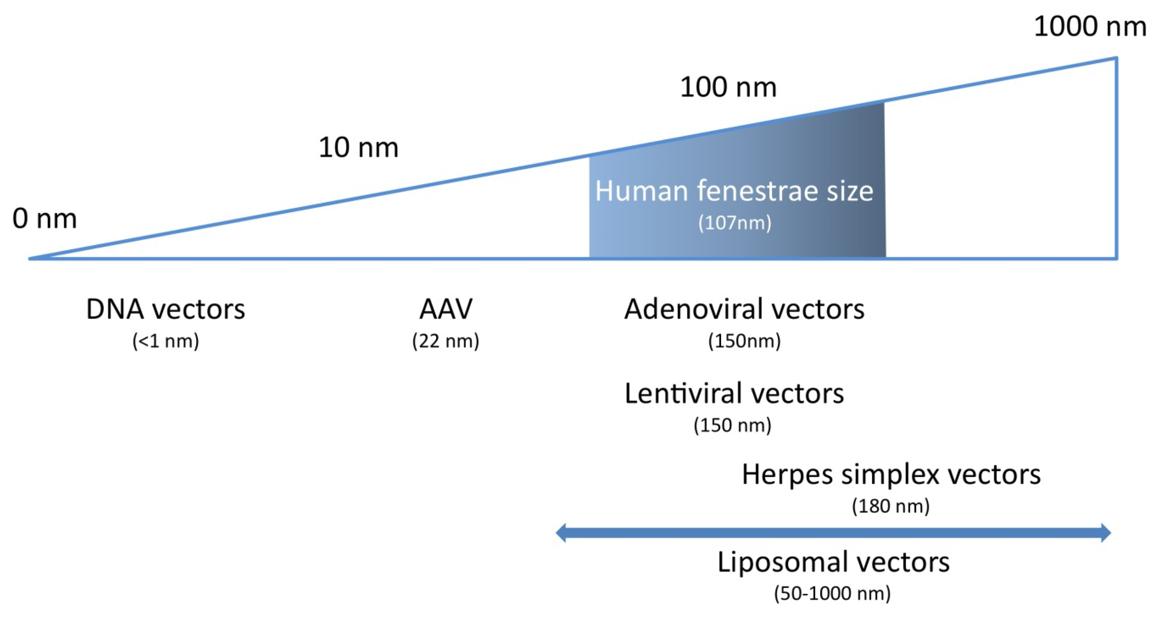
3. Parenchymal Liver cells as a Gene Transfer Target: the Role of Sinusoidal Fenestrae

Fenestrae are clustered in sieve plates and provide an open pathway between the sinusoidal lumen and the space of Disse, in which numerous microvilli from parenchymal liver cells protrude [4,27]. Whereas the Kupffer cells and liver sinusoidal endothelial cells constitute a barrier for access to the parenchymal liver cells, sinusoidal fenestrae form an escape route to the space of Disse and the microvillous surface of hepatocytes. Sinusoidal fenestrae have no diaphragm. Although fenestrae constitute an open communication between the sinusoidal lumen and the space of Disse, they will act as a sieve and will mechanically restrict the transendothelial transport of gene transfer vectors according to their size.
Fenestrae measure between 100 nm and 200 nm and significant species differences in their size exist [4,27,28,29,30,31]. Using state of the art transmission electron microscopy measurements, we have previously demonstrated that the average diameter of fenestrae is significantly larger in Dutch Belt rabbits (124 nm) [32], in Sprague Dawley rats (150 nm in the pericentral area and 175 nm in the periportal area) [28], and in C57BL/6 mice (141 nm) [33] than in humans with a healthy liver (107 nm) [34]. In contrast, the average diameter in New Zealand White rabbits (103 nm) [33] and Fauve de Bourgogne rabbits (105 nm) [32] was similar to humans. Importantly, interindividual variation of the average diameter of fenestrae within the same species or strain is low, as indicated by coefficients of variation between 3%–8%. However, within the same animal or individual, there is a Gaussian distribution of diameters around the average with skewing to the right [29].
Before summarizing experimental evidence that these diameters are a determinant of gene transfer efficiency into hepatocytes, a discussion of the diameter of gene transfer vectors is warranted. Using cryo-electron microscopy, adenoviral serotype 5 virions were shown to have a diameter of 93 nm with protruding fibers of 30 nm [33], whereas the diameter of a vesicular stomatitis virus-G pseudotyped human immunodeficiency virus-1 derived lentiviral vector was found to be 150 nm [33]. Adeno-associated viral (AAV) serotype 2 vectors have an average diameter of 22 nm [35] and this size does not vary significantly between different serotypes of AAV vectors [36,37,38,39]. Herpes simplex virions have been reported to be as large as 180 nm [40]. The diameter of liposomes used for non-viral gene transfer varies between 50 nm and 1000 nm and is highly dependent on production parameters [41,42]. All in all, from a theoretical point of view, it is clear that fenestrae in humans may act as a sieve for most vectors with the obvious exception of AAV vectors (Figure 1).
Figure 1. Relevance of gene therapy vector dimensions for gene transfer efficiency into hepatocytes. Human liver sinusoidal fenestrae show a Gaussian size distribution (shaded box) and have an average diameter of 107 nm. To ensure efficient transendothelial passage of gene transfer vectors, their size should be small enough to pass through the fenestrae into the space of Disse. The only gene therapy systems that display this highly desirable feature are naked DNA transfer and AAV-mediated transfer.
Pharmaceuticals 05 01372 g001
The experimental evidence for the role of the diameter of fenestrae in hepatocyte transduction by adenoviral vectors is in essence based on two seminal observations [32,33]. Firstly, the ratio of transgene DNA in parenchymal liver cells versus transgene DNA in non-parenchymal liver cells early after gene transfer correlates directly with the diameter of fenestrae [33]. Secondly, interventions that increase the diameter of fenestrae induce enhanced transgene DNA uptake in hepatocytes and augmented transgene expression [32,33].
The diameter of fenestrae in New Zealand White rabbits and Fauve de Bourgogne rabbits resembles the size in humans. Adenoviral gene transfer into hepatocytes is highly inefficient in Fauve de Bourgogne rabbits and New Zealand White rabbits [32,33]. Therefore, one would predict a similar low efficiency in humans. Evidence supporting this hypothesis comes from the ornithine transcarbamylase deficiency trial, which remains the only hepatocyte-directed clinical trial in humans using adenoviral vectors [43]. Levels of transgene expression in this trial were indeed much lower than what would have been predicted based on preclinical animal models [43].

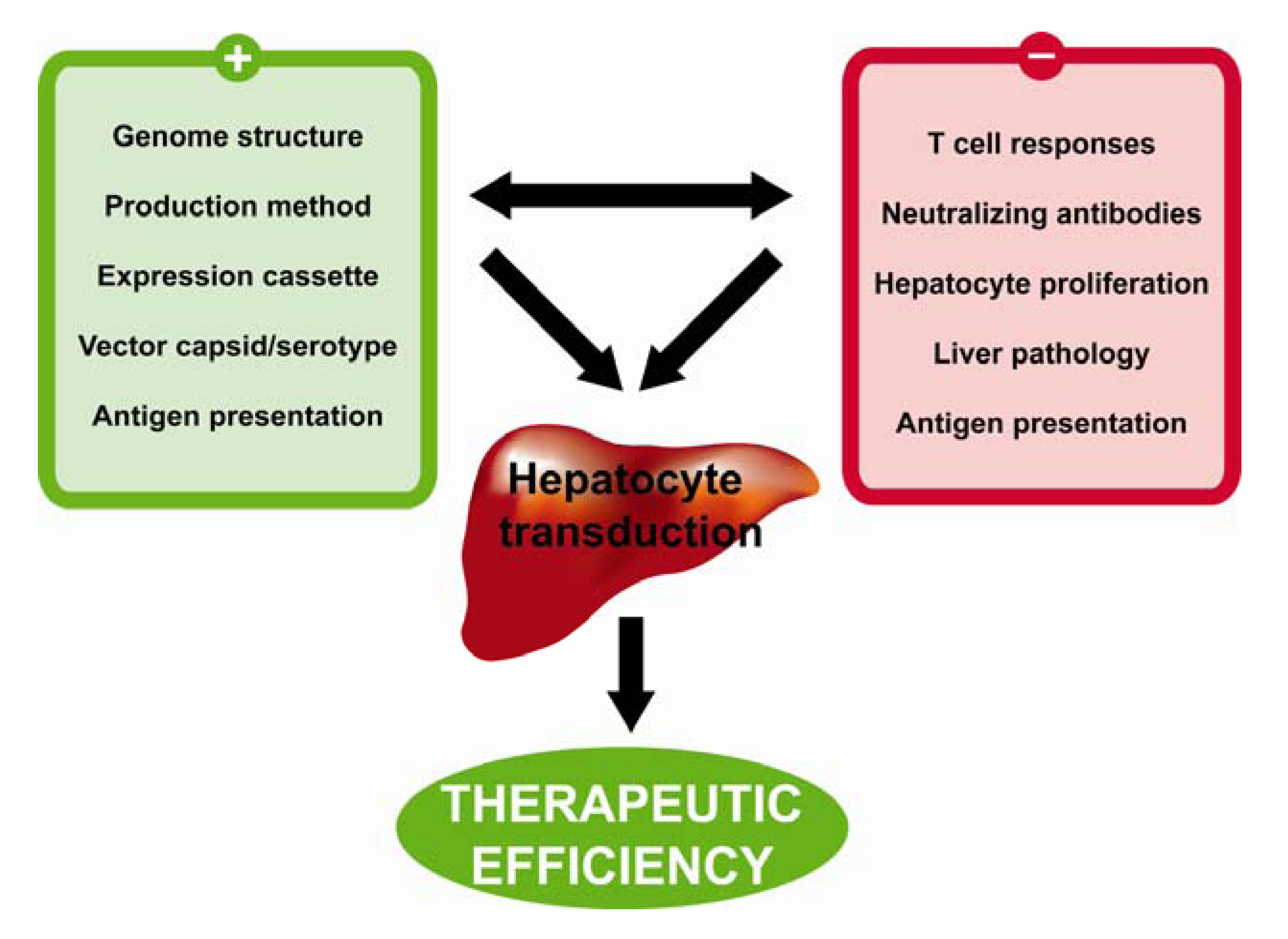
4. Characteristics of the Ideal Vector for Hepatocyte-Directed Gene Transfer

Table 1 summarizes the characteristics of the ideal vector for hepatocyte-directed gene transfer. Based on the discussion in the previous two sections, a conditio sine qua non for efficient hepatocyte-directed gene transfer is facilitated passage of vectors through fenestrae. Therefore, this criterion is listed on top in the Table 1. Considering that the diameter of fenestrae in humans with a healthy liver is 107 nm [34], only AAV vectors and some non-viral gene transfer vectors fulfil this criterion in humans. The second criterion implies efficient transduction or transfection of hepatocytes. A vector not only has to pass the fenestrae but the transgene DNA must be subsequently delivered from the extracellular space of Disse into the nucleus of hepatocytes with high efficiency. This requires efficient cellular uptake and efficient nuclear delivery without annihilating intracellular degradation of transgene DNA. Using these first two criteria as decisive parameters for selection of the ideal gene transfer vector system, AAV vector technology remains the only valid option at present for hepatocyte-directed gene transfer in humans. However, one non-viral gene transfer strategy has been demonstrated to result in transfection of a high percentage of hepatocytes in mice. This method is hydrodynamic gene transfer. Considering the theoretically high potential of this technology, a more detailed discussion of this technology is indicated.
Table 1. Characteristics of the ideal vector for hepatocyte-directed gene transfer.
PropertiesVector
Facilitated passage through fenestrae AAV, some non-viral gene transfer vectors
Efficient transduction or transfection AAV, adenoviral vectors
Target specificNo vector system, apply transcriptional targeting
Repeated use possibleMinicircles, non-viral gene transfer vectors, apply serotype switch
No or minor vector-induced innate immune responsesAAV, minicircles?
No or minor vector-induced adaptive immune responsesMinicircles, non-viral gene transfer vectors
No non-immune toxicityAAV, some non-viral gene transfer vectors
High insertion capacityAdenoviral vectors, minicircles, non-viral gene transfer systems
In 1999, Liu et al. [44] and Zhang et al. [45] independently demonstrated that hydrodynamic gene transfer method results in high transgene expression in the liver that is in a similar order of magnitude as levels obtained after adenoviral or AAV gene transfer. This non-viral gene transfer method in mice entails the injection of a large bolus of a physiological solution, equivalent to 8 to 10% of the body weight, into the tail vein within a period of 5 to 7 seconds. The subsequent right heart volume overload results in a retrograde flow through the vena cava and in particular in a retrograde flow into the hepatic veins. Consequently, intrahepatic pressure increases and the DNA containing solution is forced out of the hepatic sinusoids into the parenchymal cells of the liver. Following systemic hydrodynamic gene transfer in mice and rats, more than 90% of the injected DNA can be retrieved in the liver [46]. In addition, microscopic analysis has indicated that transduced hepatocytes are predominantly located in the pericentral region [47]. This predilection may be explained by the fact that sinusoids are wider and straighter and contain more fenestrae per unit of surface in the pericentral area than in the periportal area [46,48].
Hydrodynamic gene transfer of minicircles may result in persistent transgene expression in mice [49,50,51]. Minicircles are devoid of bacterial sequences that cause transcriptional silencing [49,50]. In addition, repeated administration of minicircle DNA is possible. Readministration may be required since episomal DNA instability in the long-term cannot be excluded. Notwithstanding the attractive features of hydrodynamic gene transfer, application of this technology in larger species is cumbersome. The degree of systemic volume overload as applied in rodents obviously cannot be extrapolated to larger species. Therefore, great efforts have been made to develop innovative hydrodynamic gene transfer techniques that require lower volumes. Zhang et al. [52] demonstrated that, using localized hydrodynamic gene transfer via the hepatic vein in rats, the required volume for injection could be reduced to less than 2% of the body weight. Eastman et al. [53] showed that in rabbits a volume of 15 ml/kg (i.e. 1.5% of the body weight) suffices to successfully transduce hepatocytes when using catheter-based isolation of the liver. However, levels of transgene expression were 10- to 20-fold lower compared to mice [53]. Yoshino et al. [54], Alino et al. [55], and Fabre et al. [56] reported hydrodynamic gene transfer into pig liver or isolated liver segments using catheterization techniques. These procedures were well tolerated and resulted in intrahepatic vascular pressures up to 4-fold higher than observed in mice and rats. Nonetheless, levels of transgene expression were 200-fold [54,56] to 10 000-fold [55] lower following hydrodynamic gene transfer in pigs when compared to mice. Suda et al. [57] developed a computer-assisted hydrodynamic gene transfer device (a so-called ‘hydrojector’), wherein all injection parameters (volume, speed, …) are coordinated via an intrahepatic pressure sensor. This technology was applied in mice, rats, and pigs, but again, large species differences in efficiency were observed. The consistent failure of hydrodynamic gene transfer in larger species may be due to anatomical differences of the liver, in particular differences in hepatic compliance and differences in the structure of the hepatic sinusoids.
The third criterion, absolute target specificity, is not met by any vector system. However, transgene expression can be restricted to hepatocytes by use of hepatocyte-specific expression cassettes [58,59,60,61,62,63]. This transcriptional targeting has been shown to be a successful strategy to avoid the development of a humoral immune response against a potentially immunogenic transgene product in several studies [58,59,60,61,62,64,65]. From an immunological point of view, the liver is a particular gene transfer target. It has been described as a tolerogenic organ more than 40 years ago [66]. Naive T cells encounter liver antigens initially in the liver, rather than in lymphoid tissue [67]. Whereas lymph nodes and the spleen are anatomical compartments that provide a particular microarchitecture and microenvironment for the induction of immunity, antigen presentation in the liver takes places in a completely different setting. The liver microenvironment determines the tolerogenic properties of liver dendritic cells [68,69]. This is a key aspect of the hepatic adaptive immune tolerance induction [70].
Repeated administration (criterion 4) is possible in case of non-viral gene transfer vectors and in case of hydrodynamic gene transfer of naked DNA. Readministration is not possible for viral vectors but this problem may be circumvented by injection of a vector of a different serotype [71,72]. The requirement for readministration after AAV-mediated gene transfer will depend on the long-term episomal transgene DNA stability. In general, this stability is remarkably good for AAV episomes.
In contrast to potentially very serious innate immune responses after adenoviral gene transfer [43,73,74,75], AAV gene transfer induces very weak or absent innate immune responses to viral capsids both in mice [76] and monkeys [77]. Serum levels of five major inflammatory cytokines (tumor necrosis factor-α, interferon-γ, interleukin-6, interleukin-10, and interleukin-12) were not elevated in macaques after gene transfer with vectors based on AAV serotypes 2, 7, and 8 [77]. Although non-viral gene transfer systems could theoretically be devoid of innate immune responses, serious innate immunity may occur [78,79,80], which is not only related to the presence of unmethylated CpG motifs. Further criteria of the ideal vector will be discussed within the framework of the next sections on AAV-mediated hepatocyte-directed gene transfer.

5. AAV-Mediated Hepatocyte-Directed Gene Transfer: Introduction

AAV are small, nonenveloped, single-stranded DNA viruses and belong to the Dependovirus genus of the Parvoviridae family. Up to date, 11 different serotypes comprising over 120 capsid variants have been isolated from humans, nonhuman primates, cows, dogs, horses, and birds [81,82,83,84,85]. Compared to the genome size and structure of other viral vectors, the AAV genome is relatively small and simple, at approximately 4.7 kb, and contains only two genes, rep and cap, that encode four Rep proteins and three capsid proteins, flanked by two 145-base inverted terminal repeats (ITRs). Recombinant and replication defective AAV vectors used for gene transfer applications retain only the ITRs, which are necessary for replication, packaging, and integration, while all viral coding sequences are entirely removed [86]. In light of the AAV genome size, the insertion capacity of AAV vectors is clearly limited.
Several favorable features have made AAV a valid candidate for hepatocyte-directed gene transfer. Most importantly, AAV have not been associated with human disease. Moreover, AAV are naturally replication defective and require co-infection with a helper virus, such as adenovirus or herpes virus, in order to replicate. Furthermore, AAV transduce both dividing and non-dividing cells such as hepatocytes and have been shown to maintain long-term expression in hepatocytes in the absence of chromosomal integration [80]. Whereas wild-type AAV possess the ability for site-specific integration into the human genome, this feature is absent from recombinant AAV vectors and integration into the host genome is random but relatively infrequent [87]. As already discussed, the small diameter of AAV vectors [35] is important for facilitated passage through fenestrae.

6. AAV2-Mediated Hepatocyte-Directed Gene Transfer

Systemic liver-directed AAV serotype 2 mediated gene transfer resulted in persistent therapeutic factor IX (FIX) expression in immunocompetent mice, dogs, and nonhuman primates [88,89,90]. Complete correction of the bleeding diathesis in inhibitor-prone hemophilia dogs with a complete deficiency of FIX has been achieved after AAV2 gene transfer for more than 9 years [89]. Of note, onset of transgene expression following AAV2 gene transfer to the liver is slow, with a lag phase of 4 to 6 weeks, followed by a gradual increase over a period of 4-10 weeks before reaching a stabile plateau. Long-term expression in the liver has been shown to be attributable to episomally maintained vector genomes that mostly exist in the nucleus in the form of high-molecular concatamers [91,92].
The first clinical trial for hepatocyte-directed AAV2 gene transfer [93] was an open-label, dose-escalation study of a human FIX expressing vector delivered through the hepatic artery in patients with severe hemophilia B. One subject in the highest dose cohort receiving 2 x 10¹² vector genomes/kg showed a therapeutic increase in FIX activity of up to 10%–12% of normal levels. In contrast to long-lasting expression in hemophilic dogs and non-human primates using similar vectors, expression at therapeutic levels in this subject was short lived. Starting at 5 weeks after vector administration, FIX gradually declined to baseline at 10 weeks post vector infusion. This decline coincided with a rise in transaminase levels between 4 and 8 weeks after vector administration. Antibodies directed against the human FIX were undetectable and no toxic or infectious causes for the increase in liver enzymes were found. Another subject that was dosed at a 5-fold lower dose also displayed a self-limited rise in liver enzymes with similar kinetics. A series of follow-up experiments suggested that T cell–mediated immunity to AAV capsid antigens may have induced destruction of AAV2–transduced hepatocytes [93,94]. However, this remains a matter of debate.
Overall, despite initial success in pre-clinical studies, several limitations of AAV2 vectors have emerged including limited transduction efficiency of hepatocytes, delayed onset of transgene expression [95], high prevalence of neutralizing antibodies in humans, and potentially destructive T-cell responses to capsids. These observations have underscored the need for further refinement of AAV vectors.

7. Hepatocyte-Directed Gene Transfer Using Alternative AAV Serotypes

In the past decade, the Wilson lab has spearheaded the identification and characterization of novel AAV serotypes [81,82]. Recent studies have demonstrated the superiority of these novel serotypes for gene transfer applications based on enhanced hepatocyte transduction, reduced prevalence of neutralizing antibodies, and diminished capsid immune responses [96,97,98]. The most salient increase in liver transduction and transgene expression has been observed for AAV8 and AAV9 [99,100]. In general, transgene expression levels following intravenous injection mice are 10 to 100-fold higher compared to AAV2 [82]. However, the observed advantages of AAV8 over AAV2 seem to be reduced in larger animal species such as dogs and nonhuman primates [77,101,102].
Mechanisms underlying the distinct biological performance of different AAV serotypes have been attributed to differences in viral entry, trafficking, uncoating, and genome processing [103,104]. Cunningham et al. showed that following systemic administration of AAV8 to the liver, all dsAAV8 genomes are formed within 3 days [105]. In contrast, AAV2 shows a gradual increase of dsAAV2 genomes over a period of several weeks [91]. Nakai et al. demonstrated that up to 90% hepatocyte transduction could be achieved after systemic AAV8-mediated gene transfer [95]. AAV8-mediated gene transfer has also been proven to be more potent in inducing immune tolerance for the transgene product [106].
Nathwani et al. [107] performed a landmark study that demonstrated for the first time long-term expression of a transgene product following systemic gene transfer to the liver at therapeutic levels in humans. In this study [107], hemophilia B was successfully treated with an AAV8 human FIX expressing vector. The vector genome was designed as a self-complementary vector in order to bypass the synthesis of a second DNA strain following hepatocyte transduction [108]. The authors also applied codon optimalisation of the transgene cDNA sequence in order to improve the efficiency of translation [109]. A dose-escalation study in six patients with severe hemophilia B was performed and patients have been followed 6-16 months [107]. Interestingly, all patients showed a therapeutic response, which was roughly dose-dependent. Human FIX levels ranged from 2%–11% of physiological levels in normal subjects. Four out of six patients discontinued prophylactic FIX treatment and remained free of spontaneous hemorrhages. The remaining two patients that remained on FIX protein therapy were able to extend the time interval between prophylactic injections. Intriguingly, one of the two patients that had received the highest vector dose displayed an asymptomatic increase in serum transaminases, which coincided with the presence of AAV8-capsid specific T cells in the peripheral blood. The other patient in the same cohort also displayed a slight increase in liver enzyme levels, although the cause hereof was less clear. Both patients received a short course of glucocorticoid therapy. Following administration of glucocorticoids, serum transaminase levels quickly returned to normal and FIX expression levels remained in the range of 3%–11% of normal. The gene transfer vector production costs for this study have been estimated at $30,000 for a patient treated with the middle dose of vector. In light of the annual costs of protein infusion therapy, it is clear that dramatic cost reductions may be achieved by widespread use of this therapy. If further studies indicate that this approach is safe, it may eventually replace the cumbersome and expensive prophylactic FIX therapy. Moreover, the use of similar vectors may spark significant progress in gene therapy for other disorders, including familial hypercholesterolemia [110], lysosomal storage diseases, and urea cycle defects [111].

8. Challenges for AAV-Mediated Hepatocyte-Directed Gene Transfer

Key determinants of efficient AAV-mediated hepatocyte-directed gene transfer and of persistent transgene expression in parenchymal liver cells are summarized in Figure 2. As already mentioned, if major histocompatibility complex 1 presentation of AAV epitopes on hepatocytes occurs in vivo, this might be a cause of destruction of hepatocytes by cytotoxic T cells and of decline of transgene expression. Interindividual differences in antigen processing and presentation may influence the type and magnitude of the immunological response against the AAV particles [112]. Furthermore, pre-exposure to natural AAV infections may vary highly between patients, resulting in a completely different repertoire of memory T cells.
Seroneutralisation of AAV vectors in patients with prior exposure to AAV viruses is a serious obstacle for a successful outcome of the treatment. Prior exposure in mice, dogs, nonhuman primates, and humans results in the formation of complex profiles of serum antibodies capable of binding and neutralizing specific AAV serotypes [96,113]. Although human data on the effects of neutralizing antibodies on hepatocyte transduction are scarce, the absence of efficient transduction in at least one patient of the FIX clinical trial has been attributed to the higher level of neutralizing antibodies [93]. A large proportion of the human population has been exposed to AAV, with at least one-third having neutralizing antibodies against AAV2 [114]. In a recent study by the Wilson group, the authors analyzed serum samples from 888 persons, spanning four continents and 10 different countries, for the presence of neutralizing antibodies directed against a wide array of AAV serotypes [96]. In most regions, the prevalence of neutralizing antibody titers exceeding 1:10 was lowest for AAV7 (15%–30%) and AAV8 (15%–30%). In contrast, the prevalence of neutralizing antibodies against AAV1 (20%–45%) and AAV2 (30%–60%) was significantly higher. These numbers are in accordance with a recent study by Boutin et al., showing the prevalence of AAV-binding Ab against AAV1 (51%) and AAV2 (59%) that were significantly higher than those for AAV5 (3.2%), AAV6 (37%), AAV8 (19%), and AAV9 (36%) [115]. The observed differences in seroprevalence against distinct serotypes most likely reflect their natural host tropism. Humans are not commonly pre-exposed to serotypes derived from nonhuman primates, resulting in lower titers of neutralizing antibodies. Finally, a significant degree of coprevalence of neutralizing antibodies against different serotypes has been observed.
Figure 2. Key determinants of efficient AAV-mediated hepatocyte-directed gene transfer and of persistent transgene expression in parenchymal liver cells. Efficient gene transfer and the maintenance of persistent transgene expression in hepatocytes are influenced by many factors in both positive and negative ways. Pre-existing neutralizing antibody titers in excess of 1:10 will greatly affect hepatocyte transduction by AAV vectors. In addition, depending on the context, transduction of antigen presenting cells may contribute to the generation of cytotoxic T cells, resulting in the loss of transgene expression over time. Pathological sequelae of the liver, such as inflammation or pseudocappilarization, may also interfere with hepatocyte transduction or contribute to the priming of reactive T cells. Antigen presentation in the microenvironment of the liver plays a critical role in the generation of a tolerogenic state and may induce tolerance for the transgene product. On the other hand, hepatocyte destruction by cytotoxic T cells might occur as a consequence of MHCI presentation of capsid epitopes. In pediatric patients, fast liver growth will dilute vector genomes, resulting in a quick decline of transgene expression. Finally, different facets of vector design will greatly influence the outcome of hepatocyte-directed gene transfer strategies. Several capsid variants have shown increased hepatotropsim when compared to AAV2. Moreover, the purity and quality of AAV vector preps should be carefully monitored to ensure reproducibility and efficiency. With regard to vector expression cassette design, several studies have underscored the importance of promoter selection, optimization of coding sequences (codon optimization, Kozak sequence), intron and polyadenylation signal selection, and vector genome structure (i.e. scAAV vectors versus ssAAV vectors).
Pharmaceuticals 05 01372 g002
Pre-exposure to AAV has been mimicked by readministration studies in animal models. However, neutralizing antibodies generated in this context are significantly higher than could be expected following natural exposure to AAV in humans. Additionally, since neutralizing antibodies are generated in the absence of co-infection of the helper virus, they may vary in their affinity and/or breadth towards AAV. Scallan et al. used a model of passive transfer of pooled human IgG in mice injected with AAV2 to overcome these limitations and showed that neutralizing antibodies titers as low as 1:10 could efficiently block hepatocyte transduction in mice [116]. Importantly, sero-neutralization in this model could be circumvented by administration of vectors of serotypes AAV6 and AAV8. In this respect, AAV8 gene transfer also resulted in sustained transgene expression in dogs that had previously been treated with an AAV2 vector [102]. In light of these results, routine screening of neutralizing antibody titers in larger animals such as nonhuman primates has become general practice in preclinical studies over the past years. Moreover, the absence of detectable levels of neutralizing antibodies towards a given AAV serotype is commonly included in the criteria of eligibility for human patients participating in phase I clinical trials.
A particular concern for hepatocyte-directed gene transfer in pediatric patients is the impact of liver growth on transgene DNA persistence. Most AAV genomes reside within the nucleus in an episomal form. Consequently, transgene DNA is lost during mitosis when the nuclear envelope is degraded. In the adult mouse liver, individual hepatocytes are slowly-dividing, completing a single population turnover every 180–400 days [117]. A rapid decline of transgene expression has been observed in the context of extensive hepatocyte proliferation following partial hepatectomy in adult mice [92] or following AAV administration into neonatal mice [105,118]. Interestingly, when vector is delivered at serially higher ages up to adulthood, transgene expression is progressively more durable, correlating with the deceleration of liver growth [105]. Taken together, these studies indicate that proliferation of hepatocytes results in a nearly complete clearance of AAV genomes from the liver within two population doublings [92,105,118,119]. In humans, it is estimated that a first doubling of the liver mass occurs within 9 months following birth followed by a second doubling in mass taking up to 3 to 4 years [120]. Thus, vector readministration is likely to be necessary. This may be achieved by using distinct AAV serotypes to circumvent pre-existing immunity [102,121,122].
Another challenge is to scale-up AAV production and purification. Several innovative production systems have recently been developed that are compatible with large-scale production for clinical application [123,124,125]. These new systems use adenovirus, herpesvirus, and baculovirus hybrids to deliver the rAAV genome and trans-acting helper functions to the producer cells. However, whereas these systems are particularly suited for production of clinical grade candidate vectors, the need to make the hybrid viruses for each vector makes them less suited to early development and preclinical studies in which several combinations of transgene and vector serotype may need to be evaluated.
Interestingly, Vandenberghe et al. recently reported a novel AAV isolation method that is based on the significant release of AAV from the cells during vector production [126,127]. The option of harvesting from supernatants rather than cell lysates should greatly simplify downstream processing of AAV vectors for research and clinical applications. In addition, using this system most AAV serotypes can be produced in the absence of serum, thus limiting concerns about possible contamination of AAV vector preparations.
Irrespective of the modus of production, several studies have identified vector purity as a key determinant of vector potency in vivo [127,128]. Increasing vector purity and potency is predicted to reduce the risk of deleterious immune responses after administration of recombinant AAV to human subjects. Standard purification of AAV vectors depends on multiple rounds of CsCl ultracentrifugation. However, these gradients are limited in terms of their loading capacity and prolonged exposure to CsCl has been reported to compromise AAV vector potency. Large-scale purification of AAV has been based on ion-exchange chromatography, hydrophobic interaction, and affinity purification methods, resulting in increased purity and yields. However, these methods are generally serotype dependent and lack the ability to discern between fully functional AAV particles and empty capsids. In contrast, density centrifugation using iodixanol gradients effectively separates infectious particles from empty capsids [127].

9. Conclusions and Future Perspectives

Initial gene transfer studies heralded great promise for this novel class of therapeutic agents. However, initial progress was hyped and too little effort was oriented towards understanding basic aspects of vector biology and host immunology, resulting in a general failure of the field to deliver clinically applicable gene transfer strategies. Nevertheless, under the impulse of unremitting efforts by many researchers, the past decade has seen great progress in the detailed characterization of many facets of gene transfer vectors. To date, AAV represent the most promising gene transfer vehicle and studies have shown great efficiency in various animal models. With the advent of large scale AAV production, these advances have recently finally been extrapolated to humans, resulting in the successful completion of numerous phase I clinical trials for mendelian disorders including Leber's congenital amaurosis and hemophilia B. These studies constitute an important landmark underscoring the vast improvements of gene transfer technologies over the past decade. In light of these achievements, the recent positive assessment by the European Medicines Agency's Committee for Medicinal Products for Human Use (CHMP) of the marketing authorization for the first gene transfer product in Europe (alipogene tiparvovec; Glybera®) highlights an important paradigm shift by regulatory agencies, as well as the biotechnological entrepreneurs and investors. However, despite this significant progress, clinical experience with gene transfer technologies remains very limited and many unanswered questions warrant further study. Therefore, the field should continue to progress as it has over the past decade, cautiously and diligently.

Acknowledgments

Experimental work of the authors was supported by grant G.0599.09N the Fonds voor Wetenschappelijk Onderzoek-Vlaanderen and by Onderzoekstoelagen OT/11/083 of the K.U. Leuven. Stephanie C. Gordts is a Research Assistant of the Agentschap voor Innovatie door Wetenschap en Technologie (IWT).

Conflict of Interest

The authors declare no conflict of interest.

References

  1. Nguyen, T.H.; Ferry, N. Liver gene therapy: Advances and hurdles. Gene Ther. 2004, 11 (Suppl. 1), S76–S84. [Google Scholar] [CrossRef]
  2. Hernandez-Alcoceba, R.; Sangro, B.; Prieto, J. Gene therapy of liver cancer. Ann. Hepatol. 2007, 6, 5–14. [Google Scholar]
  3. Grimm, D.; Kay, M.A. Therapeutic short hairpin RNA expression in the liver: viral targets and vectors. Gene Ther. 2006, 13, 563–575. [Google Scholar] [CrossRef]
  4. Wisse, E. An electron microscopic study of the fenestrated endothelial lining of rat liver sinusoids. J. Ultrastruct. Res. 1970, 31, 125–150. [Google Scholar] [CrossRef]
  5. Do, H.; Healey, J.F.; Waller, E.K.; Lollar, P. Expression of factor VIII by murine liver sinusoidal endothelial cells. J. Biol. Chem. 1999, 274, 19587–19592. [Google Scholar]
  6. Sasse, D.; Spornitz, U.M.; Maly, I.P. Liver architecture. Enzyme 1992, 46, 8–32. [Google Scholar]
  7. Blouin, A.; Bolender, R.P.; Weibel, E.R. Distribution of organelles and membranes between hepatocytes and nonhepatocytes in the rat liver parenchyma. A stereological study. J. Cell Biol. 1977, 72, 441–455. [Google Scholar] [CrossRef]
  8. Knook, D.L.; Sleyster, E.C. Isolated parenchymal, Kupffer and endothelial rat liver cells characterized by their lysosomal enzyme content. Biochem. Biophys. Res. Commun. 1980, 96, 250–257. [Google Scholar] [CrossRef]
  9. Arii, S.; Imamura, M. Physiological role of sinusoidal endothelial cells and Kupffer cells and their implication in the pathogenesis of liver injury. J. Hepatobiliary Pancreat Surg. 2000, 7, 40–48. [Google Scholar]
  10. Shiratori, Y.; Tananka, M.; Kawase, T.; Shiina, S.; Komatsu, Y.; Omata, M. Quantification of sinusoidal cell function in vivo. Semin. Liver Dis. 1993, 13, 39–49. [Google Scholar] [CrossRef]
  11. Alemany, R.; Suzuki, K.; Curiel, D.T. Blood clearance rates of adenovirus type 5 in mice. J. Gen. Virol. 2000, 81, 2605–2609. [Google Scholar]
  12. Tao, N.; Gao, G.P.; Parr, M.; Johnston, J.; Baradet, T.; Wilson, J.M.; Barsoum, J.; Fawell, S.E. Sequestration of adenoviral vector by Kupffer cells leads to a nonlinear dose response of transduction in liver. Mol. Ther. 2001, 3, 28–35. [Google Scholar]
  13. Wolff, G.; Worgall, S.; van Rooijen, N.; Song, W.R.; Harvey, B.G.; Crystal, R.G. Enhancement ofin vivo adenovirus-mediated gene transfer and expression by prior depletion of tissue macrophages in the target organ. J. Virol. 1997, 71, 624–629. [Google Scholar]
  14. Snoeys, J.; Mertens, G.; Lievens, J.; van Berkel, T.; Collen, D.; Biessen, E.A.; De Geest, B. Lipid emulsions potently increase transgene expression in hepatocytes after adenoviral transfer. Mol. Ther. 2006, 13, 98–107. [Google Scholar]
  15. Waddington, S.N.; Parker, A.L.; Havenga, M.; Nicklin, S.A.; Buckley, S.M.; McVey, J.H.; Baker, A.H. Targeting of adenovirus serotype 5 (Ad5) and 5/47 pseudotyped vectors in vivo: fundamental involvement of coagulation factors and redundancy of CAR binding by Ad5. J. Virol. 2007, 81, 9568–9571. [Google Scholar] [CrossRef]
  16. Stone, D.; Liu, Y.; Shayakhmetov, D.; Li, Z.Y.; Ni, S.; Lieber, A. Adenovirus-platelet interaction in blood causes virus sequestration to the reticuloendothelial system of the liver. J. Virol. 2007, 81, 4866–4871. [Google Scholar]
  17. Xu, Z.; Tian, J.; Smith, J.S.; Byrnes, A.P. Clearance of adenovirus by Kupffer cells is mediated by scavenger receptors, natural antibodies, and complement. J. Virol. 2008, 82, 11705–11713. [Google Scholar] [CrossRef]
  18. Di Paolo, N.C.; van Rooijen, N.; Shayakhmetov, D.M. Redundant and synergistic mechanisms control the sequestration of blood-born adenovirus in the liver. Mol. Ther. 2009, 17, 675–684. [Google Scholar] [CrossRef]
  19. Stone, D.; Ni, S.; Li, Z.Y.; Gaggar, A.; DiPaolo, N.; Feng, Q.; Sandig, V.; Lieber, A. Development and assessment of human adenovirus type 11 as a gene transfer vector. J. Virol. 2005, 79, 5090–5104. [Google Scholar]
  20. Sakurai, F.; Mizuguchi, H.; Yamaguchi, T.; Hayakawa, T. Characterization of in vitro and in vivo gene transfer properties of adenovirus serotype 35 vector. Mol. Ther. 2003, 8, 813–821. [Google Scholar] [CrossRef]
  21. Stone, D.; Liu, Y.; Li, Z.Y.; Tuve, S.; Strauss, R.; Lieber, A. Comparison of adenoviruses from species B, C, E, and F after intravenous delivery. Mol. Ther. 2007, 15, 2146–2153. [Google Scholar] [CrossRef]
  22. Kuzmin, A.I.; Finegold, M.J.; Eisensmith, R.C. Macrophage depletion increases the safety, efficacy and persistence of adenovirus-mediated gene transfer in vivo. Gene Ther. 1997, 4, 309–316. [Google Scholar]
  23. Schiedner, G.; Hertel, S.; Johnston, M.; Dries, V.; van Rooijen, N.; Kochanek, S. Selective depletion or blockade of Kupffer cells leads to enhanced and prolonged hepatic transgene expression using high-capacity adenoviral vectors. Mol. Ther. 2003, 7, 35–43. [Google Scholar] [CrossRef]
  24. Niwano, M.; Arii, S.; Monden, K.; Ishiguro, S.; Nakamura, T.; Mizumoto, M.; Takeda, Y.; Fujioka, M.; Imamura, M. Amelioration of sinusoidal endothelial cell damage by Kupffer cell blockade during cold preservation of rat liver. J. Surg. Res. 1997, 72, 36–48. [Google Scholar] [CrossRef]
  25. Deaciuc, I.V.; Bagby, G.J.; Niesman, M.R.; Skrepnik, N.; Spitzer, J.J. Modulation of hepatic sinusoidal endothelial cell function by Kupffer cells: an example of intercellular communication in the liver. Hepatology 1994, 19, 464–470. [Google Scholar]
  26. Haisma, H.J.; Kamps, J.A.; Kamps, G.K.; Plantinga, J.A.; Rots, M.G.; Bellu, A.R. Polyinosinic acid enhances delivery of adenovirus vectors in vivo by preventing sequestration in liver macrophages. J. Gen. Virol. 2008, 89, 1097–1105. [Google Scholar] [CrossRef]
  27. Braet, F.; Wisse, E. Structural and functional aspects of liver sinusoidal endothelial cell fenestrae: a review. Comp. Hepatol 2002, 1, 1. [Google Scholar] [CrossRef] [Green Version]
  28. Wisse, E.; De Zanger, R.B.; Charels, K.; Van Der Smissen, P.; McCuskey, R.S. The liver sieve: considerations concerning the structure and function of endothelial fenestrae, the sinusoidal wall and the space of Disse. Hepatology 1985, 5, 683–692. [Google Scholar] [CrossRef]
  29. Jacobs, F.; Wisse, E.; De Geest, B. The role of liver sinusoidal cells in hepatocyte-directed gene transfer. Am. J. Pathol. 2010, 176, 14–21. [Google Scholar]
  30. Jacobs, F.; Feng, Y.; Van Craeyveld, E.; Lievens, J.; Snoeys, J.; De Geest, B. Species differences in hepatocyte-directed gene transfer: implications for clinical translation. Curr. Gene Ther. 2009, 9, 83–90. [Google Scholar] [CrossRef]
  31. Wisse, E.; Braet, F.; Duimel, H.; Vreuls, C.; Koek, G.; Olde Damink, S.W.; van den Broek, M.A.; De Geest, B.; Dejong, C.H.; Tateno, C.; et al. Fixation methods for electron microscopy of human and other liver. World J. Gastroenterol. 2010, 16, 2851–2866. [Google Scholar] [CrossRef]
  32. Lievens, J.; Snoeys, J.; Vekemans, K.; Van Linthout, S.; de Zanger, R.; Collen, D.; Wisse, E.; De Geest, B. The size of sinusoidal fenestrae is a critical determinant of hepatocyte transduction after adenoviral gene transfer. Gene Ther. 2004, 11, 1523–1531. [Google Scholar]
  33. Snoeys, J.; Lievens, J.; Wisse, E.; Jacobs, F.; Duimel, H.; Collen, D.; Frederik, P.; De Geest, B. Species differences in transgene DNA uptake in hepatocytes after adenoviral transfer correlate with the size of endothelial fenestrae. Gene Ther. 2007, 14, 604–612. [Google Scholar] [CrossRef]
  34. Wisse, E.; Jacobs, F.; Topal, B.; Frederik, P.; De Geest, B. The size of endothelial fenestrae in human liver sinusoids: implications for hepatocyte-directed gene transfer. Gene Ther. 2008, 15, 1193–1199. [Google Scholar]
  35. Chen, H. Comparative observation of the recombinant adeno-associated virus 2 using transmission electron microscopy and atomic force microscopy. Microsc. Microanal. 2007, 13, 384–389. [Google Scholar]
  36. Xie, Q.; Bu, W.; Bhatia, S.; Hare, J.; Somasundaram, T.; Azzi, A.; Chapman, M.S. The atomic structure of adeno-associated virus (AAV-2), a vector for human gene therapy. Proc. Nat. Acad. Sci. USA 2002, 99, 10405–10410. [Google Scholar]
  37. Govindasamy, L.; Padron, E.; McKenna, R.; Muzyczka, N.; Kaludov, N.; Chiorini, J.A.; Agbandje-McKenna, M. Structurally mapping the diverse phenotype of adeno-associated virus serotype 4. J. Virol. 2006, 80, 11556–11570. [Google Scholar]
  38. DiMattia, M.A.; Nam, H.J.; Van Vliet, K.; Mitchell, M.; Bennett, A.; Gurda, B.L.; McKenna, R.; Olson, N.H.; Sinkovits, R.S.; Potter, M.; et al. Structural insight into the unique properties of adeno-associated virus serotype 9. J. Virol. 2012, 86, 6947–6958. [Google Scholar]
  39. Nam, H.J.; Lane, M.D.; Padron, E.; Gurda, B.; McKenna, R.; Kohlbrenner, E.; Aslanidi, G.; Byrne, B.; Muzyczka, N.; Zolotukhin, S.; et al. Structure of adeno-associated virus serotype 8, a gene therapy vector. J. Virol. 2007, 81, 12260–12271. [Google Scholar]
  40. Szilagyi, J.F.; Berriman, J. Herpes simplex virus L particles contain spherical membrane-enclosed inclusion vesicles. J. Gen. Virol. 1994, 75, 1749–1753. [Google Scholar] [CrossRef]
  41. Li, S.; Rizzo, M.A.; Bhattacharya, S.; Huang, L. Characterization of cationic lipid-protamine-DNA (LPD) complexes for intravenous gene delivery. Gene Ther. 1998, 5, 930–937. [Google Scholar]
  42. Banerjee, R. Liposomes: applications in medicine. J. Biomater. Appl. 2001, 16, 3–21. [Google Scholar] [CrossRef]
  43. Raper, S.E.; Yudkoff, M.; Chirmule, N.; Gao, G.P.; Nunes, F.; Haskal, Z.J.; Furth, E.E.; Propert, K.J.; Robinson, M.B.; Magosin, S.; et al. A pilot study of in vivo liver-directed gene transfer with an adenoviral vector in partial ornithine transcarbamylase deficiency. Hum. Gene Ther. 2002, 13, 163–175. [Google Scholar] [CrossRef]
  44. Liu, F.; Song, Y.; Liu, D. Hydrodynamics-based transfection in animals by systemic administration of plasmid DNA. Gene Ther. 1999, 6, 1258–1266. [Google Scholar]
  45. Zhang, G.; Budker, V.; Wolff, J.A. High levels of foreign gene expression in hepatocytes after tail vein injections of naked plasmid DNA. Hum. Gene Ther. 1999, 10, 1735–1737. [Google Scholar] [CrossRef]
  46. Herweijer, H.; Wolff, J.A. Gene therapy progress and prospects: hydrodynamic gene delivery. Gene Ther. 2007, 14, 99–107. [Google Scholar]
  47. Suda, T.; Gao, X.; Stolz, D.B.; Liu, D. Structural impact of hydrodynamic injection on mouse liver. Gene Ther. 2007, 14, 129–137. [Google Scholar]
  48. Wisse, E.; De Zanger, R.B.; Jacobs, R.; McCuskey, R.S. Scanning electron microscope observations on the structure of portal veins, sinusoids and central veins in rat liver. Scan Electron. Microsc. 1983, 1441–1452. [Google Scholar]
  49. Chen, Z.Y.; He, C.Y.; Ehrhardt, A.; Kay, M.A. Minicircle DNA vectors devoid of bacterial DNA result in persistent and high-level transgene expression in vivo. Mol. Ther. 2003, 8, 495–500. [Google Scholar] [CrossRef]
  50. Chen, Z.Y.; He, C.Y.; Meuse, L.; Kay, M.A. Silencing of episomal transgene expression by plasmid bacterial DNA elements in vivo. Gene Ther. 2004, 11, 856–864. [Google Scholar] [CrossRef]
  51. Jacobs, F.; Snoeys, J.; Feng, Y.; Van Craeyveld, E.; Lievens, J.; Armentano, D.; Cheng, S.H.; De Geest, B. Direct comparison of hepatocyte-specific expression cassettes following adenoviral and nonviral hydrodynamic gene transfer. Gene Ther. 2008, 15, 594–603. [Google Scholar]
  52. Zhang, X.; Dong, X.; Sawyer, G.J.; Collins, L.; Fabre, J.W. Regional hydrodynamic gene delivery to the rat liver with physiological volumes of DNA solution. J. Gene Med. 2004, 6, 693–703. [Google Scholar] [CrossRef]
  53. Eastman, S.J.; Baskin, K.M.; Hodges, B.L.; Chu, Q.; Gates, A.; Dreusicke, R.; Anderson, S.; Scheule, R.K. Development of catheter-based procedures for transducing the isolated rabbit liver with plasmid DNA. Hum. Gene Ther. 2002, 13, 2065–2077. [Google Scholar]
  54. Yoshino, H.; Hashizume, K.; Kobayashi, E. Naked plasmid DNA transfer to the porcine liver using rapid injection with large volume. Gene Ther. 2006, 13, 1696–1702. [Google Scholar]
  55. Alino, S.F.; Herrero, M.J.; Noguera, I.; Dasi, F.; Sanchez, M. Pig liver gene therapy by noninvasive interventionist catheterism. Gene Ther. 2007, 14, 334–343. [Google Scholar] [CrossRef]
  56. Fabre, J.W.; Grehan, A.; Whitehorne, M.; Sawyer, G.J.; Dong, X.; Salehi, S.; Eckley, L.; Zhang, X.; Seddon, M.; Shah, A.M.; et al. Hydrodynamic gene delivery to the pig liver via an isolated segment of the inferior vena cava. Gene Ther. 2008, 15, 452–462. [Google Scholar] [CrossRef]
  57. Suda, T.; Suda, K.; Liu, D. Computer-assisted hydrodynamic gene delivery. Mol. Ther. 2008, 16, 1098–1104. [Google Scholar] [CrossRef]
  58. De Geest, B.R.; Van Linthout, S.A.; Collen, D. Humoral immune response in mice against a circulating antigen induced by adenoviral transfer is strictly dependent on expression in antigen-presenting cells. Blood 2003, 101, 2551–2556. [Google Scholar]
  59. Follenzi, A.; Battaglia, M.; Lombardo, A.; Annoni, A.; Roncarolo, M.G.; Naldini, L. Targeting lentiviral vector expression to hepatocytes limits transgene-specific immune response and establishes long-term expression of human antihemophilic factor IX in mice. Blood 2004, 103, 3700–3709. [Google Scholar] [CrossRef]
  60. Pastore, L.; Morral, N.; Zhou, H.; Garcia, R.; Parks, R.J.; Kochanek, S.; Graham, F.L.; Lee, B.; Beaudet, A.L. Use of a liver-specific promoter reduces immune response to the transgene in adenoviral vectors. Hum. Gene Ther. 1999, 10, 1773–1781. [Google Scholar] [CrossRef]
  61. Feng, Y.; Jacobs, F.; Van Craeyveld, E.; Lievens, J.; Snoeys, J.; Van Linthout, S.; De Geest, B. The impact of antigen expression in antigen-presenting cells on humoral immune responses against the transgene product. Gene Ther. 2010, 17, 288–293. [Google Scholar]
  62. De Geest, B.; Van Linthout, S.; Lox, M.; Collen, D.; Holvoet, P. Sustained expression of human apolipoprotein A-I after adenoviral gene transfer in C57BL/6 mice: role of apolipoprotein A-I promoter, apolipoprotein A-I introns, and human apolipoprotein E enhancer. Hum. Gene Ther. 2000, 11, 101–112. [Google Scholar] [CrossRef]
  63. Van Linthout, S.; Lusky, M.; Collen, D.; De Geest, B. Persistent hepatic expression of human apo A-I after transfer with a helper-virus independent adenoviral vector. Gene therapy 2002, 9, 1520–1528. [Google Scholar]
  64. De Geest, B.; Van Linthout, S.; Collen, D. Sustained expression of human apo A-I following adenoviral gene transfer in mice. Gene therapy 2001, 8, 121–127. [Google Scholar] [CrossRef]
  65. Mingozzi, F.; Liu, Y.L.; Dobrzynski, E.; Kaufhold, A.; Liu, J.H.; Wang, Y.; Arruda, V.R.; High, K.A.; Herzog, R.W. Induction of immune tolerance to coagulation factor IX antigen by in vivo hepatic gene transfer. J. Clin. Invest. 2003, 111, 1347–1356. [Google Scholar]
  66. Calne, R.Y.; Sells, R.A.; Pena, J.R.; Davis, D.R.; Millard, P.R.; Herbertson, B.M.; Binns, R.M.; Davies, D.A. Induction of immunological tolerance by porcine liver allografts. Nature 1969, 223, 472–476. [Google Scholar]
  67. Thomson, A.W.; Knolle, P.A. Antigen-presenting cell function in the tolerogenic liver environment. Nat. Rev. Immunol. 2010, 10, 753–766. [Google Scholar] [CrossRef]
  68. Bamboat, Z.M.; Stableford, J.A.; Plitas, G.; Burt, B.M.; Nguyen, H.M.; Welles, A.P.; Gonen, M.; Young, J.W.; DeMatteo, R.P. Human liver dendritic cells promote T cell hyporesponsiveness. J. Immunol. 2009, 182, 1901–1911. [Google Scholar]
  69. Thomson, A.W.; Lu, L. Are dendritic cells the key to liver transplant tolerance? Immunol. Today 1999, 20, 27–32. [Google Scholar] [CrossRef]
  70. Gordts, S.C.; Van Craeyveld, E.; Jacobs, F.; De Geest, B. Gene transfer for inherited metabolic disorders of the liver: immunological challenges. Curr. Pharm. Des. 2011, 17, 2542–2549. [Google Scholar]
  71. Wang, L.; Wang, H.; Bell, P.; McMenamin, D.; Wilson, J.M. Hepatic gene transfer in neonatal mice by adeno-associated virus serotype 8 vector. Hum. Gene Ther. 2012, 23, 533–539. [Google Scholar] [CrossRef]
  72. Morral, N.; O'Neal, W.; Rice, K.; Leland, M.; Kaplan, J.; Piedra, P.A.; Zhou, H.; Parks, R.J.; Velji, R.; Aguilar-Cordova, E.; et al. Administration of helper-dependent adenoviral vectors and sequential delivery of different vector serotype for long-term liver-directed gene transfer in baboons. Proc. Nat. Acad. Sci. USA 1999, 96, 12816–12821. [Google Scholar]
  73. Nunes, F.A.; Furth, E.E.; Wilson, J.M.; Raper, S.E. Gene transfer into the liver of nonhuman primates with E1-deleted recombinant adenoviral vectors: safety of readministration. Hum. Gene Ther. 1999, 10, 2515–2526. [Google Scholar]
  74. Schnell, M.A.; Zhang, Y.; Tazelaar, J.; Gao, G.P.; Yu, Q.C.; Qian, R.; Chen, S.J.; Varnavski, A.N.; LeClair, C.; Raper, S.E.; et al. Activation of innate immunity in nonhuman primates following intraportal administration of adenoviral vectors. Mol. Ther. 2001, 3, 708–722. [Google Scholar] [CrossRef]
  75. De Geest, B.; Snoeys, J.; Van Linthout, S.; Lievens, J.; Collen, D. Elimination of innate immune responses and liver inflammation by PEGylation of adenoviral vectors and methylprednisolone. Hum. Gene Ther. 2005, 16, 1439–1451. [Google Scholar] [CrossRef]
  76. Zaiss, A.K.; Liu, Q.; Bowen, G.P.; Wong, N.C.; Bartlett, J.S.; Muruve, D.A. Differential activation of innate immune responses by adenovirus and adeno-associated virus vectors. J. Virol. 2002, 76, 4580–4590. [Google Scholar]
  77. Gao, G.; Lu, Y.; Calcedo, R.; Grant, R.L.; Bell, P.; Wang, L.; Figueredo, J.; Lock, M.; Wilson, J.M. Biology of AAV serotype vectors in liver-directed gene transfer to nonhuman primates. Mol. Ther. 2006, 13, 77–87. [Google Scholar]
  78. Sakurai, H.; Sakurai, F.; Kawabata, K.; Sasaki, T.; Koizumi, N.; Huang, H.; Tashiro, K.; Kurachi, S.; Nakagawa, S.; Mizuguchi, H. Comparison of gene expression efficiency and innate immune response induced by Ad vector and lipoplex. J. Control Release 2007, 117, 430–437. [Google Scholar] [CrossRef]
  79. Zhao, H.; Hemmi, H.; Akira, S.; Cheng, S.H.; Scheule, R.K.; Yew, N.S. Contribution of Toll-like receptor 9 signaling to the acute inflammatory response to nonviral vectors. Mol. Ther. 2004, 9, 241–248. [Google Scholar]
  80. Krieg, A.M. Direct immunologic activities of CpG DNA and implications for gene therapy. J. Gene Med. 1999, 1, 56–63. [Google Scholar]
  81. Gao, G.; Vandenberghe, L.H.; Alvira, M.R.; Lu, Y.; Calcedo, R.; Zhou, X.; Wilson, J.M. Clades of Adeno-associated viruses are widely disseminated in human tissues. J. Virol. 2004, 78, 6381–6388. [Google Scholar]
  82. Gao, G.P.; Alvira, M.R.; Wang, L.; Calcedo, R.; Johnston, J.; Wilson, J.M. Novel adeno-associated viruses from rhesus monkeys as vectors for human gene therapy. Proc. Nat. Academy Sci. USA 2002, 99, 11854–11859. [Google Scholar]
  83. Chiorini, J.A.; Kim, F.; Yang, L.; Kotin, R.M. Cloning and characterization of adeno-associated virus type 5. J. Virol. 1999, 73, 1309–1319. [Google Scholar]
  84. Muramatsu, S.; Mizukami, H.; Young, N.S.; Brown, K.E. Nucleotide sequencing and generation of an infectious clone of adeno-associated virus 3. Virology 1996, 221, 208–217. [Google Scholar] [CrossRef]
  85. Gao, G.; Vandenberghe, L.H.; Wilson, J.M. New recombinant serotypes of AAV vectors. Curr. Gene Ther. 2005, 5, 285–297. [Google Scholar] [CrossRef]
  86. Stilwell, J.L.; Samulski, R.J. Adeno-associated virus vectors for therapeutic gene transfer. Biotechniques 2003, 34, 148–159. [Google Scholar]
  87. Surosky, R.T.; Urabe, M.; Godwin, S.G.; McQuiston, S.A.; Kurtzman, G.J.; Ozawa, K.; Natsoulis, G. Adeno-associated virus Rep proteins target DNA sequences to a unique locus in the human genome. J. Virol. 1997, 71, 7951–7959. [Google Scholar]
  88. Snyder, R.O.; Miao, C.H.; Patijn, G.A.; Spratt, S.K.; Danos, O.; Nagy, D.; Gown, A.M.; Winther, B.; Meuse, L.; Cohen, L.K.; Thompson, A.R.; et al. Persistent and therapeutic concentrations of human factor IX in mice after hepatic gene transfer of recombinant AAV vectors. Nat. Genet. 1997, 16, 270–276. [Google Scholar]
  89. Niemeyer, G.P.; Herzog, R.W.; Mount, J.; Arruda, V.R.; Tillson, D.M.; Hathcock, J.; van Ginkel, F.W.; High, K.A.; Lothrop, C.D., Jr. Long-term correction of inhibitor-prone hemophilia B dogs treated with liver-directed AAV2-mediated factor IX gene therapy. Blood 2009, 113, 797–806. [Google Scholar]
  90. Nathwani, A.C.; Davidoff, A.M.; Hanawa, H.; Hu, Y.; Hoffer, F.A.; Nikanorov, A.; Slaughter, C.; Ng, C.Y.; Zhou, J.; Lozier, J.N.; et al. Sustained high-level expression of human factor IX (hFIX) after liver-targeted delivery of recombinant adeno-associated virus encoding the hFIX gene in rhesus macaques. Blood 2002, 100, 1662–1669. [Google Scholar] [CrossRef]
  91. Nakai, H.; Storm, T.A.; Kay, M.A. Recruitment of single-stranded recombinant adeno-associated virus vector genomes and intermolecular recombination are responsible for stable transduction of liver in vivo. J. Virol. 2000, 74, 9451–9463. [Google Scholar] [CrossRef]
  92. Nakai, H.; Yant, S.R.; Storm, T.A.; Fuess, S.; Meuse, L.; Kay, M.A. Extrachromosomal recombinant adeno-associated virus vector genomes are primarily responsible for stable liver transduction in vivo. J. Virol. 2001, 75, 6969–6976. [Google Scholar]
  93. Manno, C.S.; Arruda, V.R.; Pierce, G.F.; Glader, B.; Ragni, M.; Rasko, J.; Ozelo, M.C.; Hoots, K.; Blatt, P.; Konkle, B.; et al. Successful transduction of liver in hemophilia by AAV-Factor IX and limitations imposed by the host immune response. Nat. Med. 2006, 12, 342–347. [Google Scholar]
  94. Mingozzi, F.; Maus, M.V.; Hui, D.J.; Sabatino, D.E.; Murphy, S.L.; Rasko, J.E.; Ragni, M.V.; Manno, C.S.; Sommer, J.; Jiang, H.; et al. CD8(+) T-cell responses to adeno-associated virus capsid in humans. Nat. Med. 2007, 13, 419–422. [Google Scholar]
  95. Nakai, H.; Thomas, C.E.; Storm, T.A.; Fuess, S.; Powell, S.; Wright, J.F.; Kay, M.A. A limited number of transducible hepatocytes restricts a wide-range linear vector dose response in recombinant adeno-associated virus-mediated liver transduction. J. Virol 2002, 76, 11343–11349. [Google Scholar] [CrossRef]
  96. Calcedo, R.; Vandenberghe, L.H.; Gao, G.; Lin, J.; Wilson, J.M. Worldwide epidemiology of neutralizing antibodies to adeno-associated viruses. J. Infect. Dis. 2009, 199, 381–390. [Google Scholar]
  97. Davidoff, A.M.; Gray, J.T.; Ng, C.Y.; Zhang, Y.; Zhou, J.; Spence, Y.; Bakar, Y.; Nathwani, A.C. Comparison of the ability of adeno-associated viral vectors pseudotyped with serotype 2, 5, and 8 capsid proteins to mediate efficient transduction of the liver in murine and nonhuman primate models. Mol. Ther. 2005, 11, 875–888. [Google Scholar] [CrossRef]
  98. Wang, L.; Figueredo, J.; Calcedo, R.; Lin, J.; Wilson, J.M. Cross-presentation of adeno-associated virus serotype 2 capsids activates cytotoxic T cells but does not render hepatocytes effective cytolytic targets. Hum. Gene Ther. 2007, 18, 185–194. [Google Scholar] [CrossRef]
  99. Wang, L.; Wang, H.; Bell, P.; McCarter, R.J.; He, J.; Calcedo, R.; Vandenberghe, L.H.; Morizono, H.; Batshaw, M.L.; Wilson, J.M. Systematic evaluation of AAV vectors for liver directed gene transfer in murine models. Mol. Ther. 2010, 18, 118–125. [Google Scholar] [CrossRef]
  100. Zincarelli, C.; Soltys, S.; Rengo, G.; Rabinowitz, J.E. Analysis of AAV serotypes 1–9 mediated gene expression and tropism in mice after systemic injection. Mol. Ther. 2008, 16, 1073–1080. [Google Scholar]
  101. Jiang, H.; Couto, L.B.; Patarroyo-White, S.; Liu, T.; Nagy, D.; Vargas, J.A.; Zhou, S.; Scallan, C.D.; Sommer, J.; Vijay, S.; et al. Effects of transient immunosuppression on adenoassociated, virus-mediated, liver-directed gene transfer in rhesus macaques and implications for human gene therapy. Blood 2006, 108, 3321–3328. [Google Scholar]
  102. Wang, L.; Calcedo, R.; Nichols, T.C.; Bellinger, D.A.; Dillow, A.; Verma, I.M.; Wilson, J.M. Sustained correction of disease in naive and AAV2-pretreated hemophilia B dogs: AAV2/8 mediated, liver-directed gene therapy. Blood 2005.
  103. Thomas, C.E.; Storm, T.A.; Huang, Z.; Kay, M.A. Rapid uncoating of vector genomes is the key to efficient liver transduction with pseudotyped adeno-associated virus vectors. J. Virol. 2004, 78, 3110–3122. [Google Scholar] [CrossRef]
  104. Wang, J.; Xie, J.; Lu, H.; Chen, L.; Hauck, B.; Samulski, R.J.; Xiao, W. Existence of transient functional double-stranded DNA intermediates during recombinant AAV transduction. Proc. Nat. Acad. Sci. USA 2007, 104, 13104–13109. [Google Scholar]
  105. Cunningham, S.C.; Dane, A.P.; Spinoulas, A.; Logan, G.J.; Alexander, I.E. Gene delivery to the juvenile mouse liver using AAV2/8 vectors. Mol. Ther. 2008, 16, 1081–1088. [Google Scholar] [CrossRef]
  106. Vandenberghe, L.H.; Wang, L.; Somanathan, S.; Zhi, Y.; Figueredo, J.; Calcedo, R.; Sanmiguel, J.; Desai, R.A.; Chen, C.S.; Johnston, J.; et al. Heparin binding directs activation of T cells against adeno-associated virus serotype 2 capsid. Nat. Med. 2006, 12, 967–971. [Google Scholar]
  107. Nathwani, A.C.; Tuddenham, E.G.; Rangarajan, S.; Rosales, C.; McIntosh, J.; Linch, D.C.; Chowdary, P.; Riddell, A.; Pie, A.J.; Harrington, C.; et al. Adenovirus-associated virus vector-mediated gene transfer in hemophilia B. New Eng. J. Med. 2011, 365, 2357–2365. [Google Scholar]
  108. McCarty, D.M.; Monahan, P.E.; Samulski, R.J. Self-complementary recombinant adeno-associated virus (scAAV) vectors promote efficient transduction independently of DNA synthesis. Gene Ther. 2001, 8, 1248–1254. [Google Scholar]
  109. Nathwani, A.C.; Gray, J.T.; Ng, C.Y.; Zhou, J.; Spence, Y.; Waddington, S.N.; Tuddenham, E.G.; Kemball-Cook, G.; McIntosh, J.; Boon-Spijker, M.; et al. Self-complementary adeno-associated virus vectors containing a novel liver-specific human factor IX expression cassette enable highly efficient transduction of murine and nonhuman primate liver. Blood 2006, 107, 2653–2661. [Google Scholar]
  110. Van Craeyveld, E.; Jacobs, F.; Gordts, S.C.; De Geest, B. Gene therapy for familial hypercholesterolemia. Curr. Pharm. Des. 2011, 17, 2575–2591. [Google Scholar] [CrossRef]
  111. Jacobs, F.; Wang, L. Adeno-associated viral vectors for correction of inborn errors of metabolism: progressing towards clinical application. Curr. Pharm. Des. 2011, 17, 2500–2515. [Google Scholar]
  112. Mingozzi, F.; Meulenberg, J.J.; Hui, D.J.; Basner-Tschakarjan, E.; Hasbrouck, N.C.; Edmonson, S.A.; Hutnick, N.A.; Betts, M.R.; Kastelein, J.J.; Stroes, E.S.; et al. AAV-1-mediated gene transfer to skeletal muscle in humans results in dose-dependent activation of capsid-specific T cells. Blood 2009, 114, 2077–2086. [Google Scholar] [CrossRef]
  113. Murphy, S.L.; Li, H.; Mingozzi, F.; Sabatino, D.E.; Hui, D.J.; Edmonson, S.A.; High, K.A. Diverse IgG subclass responses to adeno-associated virus infection and vector administration. J. Med. Virol. 2009, 81, 65–74. [Google Scholar] [CrossRef]
  114. Chirmule, N.; Propert, K.; Magosin, S.; Qian, Y.; Qian, R.; Wilson, J. Immune responses to adenovirus and adeno-associated virus in humans. Gene Ther. 1999, 6, 1574–1583. [Google Scholar]
  115. Boutin, S.; Monteilhet, V.; Veron, P.; Leborgne, C.; Benveniste, O.; Montus, M.F.; Masurier, C. Prevalence of serum IgG and neutralizing factors against adeno-associated virus (AAV) types 1, 2, 5, 6, 8, and 9 in the healthy population: implications for gene therapy using AAV vectors. Hum. Gene Ther. 2010, 21, 704–712. [Google Scholar] [CrossRef]
  116. Scallan, C.D.; Jiang, H.; Liu, T.; Patarroyo-White, S.; Sommer, J.M.; Zhou, S.; Couto, L.B.; Pierce, G.F. Human immunoglobulin inhibits liver transduction by AAV vectors at low AAV2 neutralizing titers in SCID mice. Blood 2006, 107, 1810–1817. [Google Scholar]
  117. Magami, Y.; Azuma, T.; Inokuchi, H.; Kokuno, S.; Moriyasu, F.; Kawai, K.; Hattori, T. Cell proliferation and renewal of normal hepatocytes and bile duct cells in adult mouse liver. Liver 2002, 22, 419–425. [Google Scholar] [CrossRef]
  118. Wang, Z.; Zhu, T.; Qiao, C.; Zhou, L.; Wang, B.; Zhang, J.; Chen, C.; Li, J.; Xiao, X. Adeno-associated virus serotype 8 efficiently delivers genes to muscle and heart. Nat. Biotechnol 2005, 23, 321–328. [Google Scholar]
  119. Cunningham, S.C.; Spinoulas, A.; Carpenter, K.H.; Wilcken, B.; Kuchel, P.W.; Alexander, I.E. AAV2/8-mediated correction of OTC deficiency is robust in adult but not neonatal Spf(ash) mice. Mol. Ther. 2009, 17, 1340–1346. [Google Scholar] [CrossRef]
  120. Stocker, J.T.; Dehner, L.P. Pediatric Pathology; Lippincott Williams and Wilkins: Philadelphia, PA, USA, 2002. [Google Scholar]
  121. Nathwani, A.C.; Gray, J.T.; McIntosh, J.; Ng, C.Y.; Zhou, J.; Spence, Y.; Cochrane, M.; Gray, E.; Tuddenham, E.G.; Davidoff, A.M. Safe and efficient transduction of the liver after peripheral vein infusion of self-complementary AAV vector results in stable therapeutic expression of human FIX in nonhuman primates. Blood 2007, 109, 1414–1421. [Google Scholar]
  122. Weinstein, D.A.; Correia, C.E.; Conlon, T.; Specht, A.; Verstegen, J.; Onclin-Verstegen, K.; Campbell-Thompson, M.; Dhaliwal, G.; Mirian, L.; Cossette, H.; et al. Adeno-associated virus-mediated correction of a canine model of glycogen storage disease type Ia. Hum. Gene Ther. 2010, 21, 903–910. [Google Scholar] [CrossRef]
  123. Zhang, H.; Xie, J.; Xie, Q.; Wilson, J.M.; Gao, G. Adenovirus-adeno-associated virus hybrid for large-scale recombinant adeno-associated virus production. Hum. Gene Ther. 2009, 20, 922–929. [Google Scholar]
  124. Clement, N.; Knop, D.R.; Byrne, B.J. Large-scale adeno-associated viral vector production using a herpesvirus-based system enables manufacturing for clinical studies. Hum. Gene Ther. 2009, 20, 796–806. [Google Scholar] [CrossRef]
  125. Virag, T.; Cecchini, S.; Kotin, R.M. Producing recombinant adeno-associated virus in foster cells: overcoming production limitations using a baculovirus-insect cell expression strategy. Hum. Gene Ther. 2009, 20, 807–817. [Google Scholar] [CrossRef]
  126. Vandenberghe, L.H.; Xiao, R.; Lock, M.; Lin, J.; Korn, M.; Wilson, J.M. Efficient serotype-dependent release of functional vector into the culture medium during adeno-associated virus manufacturing. Hum. Gene Ther. 2010, 21, 1251–1257. [Google Scholar]
  127. Lock, M.; Alvira, M.; Vandenberghe, L.H.; Samanta, A.; Toelen, J.; Debyser, Z.; Wilson, J.M. Rapid, simple, and versatile manufacturing of recombinant adeno-associated viral vectors at scale. Hum. Gene Ther. 2010, 21, 1259–1271. [Google Scholar] [CrossRef]
  128. Ayuso, E.; Mingozzi, F.; Montane, J.; Leon, X.; Anguela, X.M.; Haurigot, V.; Edmonson, S.A.; Africa, L.; Zhou, S.; High, K.A.; et al. High AAV vector purity results in serotype- and tissue-independent enhancement of transduction efficiency. Gene Ther. 2010, 17, 503–510. [Google Scholar] [CrossRef]

