Article

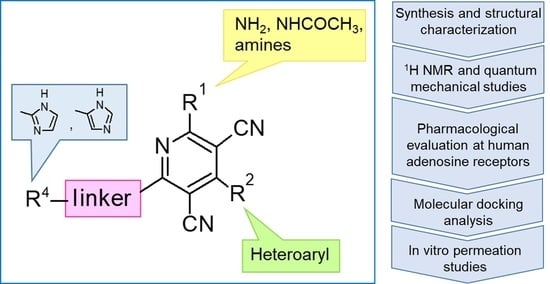
4-Heteroaryl Substituted Amino-3,5-Dicyanopyridines as New Adenosine Receptor Ligands: Novel Insights on Structure-Activity Relationships and Perspectives

1
Dipartimento di Neuroscienze, Psicologia, Area del Farmaco e Salute del Bambino, Sezione di Farmaceutica e Nutraceutica, Università degli Studi di Firenze, Via Ugo Schiff, 6, 50019 Sesto Fiorentino, Italy
2
Dipartimento di Medicina Traslazionale, Università degli Studi di Ferrara, Via Luigi Borsari 46, 44121 Ferrara, Italy
3
Dipartimento di Chimica Ugo Schiff, Università degli Studi di Firenze, Via della Lastruccia, 3, 50019 Sesto Fiorentino, Italy
4
Scuola di Scienze del Farmaco e dei Prodotti della Salute, Università degli Studi di Camerino, Via S.Agostino 1, 62032 Camerino, Italy
*
Author to whom correspondence should be addressed.
Pharmaceuticals 2022, 15(4), 478; https://doi.org/10.3390/ph15040478
Submission received: 1 March 2022 / Revised: 31 March 2022 / Accepted: 9 April 2022 / Published: 14 April 2022
(This article belongs to the Section Medicinal Chemistry)

Abstract

:
A new set of amino-3,5-dicyanopyridines was synthesized and biologically evaluated at the adenosine receptors (ARs). This chemical class is particularly versatile, as small structural modifications can influence not only affinity and selectivity, but also the pharmacological profile. Thus, in order to deepen the structure–activity relationships (SARs) of this series, different substituents were evaluated at the diverse positions on the dicyanopyridine scaffold. In general, the herein reported compounds show nanomolar binding affinity and interact better with both the human (h) A1 and A2A ARs than with the other subtypes. Docking studies at hAR structure were performed to rationalize the observed affinity data. Of interest are compounds 1 and 5, which can be considered as pan ligands as binding all the ARs with comparable nanomolar binding affinity (A1AR: 1, Ki = 9.63 nM; 5, Ki = 2.50 nM; A2AAR: 1, Ki = 21 nM; 5, Ki = 24 nM; A3AR: 1, Ki = 52 nM; 5, Ki = 25 nM; A2BAR: 1, EC50 = 1.4 nM; 5, EC50 = 1.12 nM). Moreover, these compounds showed a partial agonist profile at all the ARs. This combined AR partial agonist activity could lead us to hypothesize a potential effect in the repair process of damaged tissue that would be beneficial in both wound healing and remodeling.

Graphical Abstract

1. Introduction

The amino-3,5-dicyanopyridines have attracted much attention due to their versatility to behave as AR ligands. In fact, they are endowed with not only a wide range of affinity but also with different degrees of activities, with their profile varying from full to partial agonist or neutral antagonist at the different ARs [1,2,3,4,5,6,7]. As for other G-protein-coupled receptors [8,9,10], some of the antagonists have been proven to be inverse agonists [6]. The interest in modulating the effects of the natural ligand adenosine ensued from the evidence of its involvement in a large variety of physiological functions throughout the body. Interaction of the ubiquitous adenosine with its four G-protein-coupled A1, A2A, A2B and A3 ARs produced different responses depending on the type of AR and consequent cellular signaling involved. In fact, the A1 and A3 ARs are coupled to Gi proteins, which inhibit adenylate cyclase (AC), whereas A2A and A2B receptors signal through Gs proteins, thus activating it. Coupling to other second messenger systems has also been described [11,12].
Under stress conditions, extracellular adenosine grow from physiological nanomolar concentrations (30–200 nM) to high micromolar levels (30 µM), thus activating the diverse AR subtypes [12].
Adenosine regulates neurotransmitter release [13], synaptic plasticity [14] and neuroprotection in the central nervous system (CNS) [12,15,16] and plays different roles in a large variety of tissues and organs. In particular, adenosine controls T-cell proliferation and cytokine production [17] and produces either vasoconstriction or vasodilation [18] in the cardiovascular system. The nucleoside also inhibits lipolysis and stimulates bronchoconstriction [19,20].
Growing interest is emerging for its involvement also in wound healing and remodeling processes, including different stages, such as inflammation, neovascularization and tissue regeneration [21,22]. In fact, skin-lesion-associated diseases are one of the most common afflictions in the world, but their incidence on human health is still underestimated [23]. It is well-known that all ARs are involved, but their role has not yet been fully clarified [22,24]. AR agonists were reported to promote wound healing at different levels of the tissue-repair process, depending on the subtype activated. Thus, the use of mixed or pan agonists capable of simultaneously activating more receptor subtypes could lead to a synergistic effect [24,25].
Many publications report the amino-3,5-dicyanopyridines as AR ligands, showing nanomolar affinity and ranging from pan to selective AR ligands [1,2,3,4,5,6,7]. Moreover, this series seems to be more eclectic for pharmacological studies, because it is endowed with less species’ differences with respect to the adenosine-like AR agonists [26].
In recent years, we have produced a lot of AR ligands belonging to the amino-3,5-dicyanopyridine series [5,6,7]. In particular, in the last publication [7], a set of 4-aryl substituted dicyanopyridines bearing a 1H-imidazol-2-ylmethylsulfanyl group at R position, as in the parent LUF series [2], was described (Figure 1). The previously reported study demonstrated the key role of the R2 substituent in addressing affinity and selectivity toward specific ARs [6]. Thus, in order to deepening the SARs at this level, different heteroaryl moieties were introduced at R2, while maintaining the methylsulfanyl linker between the dicyanopyridine scaffold and the “usual” 1H-imidazol-2-yl group (compounds 110) or the “new entry” 1H-imidazol-4-yl moiety (derivative 11). Moreover, keeping constant the furan-2-yl moiety at 4-position, the R substituent was modified by varying the nature and the length of the linker bearing the imidazole as the terminal group (compounds 1217). In the meanwhile, the primary amine function (R1) was replaced by different secondary or tertiary amino-substituents (compounds 1820), or acetylated (21).

2. Results

2.1. Chemistry

The syntheses of the target compounds 121 and their intermediates followed the procedures as delineated in Scheme 1, Scheme 2, Scheme 3, Scheme 4 and Scheme 5. Derivatives 110 were prepared starting from the 6-sulfanyl-substituted compounds 3241 [6,27,28,29], which were synthesized as depicted in Scheme 1. The suitable heteroaryl aldehydes were reacted with malononitrile and thiophenol in the presence of tetrabuthylammonium fluoride hydrate (for compounds 2227, 29 [6,30,31,32] and 30) or basic alumina (for 28) [31,33] to yield the suitable 6-phenylsulfanyl-derivatives 2230 [6,30,31,32,33]. Compound 31, bearing at 4-position a 5-pyridin-2-ol moiety, was obtained from the corresponding 2-methoxy derivative 30 by treatment with hydrobromic acid (33% in glacial acetic acid), at 70 °C. When 2231 [6,30,31,32,33] were treated with anhydrous sodium sulfide in anhydrous DMF at 80 °C, followed by acidification with 1N HCl, the free thiols 3241 [6,27,28,29] were obtained in high yields. Reaction of 3234 [6,27] with equimolar amount of 2-bromomethyl-1H-imidazole hydrobromide [5] gave the hydrobromide salt of the target compounds 13. While compound 2 was isolated and characterized as such, derivatives 1 and 3 were obtained by treatment of the corresponding hydrobromide salt with sodium hydrogen carbonate at room temperature. Compounds 410 were obtained starting from the suitable sulfanyl derivatives 3541 [6,27,28,29]. Compound 11 was obtained by reacting the sulfanyl derivative 32 [6,27] with 4-chloromethyl-1H-imidazole hydrochloride 53 [34] (see Scheme 4 below), while the 2-amino-substituted derivatives 12 and 13 were generated by the microwave-assisted reaction between the phenylsulfanyl compound 22 and the commercially available (1-(1H-imidazol-2-yl)methanamine hydrochloride or 2-(1H-imidazol-4-yl)ethanamine, respectively, in DMF, at 120 °C, in the presence of Et3N.
The synthesis of the target dicyanopyridines 1417 starting from the common intermediate 32 is reported in Scheme 2. Treatment of the latter with chloroacetic acid or 4-(chloromethyl)-1,3-thiazole-2-carboxylic acid (57) [35] (see Scheme 5 below) in anhydrous DMF, in mild alkaline conditions (NaHCO3), furnished the 2-sulfanyl-acetic acid 42 and the 2-methylsulfanyl-1,3-thiazole-2-carboxylic acid derivative 43, respectively. Compound 42 was isolated from the crude mass reaction by acidification with 6M HCl. Both 42 and 43 were converted respectively into the target derivatives 1416 and 17, with variable yields, by reacting with (1-(1H-imidazol-2-yl)methanamine hydrochloride (compound 14), 1-(1H-imidazol-4-yl)methanamine (55) (see Scheme 4 below) [36] (compound 15) or 2-(1H-imidazol-4-yl)ethanamine (compounds 16 and 17), in anhydrous DMF, in the presence of 1-ethyl-3-(3-dimethylaminopropyl)carbodiimide hydrochloride, 1-hydroxybenzotriazole hydrate and Et3N.
Diazotization of the phenylsulfanyl-derivative 22 [30] with isoamyl nitrite in anhydrous CH3CN, followed by treatment with CuCl2, yielded 2-chloro-3,5-dicyanopyridine 44, which was reacted with the suitable amine in anhydrous DMF at RT to give compounds 4547 (Scheme 3). Reaction of the latter with sodium sulfide in anhydrous DMF at 80 °C furnished the 2(6)-sulfanyl-substituted pyridine derivatives 4850 as key intermediates to obtain the target compounds 1820. The reaction of 22 [30] with acetic anhydride in anhydrous pyridine at reflux gave 6-phenylsulfanyl-2-acetamide 51 [5], which was treated with anhydrous sodium sulfide in anhydrous DMF, at 50 °C, followed by 1N HCl, to yield the corresponding 6-sulfanyl derivative 52 [5]. The target compounds 1821 were obtained by treatment of 4850 and 52 [5] with 2-(bromomethyl)-1H-imidazole hydrobromide [5] in anhydrous DMF, in mild alkaline conditions.
The not commercially available reactants (53 [34], 55 [36] and 57 [35]) that are useful to synthesize compounds 11, 15 and 43 were prepared as depicted in Scheme 4 and Scheme 5. Treatment of the commercially available 1H-imidazol-4-ylmethanol with an excess of thionyl chloride at reflux gave 4-(chloromethyl)-1H-imidazole (53) [34], which was converted into the 4-(azidomethyl) intermediate 54 by reacting with sodium azide in EtOH in the presence of a catalytic amount of DMF, at 70 °C (Scheme 4). Hydrogenation of the latter in a Parr apparatus (1 atm), with 10% Pd/C as catalyst in EtOH, yielded the 1-(1H-imidazol-4-yl)methanamine (55) [36] in high yield. Reaction of ethyl thiooxamate and 1,3-dichloroacetone in anhydrous acetone at reflux gave ethyl 4-(chloromethyl)thiazole-2-carboxylate 56 [35], which was hydrolyzed with lithium hydroxide in aqueous (1:1) CH3CN, followed by acidification with citric acid, to furnish the corresponding carboxylic acid 57 [35] (Scheme 5).
The analysis of the 1H-NMR spectra of the target amino-3,5-dicyanopyridines, bearing a 2-(1H-imidazol-2-yl-methyl)sulphanyl substituent on the 6-side chain (110, 12, 1821), featured a particular behavior. In the 1H NMR spectra of both the crude and the purified compounds (13, 5, 8, 9 and 1821), the two imidazole protons at the 4 and 5 position appeared as a single signal (singlet) around 7 ppm, which integrated 2 (equivalent protons), while the NH proton was a very broad signal from 12 to 13 ppm. The same applied for compounds 4, 6, 7, 10 and 12 before purification. In this situation, it was possible to hypothesize a fast tautomeric equilibrium, which determines the equivalence of the two protons and influences the relaxation time of the imidazole NH group (broad signal). However, the pattern of the 1H NMR spectra of compounds 4, 6, 7, 10 and 12 changed after crystallization or purification by silica gel column chromatography. In fact, the two imidazole protons at 4,5 appeared as distinct signals falling around 6.8 and 7.1 ppm, while the signal of the NH proton was a singlet around 12 ppm. This experimental evidence led us to hypothesize that the two distinct signals of the imidazole protons in the purified compounds 4, 6, 7, 10 and 12 indicated the formation of an intramolecular hydrogen bond between the pyridine nitrogen at position 1 and the imidazole NH on the 6-side chain. The two distinct signals could be due to the stiffening of the sulfanylmethyl–imidazole chain as a result of which the neighborhood of the two protons at 4 and 5 becomes different. This hypothesis was supported by a 1H NMR study of compound 10, taken as reference. A detailed report of the registered spectra is reported in the Supplementary Materials. Moreover, an ab initio quantum mechanical calculation on compound 10, and also on the other congeners, namely 4, 6, 7 and 12, was performed. The program used was GAMESS (General Atomic and Molecular Electronic Structure System) [37], and the discussion is reported in the Section 3.2 (“Ab Initio Quantum Mechanical Studies”).

2.2. Pharmacological Assays

The amino-3,5-dicyanopyridines 121 were tested for their affinity at hA1, hA2A and hA3 ARs, stably transfected in Chinese Hamster Ovary (CHO) cells. Moreover, they were also studied as hA2B agonists by evaluating their stimulatory effect on cAMP production in CHO cells, stably expressing the hA2B AR. Compounds 1, 5, 16 and 17, the most interesting in terms of affinity at the hA1AR, were evaluated for their efficacy at the hA1 receptor. Due to their interesting pan profile, both compounds 1 and 5 were evaluated as AR agonists also at the hA2A and hA3 subtypes. Each compound was tested in the cAMP assay to assess its capability to modulate Forskolin-stimulated cAMP levels in the absence and/or presence of 2-chloro-N6-cyclopentyladenosine (CCPA, as an A1AR full agonist, set at 100%); 8-cyclopentyl-1,3-dipropylxanthine (DPCPX as an A1AR inverse agonist set at −100%); 2-p-(2-carboxyethyl)phenethylamino-5′-N-ethylcarboxamidoadenosine (CGS21680 as an A2AAR full agonist set at 100%); 5-[2-chloro-6-[(3-iodophenyl)methylamino]purin-9-yl]-3,4-dihydroxy-N-methyloxolane-2-carboxamide (2-Cl-IB-MECA as an A3AR full agonist set at 100%). All pharmacological data are reported in Table 1 and Table 2.

2.3. Molecular Docking Studies

Molecular docking studies were performed at the hA1AR and hA2AAR 3D structures to analyze the binding data of the synthesized compounds. The cryo-EM structure of the adenosine-bound hA1AR (pdb code: 6D9H; 3.6-Å resolution [38]) and the crystal structure of the hA2AR in complex with the inverse agonist ZM241385 (pdb code: 4EIY; 1.8-Å resolution [39]) were retrieved from the protein data bank (https://www.rcsb.org/, accessed on 6 April 2012) and employed as molecular targets. Docking analyses were performed with the CCDC Gold [40] docking algorithm by using MOE (Molecular Operating Environment, version 2019.0101) suite [41] as the interface.

2.4. In Vitro Permeation Studies

Compounds 1 and 5 were evaluated for their capability to penetrate the artificial membrane simulating the epidermal barrier. Permeation flux was assessed by using vertical Franz diffusion cells [42].

3. Discussion

3.1. Structure–Activity Relationships

The pharmacological results of the newly synthesized amino-3,5-dicyanopyridine derivatives 121 and those of the reference compound LUF5833 (2-amino-6-[(1H-imidazol-2-ylmethyl)sulfanyl]-4-phenylpyridine-3,5-dicarbonitrile) [2] are reported in Table 1 and Table 2. To better follow the discussion in both this section and in the molecular modeling one, LUF5833 was considered as a reference to define the numbering of the dicyanopyridine core to refer to. The pyridine nitrogen atom represents position 1, while the amino and the sulfanyl function occupy position 2 and 6, respectively.
Most of the reported compounds were devoid of an affinity for the hA2BAR, with the exception of derivatives 1, 4 and 58 showing EC50 values for this receptor below 63 nM (Table 1). As observed for other reported set of this series [5,6,7], the A3AR affinity was, in general, null or fell in the micromolar range. The only two exceptions were compounds 1 and 5, which displayed a nanomolar Ki value for this receptor subtype. In general, the herein reported compounds interacted better with both the hA1 and A2A ARs than with the other subtypes. However, compounds 1 and 5, which can be considered as pan ligands binding all the ARs with comparable affinity and in the nanomolar range, were interesting. The analogs 4 and 6 had a similar trend though binding the hA3AR with lower affinity with respect to 1 and 5 but similarly to the lead LUF5833. Moreover, compounds 1 and 5, when evaluated in the functional tests, showed a partial agonist profile at all the ARs (Table 2).
Table 1. Binding affinities (Ki) at hA1, hA2A and hA3 ARs and potencies (EC50) at hA2B ARs.
Pharmaceuticals 15 00478 i048
Binding Experiments acAMP
Assays
Ki (nM) or I%EC50 (nM) b
Efficacy c (%)
CompdR1R2RhA1 dhA2A ehA3 fhA2B
1NH2 Pharmaceuticals 15 00478 i001 Pharmaceuticals 15 00478 i0029.63 ± 1.6121 ± 252 ± 51.4 ± 0.2
(52%)
2 gNH2 Pharmaceuticals 15 00478 i003 Pharmaceuticals 15 00478 i004190 ± 16233 ± 19290 ± 23>1000
(8%)
3NH2 Pharmaceuticals 15 00478 i005 Pharmaceuticals 15 00478 i00610.1 ± 0.810.5 ± 0.9279 ± 21>1000
1%
4NH2 Pharmaceuticals 15 00478 i007 Pharmaceuticals 15 00478 i0080.77 ± 0.0937 ± 3274 ± 232.32 ± 0.21
(42%)
5NH2 Pharmaceuticals 15 00478 i009 Pharmaceuticals 15 00478 i0102.50 ± 0.2024 ± 225 ± 21.12 ± 0.11
(73%)
6NH2 Pharmaceuticals 15 00478 i011 Pharmaceuticals 15 00478 i0121.01 ± 0.0955 ± 6221 ± 203.15 ± 0.29
(47%)
7NH2 Pharmaceuticals 15 00478 i013 Pharmaceuticals 15 00478 i0148.85 ± 0.8281 ± 726%12.5 ± 1.3
(36%)
8NH2 Pharmaceuticals 15 00478 i015 Pharmaceuticals 15 00478 i0165.26 ± 0.48463 ± 3819%63 ± 5
(58%)
9NH2 Pharmaceuticals 15 00478 i017 Pharmaceuticals 15 00478 i0182.71 ± 0.18377 ± 32798 ± 72>1000
(7%)
10NH2 Pharmaceuticals 15 00478 i019 Pharmaceuticals 15 00478 i020149 ± 11613 ± 5323%>1000
(1%)
11 hNH2 Pharmaceuticals 15 00478 i021 Pharmaceuticals 15 00478 i02251 ± 4442 ± 37849 ± 74>1000
(4%)
12NH2 Pharmaceuticals 15 00478 i023 Pharmaceuticals 15 00478 i024315 ±28157 ± 1416%>1000
(3%)
13NH2 Pharmaceuticals 15 00478 i025 Pharmaceuticals 15 00478 i026174 ± 14125 ± 106%>1000
(9%)
14NH2 Pharmaceuticals 15 00478 i027 Pharmaceuticals 15 00478 i02887 ± 7817 ± 7133%>1000
(6%)
15NH2 Pharmaceuticals 15 00478 i029 Pharmaceuticals 15 00478 i030378 ± 26225 ± 18758 ± 66>1000
(1%)
16NH2 Pharmaceuticals 15 00478 i031 Pharmaceuticals 15 00478 i03230 ± 3138 ± 1130%>1000
(1%)
17NH2 Pharmaceuticals 15 00478 i033 Pharmaceuticals 15 00478 i03433 ± 3279 ± 22542 ± 48>1000
6%
18 Pharmaceuticals 15 00478 i035 Pharmaceuticals 15 00478 i036 Pharmaceuticals 15 00478 i037393 ± 32138 ± 11279 ± 21>1000
2%
19 Pharmaceuticals 15 00478 i038 Pharmaceuticals 15 00478 i039 Pharmaceuticals 15 00478 i040292 ± 261%18%>1000
(13%)
20 Pharmaceuticals 15 00478 i041 Pharmaceuticals 15 00478 i042 Pharmaceuticals 15 00478 i04341 ± 389 ± 7118 ± 11>1000
(8%)
21NHCOCH3 Pharmaceuticals 15 00478 i044 Pharmaceuticals 15 00478 i04534 ± 3394 ± 27768 ± 63>1000
(1%)
LUF5833iNH2 Pharmaceuticals 15 00478 i046 Pharmaceuticals 15 00478 i0472.4 ± 128 ± 1171 ± 10919 ± 7
(81%)
aKi values are means ± SEM of four separate assays each performed in triplicate. Percentage of inhibition (I%) was determined at 1 µM concentration of the tested compounds. b EC50 values are means ± SEM of four separate assays each performed in triplicate. c Efficacy of the tested compound at 1 µM concentration, in comparison with NECA (1 µM = 100%). d Displacement of specific [3H]DPCPX competition binding to hA1CHO cells. e Displacement of specific [3H]ZM241385 competition binding to hA2ACHO cells. f Displacement of specific [125I]AB-MECA competition binding to hA3CHO cells. g As hydrobromide salt. h As hydrochloride salt. i Reference [2].
Table 2. Modulation of Forskolin-stimulated cAMP levels of selected amino-3,5-dicyanopyridine derivatives on cyclic cAMP assay in hA1, hA2A and hA3 CHO cells a.
hA1ARhA2AARhA3AR
CompdEfficacy, b %
(Profile)
EC50 or IC50 (nM)Efficacy, b %EC50 (nM)Efficacy, b %IC50 (nM) c
131 ± 3
(Partial Agonist)
12.2 ± 1.2 c39 ± 315.1 ± 1.345 ± 468 ± 6
575 ± 5
(Partial Agonist)
1.95 ± 0.16 c40 ± 311.3 ± 1.034 ± 333 ± 2
110.82 ± 0.07
(Antagonist)
125 ± 11 dNT eNTNTNT
121.31 ± 0.11
(Antagonist)
768 ± 67 dNTNTNTNT
16−43 ± 4
(Inverse Agonist)
59 ± 4 cNTNTNTNT
1754 ± 4
(Partial Agonist)
38 ± 3 cNTNTNTNT
a Potency values (EC50 or IC50) are expressed as means ± SEM of four independent cAMP experiments, each performed in triplicate. b Efficacy of the novel compounds was normalized by using the efficacy value of the reference compounds: CCPA as A1AR full agonist (set at 100%); DPCPX as A1AR inverse agonist (set at −100%); CGS21680 as A2AAR full agonist (set at 100%); 2-Cl-IB-MECA as A3AR full agonist (set at 100%). c Potency of the novel compounds to modulate Forskolin-stimulated cAMP levels. d Potency of the novel compounds to inhibit the effect of CCPA 10 nM. e NT = not tested.
In general, keeping constant both the 2-amino function and the 6-(1H-imidazol-2-ylmethyl)sulfanyl side chain on the dicyanopyridine core, we can see that the introduction of different heteroaryl groups at the 4 position (compounds 110) influenced the binding affinity at the diverse ARs differently. All of these compounds showed an hA1AR Ki value below 10 nM, with the only two exceptions being compounds 2 and 10. A common feature of these latter two compounds was a hydrophilic hydroxyl group appended on the 4-heteroaryl moiety. Regarding the A2A receptor, the presence at the 4-position of either furanyl or thienyl rings (compounds 1–6) seemed to better promote the binding interaction with this subtype than a pyridine substituent (derivatives 710). Moreover, as observed in previously reported set of compounds of this series [5,6,7], the presence of a substituent on the 4-(hetero)aryl moiety dramatically influenced the hA2B AR activity (compare compound 1 to 2, 3, and compound 8 to 9, 10, respectively).
As previously reported [6], replacement of the methylsulfanyl linker with a methylamino one led to a high decrease of affinity at all the ARs (compare compound 1 to 12). Moreover, the presence of a longer 6-linker between the 1H-imidazol-2yl group and the dicyanopyridine scaffold in general negatively influenced the affinity at all the ARs, and, to a minor extent, the binding at the A1 subtype. In fact, compounds 16 and 17 maintained Ki values in the low nanomolar range.
The replacement of the methylsulfanyl linker on the 6-side-chain with a methylamino (compound 1 versus 12), or with a longer one (compound 1 versus 16), produced a shift of the pharmacological profile from partial agonist at the A1AR to antagonist or inverse agonist, respectively. Similarly, when the 1H-imidazol-2-yl was replaced with the 1H-imidazol-4-yl moiety (compound 1 versus 11) a change from partial agonist to antagonist profile was observed (Table 2). These data confirm that the requirements at the R position are very precise. In support of this, the inverse agonist profile (compound 16) was changed back to partial agonist (compound 17) when the 1,3-thiazol-5-yl moiety was directly attached to the methylsulfanyl linker. This result was in accordance with the literature data [4,43,44,45]. In fact, many amino-3,5-dicyanopyridines bearing the thiazole feature in this precise position were reported as potent and selective A1AR agonists.
The introduction of cycloalkyl substituents on the 2-amino function (compounds 18 and 19) and the inclusion of the latter in a (pyrrolidin-1-yl) moiety (compound 20) or its acetylation (compound 21) differently affected the AR binding affinities with respect to the unsubstituted derivative 1. Interaction with the A2B receptor was completely lost, while affinity was retained at the other AR subtypes. In particular, while compound 20 maintained A1 and A2A AR Ki values in the nanomolar range, the 2-acetylamino derivative 21 bound the A1 subtype with good affinity.

3.2. Ab Initio Quantum Mechanical Studies

The hypothesis of the intramolecular H-bond formation between the pyridine nitrogen at position 1 and the imidazole NH hydrogen was supported by ab initio quantum mechanical calculations performed on compounds 4, 6, 7, 10 and 12. The program used was GAMESS [37]. The formation energies (E1 and E2) of two conformations, 1 and 2, were calculated (Table 3) with Geometry Optimization, i.e., with conformational optimization. Conformation 1 included the formation of an intramolecular H-bond between the imidazole NH hydrogen and the pyridine nitrogen at position 1. As an example, the two minimized conformations, 1 and 2, of the reference compound 10 (10-1 and 10-2) were depicted in Figure 2. Referring to the compounds under study, negative ΔE values (Table 3) indicated that conformations 1 were slightly more stable than the 2 ones, where the H-bond was not supposed. Thus, the observed energy gain was attributable to the intramolecular H-bond formation, but its low value, which was generally observed, indicated that this hypothetical H-bond was very weak. In fact, in the presence of water, a perturbation of the system, was observed, and the intramolecular H-bond was not formed. In this situation, the imidazole NH hydrogen established a stabilizing H-bond with a water molecule, leading to an energy gain of about −11 Kcal/mol.

3.3. Molecular Modeling Studies

Analogously to previously reported dicyanopyridines [5,6,7,46,47], the best-score docking conformations generally observed for the newly synthesized compounds at the hA1AR present the pyridine scaffold being inserted between hydrophobic residues conserved among the human ARs (Phe171, Leu2506.51 and Ile2747.39) and making non-polar interaction with these amino acids (Figure 3 reports the binding mode of 9 within the hA1AR cavity). The 2-amino function of 9 makes a double polar interaction with Asn2546.55 and Glu172. The 3-cyano also binds Asn2546.55 through a polar interaction, while the 5-cyano moiety points toward the depth of the binding cavity positioning itself close to Ala662.61, Ile692.64, Val873.32 and His2787.43.
The 4-heterocyclic substituent is inserted in the depth of the cavity, close to residues belonging to TM3, EL2, TM5, TM6 and TM7 domains (Val873.32, Leu883.33, Thr913.36, Phe171, Met1805.38, Trp2476.48, Leu2506.51, Ile2747.39, Thr2777.42 and His2787.43). In general, all the AR non-nucleoside agonists reported to date bear an aromatic ring at this position. The affinity data of the compounds synthesized in this work confirm this feature, since the presence of aromatic heterocyclic groups (R2) at the 4-position affects affinity, especially for both the A1 and the A2B subtypes. In particular, as previously observed [2,7], the introduction of a substituent in the para-position of the 4-aromatic ring modulates the binding affinity. In fact, the presence of substituents on the pyridyl moiety (compounds 9 and 10) in a position corresponding to the para one in a phenyl ring, or at the 5-position of a 2-furanyl ring (2 and 3), modulates the affinity for the AR subtypes compared to the unsubstituted analogues 1 and 8. Figure 3A and Figure 4A show the interaction of the 2-methoxy-5-pyridyl substituent at 4-position of 9 with the receptor residues. The presence of the additional methoxy group allows the compound to completely fill the narrow sub-cavity in the depth of the binding site (Figure 3A). Since the amino acid residues forming such a sub-cavity are mainly hydrophobic, it is no surprise that the hA1AR affinity data of 9 are comparable to those of 8. When the methoxy group is replaced by a polar hydroxyl function, the affinity gets significantly lower (compound 10). Analogously, the presence of an additional 5-methyl group on a 4-(2-furanyl) substituent maintains high affinity for the hA1AR (derivative 3) compared to the corresponding unsubstituted analogue 1. Replacement of the methyl group with a polar hydroxymethyl function (2) leads to a decrease of the hA1AR affinity.
Similar to what was observed in previously reported docking studies [5,6,7], the 6-substituent points toward the entrance of the binding cavity (Figure 3). As already observed [6], a methylsulfanyl-linker on the 6-side-chain leads to a higher affinity with respect to the aminomethyl one (compare 1 to 12), probably due to the lack of conjugation effects of the methylsulfanyl-linker (existing instead for the aminomethyl one) that allows this structural feature to better accommodate in the binding site, assuming a non-coplanar position with respect to the dicyanopyridine scaffold. The 1H-imidazol-2yl moiety on the 6-side-chain gets positioned between residues belonging to TM2, EL2 and TM7 domains (Ile692.64, Asn702.65, Glu170, Phe171, Glu172, Tyr2717.36 and Ile2747.39). The location of this substituent, with respect to the receptor residues, modulates, to some extent, the binding at the A1AR. In fact, the presence of a longer 4-linker between this group and the dicyanopyridine scaffold generally leads to a slight decrease of affinity at this subtype with respect to the parent compound 1 (see derivatives 1417). In this sense, docking results show that, for compounds bearing a longer 4-linker, the imidazolyl moiety gets located in a more external position, leading to a different interaction with the receptor residues. The presence of a 4-methylsulfanyl linker and an imidazole-2yl group (compounds 1 and 5) on the 6-side-chain seems to be correlated to an agonist/partial agonist profile. The replacement of these features respectively with an aminomethyl linker (12) or an imidazole-4-yl moiety (11) causes a shift to antagonist behavior. The docking results do not explain the observed agonist-to-antagonist activity shift as well as the change from an inverse agonist (compound 16) to partial agonist profile when a 1,3-thiazole moiety was included into the 6-side-chain (compound 17). Figure 4C is a top view of the docking pose of 17 within the hA1 receptor cavity.
Compounds endowed with hA2AAR affinity showed a trend comparable to that observed for the hA1AR. Docking experiments performed at the hA2AAR crystal structure showed analogous arrangements of the analyzed molecules to those verified at the hA1AR (Figure 5 reports the binding mode of compound 3 within the hA2AAR cavity). The sets of residues involved in the interaction with the ligand are highly conserved at the two AR subtypes. The dicyanopyridine scaffold is inserted between Phe168, Leu2496.51 and Ile2747.39, with the exocyclic 2-amino function giving polar interaction with Glu169 and Asn2536.55. The 4-heterocyclic substituent is positioned close to residues belonging to TM3 (Val843.32, Leu853.33 and Thr883.36), EL2 (Phe168), TM5 (Met1775.38), TM6 (Trp2466.48 and Leu2496.51) and TM7 (Ile2747.39, Ser2777.42 and His2787.43) domains. These residues are conserved between hA2AAR and hA1AR. Introduction of substituents on the 4-heteroaryl moiety modulates hA2AAR affinity to a lesser extent than what is observed for the A1 subtype. However, the hA2AAR affinity seems generally higher for compounds bearing a 4-furan-2yl moiety (compounds 1–3) with respect to those substituted with a 4-pyridyl group. This behavior may be interpreted considering steric factors. A potential slightly smaller cavity at the hA2AAR, compared to that of the hA1AR, could accommodate slightly smaller substituents. Even the results observed for the 6-side-chain are similar to those obtained on the hA1AR. The hA2AAR residues involved in the interaction with the 6-substituent are quite conserved with the hA1AR subtype. In detail, the imidazolyl group gets located between residues belonging to the TM2 (Ile662.64 and Ser672.65), EL2 (Leu167, Phe168 and Glu169) and TM7 (Tyr2717.36 and Ile2747.39) domains.
Generally, the introduction of substituents on the exocyclic amine at 2-position of the dicyanopyridine scaffold leads to a decrease of affinity at both AR subtypes with respect to the unsubstituted derivative 1. Only a slight reduction of A1AR affinity was observed for compounds 20 and 21. As previously observed [6], docking results on the herein reported compounds showed that substituents on the 2-amino function lead to a partial disruption of the interaction with the EL2 glutamate and TM6 asparagine residues (see above), with a consequent decrease of affinity. Furthermore, docking results suggested also various binding modes of these compounds at both the AR subtypes, making it difficult to make a clear interpretation of the biological results.

3.4. In Vitro Permeation Studies

The stratum corneum is a lipophilic membrane which represents the most important barrier to drug skin diffusion. In vitro permeation studies conducted on compounds 1 and 5 through Franz cells [42] showed an amount of permeated drug after 24 h respectively of 15.0 ± 1.4 μg/mL and 22.7 ± 1.0 µg/mL.
These permeation data were in agreement with the results obtained through solubility studies, carried out on both compounds in testing medium (PBS + Tween 80 (2% w/w)). On the basis of these experiments, compound 5 resulted in being less soluble (27.3 ± 2.52 µg/mL) than the analogous 1 (40.6 ± 0.16 µg/mL).
Therefore, it can be hypothesized that the dicyanopyridine 5, due to its higher lipophilicity with respect to 1 (pkCSM calculated logP, 3.15 versus 2.68, respectively) [48,49] and, consequently, its affinity for stratum corneum, accumulates inside the membrane, generating an into/out concentration gradient, which represents the driving force to its permeation.
From the perspective of a possible application of the two compounds in wound healing [50] as promoters of endothelial cell proliferation and migration [21,22], the lipophilicity of compound 5 could be a desirable property, allowing an accumulation of the compound on the injured skin leading to a limited systemic absorption.

4. Materials and Methods

4.1. Chemistry

4.1.1. General Methods

The microwave-assisted syntheses were performed by using an Initiator EXP Microwave Biotage instrument (frequency of irradiation: 2.45 GHz). Analytical silica gel plates (Merck F254, Kenilworth, NJ, USA), preparative silica gel plates (Merck F254, 2 mm) and silica gel 60 (Merck, 70–230 mesh) were used for analytical and preparative TLC and for column chromatography, respectively. All melting points were determined on a Gallenkamp melting-point apparatus and are uncorrected. Elemental analyses were performed with a Flash E1112 Thermofinnigan elemental analyzer for C, H and N, and the results were within ±0.4% of the theoretical values. All final compounds revealed purity not less than 95%. The IR spectra were recorded with a Perkin-Elmer Spectrum RX I spectrometer in Nujol mulls and are expressed in cm−1. NMR spectra were recorded on a Bruker Avance 400 spectrometer (400 MHz for 1H NMR and 100 MHz for 13C NMR). The chemical shifts are reported in δ (ppm) and are relative to the central peak of the residual non-deuterated solvent, which was CDCl3 or DMSOd6. The following abbreviations are used: s = singlet, d = doublet, t = triplet, q = quartet, m = multiplet, br = broad, Ar = aromatic protons. Compounds 22 [30], 25, 26 [30], 27 [31], 29 [32], 32 [27], 35, 36 [27], 37 [28], 39 [29], 51, 52 and 56 were synthesized by following the procedures reported by us in Reference [6]. When available, melting point and/or 1H NMR values were in accordance with the literature data.

4.1.2. General Procedure for the Synthesis of 2-Amino-4-(heteroaryl)-6-[(1H-imidazol-2-ylmethyl)sulfanyl]pyridine-3,5-dicarbonitriles 1, 3

Equimolar amounts of sodium hydrogen carbonate and commercially available 2-(bromomethyl)-1H-imidazole hydrobromide (4.0 mmol) were consequentially added to a solution of the mercapto-compound (32 [6,27], 34, 3.8 mmol) in anhydrous DMF (1 mL). The reaction mixture was stirred at room temperature, under a nitrogen atmosphere, for 4 h. At reaction completion, water was added (30 mL) to precipitate a solid which was collected by filtration and triturated with Et2O (2 mL). A suspension of the intermediate (hydrobromide salt of compounds 1 and 3) and sodium hydrogen carbonate (4.0 mmol) in a mixture of DMF/H2O (2:1, 2 mL) was stirred at room temperature for a few minutes. Then the solid was collected by filtration, washed with water and recrystallized.
2-Amino-4-(furan-2-yl)-6-[(1H-imidazol-2-ylmethyl)sulfanyl]pyridine-3,5-dicarbonitrile (1). Yield 55%; mp 233–234 °C. 1H NMR (DMSO-d6) 8.12 (br s, 2H, NH2), 8.11 (d, 1H, Ar, J = 1.2 Hz), 7.41 (d, 1H, Ar, J = 3.6 Hz), 7.11 (s, 2H, Ar), 6.84 (dd, 1H, Ar, J = 1.7, 3.6 Hz), 4.55 (s, 2H, CH2). Anal. Calc. for C15H10N6OS.
Hydrobromide salt of compound 1: Yield 75%; mp > 300 °C dec (MeOH). 1H NMR (DMSO-d6) 13.82 (br s, 1H, NH), 8.14 (br s, 2H, NH2), 8.12 (d, 1H, Ar, J = 1.2 Hz), 7.63 (s, 2H, Ar), 7.43 (d, 1H, Ar, J = 3.6 Hz), 6.85 (dd, 1H, Ar, J = 1.7, 3.6 Hz), 4.73 (s, 2H, CH2); IR 3400, 3320, 3140, 2217. Anal. Calc. for C15H11BrN6OS.
2-Amino-6-[(1H-imidazol-2-ylmethyl)sulfanyl]-4-(5-methylfuran-2-yl)pyridine-3,5-dicarbonitrile (3): Yield 86%; mp 276–277 °C; (DMF); 1H NMR (DMSO-d6) 11.87 (s, 1H, NH), 8.07 (s, 2H, NH2), 7.35 (d, 1H, Ar, J = 3.5 Hz), 6.96 (s, 2H, Ar), 6.49 (d, 1H, Ar, J = 2.8 Hz), 4.48 (s, 2H, CH2), 2.40 (s, 3H, CH3); 13C NMR (DMSO-d6) 167.67, 160.83, 156.66, 143.92, 143.60, 142.99, 118.40, 116.36, 116.26, 110.00, 88.76, 81.09, 27.31, 13.88; IR 3289, 2206. Anal. Calc. for C16H12N6OS.
Hydrobromide salt of compound 3: 1H NMR (DMSO-d6) 14.02 (br s, 1H, NH), 8.1 (br s, 2H, NH2), 7.60 (s, 2H, Ar), 7.38 (d, 1H, Ar, J = 3.4 Hz), 6.51 (d, 1H, Ar, J = 2.8 Hz), 4.69 (s, 2H, CH2), 2.40 (s, 3H, CH3).

4.1.3. 2-Amino-4-[5-(hydroxymethyl)furan-2-yl]-6-[(1H-imidazol-2-ylmethyl)sulfanyl]pyridine-3,5-dicarbonitrile hydrobromide (2)

Sodium hydrogen carbonate (9.3 mmol) and an equimolar amount of the commercially available 2-(bromomethyl)-1H-imidazole hydrobromide were added to a solution of the suitable mercapto-compound (33, 9.3 mmol) in anhydrous DMF (3 mL). The reaction mixture was stirred at RT, in a nitrogen atmosphere, until the disappearance of the starting material (TLC monitoring). Then water was added (20 mL) to precipitate a solid which was collected by filtration and washed with water. For compound 1, the crude product was treated with Et2O (2 mL), collected by filtration and recrystallized. For derivative 2, a second crop of product was obtained by extracting the aqueous solution with EtOAc (4 × 10 mL). The collected organic layers were dried (Na2SO4), and the solvent was removed under reduced pressure. The two crops of product were triturated with Et2O (2 mL) collected by filtration and purified by crystallization.
Yield 55%; mp 293–294 °C; (EtOH); 1H NMR (DMSO-d6) 13.97 (s, 1H, NH), 8.10 (s, 2H, NH2), 7.58 (s, 2H, Ar), 7.38 (s, 1H, Ar), 6.66 (s, 1H, Ar), 5.49 (s, 1H, OH), 4.69 (s, 2H, CH2), 4.50 (s, 2H, CH2); 13C NMR (DMSO-d6) 166.10, 160.78, 160.15, 144.27, 144.02, 143.90, 119.97, 118.14, 115.96, 110.35, 89.18, 82.15, 56.22, 24.43; IR 3313, 2212, 1208. Anal. Calc. for C16H13BrN6O2S.

4.1.4. General Procedure for the Synthesis of 2-Amino-4-(heteroaryl)-6-[(1H-imidazol-2-ylmethyl)sulfanyl]pyridine-3,5-dicarbonitriles 410

Sodium hydrogen carbonate (2 mmol) and the commercially available 2-(bromomethyl)-1H-imidazole hydrobromide (1 mmol) were added to a solution of the suitable mercapto-compound (3541 [6,27,28,29] 1 mmol) in anhydrous DMF (1 mL). The reaction mixture was stirred at RT until disappearance of the starting material (TLC monitoring). Then, water was added (25 mL) to precipitate a solid which was collected by filtration and washed with water. The crude product was triturated with Et2O (5 mL), collected by filtration and purified by crystallization (compounds 49) or silica gel column chromatography, eluting system cyclohexane/EtOAc/MeOH 2:6:2 (compound 10).
2-Amino-4-(furan-3-yl)-6-[(1H-imidazol-2-ylmethyl)sulfanyl]pyridine-3,5-dicarbonitrile (4): Yield 53%; mp 271–273 °C; (EtOH); 1H NMR (DMSO-d6) 11.83 (s, 1H, NH), 8.26 (s, 1H, Ar), 8.02 (s, 2H, NH2), 7.92 (s, 1H, Ar), 7.07 (s, 1H, Ar) 6.88 (s, 1H, Ar), 6.86 (s, 1H, Ar), 4.50 (s, 2H, CH2); IR 3328, 2220, 2211. Anal. Calc. for C15H10N6OS.
2-Amino-6-[(1H-imidazol-2-ylmethyl)sulfanyl]-4-(thiophen-2-yl)pyridine-3,5-dicarbonitrile (5): Yield 76%; mp 246–248 °C; (MeOH); 1H NMR (DMSO-d6) 11.84 (s, 1H, NH), 8.07 (s, 2H, NH2), 7.96 (d, 1H, Ar, J = 4.28 Hz), 7.56 (d, 1H, Ar, J = 1.88 Hz), 7.28 (s, 1H, Ar), 6.97 (s, 2H, Ar), 4.50 (s, 2H, CH2); IR 3305, 2212. Anal. Calc. for C15H10N6S2.
2-Amino-6-[(1H-imidazol-2-ylmethyl)sulfanyl]-4-(thiophen-3-yl)pyridine-3,5-dicarbonitrile (6): Yield 73%; mp 239–241 °C dec; (CH3CN); 1H NMR (DMSO-d6) 11.83 (br s, 1H, NH), 8.05 (dd, 1H, Ar, J = 2.9, 1.2 Hz), 8.04 (br s, 2H, NH2), 7.78 (dd, 1H, Ar, J = 5.0, 2.9 Hz), 7.39 (dd, 1H, Ar, J = 5.0, 1.2 Hz), 7.07 (br s, 1H, Ar), 6.97 (br s, 1H, Ar), 4.50 (s, 2H, CH2); 13C NMR (DMSO-d6) 166.79, 160.36, 153.48, 142.99, 133.83, 129.32, 128.16, 127.92, 115.88, 93.29, 86.01, 27.24. IR 3400, 3321, 2218. Anal. Calc. for C15H10N6S2.
2-Amino-6-[(1H-imidazol-2-ylmethyl)sulfanyl]-4,4′-bipyridine-3,5-dicarbonitrile (7): Yield 20%; mp 230 °C dec; (EtOH); 1H NMR (DMSO-d6) 11.87 (br s, 1H, NH), 8.80 (d, 2H, Ar, J = 4.7 Hz), 8.2 (br s, 2H, NH2), 7.59 (d, 2H, Ar, J = 4.7 Hz), 7.09 (br s, 1H, Ar), 6.86 (br s, 1H, Ar), 4.52 (s, 2H, CH2). IR 3312, 3175, 2210. Anal. Calc. for C16H11N7S.
2′-Amino-6′-[(1H-imidazol-2-ylmethyl)sulfanyl]-3,4′-bipyridine-3′,5′-dicarbonitrile (8): Yield 64%; mp 244–245 °C dec; (EtOH); 1H NMR (DMSO-d6) 12.18 (br s, 1H, NH), 8.78–8.74 (m, 2H, Ar), 8.25 (br s, 2H, NH2), 8.04–8.02 (m, 1H, Ar), 7.64–7.61 (m, 1H, Ar), 7.02 (s, 2H, Ar), 4.54 (s, 2H, CH2). IR 3379, 3308, 2212. Anal. Calc. for C16H11N7S.
2′-Amino-6′-[(1H-imidazol-2-ylmethyl)sulfanyl]-6-methoxy-3,4′-bipyridine-3′,5′-dicarbonitrile (9): Yield 82%; mp 232–233 °C (EtOH); 1H NMR (DMSO-d6) 12.06 (s, 1H, NH), 8.38 (d, 1H, Ar, J = 1.7 Hz), 8.18 (s, 2H, NH2), 7.93 (dd, 1H, Ar, J = 8.6, 2.1 Hz), 7.02 (d, 1H, Ar, J = 8.7 Hz), 7.00 (s, 2H, Ar), 4.52 (s, 2H, CH2), 3.94 (s, 3H, CH3); 13C NMR (DMSO-d6) 166.66, 165.10, 160.18, 155.69, 147.37, 139.95, 123.83, 115.72, 110.98, 93.76, 86.62, 54.14, 27.21. IR 3383, 3335, 2216. Anal. Calc. for C17H13N7OS.
2′-Amino-6-hydroxy-6′-[(1H-imidazol-2-ylmethyl)sulfanyl]-3,4′-bipyridine-3′,5′-dicarbonitrile (10): Yield 61%; mp 269–271 °C; 1H NMR (DMSO-d6) 12.14 (s, 1H, OH), 11.82 (s, 1H, NH), 8.12 (s, 2H, NH2), 7.81 (s, 1H, Ar), 7.61 (dd, 1H, Ar, J = 9.5, 2.7 Hz), 7.07 (s, 1H, Ar), 6.85 (s, 1H, Ar), 6.47 (d, 1H, Ar, J = 9.5 Hz), 4.48 (s, 2H, CH2); 13C NMR (DMSO-d6) 166.64, 165.93, 154.78, 141.10, 115.88, 93.42, 92.66, 86.18, 83.66, 29.93, 27.15. 3400, 3308, 3185, 2214. Anal. Calc. for C16H11N7OS.

4.1.5. 2-Amino-4-(furan-2-yl)-6-[(1H-imidazol-5-ylmethyl)sulfanyl]pyridine-3,5-dicarbonitrile hydrochloride (11)

Equimolar amounts of sodium hydrogen carbonate and the 4-(chloromethyl)-1H-imidazole hydrochloride 53 [34] (1.36 mmol) were consequentially added to a solution of the mercapto-compound 32 [6,27], (1.24 mmol) in anhydrous DMF (3 mL). The reaction mixture was stirred at RT for 3 h. At reaction completion, water was added (30 mL) to precipitate a solid which was collected by filtration, triturated with Et2O (2 mL) and then crystallized.
Yield 75%; mp 147–148 °C; (EtOH/Et2O/acetone); 1H NMR (DMSO-d6) 8.6–7.6 (br s, 2H, NH2), 8.28 (s, 1H, Ar), 8.10 (d, 1H, Ar, J = 1.3 Hz), 7.48 (s, 1H, Ar), 7.39 (d, 1H, Ar, J = 3.6 Hz), 6.84 (dd, 1H, Ar, J = 3.6, 1.7 Hz), 4.45 (s, 2H, CH2); 13C NMR (DMSO-d6) 167.71, 160.64, 145.52, 144.21, 116.84, 116.18, 116.13, 113.31, 89.73, 81.98, 26.02. IR 3325, 3211, 2210. Anal. Calc. for C15H11ClN6OS

4.1.6. General Procedure for the Synthesis of 2-Amino-substituted Derivatives 12, 13

A solution of the phenylsulfanyl-derivative 22 [6,30] (0.47 mmol), the commercially available 1-(1H-imidazol-2-yl)methanamine hydrochloride for compound 12 or 2-(1H-imidazol-4-yl)ethanamine for 13 (0.56 mmol), and Et3N (1.2 mmol) in DMF (1 mL) was heated at 120 °C, under microwave irradiation, until the disappearance of the starting material (TLC monitoring). After cooling at RT, water (20 mL) was added, and the resulting precipitate was collected by filtration and washed with water (10 mL) and Et2O (5 mL). The crude product was purified by silica gel column chromatography, eluting system cyclohexane/EtOAc 1:1.
2-Amino-4-(furan-2-yl)-6-[(1H-imidazol-2-ylmethyl)amino]pyridine-3,5-dicarbonitrile (12): Yield 14%; mp 265–266 °C; 1H NMR (DMSO-d6) 11.63 (s, 1H, NH), 8.03 (s, 1H, Ar), 7.84 (t, 1H, NH, J = 5.5 Hz), 7.49 (s, 2H, NH2), 7.27 (d, 1H, Ar, J = 3.5 Hz), 7.04 (s, 1H, Ar), 6.82 (s, 1H, Ar), 6.79 (dd, 1H, Ar, J = 3.6, 1.7 Hz), 4.57 (d, 2H, CH2, J = 5.5 Hz,); 13C NMR (DMSO-d6) 162.02, 159.93, 145.51, 145.03, 117.29, 117.09, 115.50, 112.88, 77.55, 76.11, 65.37. IR 3337, 2203. Anal. Calc. for C15H11N7O.
2-Amino-4-(furan-2-yl)-6-{[2-(1H-imidazol-4-yl)ethyl]amino}pyridine-3,5-dicarbonitrile (13): Yield 37%; mp 242 °C dec;1H-NMR (DMSO-d6) 11.83 (br s, 1H, NH), 8.02 (d, 1H, Ar, J = 1.8 Hz), 7.57–7.53 (m, 2H, NH + Ar), 7.41 (br s, 2H, NH2), 7.25 (d, 1H, Ar, J = 3.6 Hz), 6.85 (s, 1H, Ar), 6.78 (dd, 1H, Ar, J = 3.6, 1.8 Hz), 3.62 (q, 2H, CH2, J = 7.2 Hz), 2.79 (t, 2H, CH2, J = 7.2 Hz). 13C NMR (DMSO-d6) 162.05; 159.92; 146.58; 146.02; 145.50; 135.08; 117.39; 117.17; 115.37; 112.81; 77.35; 75.71; 41.40. Anal. Calc. for C16H13N7O.

4.1.7. General Procedure for the Synthesis of 2-{[6-Amino-3,5-dicyano-4-(furan-2-yl)pyridin-2-yl]sulfanyl}-N-acetamides 14–16

The suitable 1-(1H-imidazol-2-yl)methanamine hydrocloride (compound 14) or 1-(1H-imidazol-4-yl)methanamine (55) [36] (compound 15) or 2-(1H-imidazol-4-yl)ethanamine (compounds 16, 17) (1.1 mmol); the intermediate compound 42 (1.0 mmol); 1-ethyl-3-(3-dimethylaminopropyl)carbodiimide hydrochloride (1.48 mmol); and 1-hydroxybenzotriazole hydrate (1.3 mmol) were subsequently added to a solution of anhydrous Et3N (0.5 mL) in anhydrous DMF (5 mL). The reaction mixture was stirred at RT, under a nitrogen atmosphere, until the disappearance of the starting material (TLC monitoring). After dilution with cold water (50 mL), the suspension was stirred for 15 min, and the resulting solid was collected by filtration and washed with water and Et2O. The crude derivative 14 was then purified by treatment with boiling EtOH (20 mL). For compound 15, a second crop of product was obtained by extracting the aqueous phase with EtOAc (3 × 30 mL). Then the collected organic layers were anhydrified (Na2SO4) and evaporated under reduced pressure to yield a solid which was then washed with a 10% solution of Na2CO3 (30 mL) and with water. Washing with 10% solution of Na2CO3 (30 mL) was performed also on the crude compound 16. Compounds 15 and 16 were purified by crystallization.
2-{[6-Amino-3,5-dicyano-4-(furan-2-yl)pyridin-2-yl]sulfanyl}-N-(1H-imidazol-2-ylmethyl)acetamide (14): Yield 72%; mp 274–276 °C; 1H NMR (DMSO-d6) 11.88 (br s, 1H, NH), 9.0–7.5 (br s, 2H, NH2), 8.60 (t, 1H, NH, J = 5.5 Hz), 8.11 (d, 1H, Ar, J = 1.3 Hz), 7.41 (d, 1H, Ar, J = 3.6 Hz), 6.98 (s, 2H, Ar), 6.85 (dd, 1H, Ar, J = 3.7, 1.7 Hz), 4.34 (d, 2H, CH2, J = 5.5 Hz), 3.91 (s, 2H, CH2); 13C NMR (DMSO-d6) 168.11, 167.43, 160.64, 145.55, 145.10, 144.09, 116.83, 116.21, 113.32, 89.40, 81.95, 37.14, 34.39. Anal. Calc. for C17H13N7O2S.
2-{[6-Amino-3,5-dicyano-4-(furan-2-yl)pyridin-2-yl]sulfanyl}-N-(1H-imidazol-5-ylmethyl)acetamide (15): Yield 12%; mp 269–270 °C (EtOH). 1H-NMR (DMSO-d6) 11.93 (br s, 1H, NH), 8.4–7.6 (br s, 2H, NH2), 8.34 (t, 1H, NH), 8.12–8.11 (m, 1H, Ar), 7.61 (s, 1H, Ar), 7.41 (d, 1H, Ar, J = 3.6 Hz), 6.94 (s, 1H, Ar), 6.85 (dd, 1H, Ar, J = 3.6, 1.7 Hz), 4.21 (d, 2H, CH2, J = 5 Hz), 3.89 (s, 2H, CH2). Anal. Calc. for C17H13N7O2S.
2-{[6-Amino-3,5-dicyano-4-(furan-2-yl)pyridin-2-yl]sulfanyl}-N-[2-(1H-imidazol-5-yl)ethyl]acetamide (16): Yield 28%; mp 223–226 °C (CH3CN);1HNMR (DMSO-d6) 11.80 (br s, 1H, NH); 8.13–8.07 (m, 2H, Ar + NH); 8.5–7.7 (s br, 2H, NH2), 7.50 (s, 1H, Ar); 7.41 (d, 1H, Ar, J = 3.6 Hz), 6.85 (dd, 1H, Ar, J = 3.6, 1.7 Hz), 6.82 (br s, 1H, Ar), 3.87 (s, 2H, CH2), 3.31–3.27 (m, 2H, CH2), 2.68–2.61 (m, 2H, CH2). 13C NMR (DMSO-d6) 167.74; 167.14; 160.57; 146.08; 145.54; 144.19; 116.88; 116.14; 113.33; 89.63; 82.09; 34.10. IR: 2922, 2362, 2212, 1653, 1458, 1377. Anal. Calc. for C18H15N7O2S.

4.1.8. 4-({[6-Amino-3,5-dicyano-4-(furan-2-yl)pyridin-2-yl]sulfanyl}methyl)-N-[2-(1H-imidazol-5-yl)ethyl]-1,3-thiazole-2-carboxamide (17)

Then 1-hydroxybenzotriazole hydrate (0.9 mmol), 1-ethyl-3-(3-dimethylaminopropyl)carbodiimide hydrochloride (0.98 mmol), Et3N (0.85 mmol), and 2-(1H-imidazol-4-yl)ethanamine (2.61 mmol) were subsequently added to a solution of compound 43 (0.85 mmol) in anhydrous DMF (2 mL). The reaction mixture was stirred at RT, under a nitrogen atmosphere, for 3 h. After dilution with cold water (30 mL), a solid precipitated and was collected by filtration and washed with water and Et2O. A second crop of product was obtained by extracting the aqueous phase with EtOAc (3 × 30 mL). Then the collected organic layers were anhydrified (Na2SO4) and evaporated under reduced pressure to yield a solid which was treated with Et2O (5 mL) and then collected by filtration. The crude product was purified by silica gel column chromatography, eluting system CH2Cl2/MeOH 8:2.
Yield 40%; 1H NMR (DMSO-d6) 11.85 (s, 1H, NH), 8.88 (t, 1H, NH, J = 5.9 Hz), 8.10 (s, 2H, Ar), 8.4–7.8 (br s, 2H, NH2), 7.54 (s, 1H, Ar), 7.39 (d, 1H, Ar, J = 3.7 Hz), 6.83 (m, 2H, Ar), 4.61 (s, 2H, CH2), 3.50 (dd, 2H, CH2, J = 13.7, 7.1 Hz), 2.77 (t, 2H, CH2, J = 7.3 Hz). IR 3300, 3120, 2200, 1539, 1519,1456. Anal. Calc. for C21H16N8O2S2.

4.1.9. General Procedure for the Synthesis of the Target Compounds 18–21

Sodium hydrogen carbonate (5.5 mmol) and an equimolar amount of the commercially available 2-(bromomethyl)-1H-imidazole hydrobromide were added to a solution of the suitable mercapto-compound (4850, 52 [5], 5.0 mmol) in anhydrous DMF (2 mL). The reaction mixture was stirred at RT, a under nitrogen atmosphere, until the disappearance of the starting material (TLC monitoring). Then, water was added (30 mL) to precipitate a solid which was collected by filtration and washed with water. A second crop of product was obtained by extracting the aqueous solution with EtOAc (3 × 10 mL). The collected organic layers were dried (Na2SO4) and the solvent removed under reduced pressure. The two crops of product were triturated with Et2O (2 mL), collected by filtration and purified by crystallization.
2-(Cyclopentylamino)-4-(furan-2-yl)-6-[(1H-imidazol-5-ylmethyl)sulfanyl]pyridine-3,5-dicarbonitrile (18): Yield 73%; mp 221–222 °C (CH3NO2); 1H NMR (DMSO-d6) 12.62 (s, 1H, NH), 8.12 (s, 1H, Ar), 7.93 (d, 1H, NH, J = 7.1 Hz), 7.39 (d, 1H, Ar, J = 3.4 Hz), 7.09 (s, 2H, Ar), 6.85 (d, 1H, Ar, J = 1.8 Hz), 4.61 (s, 2H. CH2), 4.42 (d, 1H, CH, J = 6.7 Hz), 1.85 (s, 2H, CH2), 1.69 (s, 2H, CH2), 1.64–1.45 (m, 4H, 2CH2); 13C NMR (DMSO-d6): 167.39, 157.87, 147.03, 145.48, 144.14, 122.31, 116.84, 116.18, 115.96, 113.28, 89.32, 83.45, 53.68, 32.10, 27.41, 24.20; IR: 3312, 2216. Anal. Calc. for C20H18N6OS.
2-(Cyclopropylamino)-4-(furan-2-yl)-6-[(1H-imidazol-5-ylmethyl)sulfanyl]pyridine-3,5-dicarbonitrile (19): Yield 40%; mp 241–242 °C; (EtOH/Et2O); 1H NMR (DMSO-d6): 12.41 (s, 1H, NH), 8.36 (s, 1H, NH), 8.12 (s, 1H, Ar), 7.39 (d, 1H, Ar, J = 3.5 Hz), 7.04 (s, 2H, Ar), 6.85 (dd, 1H, Ar, J = 3.5, 1.7 Hz), 4.66 (s, 2H, CH2), 2.93 (s, 1H, CH), 0.77–0.70 (m, 2H, CH2), 0.68 (s, 2H, CH2); 13C NMR (DMSO-d6) 167.16, 159.65, 145.42, 144.09, 143.03, 122.05, 116.93, 116.10, 115.77, 113.32, 89.97, 83.55, 27.05, 25.51, 6.81; IR 3312, 2212. Anal. Calc. for C18H14N6OS.
4-(Furan-2-yl)-2-[(1H-imidazol-5-ylmethyl)sulfanyl]-6-(pyrrolidin-1-yl)pyridine-3,5-dicarbonitrile (20): Yield 32%; mp 232–234 °C (EtOH); 1H NMR (DMSO-d6) 11.94 (s, 1H, NH), 8.10 (s, 1H, Ar), 7.34 (d, 1H, Ar, J = 3.3 Hz), 7.07 (s, 1H, Ar), 6.82 (s, 2H, Ar), 4.56 (s, 2H, CH2), 3.82 (s, 4H, 2CH2), 1.96 (s, 4H, 2CH2); 13C NMR (DMSO-d6) 165.61, 156.04, 146.97, 146.33, 145.57, 122.28, 117.41, 117.03, 116.00, 113.14, 90.10, 82.88, 56.30, 50.84, 50.33, 49.63, 30.06, 27.19, 25.40; IR 2208. Anal. Calc. for C19H16N6OS.
N-{3,5-dicyano-4-(furan-2-yl)-6-[(1H-imidazol-2-ylmethyl)sulfanyl]pyridin-2-yl}acetamide (21): Yield 27%; mp 203–204 °C (EtOH/Et2O); 1H NMR (DMSO-d6) 12.28 (s, 1H, NH), 11.20 (s, 1H, NH), 8.21 (d, 1H, Ar, J = 4.2 Hz), 7.55 (d, 1H, Ar, J = 3.9 Hz), 7.04 (s, 2H, Ar), 6.96–6.77 (m, 1H, Ar), 4.65 (d, 2H, CH2, J = 4.2 Hz), 2.24 (d, 3H, CH3, J = 4.8 Hz,); 13C NMR (DMSO-d6) 169.70, 167.18, 155.21, 148.16, 144.88, 144.44, 142.59, 122.63, 118.47, 115.13, 114.88, 113.86, 98.26, 94.65, 27.32, 24.24. IR 3219, 2231, 2214, 1710, 1604. Anal. Calc. for C17H12N6O2S.

4.1.10. General Procedure for the Synthesis of 2-Amino-4-heteroaryl-6-(phenylsulfanyl)pyridine-3,5-dicarbonitriles 23, 24, 30

A solution of the suitable aldehyde (10 mmol), malononitrile (20 mmol) and tetrabutylammonium fluoride hydrate (10% mol) in water (50 mL) was stirred at RT for 20 min. Then thiophenol (10 mmol) was added and the mixture was heated at 80 °C for 2 h. After cooling at RT, the water was removed under reduced pressure, the residue dissolved in EtOAc (100 mL) and the resulting solution dried (Na2SO4). After distillation of the solvent, the crude product was triturated with a mixture of Et2O/EtOH (10:1), collected by filtration and recrystallized.
2-Amino-4-[5-(hydroxymethyl)furan-2-yl]-6-(phenylsulfanyl)pyridine-3,5-dicarbonitrile (23): Yield 22%; mp 161–162 °C (CH3NO2). 1H NMR (DMSO-d6) 7.78 (br s, 2H, NH2), 7.60 (dd, 2H, Ar, J = 7.0, 2.4 Hz), 7.53–7.44 (m, 3H Ar), 7.37 (d, 1H, Ar, J = 3.5 Hz), 6.66 (d, 1H, Ar, J = 3.5 Hz); 5.51 (t, 1H, OH, J = 5.8 Hz), 4.52 (d, 2H, CH2, J = 5.8 Hz). IR 3414, 3296, 2206, 1026. Anal. Calc. for C18H12N4O2S.
2-Amino-4-(5-methylfuran-2-yl)-6-(phenylsulfanyl)pyridine-3,5-dicarbonitrile (24): Yield 14%; mp 170–172 °C (EtOH); 1H NMR (DMSO d6) 7.72 (br s, 2H, NH2); 7.63–7.55 (m, 2H, Ar), 7.49 (d, 3H, Ar, J = 49 Hz), 7.36 (d, 1H, Ar, J = 3.3 Hz), 6.50 (d, 1H Ar, J = 2.7 Hz), 2.41 (s, 3H CH3), IR 3456, 3331, 2214. Anal. Calc. for C18H12N4OS.
2′-Amino-6-methoxy-6′-(phenylsulfanyl)-3,4′-bipyridine-3′,5′-dicarbonitrile (30): Yield 22%; mp 244–245 °C (CH3NO2); 1H NMR (DMSO-d6) 8.41 (d, 1H, Py, J = 2.3 Hz), 7.96 (dd, 1H, Py, J = 8.6, 2.5 Hz), 7.87 (br s, 2H, NH2), 7.61 (dd, 2H, Ar, J = 6.4, 2.9Hz), 7.53–7.47 (m, 3H, Ar), 7.06 (d, 1H, Py, J = 8.6 Hz), 3.95 (s, 3H, CH3). IR 3448, 3344, 3230, 2214. Anal. Calc. for C19H13N5OS.

4.1.11. The 2-Amino-6-(phenylsulfanyl)-4,4′-bipyridine-3,5-dicarbonitrile (28)

A suspension of the suitable aldehyde (8.9 mmol), malononitrile (20.8 mmol), thiophenol (10.4 mmol) and basic alumina (0.019 mol) in water (20 mL) was stirred at 100 °C until the disappearance of the starting material (TLC monitoring). After cooling at RT, the aqueous layer was separated by decantation from the yellow sticky mass. The residue was treated with a mixture EtOH/Et2O 1:2 (3 mL), yielding a solid which was then collected by filtration and extracted with boiling EtOAc (100 mL×2). After distillation of the combined and dried (Na2SO4) organic layers, the crude product was triturated with a little of 1:1 mixture of Et2O/EtOH and collected by filtration. The compound was used without further purification. Yield 70%; mp > 300 °C. 1H NMR (DMSO-d6) 8.82 (d, 2H, Ar, J = 4.7 Hz), 7.95 (br s, 2H, NH2), 7.64–7.58 (m, 4H, Ar), 7.55–7.49 (m, 3H, Ar) [31]. IR 3335, 3224, 2223.

4.1.12. The 2′-Amino-6-hydroxy-6′-(phenylsulfanyl)-3,4′-bipyridine-3′,5′-dicarbonitrile (31)

A solution 33% HBr in acetic acid (8 mL) containing 30 (1.7 mmol) was heated at 70 °C for 8 h. After evaporation of the solvent under reduced pressure, a yellow solid was obtained and was then treated with Et2O (10 mL) and collected by filtration.
Yield 76%; mp > 300 °C (CH3NO2). 1H NMR (DMSO-d6) 7.86–7.85 (m, 1H, Ar), 7.80 (br s, 2H, NH2), 7.64 (dd, 1H, Ar, J = 9.5, 2.2 Hz), 7.60–7.54 (m, 2H, Ar), 7.50 (d, 3H, Ar, J = 4.9 Hz), 6.49 (d, 1H, Ar, J = 9.5 Hz); IR 3323, 3221, 2214. Anal. Calc. for C18H11N5OS.

4.1.13. General Procedure for the Synthesis of 2-Amino-4-(heteroaryl)-6-sulfanylpyridine-3,5-dicarbonitriles 33, 34, 38, 40, 41

To a stirred solution of the suitable phenyl-sulfanyl derivative 23, 24, 28 [6,31], 30 and 31 (10 mmol) in anhydrous DMF (1 mL) maintained at RT and under nitrogen atmosphere, an excess of anhydrous sodium sulfide (33 mmol) was added. The reaction mixture was heated at 80 °C for 2 h. Then 1N HCl (25 mL), was drop-by-drop added to obtain a precipitate which was then collected by filtration and washed with water (20 mL) and Et2O (5 mL). Compounds 33, 34, 38 and 41 were recrystallized, while derivative 40 was purified by silica gel column chromatography, eluting system: cyclohexane/MeOH/EtOAc, 2:2:6.
2-Amino-4-[5-(hydroxymethyl)furan-2-yl]-6-sulfanylpyridine-3,5-dicarbonitrile (33): Yield 97%; mp 280–282 °C (EtOH/2-Methoxyethanol). 1H NMR (DMSO-d6) 12.90 (br s, 1H, SH), 7.85 (br s, 2H, NH2), 7.40 (d, 1H, Ar, J = 3.5 Hz), 6.65 (d, 1H, Ar, J = 3.4 Hz), 5.50 (br s, 1H, OH), 4.51 (s, 2H, CH2). IR 3286, 3213, 2204. Anal. Calc. for C12H8N4O2S.
2-Amino-4-(5-methylfuran-2-yl)-6-sulfanylpyridine-3,5-dicarbonitrile (34): Yield 96%; mp > 300 °C; (EtOH/2-Methoxyethanol). 1H NMR (DMSO-d6) 12.85 (br s, 1H, SH), 7.81 (br s, 2H, NH2), 7.41 (d, 1H, Ar, J = 3.2 Hz), 6.51 (d, 1H, Ar, J = 2.9 Hz), 2.41 (s, 3H, CH3); IR 3383, 3298, 2214. Anal. Calc. for C12H8N4OS.
2-Amino-6-sulfanyl-4,4′-bipyridine-3,5-dicarbonitrile (38): Yield 89%; mp > 300 °C (MeOH); 1HNMR (DMSO-d6) 13.2 (br s, 1H, SH); 8.77 (d, 2H, Ar, J = 4.7 Hz); 7.53 (d, 2H, Ar, J = 4.7 Hz); IR 3310, 3193, 2225. Anal. Calc. for C12H7N5S.
2′-Amino-6-methoxy-6′-sulfanyl-3,4′-bipyridine-3′,5′-dicarbonitrile (40): Yield 50%; mp > 300 °C; 1H NMR (DMSO-d6) 13.06 (br s, 1H, SH), 8.34 (s, 1H, Ar), 7.88 (d, 1H, Ar, J = 8.6 Hz), 7.71 (br s, 2H, NH2), 7.00 (d, 1H, Ar, J = 8.6 Hz), 3.94 (s, 3H, CH3); IR 3315, 3209, 2216. Anal. Calc. for C13H9N5OS.
2′-Amino-6-hydroxy-6′-sulfanyl-3,4′-bipyridine-3′,5′-dicarbonitrile (41): Yield 74%; mp > 300 °C (MeOH). 1H NMR (DMSO-d6) 12.98 (br s, 1H, SH), 12.16 (br s, 1H, OH), 7.87 (br s, 2H, NH2), 7.81 (s, 1H, Ar), 7.59 (dd, 1H, Ar, J = 9.5, 2.5 Hz), 6.47 (d, 1H, Ar, J = 9.5 Hz). IR 3288, 3172, 2208. Anal. Calc. for C12H7N5OS.

4.1.14. {[6-Amino-3,5-dicyano-4-(furan-2-yl)pyridin-2-yl]sulfanyl}acetic acid (42)

A suspension of 32 (1.44 mmol) in anhydrous DMF (2 mL) containing NaHCO3 (2.9 mmol) was stirred at RT, under a nitrogen atmosphere, for 30 min. After an addition of chloroacetic acid (1.44 mmol), the reaction mixture was stirred at RT, under a nitrogen atmosphere, for 5 h. The resulting brown suspension was diluted with water (30 mL) and acidified (pH 2) with 6N HCl. A solid precipitate was collected by filtration and washed with water. The product was used for the next step, without further purification.
Yield 80%; mp 260–262 °C; 1H-NMR (DMSO-d6) 12.97 (br s, 1H, OH), 8.12 (d, 1H, Ar, J = 1.2 Hz), 8.00 (br s, 2H, NH2), 7.41 (d, 1H, Ar, J = 3.6 Hz), 6.85 (dd, 1H, Ar, J = 1.7, 3.6 Hz), 4.12 (s, 2H, CH2).

4.1.15. 4-({[6-Amino-3,5-dicyano-4-(furan-2-yl)pyridin-2-yl]sulfanyl}methyl)-1,3-thiazole-2-carboxylic acid (43)

Sodium hydrogen carbonate (7.5 mmol) and an equimolar amount of compound 57 [35] were added to a solution of the mercapto-derivative 32 (6.8 mmol) in anhydrous DMF (2 mL). The reaction mixture was stirred at RT under nitrogen atmosphere until the disappearance of the starting material (TLC monitoring). Then water was added (30 mL) to precipitate a solid which was collected by filtration and washed with water. A second crop of product was obtained by extracting the aqueous solution with EtOAc (3 × 30 mL). The collected organic layers were washed with brine (3 × 30 mL), dried (Na2SO4) and the solvent was removed under reduced pressure. The crude product was used for the next step, without further purification.
Yield 30%; 1H NMR (DMSO-d6) 8,16 (s, 1H, Ar), 8.10 (d, 1H, Ar, J = 1.3 Hz), 7.40 (d, 1H, Ar, J = 3.7 Hz), 6.84 (dd, 1H, Ar, J = 3.6, 1.8 Hz), 4.64 (s, 2H, CH2).

4.1.16. 2-Chloro-4-(furan-2-yl)-6-(phenylsulfanyl)pyridine-3,5-dicarbonitrile (44)

A suspension of CuCl2 (56.5 mmol) and isoamyl nitrite (56.5 mmol) in anhydrous acetonitrile (10 mL) was stirred at RT for 20 min. After the addition of the 2-amino-derivative 22 [30] (94.2 mmol), the mixture was then left under stirring at RT and nitrogen atmosphere for 48 h. Then it was acidified with 1N HCl and extracted with EtOAc (50 mL × 3). The collected organic layers were washed with water (100 mL × 2) to a neutral pH and dried over Na2SO4. The solvent was evaporated under reduced pressure, yielding a solid which was recovered with Et2O (5 mL) and collected by filtration.
Yield 57%; mp 196–198 °C (EtOH); 1H NMR (DMSO-d6) 8.30 (d, 1H, Ar, J = 1.7 Hz), 7.70 (d, 1H, Ar, J = 3.7 Hz), 7.64 (dd, 2H, Ar, J = 6.4, 1.5 Hz), 7.58 (t, 3H, Ar, J = 6.3 Hz), 6.97 (dd, 1H, Ar, J = 3.7, 1.7 Hz); IR 2232. Anal. Calc. for C17H8ClN3OS.

4.1.17. General Procedure for the Synthesis of 2(6)-Substituted-4-(furan-2-yl)-6(2)-(phenylsulfanyl)pyridine-3,5-dicarbonitriles 45–47

A solution of the 2-chloro-derivative 44 (7.4 mmol) and the suitable amine (14.8 mmol) in anhydrous DMF (2 mL) was left under stirring at RT and a nitrogen atmosphere for 2 h. Then water (50 mL) was added, and the mixture was stirred at RT for 10 min. The resulting precipitate was collected by filtration, washed with water and then with a mixture of diethyl ether and petroleum ether.
2-(Cyclopentylamino)-4-(furan-2-yl)-6-(phenylsulfanyl)pyridine-3,5-dicarbonitrile (45): Yield 79%; mp 224–225 °C; (EtOH); 1H NMR (CDCl3): 7.74 (d, 1H, Ar, J = 1.7 Hz), 7.61–7.58 (m, 3H, Ar), 7.48–7.44 (m, 3H, Ar), 6.66 (dd, 1H, Ar, J = 3.6, 1.7 Hz), 5.59 (d, 1H, NH, J = 6.3 Hz), 3.70–3.63 (m, 1H, CH), 1.65–1.60 (m, 4H, CH), 1.45–1.41 (m, 2H, CH), 1.23–1.20 (m, 2H, CH); IR 3317, 2208. Anal. Calc. for C22H18N4OS.
2-(Cyclopropylamino)-4-(furan-2-yl)-6-(phenylsulfanyl)pyridine-3,5-dicarbonitrile (46): Yield 94%; mp 199–200 °C (EtOH); 1H NMR (CDCl3): 7.74 (s, 1H, Ar), 7.63–7.61 (m, 2H, Ar), 7.57 (d, 1H, Ar, J = 3.6 Hz), 7.47–7.43 (m, 3H, Ar), 6.67 (dd, 1H, Ar, J = 3.6, 1.7 Hz), 5.8 (br s, 1H, NH) 2.34–2.30 (m, 1H, CH), 0.45– 0.42 (m, 2H, CH), 0.39–0.37 (m, 2H, CH); IR 3327, 2210. Anal. Calc. for C20H14N4OS.
4-(Furan-2-yl)-2-(phenylsulfanyl)-6-(pyrrolidin-1-yl)pyridine-3,5-dicarbonitrile (47): Yield 84%; mp 172–173 °C (EtOH); 1H NMR (CDCl3): 7.73 (d, 1H, Ar, J = 1.7 Hz), 7.59–7.57 (m, 2H, Ar), 7.48–7.43 (m, 3H, Ar), 7.39 (d, 1H, Ar, J = 3.6 Hz), 6.65 (dd, 1H, Ar, J = 3.6, 1.7 Hz), 3.6–3.3 (br s, 4H, CH), 1.9–1.8 (br s, 4H, CH); IR 2210. Anal. Calc. for C21H16N4OS.

4.1.18. General Procedure for the Synthesis of 2(6)-Substituted-4-(furan-2-yl)-6(2)-sulfanylpyridine-3,5-dicarbonitriles 48–50

An excess of sodium sulfide (5.6 mmol) was added to a solution of the suitable phenylsulfanyl derivative 4547 (1.9 mmol) in anhydrous DMF (2 mL). The reaction mixture was stirred at 80 °C, under a nitrogen atmosphere, for 2 h. Then 1 N HCl (30 mL), followed by 6 N HCl (1 mL), was dropwise added to the cold solution to yield a solid which was then collected by filtration and washed with water (50 mL) and Et2O (5 mL). The residue was suspended in a mixture of petroleum ether and Et2O (30 mL, 1:1) and maintained under stirring for 30 min. Thus, the orange solid was collected by filtration and washed with a little of petroleum ether.
2-(Cyclopentylamino)-4-(furan-2-yl)-6-sulfanylpyridine-3,5-dicarbonitrile (48): Yield 95%; mp 219–220 °C (MeOH); 1H NMR (DMSO-d6) 8.10 (s, 1H, Ar), 8.06 (br s, 1H, NH), 7.34 (d, 1H, Ar, J = 3.5 Hz), 6.82 (s, 1H, Ar), 4.54–4.51 (m, 1H, CH), 2.00–1.93 (m, 2H, CH), 1.74–1.55 (m, 6H, CH); IR 3312, 2212. Anal. Calc. for C16H14N4OS.
2-(Cyclopropylamino)-4-(furan-2-yl)-6-sulfanylpyridine-3,5-dicarbonitrile (49): Yield 89%; mp 265–266 °C; (EtOH/2-Methoxyethanol); 1H NMR DMSO-d6) 8.84 (s, 1H, NH), 8.11 (s, 1H, Ar), 7.36 (d, 1H, Ar, J = 3.5 Hz), 6.83 (dd, 1H, Ar, J = 3.5, 1.6 Hz), 2.97 (d, 1H, CH, J = 2.2 Hz), 0.95–0.86 (m, 2H, CH), 0.79 (d, 2H, CH, J = 2.8 Hz); IR 3231, 2220. Anal. Calc. for C14H10N4OS.
4-(Furan-2-yl)-2-(pyrrolidin-1-yl)-6-sulfanylpyridine-3,5-dicarbonitrile (50): Yield 78%; mp 175–176 °C; (EtOH); 1H NMR (DMSO-d6) 8.07 (s, 1H, Ar), 7.29 (d, 1H, Ar, J = 3.3 Hz), 6.80 (dd, 1H, Ar, J = 3.4, 1.6 Hz), 3.82 (br s, 4H, CH), 1.95 (br s, 4H, CH); IR 2220. Anal. Calc. for C15H12N4OS.

4.1.19. The 4-(Chloromethyl)-1H-imidazole Hydrochloride (53)

A solution of commercially available 1H-imidazol-4-ylmethanol (8.15 mmol) in a large excess of thionyl chloride (8 mL) was heated at reflux for 3 h. After evaporation under reduced pressure of the excess of the reagent, cyclohexane (10 mL ×2) was added to the residue and then removed under reduced pressure to yield the crude product. The latter was then treated with diethyl ether (5 mL), collected by filtration and used without further purification.
Yield 93%; mp 141–143 °C; 1H NMR (DMSO-d6) 9.15 (s, 1H, Ar), 7.75 (s, 1H, Ar), 4.89 (s, 2H, CH2) [34].

4.1.20. The 4-(Azidomethyl)-1H-imidazole (54)

A suspension of 4-(chloromethyl)-1H-imidazole (53) [34] (7.2 mmol) and sodium azide (21.6 mmol) in ethanol (10 mL) and DMF (0.1 mL) was heated at 70 °C, under stirring, for 12 h. After cooling at room temperature and removing by filtration of the resulting solid, the filtrate was evaporated under reduced pressure to yield an oily residue which was then purified by silica gel column chromatography, eluting system CH2Cl2/MeOH 9:1. Yield 30%; 1H-NMR (DMSO-d6) 12.11 (br s, 1H, NH), 7.66 (s, 1H, Ar), 7.15 (s, 1H, Ar), 4.27 (s, 2H, CH2) [36].

4.1.21. The 1-(1H-Imidazol-4-yl)methanamine (55)

A solution of the azido derivative 54 in ethanol containing Pd/C (10%, 150 mg) was hydrogenated in the Parr apparatus at 15 Psi for 12 h. After removing of the catalyst by filtration, the solvent was distilled under reduced pressure to yield a solid which was utilized for the next step, without further purification. Yield 88%; 1H-NMR (CDCl3) 7.60 (s, 1H, Ar), 6.91 (s, 1H, Ar), 3.89 (s, 2H, CH2) [36].

4.1.22. Ethyl 4-(Chloromethyl)-1,3-thiazole-2-carboxylate (56)

A solution of ethyl 2-amino-2-thioxoacetate (11.3 mmol) and 1,3-dichloroacetone (14.7 mmol) in anhydrous acetone (20 mL) was heated at reflux under stirring and nitrogen atmosphere for 24 h. The resulting solution was concentrated to a small volume and then diluted with EtOAc (30 mL). The mixture was washed with a saturated solution of NaHCO3 (30 mL × 3) and water (30 mL × 3), dried (Na2SO4) and evaporated under reduced pressure to afford an orange oil. Yield 87%; 1H NMR (DMSO-d6): 8.17 (s, 1H, Ar), 4.92 (s, 2H, CH2), 4.39 (q, 2H, CH2, J = 7.1 Hz), 1.34 (t, 3H, CH3, J = 7.1 Hz). IR 1732, 1712 [35].

4.1.23. The 4-(Chloromethyl)-1,3-thiazole-2-carboxylic acid (57)

An excess of lithium hydroxide monohydrate (18.8 mmol) was added to a solution of the ethyl carboxylate ester (56) [35] (8.42 mmol) in a mixture of acetonitrile and water (150 mL, 1:1). The reaction mixture was stirred at RT until the disappearance of the starting material (10–60 min), and then, after ice-cooling, a 1M solution of citric acid was dropwise added up to pH = 2–3. After stirring for 1 h, the resulting solution was extracted with EtOAc (140 mL × 3), and the collected organic layers were washed with water (420 mL × 3) up to the neutrality, dried (Na2SO4) and evaporated under reduced pressure. The resulting oily residue was used without further purification. Yield 44%; 1H NMR (DMSO-d6) 14.14 (br s, 1H, OH), 8.11 (s, 1H, Ar), 4.9 (s, 2H, CH2) [35].

4.2. Pharmacological Assays

4.2.1. Cell Culture and Membrane Preparation

CHO cells transfected with hA1, hA2A, hA2B and hA3 ARs (Perkin Elmer, Boston, MA, USA) were grown adherently and maintained in Dulbecco’s modified Eagle’s medium with nutrient mixture F12, containing 10% fetal calf serum, penicillin (100 U/mL), streptomycin (100 μg/mL), l-glutamine (2 mM) and geneticine (G418; 0.2 mg/mL), at 37 °C, in 5% CO2/95% air, until their use in cAMP assays [51]. For membrane preparation, the culture medium was removed, and the cells were washed with phosphate-buffered saline and scraped off T75 flasks in ice-cold hypotonic buffer (5 mM Tris HCl, 1 mM EDTA, pH 7.4). The cell suspension was homogenized with a Polytron, centrifuged for 30 min at 40,000× g, at 4 °C, and the resulting membrane pellet was used for competition binding experiments [51].

4.2.2. Competition Binding Experiments

All synthesized compounds were tested for their affinity to hA1, hA2A and hA3 ARs. Competition experiments to hA1AR were carried out by incubating 1 nM [3H]-8-cyclopentyl-1,3-dipropylxanthine ([3H]-DPCPX) with membrane suspension (50 μg of protein/100 μL) and different concentrations of the examined compounds at 25 °C for 90 min in 50 mM Tris HCl, pH 7.4. Non-specific binding was defined as binding in the presence of 1 μM DPCPX and was always <10% of the total binding [51]. Inhibition experiments to hA2A ARs were performed by incubating the radioligand [3H]-4-(-2-[7-amino-2-{2-furyl}{1,2,4}triazolo{2,3-a} {1,3,5}triazin-5-yl-amino]ethyl)phenol ([3H]-ZM241385) (1 nM) with the membrane suspension (50 μg of protein/100 μL) and different concentrations of the examined compounds for 60 min, at 4 °C, in 50 mM Tris HCl (pH 7.4), 10 mM MgCl2. Non-specific binding was determined in the presence of ZM241385 (1 μM) and was about 20% of the total binding [52]. Competition binding experiments to A3ARs were carried out by incubating the membrane suspension (50 μg of protein/100 μL) with 0.5 nM [125I]-N6-(4-aminobenzyl)-N-methylcarboxamidoadenosine ([125I]-ABMECA) in the presence of different concentrations of the examined compounds for an incubation time of 120 min, at 4 °C, in 50 mM Tris HCl (pH 7.4), 10 mM MgCl2, 1 mM EDTA. Non-specific binding was defined as binding in the presence of 1 μM ABMECA and was always <10% of the total binding [53]. Bound and free radioactivity were separated by filtering the assay mixture through Whatman GF/B glass-fiber filters, using a Brandel cell harvester (Brandel Instruments, Unterföhring, Germany). The filter bound radioactivity was counted by Packard Tri Carb 2810 TR scintillation counter (Perkin Elmer).

4.2.3. Cyclic AMP Assays

CHO cells transfected with hAR subtypes were washed with phosphate-buffered saline, detached with tripsine and centrifuged for 10 min at 200× g. Cells were seeded in a 96-well white half-area microplate (Perkin Elmer, Boston, USA) in a stimulation buffer composed of Hank Balanced Salt Solution, 5 mM HEPES, 0.5 mM Ro 20–1724, 0.1% BSA. The cAMP levels were then quantified by using the AlphaScreen cAMP Detection Kit (Perkin Elmer, Boston, MA, USA), following the manufacturer’s instructions [54]. At the end of the experiments, the plates were read with a Perkin Elmer EnSight Multimode Plate Reader.

4.2.4. Data Analysis

The protein concentration was determined according to a Bio-Rad method, with bovine albumin as a standard reference. Inhibitory binding constant (Ki) values were calculated from those of IC50 according to the Cheng–Prusoff equation, Ki = IC50/(1 + [C*]/KD*), where [C*] is the concentration of the radioligand, and KD* its dissociation constant [53]. Ki and IC50 values were calculated by non-linear regression analysis, using the equation for a sigmoid concentration-response curve (Graph-PAD Prism, San Diego, CA, USA).

4.3. Ab Initio Quantum Mechanical Studies

The ab initio quantum mechanical calculations were performed by using the program GAMESS (General Atomic and Molecular Electronic Structure System) [37], a general ab initio quantum chemistry package which is maintained by the members of the Gordon research group (Department. of Chemistry) at Iowa State University, USA, which uses an RHF (Restricted Hartree-Fock) calculation and whose basic function for the description of atomic orbitals is average accuracy (basis set 3–21 G) [37,55,56]

4.4. Molecular Modeling

Receptor refinement and energy minimization tasks were carried out by using Molecular Operating Environment (MOE, version 2019.01) suite [41]. Docking experiments were performed with CCDC Gold [40].
A1AR and A2AAR crystal structures refinement: The recently published cryo-EM structure of the hA1AR and the crystal structure of the human A2AAR in complex with adenosine and ZM241385, respectively, were downloaded by the Protein Data Bank webpage (http://www.rcsb.org; accessed on 6 April 2012, pdb code, 6D9H, with 3.6-Å resolution [38]; and pdb code, 4EIY, with 1.8-Å resolution [39], respectively). Both structures were checked within MOE and corrected by restoring missing loops and the wild-type receptor sequences and by adding hydrogen atoms. The Homology Modeling tool of MOE was used for these tasks. The protein structures were then energetically minimized with MOE, using the AMBER99 force field, until the RMS gradient of the potential energy was less than 0.05 kJ mol-1 Å-1. The reliability and quality of the models were checked by using the Protein Geometry Monitor application within MOE.
Molecular docking analysis: Docking analyses were performed by using CCDC Gold [40], with default efficiency settings through MOE interface, by selecting ChemScore as scoring function and 50 poses to be generated for each ligand. Each docking pose was then energetically minimized within the respective receptor target within MOE by keeping fixed the receptor coordinates. The minimized docking poses were then re-scored by using ChemScore as the scoring function.

4.5. Permeation Studies

4.5.1. HPLC Assay

Compound assay was performed by HPLC (Merck Hitachi Elite LaChrom apparatus, Darmstadt, Germany), equipped with a L-2400 UV–Vis detector and an L-2130 isocratic pump. The mobile phase A was water, and the mobile phase B was methanol. Gradient steps were programmed as follows: A–B 60%–40% at time 0, ramped to 70% B in 3 min, held at 70% for 2 min and returned to initial conditions during 3 min. A Hibar Purospher® RP-8e (150 × 4.6 mm, 5 µm pore size) was the stationary phase. UV detection was performed at 292 nm. The injection volume was 20 µL, the flow rate was 1.2 mL/min and the column temperature was 40 ± 1 °C. Under these conditions, the compounds retention time was 5.30 ± 0.01 min. The method was validated for linearity (r2 = 0.998; r2 = 0.997), limit of quantification (3.03 µg/mL; 4.50 µg/mL) and limit of detection (0.91 µg/mL; 1.35 µg/mL) for P293BL and P297, respectively.

4.5.2. Evaluation of In Vitro Permeation

In vitro permeation studies were carried out by using vertical Franz diffusion cell [42] (Rofarma, Gaggiano, Italy). Artificial membranes of cellulose nitrate with a pore size of 0.45 µm (Sartorius, Gӧttingen, Germany) impregnated with lauryl alcohol as lipid phase simulating the epidermal barrier were employed for the study. Briefly, each membrane was weighted, completely saturated with lauryl alcohol, dried by a filter paper, then weighted again to check the weight increase and immediately mounted on cell. The acceptor medium, maintained at 37 °C and kept under gentle agitation with a magnetic bar at 50 rpm, consisted of pH 7.4 phosphate buffer (PBS) containing Tween 80 (2% w/w) to increase the compounds’ solubility in order to maintain sink conditions. A fixed amount (2 mL) of compounds solution (PBS pH 7.4 and Tween 80 2% w/w) was placed in the donor compartment. At predetermined time intervals (1, 2, 3, 4, 5, 6 and 24 h), 0.5 mL samples were withdrawn from the receiving chamber, and the drug concentration was assayed by HPLC. A correction for the cumulative dilution, due to the sample replacement with an equal volume of fresh medium, was calculated. All the experiments were performed in triplicate.

5. Conclusions

This study has produced a new set of amino-3,5-dicyanopyridines as AR ligands which were synthesized to deepen the SARs of this versatile series. With this in mind, molecular modeling studies supported the rationalization of the biological results obtained on this set. In general, the target compounds interacted better with both the hA1 and A2A subtypes than with the other ARs. However, compounds 1 and 5 emerged as pan ligands by binding all the subtypes with similar binding affinity in the nanomolar range. This interesting behavior, together with their partial agonist profile, suggested the need to evaluate them for their potential use in wound healing. Preliminary results on their permeation capability through artificial membrane, simulating the epidermal barrier, indicated that compound 5 could be a candidate for further evaluation as a promoter of skin-wound healing.

Supplementary Materials

The following are available online at https://www.mdpi.com/article/10.3390/ph15040478/s1. Figures S1–S6 report on the different 1H NMR spectra registered in DMSO-d6 for compound 10 [57,58,59]. Figure S1: Before purification (crude product). Figure S2: After purification (crystallization). Figure S3: Immediately after adding a drop of D2O (crystallized product). Figure S4: Twenty-four hours after adding a drop of D2O (crystallized product). Figure S5: Forty-eight hours after adding a drop of D2O (crystallized product). Figure S6: One week after adding a drop of D2O (crystallized product). Table S1: Combustion analysis data of the newly synthesized compounds 121, 23, 24, 30, 31, 33, 34, 38, 40, 41 and 4450.

Author Contributions

Conceptualization, D.C.; methodology, F.V. (Flavia Varano), F.M., K.V., F.V. (Fabrizio Vincenzi), D.D.B., R.V., N.M. and V.C.; software, F.M. and D.D.B.; validation, F.V. (Flavia Varano), F.M., K.V. and R.V.; formal analysis, D.D.B.; investigation, E.V., S.C., F.M., S.P., N.M., G.N. and D.D.B.; resources, V.C.; data curation, F.V. (Flavia Varano), K.V., R.V. and V.C.; writing—original draft preparation, D.C.; writing—review and editing, D.C.; visualization, F.V. (Flavia Varano), K.V., R.V. and V.C.; supervision, D.C.; project administration, V.C.; funding acquisition, V.C. All authors have read and agreed to the published version of the manuscript.

Funding

This research was funded by the University of Florence, grant number Fondi Ateneo Ricerca 2019.

Institutional Review Board Statement

Not applicable.

Informed Consent Statement

Not applicable.

Data Availability Statement

Data is contained within the article and supplementary material.

Conflicts of Interest

The authors declare no conflict of interest.

Abbreviations

AB-MECA: N6-(4-Aminobenzyl)-N-methylcarboxamidoadenosine; AC, adenylate cyclase; AR, adenosine receptor; BSA, bovine serum albumin; CCPA, 2-chloro-N6-cyclopentyladenosine; CHO, Chinese Hamster Ovary; DPCPX, 8-cyclopentyl-1,3-dipropylxanthine; EL, extracellular loop; EtOAc, ethyl acetate; DMF, dimethylformamide; DMSO, dimethylsulfoxide; EDTA, ethylendiaminotetracetic acid; GAMESS, General Atomic and Molecular Electronic Structure System; HEPES, 4-(2-hydroxyethyl)-1-piperazine-1-ethane sulfonic acid; MOE, molecular operating environment; RT, room temperature; RHF, Restricted Hartree-Fock; SARs, structure-activity relationships; THF, tetrahydrofuran; TM, transmembrane.

References

  1. Rosentreter, U.; Henning, R.; Bauser, M.; Krämer, T.; Vaupel, A.; Hübsch, W.; Dembowsky, K.; Salcher-Schraufstätter, O.; Stasch, J.P.; Krahn, T.; et al. Substituted 2-Thio-3,5-Dicyano-4-Aryl-6-Aminopyridines and the Use Thereof as Adenosine Receptor Ligands. Patent WO2001025210, 19 September 2000. Available online: https://worldwide.espacenet.com/ (accessed on 15 October 2019).
  2. Beukers, M.W.; Chang, L.C.W.; von Frijtag Drabbe Kunzel, J.K.; Mulder-Krieger, T.; Spanjersberg, R.F.; Brussee, J.; IJzerman, A.P. New, non-adenosine, high-potency agonists for the human adenosine A2B receptor with an improved selectivity profile compared to the reference agonist N-ethylcarboxamidoadenosine. J. Med. Chem. 2004, 47, 3707–3709. [Google Scholar] [CrossRef] [PubMed]
  3. Chang, L.C.W.; von Frijtag Drabbe Künzel, J.K.; Mulder-Krieger, T.; Spanjersberg, R.F.; Roerink, S.F.; Van den Hout, G.; Beukers, M.W.; Brussee, J.; IJzerman, A.P. A series of ligand displaying a remarkable agonistic-antagonistic profile at the adenosine A1 receptor. J. Med. Chem. 2005, 48, 2045–2053. [Google Scholar] [CrossRef] [PubMed]
  4. Meibom, D.; Albrecht-Küpper, B.; Diedrichs, N.; Hübsch, W.; Kast, R.; Krämer, T.; Krenz, U.; Lerchen, H.-J.; Mittendorf, J.; Nell, P.G.; et al. Neladenoson bialanate hydrochloride: A prodrug of a partial adenosine A1 receptor agonist for the chronic treatment of heart disease. ChemMedChem 2017, 12, 728–737. [Google Scholar] [CrossRef] [PubMed]
  5. Betti, M.; Catarzi, D.; Varano, F.; Falsini, M.; Varani, K.; Vincenzi, F.; Dal Ben, D.; Lambertucci, C.; Colotta, V. The aminopyridine-3,5-dicarbonitrile core for the design of new non-nucleoside-like agonists of the human adenosine A2B receptor. Eur. J. Med. Chem. 2018, 150, 127–139. [Google Scholar] [CrossRef]
  6. Betti, M.; Catarzi, D.; Varano, F.; Falsini, M.; Varani, K.; Vincenzi, F.; Pasquini, S.; Mannelli, L.D.C.; Ghelardini, C.; Lucarini, E.; et al. Modifications on the amino-3,5-dicyanopyridine core to obtain multifaceted adenosine receptor ligands with antineuropathic activity. J. Med. Chem. 2019, 62, 6894–6912. [Google Scholar] [CrossRef]
  7. Catarzi, D.; Varano, F.; Varani, K.; Vincenzi, F.; Pasquini, S.; Dal Ben, D.; Volpini, R.; Colotta, V. Amino-3,5-Dicyanopyridines Targeting the Adenosine Receptors. Ranging from Pan Ligands to Combined A1/A2B Partial Agonists. Pharmaceuticals 2019, 12, 159. [Google Scholar] [CrossRef] [Green Version]
  8. Bennett, K.A.; Tehan, B.; Lebon, G.; Tate, C.G.; Weir, M.; Marshall, F.H.; Langmead, C.J. Pharmacology and structure of isolated conformations of the adenosine A2A receptor define ligand efficacy. Mol. Pharmacol. 2013, 83, 949–958. [Google Scholar] [CrossRef] [Green Version]
  9. He, W.; Wilder, T.; Cronstein, B.N. Rolofylline, an adenosine A1 receptor antagonist, inhibits osteoclast differentiation as an inverse agonist. Br. J. Pharmacol. 2013, 170, 1167–1176. [Google Scholar] [CrossRef] [Green Version]
  10. Li, Q.; Ye, K.; Blad, C.C.; den Dulk, H.; Brouwer, J.; Ijzerman, A.P.; Beukers, M.W. ZM241385, DPCPX, MRS1706 are inverse agonists with different relative intrinsic efficacies on constitutively active mutants of the human adenosine A2B receptor. J. Pharmacol. Exp. Ther. 2007, 320, 637–645. [Google Scholar] [CrossRef]
  11. Fredholm, B.B.; IJzerman, A.P.; Jacobson, K.A.; Klotz, K.N.; Linden, J. International Union of Pharmacology. XXV. Nomenclature and classification of adenosine receptors. Pharmacol. Rev. 2001, 53, 527–552. [Google Scholar]
  12. Fredholm, B.B.; IJzerman, A.P.; Jacobson, K.A.; Linden, J.; Müller, C.E. International Union of Basic and Clinical Pharmacology. LXXXI. Nomenclature and classification of adenosine receptors—An update. Pharmacol. Rev. 2011, 63, 1–34. [Google Scholar] [CrossRef] [PubMed]
  13. Sebastiao, A.M.; Ribeiro, J.A. Fine-tuning neuromodulation by adenosine. Trends Pharmacol. Sci. 2000, 21, 341–346. [Google Scholar] [CrossRef]
  14. De Mendoncca, A.; Ribeiro, J.A. Adenosine and synaptic plasticity. Drug Dev. Res. 2001, 52, 283–290. [Google Scholar] [CrossRef]
  15. Cunha, R.A. Adenosine as a neuromodulator and as a homeostatic regulator in the nervous system: Different roles, different sources and different receptors. Neurochem. Int. 2001, 38, 107–125. [Google Scholar] [CrossRef]
  16. Ferreira, J.M.; Paes-de-Carvalho, R. Long-term activation of adenosine A(2a) receptors blocks glutamate excitotoxicity in cultures of avian retinal neurons. Brain Res. 2001, 900, 169–176. [Google Scholar] [CrossRef]
  17. Hasko, G.; Pacher, P. A2A receptors in inflammation and injury: Lessons learned from transgenic animals. J. Leukoc. Biol. 2008, 83, 447–455. [Google Scholar] [CrossRef] [Green Version]
  18. Li, J.; Fenton, R.A.; Wheeler, H.B.; Powell, C.C.; Peyton, B.D.; Cutler, B.S.; Dobson, J.G.J. Adenosine A2a receptors increase arterial endothelial cell nitric oxide. J. Surg. Res. 1998, 80, 357–364. [Google Scholar] [CrossRef]
  19. Bouma, M.G.; Stad, R.K.; van den Wildenberg, F.A.; Buurman, W.A. Differential regulatory effects of adenosine on cytokine release by activated human monocytes. J. Immunol. 1994, 153, 4159–4168. [Google Scholar]
  20. Van der Graaf, P.H.; Van Schaick, E.A.; Visser, S.A.; De Greef, H.J.; Ijzerman, A.P.; Danhof, M. Mechanism-based pharmacokinetic-pharmacodynamic modeling of antilipolytic effects of adenosine A(1) receptor agonists in rats: Prediction of tissue-dependent efficacy in vivo. J. Pharmacol. Exp. Ther. 1999, 290, 702–709. [Google Scholar]
  21. Borea, P.A.; Gassi, S.; Merighi, S.; Varani, K. Adenosine as a multi-signalling guardian angel in human diseases: When, where and how does it exert protective effects? Trends Pharmacol. Sci. 2016, 37, 419–434. [Google Scholar] [CrossRef]
  22. Feoktistov, I.; Biaggioni, I.; Cronstein, B.N. Adenosine receptors in wound healing, fibrosis and angiogenesis. Handb. Exp. Pharmacol. 2009, 193, 383–397. [Google Scholar]
  23. Flohr, C.; Hay, R. Putting the burden of skin diseases on the global map. Br. J. Dermatol. 2021, 184, 189–190. [Google Scholar] [CrossRef] [PubMed]
  24. Valls, M.D.; Cronstein, B.N.; Montesinos, M.C. Adenosine receptor agonists for promotion of dermal wound healing. Biochem. Pharmacol. 2009, 77, 1117–1124. [Google Scholar] [CrossRef] [PubMed] [Green Version]
  25. Bonyanian, Z.; Walker, M.; Du Toit, E.; Rose’Meyer, R.B. Multiple adenosine receptor subtypes stimulate wound healing in human EA.hy926 endothelial cells. Purinergic Signal 2019, 15, 357–366. [Google Scholar] [CrossRef] [PubMed]
  26. Müller, C.E.; Jacobson, K.A. Recent developments in adenosine receptor ligands ad their potential as novel drugs. Biochim. Biophys. Acta 2011, 1808, 1290–1308. [Google Scholar] [CrossRef] [Green Version]
  27. Eissa, A.M.F.; Ezz El-Arab, E.M.; Farag, A.M.; Moharram, H.H. Synthesis and biological evaluation of pyrido [2,3.d]pyrimidine as antitumor effect. Egypt. J. Chem. 2006, 49, 761–774. [Google Scholar]
  28. Brandt, W.; Mologni, L.; Preu, L.; Lemcke, T.; Gambacorti-Passerini, C.; Kunick, C. Inhibitors of the RET tyrosine based on a 2-(alkylsulfanyl)-4-(3-thienyl)nicotinonitrile scaffold. Eur. J. Med. Chem. 2010, 45, 2919–2927. [Google Scholar] [CrossRef]
  29. Attaby, F.A.; Elghandour, A.H.H.; Ali, M.A.; Ibrahem, Y.M. Synthesis, reactions, and antiviral activity of 6′-amino-2′-thioxo-1′,2-dihydro-3,4′-bipyridine-3′-5′-dicarbonitrile. Phosphorus Sulfur Silicon Relat. Elem. 2007, 182, 695–709. [Google Scholar] [CrossRef]
  30. Sridhar, M.; Ramanaiah, B.C.; Narsaiah, C.; Mahesh, B.; Kumaraswamy, M.; Mallu, K.K.R.; Ankathi, V.M.; Shanthan Rao, P. Novel ZnCl2-catalyzed one-pot multicomponent synthesis of 2-amino-3,5-dicarbonitrile-6-thio-pyridines. Tetrahedron Lett. 2009, 50, 3897–3900. [Google Scholar] [CrossRef]
  31. Evdokimov, N.M.; Kireev, A.S.; Yakovenko, A.A.; Antipin, M.Y.; Magedov, I.V.; Kornienko, A. One-step synthesis of heterocyclic privileged medicinal scaffolds by a multicomponent reaction of malononitrile with aldehydes and thiols. J. Org. Chem. 2007, 72, 3443–3453. [Google Scholar] [CrossRef]
  32. Evdokimov, N.M.; Magedov, I.V.; Kireev, A.S.; Kornienko, A. One-step, three-component synthesis of pyridines and 1,4-dihydropyridines with manifold medicinal utility. Org. Lett. 2006, 8, 899–902. [Google Scholar] [CrossRef] [PubMed]
  33. Manna, P.; Maiti, P.K. First report of multiple metal ions containing glass-ceramic material as a heterogeneous ditopic catalyst for the chromatography free synthesis of 2-amino-3,5-dicarbonitrile-6-arylthio-pyridines in water. Tetrahedron Lett. 2015, 56, 5094–5098. [Google Scholar] [CrossRef]
  34. Bartels-Keith, J.R.; Boggs, R.A.; Puttick, A.J.; Sofen, N.M. Photographic Reagent Tetrazoles. U.S. Patent US4847383, 11 July 1989. [Google Scholar]
  35. Inoue, T.; Tojo, T.; Morita, M. Thiazole Derivatives as VAP-1 Inhibitors, Their Preparation, Pharmaceutical Compositions, and Use in Therapy. Patent WO2006028269A2, 16 March 2006. [Google Scholar]
  36. De Roulet, D.; Devita, R. Composition and Methods Using the Same for Treatment of Neurodegenerative and Mitochondrial Disease. Patent WO2015123365, 20 August 2015. [Google Scholar]
  37. Available online: https://www.msg.chem.iastate.edu/gamess/download.html (accessed on 1 September 2021).
  38. Draper-Joyce, C.J.; Khoshouei, M.; Thal, D.M.; Liang, Y.L.; Nguyen, A.T.N.; Furness, S.G.B.; Venugopal, H.; Baltos, J.A.; Plitzko, J.M.; Danev, R.; et al. Structure of the adenosine-bound human adenosine A1 receptor-Gi complex. Nature 2018, 558, 559–563. [Google Scholar] [CrossRef] [PubMed]
  39. Liu, W.; Chun, E.; Thompson, A.A.; Chubukov, P.; Xu, F.; Katritch, V.; Han, G.W.; Roth, C.B.; Heitman, L.H.; IJzerman, A.P.; et al. Structural basis for allosteric regulation of GPCRs by sodium ions. Science 2012, 337, 232–236. [Google Scholar] [CrossRef] [PubMed] [Green Version]
  40. Jones, G.; Willett, P.; Glen, R.C.; Leach, A.R.; Taylor, R. Development and validation of a genetic algorithm for flexible docking. J. Mol. Biol. 1997, 267, 727–748. [Google Scholar] [CrossRef] [PubMed] [Green Version]
  41. Molecular Operating Environment; Chemical Computing Group: Montreal, QC, Canada, 2022.
  42. Bartosova, L.; Bajgar, J. Transdermal drug delivery in vitro using diffusion cells. Curr. Med. Chem. 2012, 19, 4671–4677. [Google Scholar] [CrossRef]
  43. Rosentreter, U.; Kramer, T.; Shimada, M.; Hebsch, W.; Diedrichs, N.; Krahn, T.; Henninger, K.; Stasch, J.-P.; Wischnat, R. New 2-Substituted Methylthio-Dicyanopyridine Derivatives, Useful for Treating or Preventing e.g., Cardiovascular Disease and Inflammation, Are Adenosine A1 Receptor Agonists. Patent WO2003053441A1, 21 August 2022. [Google Scholar]
  44. Vakalopoulos, A.; Meibom, D.; Albrecht-Kepper, B.; Zimmermann, K.; Keldenich, J.; Lerchen, H.-G.; Nell, P.; Sessmeier, F.; Krenz, U. Preparation of Alkylaminodicyanopyridines as Adenosine Receptor Antagonists. Patent WO2010086101A1, 19 January 2010. [Google Scholar]
  45. Dal Ben, D.; Lambertucci, C.; Buccioni, M.; Marti Navia, A.; Marucci, G.; Spinaci, A.; Volpini, R. Non-Nucleoside Agonists of the Adenosine Receptors: An Overview. Pharmaceuticals 2019, 12, 150. [Google Scholar] [CrossRef] [Green Version]
  46. Rodriguez, D.; Gao, Z.G.; Moss, S.M.; Jacobson, K.A.; Carlsson, J. Molecular docking screening using agonist-bound GPCR structures: Probing the A2A adenosine receptor. J. Chem. Inf. Model 2015, 55, 550–563. [Google Scholar] [CrossRef] [Green Version]
  47. Dal Ben, D.; Buccioni, M.; Lambertucci, C.; Marucci, G.; Santinelli, C.; Spinaci, A.; Thomas, A.; Volpini, R. Simulation and Comparative Analysis of Different Binding Modes of Non-nucleoside Agonists at the A2A Adenosine Receptor. Mol. Inform. 2016, 35, 403–413. [Google Scholar] [CrossRef]
  48. Available online: http://biosig.unimelb.edu.au/pkcsm/ (accessed on 10 April 2015).
  49. Pires, D.E.V.; Blundell, T.L.; Ascher, D.B. pkCSM: Predicting Small-Molecule Pharmacokinetic and Toxicity Properties Using Graph-Based Signatures. J. Med. Chem. 2015, 58, 4066–4072. [Google Scholar] [CrossRef]
  50. Wilkinson, H.N.; Hardman, M.J. Wound healing: Cellular mechanisms and pathological outcomes. Open Biol. 2020, 10, 200223. [Google Scholar] [CrossRef] [PubMed]
  51. Vincenzi, F.; Targa, M.; Romagnoli, R.; Merighi, S.; Gessi, S.; Baraldi, P.G.; Borea, P.A.; Varani, K. TRR469, a potent A1 adenosine receptor allosteric modulator, exhibits anti-nociceptive properties in acute and neuropathic pain models in mice. Neuropharmacology 2014, 81, 6–14. [Google Scholar] [CrossRef] [PubMed]
  52. Varani, K.; Massara, A.; Vincenzi, F.; Tosi, A.; Padovan, M.; Trotta, F.; Borea, P.A. Normalization of A2A and A3 adenosine receptor up-regulation in rheumatoid arthritis patients by treatment with anti-tumor necrosis factor alpha but not methotrexate. Arthritis Rheum. 2009, 60, 2880–2891. [Google Scholar] [CrossRef] [PubMed]
  53. Varani, K.; Merighi, S.; Gessi, S.; Klotz, K.-N.; Leung, E.; Baraldi, P.G.; Cacciari, B.; Romagnoli, R.; Spalluto, G.; Borea, P.A. [3H]MRE 3008F20: A novel antagonist radioligand for the pharmacological and biochemical characterization of human A3 adenosine receptors. Mol. Pharmacol. 2000, 57, 968–975. [Google Scholar]
  54. Ravani, A.; Vincenzi, F.; Bortoluzzi, A.; Padovan, M.; Pasquini, S.; Gessi, S.; Merighi, S.; Borea, P.A.; Govoni, M.; Varani, K. Role and Function of A2A and A3 Adenosine Receptors in Patients with Ankylosing Spondylitis, Psoriatic Arthritis and Rheumatoid Arthritis. Int. J. Mol. Sci. 2017, 18, 697. [Google Scholar] [CrossRef]
  55. Schmidt, M.W.; Baldridge, K.K.; Boatz, J.A.; Elbert, S.T.; Gordon, M.S.; Jensen, J.H.; Koseki, S.; Matsunaga, N.; Nguyen, K.A.; Su, S.; et al. General Atomic and Molecular Electronic Structure System. J. Comput. Chem. 1993, 14, 1347–1363. [Google Scholar] [CrossRef]
  56. Gordon, M.S.; Schmidt, M.W. Advances in electronic structure theory: GAMESS a decade later. In Theory and Applications of Computational Chemistry: The First Forty Years; Dykstra, C.E., Frenking, G., Kim, K.S., Scuseria, G.E., Eds.; Elsevier: Amsterdam, The Netherlands, 2005; pp. 1167–1189. [Google Scholar]
  57. Dougan, L.; Koti, A.S.R.; Genchev, G.; Lu, H.; Fernandez, J.M. A Single-Molecule Perspective on the Role of Solvent Hydrogen Bonds in Protein Folding and Chemical Reactions. Chem. Phys. Chem. 2008, 9, 2836–2847. [Google Scholar] [CrossRef]
  58. Bachovchin, W.W. Contributions of NMR spectroscopy to the study of hydrogen bonds in serine protease active sites. Magn. Reson. Chem. 2001, 39, S199–S213. [Google Scholar] [CrossRef]
  59. Patel, A.K.; Mishra, S.K.; Krishnamurthy, K.; Suryaprakash, N. Retention of strong intramolecular hydrogen bonds in high polarity solvents in binaphthalene–benzamide derivatives: Extensive NMR studies. RSC Adv. 2019, 9, 32759–32770. [Google Scholar] [CrossRef] [Green Version]
Figure 1. Lead structures and currently reported amino-3,5-dicyanopyridine-based adenosine receptor ligands.
Pharmaceuticals 15 00478 g001
Scheme 1. Reagents and conditions: (a) malononitrile, thiophenol, TBAF·H2O, H2O, 80 °C (compounds 2227, 29, 30); malononitrile, thiophenol, basic Al2O3, H2O, 100 °C (compound 28); (b) HBr 33% solution in CH3COOH, 70 °C; (c) Na2S, anhydrous DMF, 80 °C; 1N HCl, RT; (d) 2-bromomethyl)-1-H-imidazole hydrobromide (for compounds 110) or 4-(chloromethyl)-1H-imidazole hydrochloride (53) (for compound 11), NaHCO3, anhydrous DMF, RT (see experimental for details); (e) anhydrous Et3N, anhydrous DMF, RCH2NH2 (1-(1H-imidazol-2-yl)methanamine hydrochloride for compound 12, or 2-(1H-imidazol-4-yl)ethanamine for 13), MW, 120 °C.
Pharmaceuticals 15 00478 sch001
Scheme 2. Reagents and conditions: (a) ClCH2COOH, NaHCO3, anhydrous DMF, RT, N2 atmosphere; H2O, 6 M HCl (pH 2); (b) R-CH2-NH2 (1-(1H-imidazol-2-yl)methanamine hydrocloride, for compound 14; or 1-(1H-imidazol-4-yl)methanamine (55), for compound 15); or 2-(1H-imidazol-4-yl)ethanamine, for compounds 16, 17), 1-ethyl-3-(3-dimethylaminopropyl)carbodiimide hydrochloride, 1-hydroxybenzotriazole hydrate, Et3N, anhydrous DMF, RT; (c) 4-(chloromethyl)-1,3-thiazole-2-carboxylic acid (57), NaHCO3, anhydrous DMF, RT.
Pharmaceuticals 15 00478 sch002
Scheme 3. Reagents and conditions: (a) isoamyl nitrite, CuCl2, anhydrous CH3CN, RT; (b) R1-H, anhydrous DMF, RT; (c) Na2S, anhydrous DMF, 80 °C; 1 N HCl, RT; (d) acetic anhydride, anhydrous pyridine, reflux; (e) Na2S, anhydrous DMF, 50 °C; 1 N HCl, RT; (f) 2-(bromomethyl)-1H-imidazole hydrobromide, NaHCO3, anhydrous DMF, RT.
Pharmaceuticals 15 00478 sch003
Scheme 4. Reagents and conditions: (a) SOCl2, reflux; (b) NaN3, EtOH, DMF (catalytic amount), 70 °C; (c) H2 (1 Atm), 10% Pd/C, EtOH.
Pharmaceuticals 15 00478 sch004
Scheme 5. Reagents and conditions: (a) 1,3-dichloroacetone, anhydrous acetone, reflux; (b) LiOH, 1:1 CH3CN/H2O, RT; citric acid, RT.
Pharmaceuticals 15 00478 sch005
Figure 2. Minimized conformations 1 and 2 of compound 10.
Pharmaceuticals 15 00478 g002
Figure 3. (A) Binding mode of the synthesized compounds at the hA1AR cavity; 9-receptor interaction is shown, with indication of key receptor residues. (B) Schematic view of the ligand–target interaction (developed with the Ligand Interaction tool within MOE).
Pharmaceuticals 15 00478 g003
Figure 4. (A,B) Detailed view of the binding mode of compound 9 within the hA1AR cavity. (A) Detail of the interaction of the 4-heteroaryl substituent with the receptor residues in proximity. (B) Top view of the docking pose of 9 within the hA1AR cavity, with indication of the receptor residues in proximity of the 2-substituent. (C) Top view of the docking pose of derivative 17 within the hA1AR cavity, with indication of the receptor residues in proximity of the 6-side-chain.
Pharmaceuticals 15 00478 g004
Figure 5. (A,B) Binding mode of compound 3 at the hA2AAR cavity. (A) Compound 3–receptor interaction is shown, with indication of key receptor residues.
Pharmaceuticals 15 00478 g005
Table 3. Formation energies of selected dicyanopyridine derivatives and evaluation of the relative intramolecular H-bond energy.
CompdFormation Energies
Conformation 1
(Intramolecular H-Bond)
Conformation 2
(No Intramolecular H-Bond
Intramolecular
H-Bond
E1
(Hartree)
E2
(Hartree)
ΔE = E1 − E2 (Kcal/mol)
4−1478.7221−1478.7188−2.1
6−1686.585−1686.5850.0
7−1383.427−1383.4216−3.4
10−1457.8725−1457.8675−3.1
12−1024.6594−1024.6409−11.6
Publisher’s Note: MDPI stays neutral with regard to jurisdictional claims in published maps and institutional affiliations.

