Protein Arrays for Multidrug-resistance in Human Leukemia Cell Determination
by
Juan Du
1,
Baoan Chen
1,*, 
Chunxiu Zhang
2,
Xiaoxing Xu
3,
Jian Cheng
1,
Feng Gao
1 and
Zuhong Lu
2 1
Department of Hematology, Institute of MDS, Zhongda Hospital, Southeast University Medical College, 87 Dingjia Bridge Hunan Road, Nanjing, 210009, PR China
2
National Laboratory for Molecular and Biomolecular Electronics (Jian-Xiong Wu Laboratory), Southeast University, Nanjing, 210096, PR China
3
Department of Chemistry, Changshu College of Jiangsu, Changshu, 215500, PR China
*
Author to whom correspondence should be addressed.
Sensors 2005, 5(4), 250-257; https://doi.org/10.3390/s5040259
Submission received: 21 May 2004
/
Accepted: 11 November 2004
/
Published: 3 May 2005
(This article belongs to the Special Issue Papers presented at I3S2004, Nanjing)
Abstract
:A novel technique was developed, that was high throughput simultaneous screening of multiple resistance protein expression based on a protein array system. The method combined the advantage of the specificity of enzyme-linked immunosorbent assays with the sensitivity and high throughput of microspot. In this system, the multiple resistance protein arrays were created by spotting the captured antibodies onto the glass slide. The arrays were then incubated with cell samples of leukemia patients. The bound proteins were recognized by biotin-conjugated antibodies and detected by CCD. Experiments demonstrated that three multiple resistance proteins, including Pgp, MRP and BCRP which are members of the ATP-binding-cassette (ABC) superfamily of membrane transporters could be simultaneously detected using this new approach. Research work shows the result is coincident with flow cytometry (FCM) (P>0.01). It provided a methodology to develop many high-density protein array systems to detect a variety of proteins. The protein arrays will provide a powerful tool to identify the leukemia cell protein expression and rapidly validate their MDR determination.
Introduction
The emergence of multidrug resistance (MDR) plays a crucial role in the failure of chemotherapy of leukemia patients [1,2]. One of the most common mechanisms implicated in causing MDR is in the multidrug proteins P-gp, MRP1, and BCRP — all belong to the ATP-binding cassette (ABC) transporter family, which is the ATP dependent, transmembrane drug efflux pump [3]. Accurate determination of these MDR proteins is necessary as they may have important clinical implications. Until now, there have been several methods in the determination of MDR such as immunohistochemistry [4], PCR [5], RT-PCR [6,7], FCM [8] etc. Although refinements in these methods have been developed continually, there are still several insufficiencies. Immunohistochemistry is difficult to quantitate the increased range of multidrug gene expression. PCR-based methods are multistep processes, which makes them challenging to initiate. They are prone to false positives and can be expensive and/or tedious and time consuming. Although DNA is an information archive, almost all cell functions are executed by protein, which cannot be assessed by evaluation of DNA and RNA alone. The RT-PCR permits the amplification of mRNA, however, there is no analogous method to amplify protein expression. Experimental evidence clearly shows a disparity between the relative expression levels of mRNA and their corresponding proteins [9]. Furthermore, post-translational protein modification, protein–protein interactions, and protein–DNA interaction, which are all vital for cellular activity, cannot be understood by studies of DNA and mRNA alone. Protein-based analyses are required to address these questions. FCM is sensitive and rapid, but it usually involves multi-stage processes and a relatively large and heavy apparatus. Therefore, there is a strong interest and need for sensitive and rapid determination methods for MDR of leukemia cells to provide in-time warnings, low sample volume, and low cost and facilitate early clinical reverse treatment. Array-based assays using nucleic acid-nucleic acid interactions (DNA chips) are well established and protein assays are just becoming popular [10]. Protein arrays rely on the immobilization of specific protein, such as antibodies, onto a support matrix glass [10,11]. The glass surfaces were modified with spacers for covalent bindings of protein to it. Protein arrays have the advantage of high throughput, high specificity, low sample volume, and low cost. The present work provided a novel method to detect multiple MDR proteins of leukemia cells simultaneously by protein arrays. In this paper, three monoclonal antibodies of P-gp, MRP1 and BCRP were immobilized on a modified glass slide. Leukemia cells were incubated with the protein array, and then detected and imaged by a CCD.
Experimental
Apparatus
Result determination was accomplished with an inverted microscope (TE-300, Nikon, Japan), which has a halogen lamp as the light source, and the image was captured with a CCD camera (WV-CL 350/G, Panasonic, Japan).
Reagents
Three monoclonal antibodies: JSB-1, MRPm6, BXP-34 were purchased from Alexis Inc. Agarose and bovine serum albumin (BSA) were obtained from Sigma Inc. The second-step antibodies (APC-conjugated streptavidin, phycoerythrin [PE]-conjugated goat anti-mouse IgG) were purchased from Pharmingen (San Diego,CA). Chemicals used were obtained from Shanghai Chemical Company. The water used was produced by Milli-Q (Millipore, USA).
Modified glass slide preparation
Glass slides were cleaned for one hour in a solution consisting of one third hydrogen peroxide (30%) and two-thirds sulfuric acid (18 M) [12], rinsed three times in deionized distilled water (ddH2O), left for ten minutes in boiling ddH2O, dried under an argon flow. The above glass slide was dunked in 95% acetone solution including 1% (3-aminopropyl) triethoxysilane left for ten minutes, rinsed three times, each time lasting three minutes using acetone, then washed three times in ddH2O, dried in 120°C. The dried slides can be stored in desiccation condition. Agarose solution was matched by adding 1g agarose to 100 mL ddH2O, completely mixing and boiling for three minutes. Then, 2 mL agarose solution was poured over each of the aminosilane derived glass slide. After agarose gelating, the slides were dried at 37°C overnight. The dried slides can be stored at room temperature for future use. Before immobilization of the antibody, the agarose films were immersed for 30 minutes in a 20 mM solution of NaIO4 at room temperature, then thoroughly washed three times with ddH2O and dried in an argon flow. The proteins and BSA were dissolved in a phosphate buffered saline (0.01 M pH 7.4) with 20% glycerol. Three monoclonal antibodies (0.2 μl of 200 μg/mL) were immobilized onto glass slides as described above. The arrays were blocked with 5% BSA (Bovine Serum Albumin) /TBS (0.01 M Tris HCl pH 7.6/0.15 M NaCl) for 1 h at room temperature, then stored in boxes at 4°C for future use.
Procedure
Leukemia samples consisting of peripheral blood or bone marrow specimens from 5 healthy controls (Group1), 5 untreated acute leukemia (AL) patients (Group2) with the FAB classification, and 5 relapsed-refractory AL patients (Group3) were analyzed. The mononuclear fraction isolated and cryopreserved as described [13].
Cells were centrifuged for 10 min at 1000 rpm/min and then washed with pH 7.4 PBS buffer, which process was repeated three times. Then, the cells were suspended at varying concentrations of 1.0 ×10³ cells/mL, 1.0 ×10⁴ cells/mL, 1.0 ×10⁵ cells/mL, 1.0 ×10⁶ cells/mL and incubated with the arrays in PBS for 1 h, 2 h, and 3 h at room temperature in a shaker. The slide was washed five times with PBST. After the arrays were washed to remove unbound cells, a solution containing an energy-absorbing molecule (typically referred to as matrix) was added and allowed to crystallize, embedding retained cells within spots. The signals were observed by an inverted microscope and imaged with a CCD camera. The number of captured cells was calculated with software. Finally, the percentages of captured cells were determined.
Flow cytometry
For flow cytometric determination of cell-surface Pgp, MRP1 and BCRP, sample cells were washed and incubated for 30 min at 4°C in the presence of 50 mL of 10 mg/mL of the MAb JSB-1, MRPm6, BXP-34 which recognizes an external epitope of these MDR proteins. After washing with ice-cold PBS containing 1% BSA, cells were incubated for 30 min at 4°C with the goat anti-mouse immunoglobulins. After washing, the cells were immediately analysed with a FACStar Plus flow cytometer (Becton Dickinson) with an argon laser set at 488 nm. Live cells were gated by eye on the basis of forward scatter and side scatter characteristics, analysed using CellQuest software (Beckton Dickinson). The fluorescence was measured on a logarithmic scale.
Statistical analyses
Chi-square test was used for testing significance and P<0.01 values were considered significant.
Results and Discussion
MDR protein arrays for multiplex characterization of leukemia cells autoantibody responses
Several types of protein chips have been designed, including glass slides, porous gel pad slides and microwells. Glass slides have the advantage that they can be used with standard microrrayers and scanners used for DNA chips, with less background, are more amenable to high through work, inexpensive, possible cross-contamination and scanners by CCD [9]. A lot of methods for antibody immobilization have been studied before Piehler used dextran as spacer molecules to attach antibodies to a silica surface [14]. This spacer improved surface properties for antigen-antibody interaction because of their length, orientation and flexibility. Agarose was used as a spacer linker because of its length and previous applications in antigen-antibody interaction, which is soluble in water in any proportion, resulting in highly hydrated hydrogels. Agarose film can provide a compatible environment for macromolecules interaction. In this study, immobilization of P-gp, MRP1 and BCRP proteins on the modified glass slide was used.
The microscope showed that antibodies were immobilized onto the glass surface efficiently and the bioactivity was sustained. Leukemia cells peculiarly captured the cell membrane antibodies on the slides. Every antibody was immobilized 1 row and 6 points, therefore three antibodies formed 3×6 arrays in a glass slide.
We detected different concentration of the cells from 1.0×10³ cells/mL to1.0×106 cells/mL and different incubation time from 1 h to 3 h. In 1.0×10³ cells/mL concentration or incubation 1 h, the small quantity cell mounts made the bind signal indistinct. However, the higher cell concentration will cause a higher background. It appeared as clusters of cells on 1.0×10⁶ cells/mL or incubation 3 h. In this system, the image with a concentration of 1.0×10⁴ cells/mL was clear and satisfied. The captured cells intensity signals showed with a fixed concentration of 1.0×10⁴ cells/mL in the sample. The CCD image indicated that the antibodies combined leukemia cells of Group 3 patient on the protein array (Fig. 1).
Cells combine antibody on a spot
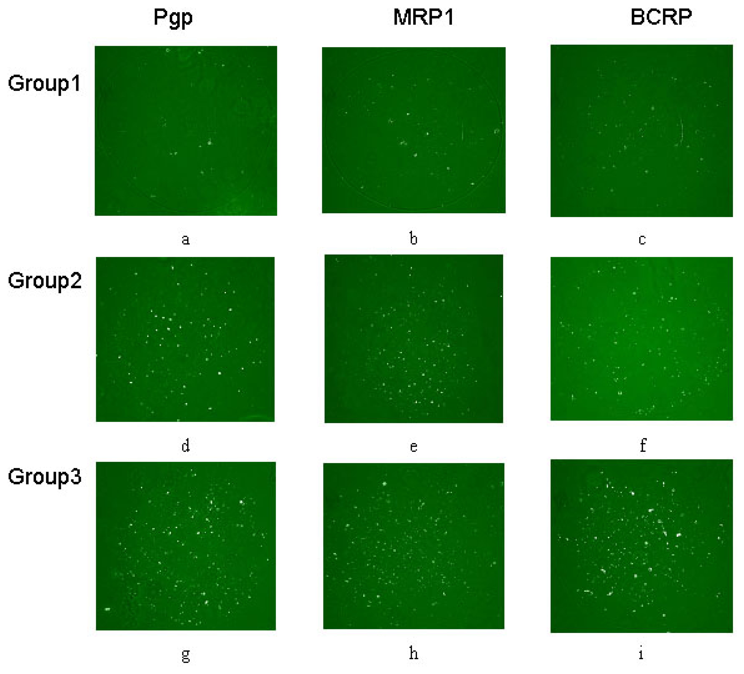
The concentration of leukemia and normal cells was studied at 1.0×10⁴ cells/mL. The cells captured three monoclonal antibodies respectively, which are shown in Figure 2 from a to i (took 1 case/group binding P-gp antibody for illustration).
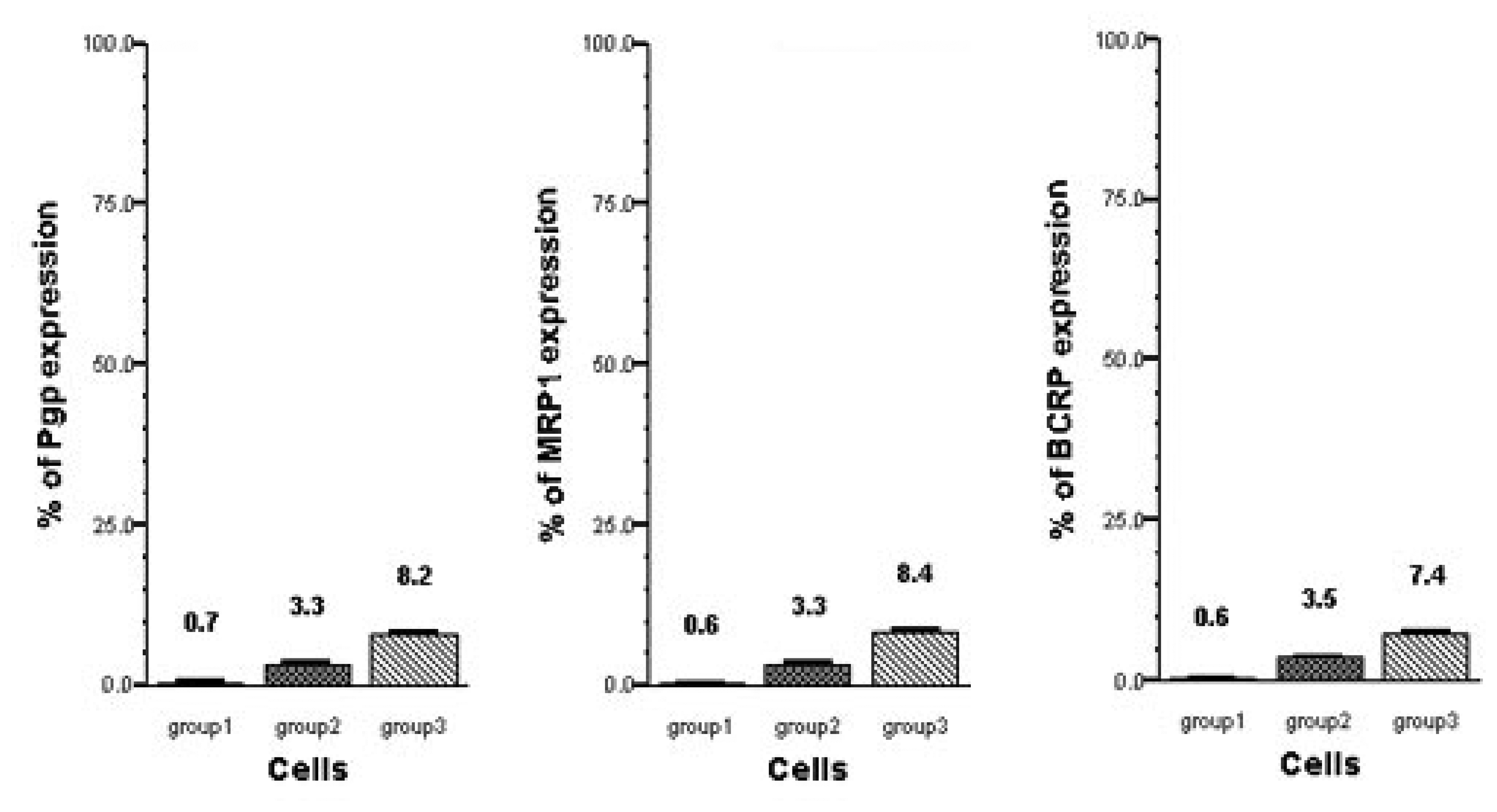
Figures 2a-c showed the result of normal cells binding P-gp antibody. Figures d-f displayed untreated leukemia cells, respectively. The results of relapsed-refractory AL patients' cells were revealed on Figures 2g-i. The numbers of captured cells of every spot were calculated with the computer software and the MDR protein expression difference was analyzed in three groups (Fig. 3). The analysis demonstrated that P-gp, MRP1 and BCRP were overexpressed in leukemia cells and the level of these proteins expression in relapsed-refractory group was significantly higher than those in untreated and normal control groups. Cells expressions of the untreated group were significantly higher than normal control cells which expressed very low.
Flow cytometry determination results
Conclusions
The images show that the method was sensitive and specific. The preparation of protein arrays is simple and cost-efficient. The detection process can be achieved rapidly in 2 hours. The modification method to modify the glass slide for protein imprinting is easy to do. For the determination of captured cells, only a microscope was required to observe the results and a CCD camera to capture the images. Protein arrays could be designed in various patterns by spotting different proteins on the same chip. Different cells could be detected simultaneously with the same chip. This novel method for determination of human leukemia cells has the advantage of high throughput, high specificity, low sample consume, and low cost. To sum up, protein arrays have a brighter and better future in medical applications.
Acknowledgments
This work was supported by the National Natural Science Foundation of China (Grant No. 39970832).
References
- Arceci, R.J. Tumor cell survival and resistance to therapy. Curr. Opin. Hematol. 1996, 3, 279–287. [Google Scholar]
- Moscow, J.A.; Cowan, K.H. Multidrug resistance. J. Natl. Cancer Inst. 1988, 80, 14–20. [Google Scholar]
- Schneider, E.; Hunke, S. ATP-binding–cassette (ABC) transporter systems functional and structural aspects of the ATP-hyfrolyzing subunits/domains. FEMS Microbiol Rev. 1998, 22, 1–20. [Google Scholar]
- Lazaris, A.C.; Kavantzas, N.G.; Zorzos, H.S.; Tsavaris, N.V.; Davaris, P.S. Markers of drug resistance in relapsing colon cancer. J. Cancer Res. Clin. Oncol. 2002, 128, 114–118. [Google Scholar]
- Liang, P.; Pardee, A.B. Differential display of eukaryotic messenger RNA by means of the polymerase chain reaction. Science 1992, 257, 967–971. [Google Scholar]
- Markova, V. Quantitative analysis of the expression of apoptosis-related genes. Folia Med. 1998, 40, 51–57. [Google Scholar]
- Yang, Z.; Woodahl, E.L.; Wang, X.Y.; Bui, T.; Shen, D.D.; Ho, R.J. Semi-quantitative RT-PCR method to estimate full-length mRNA levels of the multidrug resistance gene. Biotechniques 2002, 33. [Google Scholar]
- Gygi, S.P.; Rochon, Y.; Franza, B.R.; Aebersold, R. Correlation between protein and mRNA abundance in yeast. Mol. Cell Biol. 1999, 19, 1720–1730. [Google Scholar]
- MacBeath, G.; Schreiber, S.L. Printing proteins as microarrays for high-throughput function determination. Science 2000, 289, 1760–1763. [Google Scholar]
- Ge, H. A universal protein array system for quantitative detection of protein-protein, protein-DNA, protein-RNA and protein-ligand interactions. Nucleic Acids Res. 2000, 28, e3. [Google Scholar]
- Haab, B.B.; Dunham, M.J.; Brown, P.O. Protein microarrays for highly parallel detection and quantitation of specific proteins and antibodies in complex solutions. Genome Biol. 2001, 2, 1–13. [Google Scholar]
- Cras, J.J.; Rowe-Taitt, C.A.; Nivens, D.A.; Ligler, F.S. Comparison of chemical cleaning methods of glass in preparation for silanization. Bioelectrons 1999, 14, 683–688. [Google Scholar]
- Cole, S.R.; Aylett, G.W.; Harvey, N.L.; Cambareri, A.C.; Ashman, L.K. Increased expression of c-Kit or its ligand Steel Factor is not a common feature of adult acute myeloid leukaemia. Leukemia 1996, 10, 288–231. [Google Scholar]
- Piehler, J.; Brecht, A.; Hehl, K.; Gauglitz, G. Colloids and Surfaces. Biointerfaces 1999, 3, 325–336. [Google Scholar]
- Musto, P.; Melillo, L.; Lombardi, G. High risk of early resistant relapse for leukemia patients with presence of multidrug resistance associated P-glycoprotein positive cells in complete remission. Br. J. Haematol. 1991, 77, 50–53. [Google Scholar]
- Poeta, G.D.; Stasi, R.; Venditti, A. Prognostic value of cell marker analysis in denove acute myeloid leukemia. Leukemia 1994, 8, 38–41. [Google Scholar]
- Campos, L.; Guyotat, D.; Archimbaud, E. Clinical significance of multidrug resistance P-glycoprotein expression on acute non-lymphoblastic leukemia cells at diagnosis. Blood 1992, 79, 473–476. [Google Scholar]
- Ross, D.D.; Karp, J.E.; Chen, T.T. Expression of breast cancer resistance protein in blast cells from patients with acute leukemia. Blood 2000, 96, 365–368. [Google Scholar]
Figure 1.
The relapsed-refractory AL patients' cells combine antibodies onto the agarose film modified glass.
Figure 1.
The relapsed-refractory AL patients' cells combine antibodies onto the agarose film modified glass.
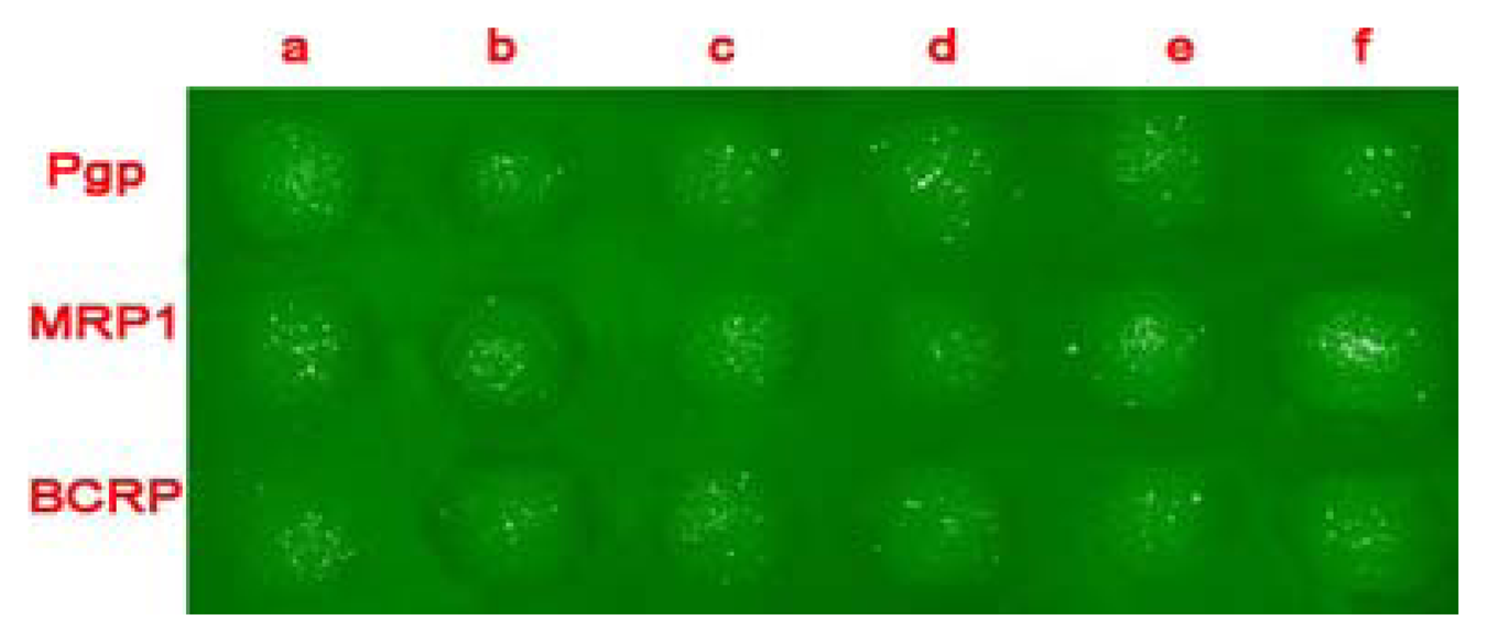
Figure 2.
(a to i). Protein array determined Pgp, MRP1 and BCRP in sample cells. Fig.a-c showed the result of normal cells binding P-gp antibody. Fig. d-f displayed untreated leukemia cells. The results of relapsed-refractory AL patients' cells were revealed on figures g-i.
Figure 2.
(a to i). Protein array determined Pgp, MRP1 and BCRP in sample cells. Fig.a-c showed the result of normal cells binding P-gp antibody. Fig. d-f displayed untreated leukemia cells. The results of relapsed-refractory AL patients' cells were revealed on figures g-i.

Figure 3.
Protein array determined every MDR protein expression level which was analyzed in three groups. The data represent the mean of 5 experiments.
Figure 3.
Protein array determined every MDR protein expression level which was analyzed in three groups. The data represent the mean of 5 experiments.

Figure 4.
Pgp, MRP1 and BCRP were assayed by flow cytometry.

© 2005 by MDPI ( http://www.mdpi.org). Reproduction is permitted for noncommercial purposes.
Share and Cite
MDPI and ACS Style
Du, J.; Chen, B.; Zhang, C.; Xu, X.; Cheng, J.; Gao, F.; Lu, Z. Protein Arrays for Multidrug-resistance in Human Leukemia Cell Determination. Sensors 2005, 5, 250-257. https://doi.org/10.3390/s5040259
AMA Style
Du J, Chen B, Zhang C, Xu X, Cheng J, Gao F, Lu Z. Protein Arrays for Multidrug-resistance in Human Leukemia Cell Determination. Sensors. 2005; 5(4):250-257. https://doi.org/10.3390/s5040259
Chicago/Turabian StyleDu, Juan, Baoan Chen, Chunxiu Zhang, Xiaoxing Xu, Jian Cheng, Feng Gao, and Zuhong Lu. 2005. "Protein Arrays for Multidrug-resistance in Human Leukemia Cell Determination" Sensors 5, no. 4: 250-257. https://doi.org/10.3390/s5040259