Development of Graphene Quantum Dots-Based Optical Sensor for Toxic Metal Ion Detection

Abstract
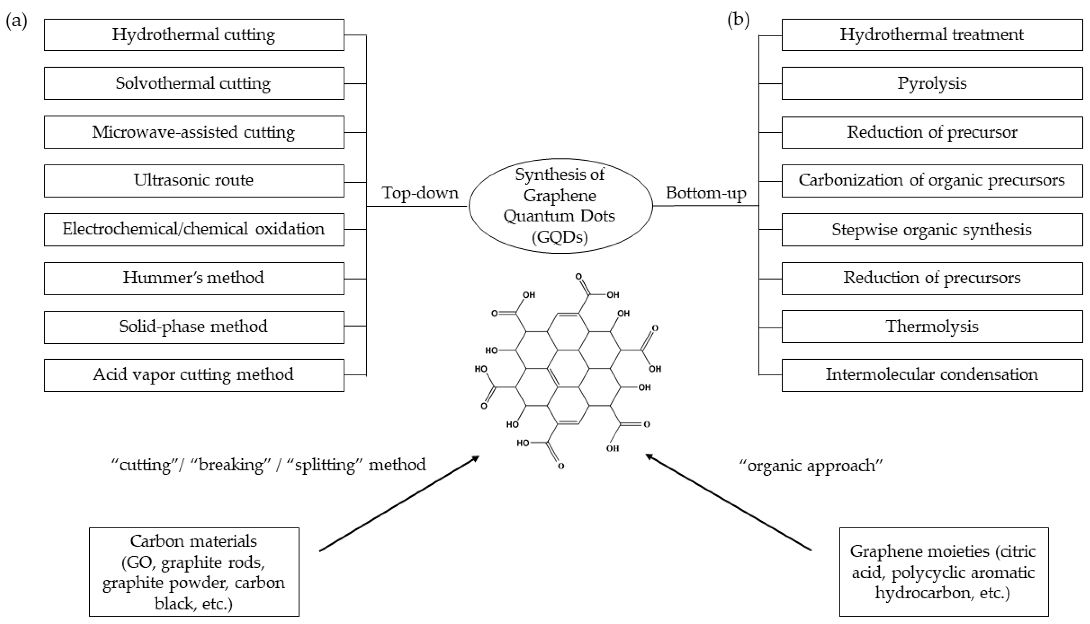
:1. Introduction
2. Incorporation of Graphene Quantum Dots with Optical Sensor for Toxic Metal Ion Detection
2.1. Ferric Ion (Fe3+)
2.2. Mercury Ion (Hg2+)
2.3. Lead Ion (Pb2+)
2.4. Copper Ion (Cu2+)
2.5. Silver Ion (Ag+)
2.6. Other Toxic Metal Ions
3. Emergence of Surface Plasmon Resonance as Alternative Optical Sensors for Metal Ion Detection
4. Future Trends in the Development of Graphene Quantum Dots-Based Surface Plasmon Resonance Optical Sensor for Toxic Metal Ion Detection
5. Conclusions
Author Contributions
Funding
Acknowledgments
Conflicts of Interest
References
- Sukumaran, L. A study of graphene. Int. J. Educ. Manag. Eng. 2014, 4, 9–14. [Google Scholar] [CrossRef]
- Ponomarenko, L.A.; Schedin, F.; Katsnelson, M.I.; Yang, R.; Hill, E.W.; Novoselov, K.S.; Geim, A.K. Chaotic Dirac billiard in graphene quantum dots. Science 2008, 320, 356–358. [Google Scholar] [CrossRef] [PubMed]
- Wei, D.; Liu, Y.; Wang, Y.; Zhang, H.; Huang, L.; Yu, G. Synthesis of N-doped graphene by chemical vapor deposition and its electrical properties. Nano Lett. 2009, 9, 1752–1758. [Google Scholar] [CrossRef] [PubMed]
- Sun, H.; Wu, L.; Wei, W.; Qu, X. Recent advances in graphene quantum dots for sensing. Mater. Today 2013, 16, 433–442. [Google Scholar] [CrossRef]
- Das, R.; Dhar, N.; Bandyopadhyay, A.; Jana, D. Size dependent magnetic and optical properties in diamond shaped graphene quantum dots: A DFT study. J. Phys. Chem. Solids 2016, 99, 34–42. [Google Scholar] [CrossRef]
- Pan, D.; Guo, L.; Zhang, J.; Xi, C.; Xue, Q.; Huang, H.; Li, J.; Zhang, Z.; Yu, W.; Chen, Z.; et al. Cutting sp2 clusters in graphene sheets into colloidal graphene quantum dots with strong green fluorescence. J. Mater. Chem. 2012, 22, 3314–3318. [Google Scholar] [CrossRef]
- Peng, J.; Gao, W.; Gupta, B.K.; Liu, Z.; Romero-Aburto, R.; Ge, L.; Song, L.; Alemany, L.B.; Zhan, X.; Gao, G.; et al. Graphene quantum dots derived from carbon fibers. Nano Lett. 2012, 12, 844–849. [Google Scholar] [CrossRef]
- Li, Y.; Hu, Y.; Zhao, Y.; Shi, G.; Deng, L.; Hou, Y.; Qu, L. An electrochemical avenue to green-luminescent graphene quantum dots as potential electron-acceptors for photovoltaics. Adv. Mater. 2011, 23, 776–780. [Google Scholar] [CrossRef]
- Dong, Y.; Shao, J.; Chen, C.; Li, H.; Wang, R.; Chi, Y.; Lin, X.; Chen, G. Blue luminescent graphene quantum dots and graphene oxide prepared by tuning the carbonization degree of citric acid. Carbon 2012, 50, 4738–4743. [Google Scholar] [CrossRef]
- Tang, L.; Ji, R.; Cao, X.; Lin, J.; Jiang, H.; Li, X.; Teng, K.S.; Luk, C.M.; Zeng, S.; Hao, J.; et al. Deep ultraviolet photoluminescence of water-soluble self-passivated graphene quantum dots. ACS Nano 2012, 6, 5102–5110. [Google Scholar] [CrossRef]
- Karimzadeh, A.; Hasanzadeh, M.; Shadjou, N.; de la Guardia, M. Optical bio(sensing) using nitrogen doped graphene quantum dots: Recent advances and future challenges. Trends Anal. Chem. 2018, 108, 110–121. [Google Scholar] [CrossRef]
- Suvarnaphaet, P.; Pechprasarn, S. Graphene-based materials for biosensors: A review. Sensors 2017, 17, 2161. [Google Scholar] [CrossRef] [PubMed]
- Li, M.; Gou, H.; Al-Ogaidi, I.; Wu, N. Nanostructured sensors for detection of heavy metals: A review. ACS Sustain. Chem. Eng. 2013, 1, 713–723. [Google Scholar] [CrossRef]
- Singh, R.; Gautam, N.; Mishra, A.; Gupta, R. Heavy metals and living systems: An overview. Indian J. Pharmacol. 2011, 43, 246. [Google Scholar] [CrossRef] [PubMed]
- Ochsenkuhn-Petropoulou, M.; Ochsenkuhn, K.M. Comparison of inductively coupled plasma-atomic emission spectrometry, anodic stripping voltammetry and instrumental neutron-activation analysis for the determination of heavy metals in airborne particulate matter. Fresenius J. Anal. Chem. 2001, 369, 629–632. [Google Scholar] [CrossRef] [PubMed]
- Rosolina, S.M.; Chambers, J.Q.; Lee, C.W.; Xue, Z.L. Direct determination of cadmium and lead in pharmaceutical ingredients using anodic stripping voltammetry in aqueous and DMSO/water solutions. Anal. Chim. Acta 2015, 893, 25–33. [Google Scholar] [CrossRef] [PubMed]
- Tarley, C.R.T.; Andrade, F.N.; De Oliveira, F.M.; Corazza, M.Z.; De Azevedo, L.F.M.; Segatelli, M.G. Synthesis and application of imprinted polyvinylimidazole-silica hybrid copolymer for Pb2+ determination by flow-injection thermospray flame furnace atomic absorption spectrometry. Anal. Chim. Acta 2011, 703, 145–151. [Google Scholar] [CrossRef] [PubMed]
- Ting, S.L.; Ee, S.J.; Ananthanarayanan, A.; Leong, K.C.; Chen, P. Graphene quantum dots functionalized gold nanoparticles for sensitive electrochemical detection of heavy metal ions. Electrochim. Acta 2015, 172, 7–11. [Google Scholar] [CrossRef]
- Ananthanarayanan, A.; Wang, X.; Routh, P.; Sana, B.; Lim, S.; Kim, D.H.; Lim, K.H.; Li, J.; Chen, P. Facile synthesis of graphene quantum dots from 3D graphene and their application for Fe3+ sensing. Adv. Funct. Mater. 2014, 24, 3021–3026. [Google Scholar] [CrossRef]
- Yu, C.; Guo, Y.; Liu, H.; Yan, N.; Xu, Z.; Yu, G.; Fang, Y.; Liu, Y. Ultrasensitive and selective sensing of heavy metal ions with modified graphene. Chem. Commun. 2013, 49, 6492–6494. [Google Scholar] [CrossRef] [PubMed]
- Chen, L.; Wu, C.; Du, P.; Feng, X.; Wu, P.; Cai, C. Electrolyzing synthesis of boron-doped graphene quantum dots for fluorescence determination of Fe3+ ions in water samples. Talanta 2017, 164, 100–109. [Google Scholar] [CrossRef] [PubMed]
- Zhou, L.; Geng, J.; Liu, B. Graphene quantum dots from polycyclic aromatic hydrocarbon for bioimaging and sensing of Fe3+ and hydrogen peroxide. Part. Part. Syst. Charact. 2013, 30, 1086–1092. [Google Scholar] [CrossRef]
- Ju, J.; Chen, W. Synthesis of highly fluorescent nitrogen-doped graphene quantum dots for sensitive, label-free detection of Fe (III) in aqueous media. Biosens. Bioelectron. 2014, 58, 219–225. [Google Scholar] [CrossRef] [PubMed]
- Xu, H.; Zhou, S.; Xiao, L.; Wang, H.; Li, S.; Yuan, Q. Fabrication of a nitrogen-doped graphene quantum dot from MOF-derived porous carbon and its application for highly selective fluorescence detection of Fe3+. J. Mater. Chem. C 2015, 3, 291–297. [Google Scholar] [CrossRef]
- Tam, T.V.; Trung, N.B.; Kim, H.R.; Chung, J.S.; Choi, W.M. One-pot synthesis of N-doped graphene quantum dots as a fluorescent sensing platform for Fe3+ ions detection. Sens. Actuators B Chem. 2014, 202, 568–573. [Google Scholar] [CrossRef]
- Li, S.; Li, Y.; Cao, J.; Zhu, J.; Fan, L.; Li, X. Sulfur-doped graphene quantum dots for highly selective and sensitive detection of Fe3+. Anal. Chem. 2014, 86, 10201–10207. [Google Scholar] [CrossRef] [PubMed]
- Li, L.; Li, L.; Wang, C.; Liu, K.; Zhu, R.; Qiang, H.; Lin, Y. Synthesis of nitrogen-doped and amino acid-functionalized graphene quantum dots from glycine, and their application to the fluorometric determination of ferric ion. Microchim. Acta 2014, 182, 763–770. [Google Scholar] [CrossRef]
- Xu, H.; Zhou, S.; Xiao, L.; Li, S.; Song, T.; Wang, Y.; Yuan, Q. Nanoreactor-confined synthesis and separation of yellow-luminescent graphene quantum dots with a recyclable SBA-15 template and their application for Fe(III) sensing. Carbon 2015, 87, 215–225. [Google Scholar] [CrossRef]
- Xu, F.; Shi, H.; He, X.; Wang, K.; He, D.; Yan, L.; Ye, X.; Tang, J.; Shangguan, J.; Luo, L. Masking agent-free and channel-switch-mode simultaneous sensing of Fe3+ and Hg2+ using dual-excitation graphene quantum dots. Analyst 2015, 140, 3925–3928. [Google Scholar] [CrossRef]
- Guo, R.; Zhou, S.; Li, Y.; Li, X.; Fan, L.; Voelcker, N.H. Rhodamine-functionalized graphene quantum dots for detection of Fe3+ in cancer stem cells. ACS Appl. Mater. Interfaces 2015, 7, 23958–23966. [Google Scholar] [CrossRef]
- Zhang, C.; Cui, Y.; Song, L.; Liu, X.; Hu, Z. Microwave assisted one-pot synthesis of graphene quantum dots as highly sensitive fluorescent probes for detection of iron ions and pH value. Talanta 2016, 150, 54–60. [Google Scholar] [CrossRef] [PubMed]
- Xu, L.; Mao, W.; Huang, J.; Li, S.; Huang, K.; Li, M.; Xia, J.; Chen, Q. Economical, green route to highly fluorescence intensity carbon materials based on ligninsulfonate/graphene quantum dots composites: Application as excellent fluorescent sensing platform for detection of Fe3+ ions. Sens. Actuators B Chem. 2016, 230, 54–60. [Google Scholar] [CrossRef]
- Zhang, W.; Gan, J. Synthesis of blue-photoluminescent graphene quantum dots/polystyrenic anion-exchange resin for Fe(III) detection. Appl. Surf. Sci. 2016, 372, 145–151. [Google Scholar] [CrossRef]
- Chowdhury, A.D.; Doong, R.A. Highly sensitive and selective detection of nanomolar ferric ions using dopamine functionalized graphene quantum dots. ACS Appl. Mater. Interfaces 2016, 8, 21002–21010. [Google Scholar] [CrossRef] [PubMed]
- Ma, Q.; Song, J.; Wang, S.; Yang, J.; Guo, Y.; Dong, C. A general sensing strategy for detection of Fe3+ by using amino acid-modified graphene quantum dots as fluorescent probe. Appl. Surf. Sci. 2016, 389, 995–1002. [Google Scholar] [CrossRef]
- Wang, X.; Li, R.; Fan, S.; Li, Z.; Wang, G.; Gu, Z.; Liu, J. D-penicillamine-functionalized graphene quantum dots for fluorescent detection of Fe3+ in iron supplement oral liquids. Sens. Actuators B Chem. 2017, 243, 211–220. [Google Scholar] [CrossRef]
- Xu, T.T.; Yang, J.X.; Song, J.M.; Chen, J.S.; Niu, H.L.; Mao, C.J.; Zhang, S.Y.; Shen, Y.H. Synthesis of high fluorescence graphene quantum dots and their selective detection for Fe3+ in aqueous solution. Sens. Actuators B Chem. 2017, 243, 863–872. [Google Scholar] [CrossRef]
- Shen, C.; Ge, S.; Pang, Y.; Xi, F.; Liu, J.; Dong, X.; Chen, P. Facile and scalable preparation of highly luminescent N, S co-doped graphene quantum dots and their application for parallel detection of multiple metal ions. J. Mater. Chem. B 2017, 5, 6593–6600. [Google Scholar] [CrossRef]
- Xia, C.; Hai, X.; Chen, X.W.; Wang, J.H. Simultaneously fabrication of free and solidified N, S-doped graphene quantum dots via a facile solvent-free synthesis route for fluorescent detection. Talanta 2017, 168, 269–278. [Google Scholar] [CrossRef]
- Zhu, X.; Zhang, Z.; Xue, Z.; Huang, C.; Shan, Y.; Liu, C.; Qin, X.; Yang, W.; Chen, X.; Wang, T. Understanding the selective detection of Fe3+ based on graphene quantum dots as fluorescent probes: The K a metal hydroxide-assisted mechanism. Anal. Chem. 2017, 89, 12054–12058. [Google Scholar] [CrossRef]
- Wang, W.; Wang, Z.; Liu, J.; Peng, Y.; Yu, X.; Wang, W.; Zhang, Z.; Sun, L. One-pot facile synthesis of graphene quantum dots from rice husks for Fe3+ sensing. Ind. Eng. Chem. Res. 2018, 57, 9144–9150. [Google Scholar] [CrossRef]
- Gao, X.X.; Zhou, X.; Ma, Y.F.; Wang, C.P.; Chu, F.X. A fluorometric and colorimetric dual-mode sensor based on nitrogen and iron co-doped graphene quantum dots for detection of ferric ions in biological fluids and cellular imaging. New J. Chem. 2018, 42, 14751–14756. [Google Scholar] [CrossRef]
- Wang, R.; Fan, H.; Jiang, W.; Ni, G.; Qu, S. Amino-functionalized graphene quantum dots prepared using high-softening point asphalt and their application in Fe3+ detection. Appl. Surf. Sci. 2019, 467–468, 446–455. [Google Scholar] [CrossRef]
- Xie, P.; Guo, F.; Yang, S.; Yao, D.; Yang, G.; Xie, L. A Novel ratiometric fluorescent mercury probe based on deprotonation-ICT mechanism. J. Fluoresc. 2014, 24, 473–480. [Google Scholar] [CrossRef] [PubMed]
- Zhang, H.; Wang, X.; Yang, X.; Pei, J.; Zhu, H. Synthesis of cysteamine-coated CdTe quantum dots and its application in mercury (II) detection. Anal. Chim. Acta 2012, 757, 63–68. [Google Scholar]
- Chakraborti, H.; Sinha, S.; Ghosh, S.; Pal, S.K. Interfacing water soluble nanomaterials with fluorescence chemosensing: Graphene quantum dot to detect Hg2+ in 100% aqueous solution. Mater. Lett. 2013, 97, 78–80. [Google Scholar] [CrossRef]
- Wang, B.; Zhuo, S.; Chen, L.; Zhang, Y. Fluorescent graphene quantum dot nanoprobes for the sensitive and selective detection of mercury ions. Spectrochim. Acta Part A Mol. Biomol. Spectrosc. 2014, 131, 384–387. [Google Scholar] [CrossRef]
- Li, Z.; Wang, Y.; Ni, Y.; Kokot, S. A rapid and label-free dual detection of Hg (II) and cysteine with the use of fluorescence switching of graphene quantum dots. Sens. Actuators B Chem. 2015, 207, 490–497. [Google Scholar] [CrossRef]
- Zhao, X.; Gao, J.; He, X.; Cong, L.; Zhao, H.; Li, X.; Tan, F. DNA-modified graphene quantum dots as a sensing platform for detection of Hg2+ in living cells. RSC Adv. 2015, 5, 39587–39591. [Google Scholar] [CrossRef]
- Shi, B.; Zhang, L.; Lan, C.; Zhao, J.; Su, Y.; Zhao, S. One-pot green synthesis of oxygen-rich nitrogen-doped graphene quantum dots and their potential application in pH-sensitive photoluminescence and detection of mercury(II) ions. Talanta 2015, 142, 131–139. [Google Scholar] [CrossRef]
- Liu, M.; Liu, T.; Li, Y.; Xu, H.; Zheng, B.; Wang, D.; Du, J.; Xiao, D. A FRET chemsensor based on graphene quantum dots for detecting and intracellular imaging of Hg2+. Talanta 2015, 143, 442–449. [Google Scholar] [CrossRef] [PubMed]
- Hua, M.; Wang, C.; Qian, J.; Wang, K.; Yang, Z.; Liu, Q.; Mao, H.; Wang, K. Preparation of graphene quantum dots based core-satellite hybrid spheres and their use as the ratiometric fluorescence probe for visual determination of mercury(II) ions. Anal. Chim. Acta 2015, 888, 173–181. [Google Scholar] [CrossRef] [PubMed]
- Tam, T.V.; Hong, S.H.; Choi, W.M. Facile synthesis of cysteine-functionalized graphene quantum dots for a fluorescence probe for mercury ions. RSC Adv. 2015, 5, 97598–97603. [Google Scholar] [CrossRef]
- Achadu, O.J.; Nyokong, T. Application of graphene quantum dots functionalized with thymine and thymine-appended zinc phthalocyanine as novel photoluminescent nanoprobes. New J. Chem. 2017, 41, 1447–1458. [Google Scholar] [CrossRef]
- Xiaoyan, Z.; Zhangyi, L.; Zaijun, L. Fabrication of valine-functionalized graphene quantum dots and its use as a novel optical probe for sensitive and selective detection of Hg2+. Spectrochim. Acta A Mol. Biomol. Spectrosc. 2017, 171, 415–424. [Google Scholar] [CrossRef] [PubMed]
- Achadu, O.J.; Nyokong, T. Graphene quantum dots coordinated to mercaptopyridine-substituted phthalocyanines: Characterization and application as fluorescence “turn ON” nanoprobes. Spectrochim. Acta A Mol. Biomol. Spectrosc. 2017, 174, 339–347. [Google Scholar] [CrossRef] [PubMed]
- Alvand, M.; Shemirani, F. A Fe3O4@SiO2@graphene quantum dot core-shell structured nanomaterial as a fluorescent probe and for magnetic removal of mercury(II) ion. Microchim. Acta 2017, 184, 1621–1629. [Google Scholar] [CrossRef]
- Achadu, O.J.; Nyokong, T. Graphene quantum dots anchored onto mercaptopyridine-substituted zinc phthalocyanine-Au@Ag nanoparticle hybrid: Application as fluorescence “off-on-off” sensor for Hg2+ and biothiols. Dyes Pigments 2017, 145, 189–201. [Google Scholar] [CrossRef]
- Amini, M.H.; Faridbod, F.; Ganjali, M.R.; Norouzi, P. Functionalized graphene quantum dots as a fluorescent “off–on” nanosensor for detection of mercury and ethyl xanthate. Res. Chem. Intermed. 2017, 43, 7457–7470. [Google Scholar] [CrossRef]
- Anh, N.T.N.; Chowdhury, A.D.; Doong, R. Highly sensitive and selective detection of mercury ions using N, S-codoped graphene quantum dots and its paper strip based sensing application in wastewater. Sens. Actuators B Chem. 2017, 252, 1169–1178. [Google Scholar] [CrossRef]
- Yang, L.; Qin, A.; Chen, S.; Liao, L.; Qin, J.; Zhang, K. Manganese(II) enhanced fluorescent nitrogen-doped graphene quantum dots: A facile and efficient synthesis and their applications for bioimaging and detection of Hg2+ ions. RSC Adv. 2018, 8, 5902–5911. [Google Scholar] [CrossRef]
- Yan, P.; Li, R.; Yang, Y.; Li, Z.; Gu, Z.; Wang, G.; Liu, J. Pentaethylenehexamine and D-Penicillamine co-functionalized graphene quantum dots for fluorescent detection of mercury(II) and glutathione and bioimaging. Spectrochim. Acta A Mol. Biomol. Spectrosc. 2018, 203, 139–146. [Google Scholar]
- Su, D.; Wang, M.; Liu, Q.; Qu, Z.; Su, X. A novel fluorescence strategy for mercury ions and trypsin activity assay based on nitrogen-doped graphene quantum dots. New J. Chem. 2018, 42, 17083–17090. [Google Scholar] [CrossRef]
- Qu, C.; Zhang, D.; Yang, R.; Hu, J.; Qu, L. Nitrogen and sulfur co-doped graphene quantum dots for the highly sensitive and selective detection of mercury ion in living cells. Spectrochim. Acta A Mol. Biomol. Spectrosc. 2019, 206, 588–596. [Google Scholar] [CrossRef] [PubMed]
- Yang, Y.; Xiao, X.; Xing, X.; Wang, Z.; Zou, T.; Wang, Z.; Zhao, R.; Wang, Y. Rhodamine B assisted graphene quantum dots flourescent sensor system for sensitive recognition of mercury ions. J. Lumin. 2019, 207, 273–281. [Google Scholar] [CrossRef]
- Tang, Y.; Li, J.; Guo, Q.; Nie, G. An ultrasensitive electrochemiluminescence assay for Hg2+ through graphene quantum dots and poly(5-formylindole) nanocomposite. Sens. Actuators B Chem. 2019, 282, 824–830. [Google Scholar] [CrossRef]
- Flora, G.; Gupta, D.; Tiwari, A. Toxicity of lead: A review with recent updates. Interdiscip. Toxicol. 2012, 5, 47–58. [Google Scholar] [CrossRef]
- De Mattos, G.F.; Costa, C.; Savio, F.; Alonso, M.; Nicolson, G.L. Lead poisoning: Acute exposure of the heart to lead ions promotes changes in cardiac function and Cav1.2 ion channels. Biophys. Rev. 2017, 9, 807–825. [Google Scholar] [CrossRef]
- Qi, Y.X.; Zhang, M.; Fu, Q.Q.; Liu, R.; Shi, G.Y. Highly sensitive and selective fluorescent detection of cerebral lead(II) based on graphene quantum dot conjugates. Chem. Commun. 2013, 49, 10599–10601. [Google Scholar] [CrossRef]
- Dong, Y.; Tian, W.; Ren, S.; Dai, R.; Chi, Y.; Chen, G. Graphene quantum dots/L-cysteine coreactant electrochemiluminescence system and its application in sensing lead(II) ions. ACS Appl. Mater. Interfaces 2014, 6, 1646–1651. [Google Scholar] [CrossRef]
- Qian, Z.S.; Shan, X.Y.; Chai, L.J.; Chen, J.R.; Feng, H. A fluorescent nanosensor based on graphene quantum dots-aptamer probe and graphene oxide platform for detection of lead (II) ion. Biosens. Bioelectron. 2015, 68, 225–231. [Google Scholar] [CrossRef] [PubMed]
- Bian, S.; Shen, C.; Hua, H.; Zhou, L.; Zhu, H.; Xi, F.; Liu, J.; Dong, X. One-pot synthesis of sulfur-doped graphene quantum dots as a novel fluorescent probe for highly selective and sensitive detection of lead(II). RSC Adv. 2016, 6, 69977–69983. [Google Scholar] [CrossRef]
- Niu, X.; Zhong, Y.; Chen, R.; Wang, F.; Liu, Y.; Luo, D. A “turn-on” fluorescence sensor for Pb2+ Detection based on graphene quantum dots and gold nanoparticles. Sens. Actuators B Chem. 2018, 255, 1577–1581. [Google Scholar] [CrossRef]
- Sun, X.; Peng, Y.; Lin, Y.; Cai, L.; Li, F.; Liu, B. G-quadruplex formation enhancing energy transfer in self-assembled multilayers and fluorescence recognize for Pb2+ ions. Sens. Actuators B Chem. 2018, 255, 2121–2125. [Google Scholar] [CrossRef]
- Xu, Y.; Wang, S.; Hou, X.; Sun, Z.; Jiang, Y.; Dong, Z.; Tao, Q.; Man, J.; Cao, Y. Coal-derived nitrogen, phosphorus and sulfur co-doped graphene quantum dots: A promising ion fluorescent probe. Appl. Surf. Sci. 2018, 445, 519–526. [Google Scholar] [CrossRef]
- Kaewprom, C.; Sricharoen, P.; Limchoowong, N.; Nuengmatcha, P.; Chanthai, S. Resonance light scattering sensor of the metal complex nanoparticles using diethyl dithiocarbamate doped graphene quantum dots for highly Pb(II)-sensitive detection in water sample. Spectrochim. Acta A Mol. Biomol. Spectrosc. 2019, 207, 79–87. [Google Scholar] [CrossRef] [PubMed]
- Ochoa-Herrera, V.; León, G.; Banihani, Q.; Field, J.A.; Sierra-Alvarez, R. Toxicity of copper(II) ions to microorganisms in biological wastewater treatment systems. Sci. Total Environ. 2011, 412–413, 380–385. [Google Scholar] [CrossRef]
- Georgopoulos, P.G.; Roy, A.; Yonone-Lioy, M.J.; Opiekun, R.E.; Lioy, P.J. Environmental copper: Its dynamics and human. J. Toxicol. Environ. Heal. Part B Crit. Rev. 2011, 4, 341–394. [Google Scholar] [CrossRef]
- Sun, H.; Gao, N.; Wu, L.; Ren, J.; Wei, W.; Qu, X. Highly photoluminescent amino-functionalized graphene quantum dots used for sensing copper ions. Chem. A Eur. J. 2013, 19, 13362–13368. [Google Scholar] [CrossRef]
- Wang, F.; Gu, Z.; Lei, W.; Wang, W.; Xia, X.; Hao, Q. Graphene quantum dots as a fluorescent sensing platform for highly efficient detection of copper(II) ions. Sens. Actuators B Chem. 2014, 190, 516–522. [Google Scholar] [CrossRef]
- Liu, X.; Gao, W.; Zhou, X.; Ma, Y. Pristine graphene quantum dots for detection of copper ions. J. Mater. Res. 2014, 29, 1401–1407. [Google Scholar] [CrossRef]
- Liu, Y.; Kim, D.Y. Ultraviolet and blue emitting graphene quantum dots synthesized from carbon nano-onions and their comparison for metal ion sensing. Chem. Commun. 2015, 51, 4176–4179. [Google Scholar] [CrossRef] [PubMed]
- Lin, L.; Song, X.; Chen, Y.; Rong, M.; Wang, Y.; Zhao, L.; Zhao, T.; Chen, X. Europium-decorated graphene quantum dots as a fluorescent probe for label-free, rapid and sensitive detection of Cu2+ and L-cysteine. Anal. Chim. Acta 2015, 891, 261–268. [Google Scholar] [CrossRef] [PubMed]
- Sun, X.; Liu, P.; Wu, L.; Liu, B. Graphene-quantum-dots-based ratiometric fluorescent probe for visual detection of copper ion. Analyst 2015, 140, 6742–6747. [Google Scholar] [CrossRef] [PubMed]
- Li, Y.; Liu, X.; Li, Q.; Ge, J.; Liu, H.; Li, S.; Wang, L.; Wang, J.; Ma, N. Post-oxidation treated graphene quantum dots as a fluorescent probe for sensitive detection of copper ions. Chem. Phys. Lett. 2016, 664, 127–132. [Google Scholar] [CrossRef]
- Wang, C.; Yang, F.; Tang, Y.; Yang, W.; Zhong, H.; Yu, C.; Li, R.; Zhou, H.; Li, Y.; Mao, L. Graphene quantum dots nanosensor derived from 3D nanomesh graphene frameworks and its application for fluorescent sensing of Cu2+ in rat brain. Sens. Actuators B Chem. 2018, 258, 672–681. [Google Scholar] [CrossRef]
- Choi, Y.; Kim, H.A.; Kim, K.W.; Lee, B.T. Comparative toxicity of silver nanoparticles and silver ions to Escherichia coli. J. Environ. Sci. 2018, 66, 50–60. [Google Scholar] [CrossRef]
- Patte, H.T. Bioaccumulation and toxicity of silver compounds: A review. Environ. Toxicol. Chem. 1999, 18, 89–108. [Google Scholar]
- Li, L.L.; Ji, J.; Fei, R.; Wang, C.Z.; Lu, Q.; Zhang, J.R.; Jiang, L.P.; Zhu, J.J. A facile microwave avenue to electrochemiluminescent two-color graphene quantum dots. Adv. Funct. Mater. 2012, 22, 2971–2979. [Google Scholar] [CrossRef]
- Ran, X.; Sun, H.; Pu, F.; Ren, J.; Qu, X. Ag nanoparticle-decorated graphene quantum dots for label-free, rapid and sensitive detection of Ag+ and biothiols. Chem. Commun. 2013, 49, 1079–1081. [Google Scholar] [CrossRef]
- Suryawanshi, A.; Biswal, M.; Mhamane, D.; Gokhale, R.; Patil, S.; Guin, D.; Ogale, S. Large scale synthesis of graphene quantum dots (GQDs) from waste biomass and their use as an efficient and selective photoluminescence on-off-on probe for Ag+ ions. Nanoscale 2014, 6, 11664–11670. [Google Scholar] [CrossRef] [PubMed]
- Tabaraki, R.; Nateghi, A. Nitrogen-doped graphene quantum dots: “turn-off” fluorescent probe for detection of Ag+ ions. J. Fluoresc. 2016, 26, 297–305. [Google Scholar] [CrossRef] [PubMed]
- Bian, S.; Shen, C.; Qian, Y.; Liu, J.; Xi, F.; Dong, X. Facile synthesis of sulfur-doped graphene quantum dots as fluorescent sensing probes for Ag+ ions detection. Sens. Actuators B Chem. 2017, 242, 231–237. [Google Scholar] [CrossRef]
- Kaewanan, P.; Sricharoen, P.; Limchoowong, N.; Sripakdee, T.; Nuengmatcha, P.; Chanthai, S. A fluorescence switching sensor based on graphene quantum dots decorated with Hg2+ and hydrolyzed thioacetamide for highly Ag+-sensitive and selective detection. RSC Adv. 2017, 7, 48058–48067. [Google Scholar] [CrossRef]
- Yang, X.; Wang, E. A nanoparticle autocatalytic sensor for Ag+ and Cu2+ ions in aqueous solution with high sensitivity and selectivity and its application in test paper. Anal. Chem. 2011, 83, 5005–5011. [Google Scholar] [CrossRef] [PubMed]
- Zhao, X.E.; Lei, C.; Gao, Y.; Gao, H.; Zhu, S.; Yang, X.; You, J.; Wang, H. A ratiometric fluorescent nanosensor for the detection of silver ions using graphene quantum dots. Sens. Actuators B Chem. 2017, 253, 239–246. [Google Scholar] [CrossRef]
- Jaishankar, M.; Tseten, T.; Anbalagan, N.; Mathew, B.B.; Beeregowda, K.N. Toxicity, mechanism and health effects of some heavy metals. Interdiscip. Toxicol. 2014, 7, 60–72. [Google Scholar] [CrossRef] [Green Version]
- Fan, Z.; Li, Y.; Li, X.; Fan, L.; Zhou, S.; Fang, D.; Yang, S. Surrounding media sensitive photoluminescence of boron-doped graphene quantum dots for highly fluorescent dyed crystals, chemical sensing and bioimaging. Carbon 2014, 70, 149–156. [Google Scholar] [CrossRef]
- Fang, B.Y.; Li, C.; Song, Y.Y.; Tan, F.; Cao, Y.C.; Zhao, Y.D. Nitrogen-doped graphene quantum dot for direct fluorescence detection of Al3+ in aqueous media and living cells. Biosens. Bioelectron. 2018, 100, 41–48. [Google Scholar] [CrossRef]
- Zhang, L.; Peng, D.; Liang, R.P.; Qiu, J.D. Nitrogen-doped graphene quantum dots as a new catalyst accelerating the coordination reaction between cadmium(II) and 5,10,15,20-tetrakis(1-methyl-4-pyridinio)porphyrin for cadmium(II) sensing. Anal. Chem. 2015, 87, 10894–10901. [Google Scholar] [CrossRef]
- Chen, H.; Li, W.; Wang, Q.; Jin, X.; Nie, Z.; Yao, S. Nitrogen doped graphene quantum dots based single-luminophor generated dual-potential electrochemiluminescence system for ratiometric sensing of Co2+ ion. Electrochim. Acta 2016, 214, 94–102. [Google Scholar] [CrossRef]
- Huang, H.; Liao, L.; Xu, X.; Zou, M.; Liu, F.; Li, N. The electron-transfer based interaction between transition metal ions and photoluminescent graphene quantum dots (GQDs): A platform for metal ion sensing. Talanta 2013, 117, 152–157. [Google Scholar] [CrossRef] [PubMed]
- Wood, R.W. On a remarkable case of uneven distribution of light in a diffraction grating spectrum. Proc. Phys. Soc. Lond. 1902, 18, 269. [Google Scholar] [CrossRef]
- Raether, H. Surface Plasmons on Smooth and Rough Surfaces and on Gratings; Springer: Berlin/Heidelberg, Germany, 1988; pp. 4–39. [Google Scholar]
- Cullen, D.C.; Brown, R.G.W.; Lowe, C.R. Detection of immuno-complex formation via surface plasmon resonance on gold-coated diffraction gratings. Biosensors 1987, 3, 211–225. [Google Scholar] [CrossRef]
- Homola, J. On the sensitivity of surface plasmon resonance sensors with spectral interrogation. Sens. Actuators B Chem. 1997, 41, 207–211. [Google Scholar] [CrossRef]
- Rossi, S.; Gazzola, E.; Capaldo, P.; Borile, G.; Romanato, F. Grating-coupled surface plasmon resonance (GC-SPR) optimization for phase-interrogation biosensing in a microfluidic chamber. Sensors 2018, 18, 1621. [Google Scholar] [CrossRef] [PubMed]
- Dostálek, J.; Homola, J.; Miler, M. Rich information format surface plasmon resonance biosensor based on array of diffraction gratings. Sens. Actuators B Chem. 2005, 107, 154–161. [Google Scholar] [CrossRef]
- Piliarik, M.; Homola, J. Surface plasmon resonance (SPR) sensors: Approaching their limits? Opt. Express 2009, 17, 16505–16517. [Google Scholar] [CrossRef]
- Moharam, M.; Gaylord, T.K. Rigorous coupled-wave analysis of metallic surface-relief gratings. J. Opt. Soc. Am. 1986, 3, 26–35. [Google Scholar] [CrossRef]
- Kolomenskii, A.A.; Gershon, P.D.; Schuessler, H.A. Sensitivity and detection limit of concentration surface-plasmon resonance. Appl. Opt. 1997, 36, 6539–6547. [Google Scholar] [CrossRef] [PubMed]
- Matsubara, K.; Kawata, S.; Minami, S. Optical chemical sensor based on surface plasmon measurement. Appl. Opt. 1988, 27, 1160–1163. [Google Scholar] [CrossRef] [PubMed]
- Löfås, S.; Malmqvist, M.; Rönnberg, I.; Stenberg, E.; Liedberg, B.; Lundström, I. Bioanalysis with surface plasmon resonance. Sens. Actuators B Chem. 1991, 5, 79–84. [Google Scholar] [CrossRef]
- Homola, J. Present and future of surface plasmon resonance biosensors. Anal. Bioanal. Chem. 2003, 377, 528–539. [Google Scholar] [CrossRef] [PubMed]
- Fen, Y.W.; Yunus, W.M.M. Surface plasmon resonance spectroscopy as an alternative for sensing heavy metal ions: A review. Sens. Rev. 2013, 33, 305–314. [Google Scholar]
- Chah, S.; Yi, J.; Zare, R.N. Surface plasmon resonance analysis of aqueous mercuric ions. Sens. Actuators B Chem. 2004, 99, 216–222. [Google Scholar] [CrossRef]
- Chen, H.; Gal, Y.S.; Kim, S.H.; Choi, H.J.; Oh, M.C.; Lee, J.; Koh, K. Potassium ion sensing using a self-assembled calix[4]crown monolayer by surface plasmon resonance. Sens. Actuators B Chem. 2008, 133, 577–581. [Google Scholar] [CrossRef]
- Yu, J.C.C.; Lai, E.P.C.; Sadeghi, S. Surface plasmon resonance sensor for Hg(II) detection by binding interactions with polypyrrole and 2-mercaptobenzothiazole. Sens. Actuators B Chem. 2004, 101, 236–241. [Google Scholar] [CrossRef]
- Sadrolhosseini, A.R.; Noor, A.S.M.; Bahrami, A.; Lim, H.N.; Talib, Z.A.; Mahdi, M.A. Application of polypyrrole multi-walled carbon nanotube composite layer for detection of mercury, lead and iron ions using surface plasmon resonance technique. PLoS ONE 2014, 9, 93962. [Google Scholar] [CrossRef]
- Fahnestock, K.J.; Manesse, M.; McIlwee, H.A.; Schauer, C.L.; Boukherroub, R.; Szunerits, S. Selective detection of hexachromium ions by localized surface plasmon resonance measurements using gold nanoparticles/chitosan composite interfaces. Analyst 2009, 134, 881–886. [Google Scholar] [CrossRef]
- Fen, Y.W.; Yunus, W.M.M.; Yusof, N.A. Detection of mercury and copper ions using surface plasmon resonance optical sensor. Sens. Mater. 2011, 23, 325. [Google Scholar]
- Fen, Y.W.; Yunus, W.M.M.; Yusof, N.A. Surface plasmon resonance optical sensor for detection of essential heavy metal ions with potential for toxicity: Copper, zinc and manganese ions. Sens. Lett. 2011, 9, 1704–1711. [Google Scholar] [CrossRef]
- Abdi, M.M.; Abdullah, L.C.; Sadrolhosseini, A.R.; Yunus, W.M.; Moksin, M.M.; Tahir, P.M. Surface plasmon resonance sensing detection of mercury and lead ions based on conducting polymer composite. PLoS ONE 2011, 6, 2–5. [Google Scholar] [CrossRef] [PubMed]
- Sadrolhosseini, A.R.; Noor, A.S.M.; Moksin, M.M.; Abdi, M.M.; Mohammadi, A. Application of polypyrrole-chitosan layer for detection of Zn (II) and Ni (II) in aqueous solutions using surface plasmon resonance. Int. J. Polym. Mater. Polym. Biomater. 2013, 62, 284–287. [Google Scholar] [CrossRef]
- Fen, Y.W.; Yunus, W.M.M.; Yusof, N.A. Surface plasmon resonance optical sensor for detection of Pb2+ based on immobilized p-tert-butylcalix[4]arene-tetrakis in chitosan thin film as an active layer. Sens. Actuators B Chem. 2012, 171–172, 287–293. [Google Scholar] [CrossRef]
- Fen, Y.W.; Yunus, W.M.M.; Talib, Z.A. Analysis of Pb(II) ion sensing by crosslinked chitosan thin film using surface plasmon resonance spectroscopy. Optik 2013, 124, 126–133. [Google Scholar] [CrossRef]
- Fen, Y.W.; Yunus, W.M.M.; Talib, Z.A.; Yusof, N.A. Development of surface plasmon resonance sensor for determining zinc ion using novel active nanolayers as probe. Spectrochim. Acta A Mol. Biomol. Spectrosc. 2015, 134, 48–52. [Google Scholar] [CrossRef]
- Verma, R.; Gupta, B.D. Detection of heavy metal ions in contaminated water by surface plasmon resonance based optical fibre sensor using conducting polymer and chitosan. Food Chem. 2015, 166, 568–575. [Google Scholar] [CrossRef]
- Sadrolhosseini, A.R.; Naseri, M.; Rashid, S.A. Polypyrrole-chitosan/nickel-ferrite nanoparticle composite layer for detecting heavy metal ions using surface plasmon resonance technique. Opt. Laser Technol. 2017, 93, 216–223. [Google Scholar] [CrossRef]
- Pelossof, G.; Tel-Vered, R.; Willner, I. Amplified surface plasmon resonance and electrochemical detection of Pb2+ ions using the Pb2+-dependent DNAzyme and hemin/g-quadruplex as a label. Anal. Chem. 2012, 84, 3703–3709. [Google Scholar] [CrossRef]
- Zhang, L.; Peng, D.; Liang, R.P.; Qiu, J.D. Graphene-based optical nanosensors for detection of heavy metal ions. Trends Anal. Chem. 2018, 102, 280–289. [Google Scholar] [CrossRef]
- Joshi, R.K.; Carbone, P.; Wang, F.C.; Kravets, V.G.; Su, Y.; Grigorieva, I.V.; Wu, H.A.; Geim, A.K.; Nair, R.R. Precise and ultrafast molecular sieving through graphene oxide membranes. Science 2014, 343, 752–754. [Google Scholar] [CrossRef] [PubMed]
- Lokman, N.F.; Bakar, A.A.A.; Suja, F.; Abdullah, H.; Rahman, W.B.W.A.; Huang, N.M.; Yaacob, M.H. Highly sensitive SPR response of Au/chitosan/graphene oxide nanostructured thin films toward Pb (II) ions. Sens. Actuators B Chem. 2014, 195, 459–466. [Google Scholar] [CrossRef]
- Kamaruddin, N.H.; Bakar, A.A.A.; Yaacob, M.H.; Mahdi, M.A.; Zan, M.S.D.; Shaari, S. Enhancement of chitosan-graphene oxide SPR sensor with a multi-metallic layers of Au-Ag-Au nanostructure for lead(II) ion detection. Appl. Surf. Sci. 2016, 361, 177–184. [Google Scholar] [CrossRef]
- Kamaruddin, N.H.; Bakar, A.A.A.; Mobarak, N.N.; Zan, M.S.D.; Arsad, N. Binding affinity of a highly sensitive Au/Ag/Au/chitosan-graphene oxide sensor based on direct detection of Pb2+ and Hg2+ ions. Sensors 2017, 17, 2277. [Google Scholar] [CrossRef] [PubMed]
- Saleviter, S.; Fen, Y.W.; Omar, N.A.S.; Zainudin, A.A.; Yusof, N.A. Development of optical sensor for determination of Co(II) based on surface plasmon resonance phenomenon. Sens. Lett. 2018, 15, 862–867. [Google Scholar] [CrossRef]
- Saleviter, S.; Fen, Y.W.; Omar, N.A.S.; Daniyal, W.M.E.M.M.; Abdullah, J.; Zaid, M.H.M. Structural and optical studies of cadmium sulfide quantum dot-graphene oxide-chitosan nanocomposite thin film as a novel SPR spectroscopy active layer. J. Nanomater. 2018, 2018, 4324072. [Google Scholar] [CrossRef]
- Zainudin, A.A.; Fen, Y.W.; Yusof, N.A.; Al-Rekabi, S.H.; Mahdi, M.A.; Omar, N.A.S. Incorporation of surface plasmon resonance with novel valinomycin doped chitosan-graphene oxide thin film for sensing potassium ion. Spectrochim. Acta A Mol. Biomol. Spectrosc. 2018, 191, 111–115. [Google Scholar] [CrossRef]
- Daniyal, W.M.E.M.M.; Fen, Y.W.; Abdullah, J.; Sadrolhosseini, A.R.; Saleviter, S.; Omar, N.A.S. Exploration of surface plasmon resonance for sensing copper ion based on nanocrystalline cellulose-modified thin film. Opt. Express 2018, 26, 34880–34894. [Google Scholar] [CrossRef]
- Daniyal, W.M.E.M.M.; Fen, Y.W.; Abdullah, J.; Sadrolhosseini, A.R.; Saleviter, S.; Omar, N.A.S. Label-free optical spectroscopy for characterizing binding properties of highly sensitive nanocrystalline cellulose-graphene oxide based nanocomposite towards nickel ion. Spectrochim. Acta A Mol. Biomol. Spectrosc. 2019, 212, 25–31. [Google Scholar] [CrossRef]
- Daniyal, W.M.E.M.M.; Saleviter, S.; Fen, Y.W. Development of surface plasmon resonance spectroscopy for metal ion detection. Sens. Mater. 2018, 30, 2023–2038. [Google Scholar] [CrossRef]
- Anas, N.A.A.; Fen, Y.W.; Omar, N.A.S.; Ramdzan, N.S.M.; Daniyal, W.M.E.M.M.; Saleviter, S.; Zainudin, A.A. Optical properties of chitosan/hydroxyl-functionalized graphene quantum dots thin film for potential optical detection of ferric (III) ion. Opt. Laser Technol. 2019, 120, 105724. [Google Scholar] [CrossRef]
- Ramdzan, N.S.M.; Fen, Y.W.; Omar, N.A.S.; Anas, N.A.A.; Daniyal, W.M.E.M.M.; Saleviter, S.; Zainudin, A.A. Optical and surface plasmon resonance sensing properties for chitosan/carboxyl-functionalized graphene quantum dots thin film. Optik 2019, 17, 802–812. [Google Scholar] [CrossRef]


| Optical Sensors | Advantages | Disadvantages |
|---|---|---|
| Fluorescent | High sensitivity and selectivity; real-time measurement; good reproducibility | Slightly slow detection; time consuming; limited application (small molecules) |
| Electrochemiluminescence | Good sensitivity and selectivity; stable; strong anti-interference ability; wide detection range | High cost; low compatibility; complicated preparation; frequent electrode fouling |
| Photoluminescent | High sensitivity and selectivity; real-time measurement; good reproducibility | Low precision and accuracy; time consuming; limited application (small molecules) |
| Colorimetric | Good sensitivity; fast detection; inexpensive | Low reproducibility; low stability; low selectivity |
| SPR | Very high sensitivity; simple; low cost; label-free | Low selectivity (improving) |
| Type of GQDs | Synthesis Method | Starting Materials | Optical Method | Linear Range | LOD 1 (nM) | References |
|---|---|---|---|---|---|---|
| P-GQDs | carbonization/hydrothermal | pyrene/hydrazine hydrate | Fluorescent probe | - | 5 | [22] |
| N-GQDs | pyrolysis/hydrothermal | citric acid/hydrazine | Fluorescent probe | 1–1945 µM | 90 | [23] |
| BMIM+-GQDs | electrochemical cutting | 3D graphene | Fluorescent sensor | 0–80 µM | 7220 | [19] |
| N-GQDs | acid vapor cutting | MOF-derived carbon | Fluorescent probe | 1–70 µM | 80 | [24] |
| N-GQDs | carbonization/hydrothermal | citric acid/ammonia | Fluorescent probe | 1–500 µM | 1000 | [25] |
| S-GQDs | electrolysis | graphite/sodium p-toluensulfonate | Fluorescent probe | 0–0.7 µM | 4.2 | [26] |
| NA-GQDs | thermolysis | glycine | Fluorescent sensor | 0.5–500 µM | 100 | [27] |
| GQDs | acid vapor cutting | SiO2(SBA-15) | Fluorescent probe | 3–60 µM | 300 | [28] |
| GQDs | pyrolysis | citric acid | Dual-channel fluorescent probe | 10–200 µM | 10 000 | [29] |
| RBD-GQDs | electrochemical exfoliation/acidic oxidation | graphite rod/rhodamine B | Fluorescent sensor | 0–1 µM | 20 | [30] |
| GQDs | microwave/pyrolysis | aspartic acid/NH4HCO3 | Fluorescent probe | 0–50 µM | 260 | [31] |
| SL/GQDs | pyrolysis/hydrothermal | citric acid/SL/NaOH | Fluorescent sensor | 0.005–500 µM | 0.5 | [32] |
| GQDs/PS-AER | acid oxidation/absorption | graphite/PS-AER | Fluorescent sensor | 1–7 µM | 650 | [33] |
| DA-GQDs | pyrolysis/covalent conjugation | citric acid/dopamine | Fluorescent probe | 0.02–2 µM | 7.6 | [34] |
| AL-GQDs | Solution chemistry/amidation | GO/amino acid | Fluorescent probe | 0.05–200 µM | 50 | [35] |
| DPA-GQDs | pyrolysis | citric acid/D-penicillamine | Fluorescent probe | 0.004–1.8 mM | 1200 | [36] |
| BGQDs | electrolysis | graphite rod/borax solution | Fluorescent probe | 0.01–100 µM | 5 | [21] |
| N-GQDs | hydrothermal | glutathione/Ag+ | Fluorescent probe | 50–2000 µM | 70 | [37] |
| N, S-GQDs | hydrothermal | 1,3,6-trinitropyrene/thiourea/DMF/sodium hydroxide | Fluorescent probe | 0.01–25.0 µM | 8 | [38] |
| N, S-GQDs | dehydration | citric acid/L-cysteine | Fluorescent probe | 0.01–3 μM | 3.3 | [39] |
| GQDs | oxidation | carbon black/nitric acid | Fluorescent probe | 0–60 μM | 450 | [40] |
| RH-GQDs | hydrothermal | rice husk | Fluorescent sensor | 0–1 mM | 5.8 | [41] |
| N, Fe-GQDs | hydrothermal | ammonium iron (III) citrate | Fluorometric and Colorimetric dual-mode sensor | 10–110 μM 0–450 μM | 3210 1340 | [42] |
| af-GQDs | chemical oxidation/hydrothermal | high-softening point asphalt/ammonia | Fluorescent probe | 0–50 µM | 0.51 | [43] |
| Type of GQDs | Synthesis Method | Starting Materials | Optical Method | Linear Range | LOD 1 (nM) | References |
|---|---|---|---|---|---|---|
| GQDs | carbonization | citric acid | Fluorescent chemosensor | – | 3360 | [46] |
| GQDs | ultrasonic route | graphene | Fluorescent probe | 0.8–9 µM | 100 | [47] |
| GQDs | pyrolysis | citric acid | Dual fluorescent sensor | 1–50 nM | 0.439 | [48] |
| DNA-GQDs | hydrothermal cutting | graphite powder | Fluorescent probe | 0.001–10 µM | 0.25 | [49] |
| N-OGQDs | microwave-assisted hydrothermal | citric acid/L-DOPA | Fluorescent probe | 0.04–6 µM | 8.6 | [50] |
| GQDs-SR | Hummers method | graphite powder/SR | Fluorescent chemsensor | 0.6–12 µM | 230 | [51] |
| CdTe@SiO2@GQDs | pyrolysis | citric acid | Ratiometric fluorescent probe | 0.01–22 µM | 3.3 | [52] |
| cys-GQDs | carbonization | citric acid/cysteine | Fluorescent probe | 0–500 µM | 20 | [53] |
| GQDs-T-ZnPc | Hummers method | GO/T-ZnPc | Fluorescent “turn ON” | 0.1–20 nM | 0.05 | [54] |
| Val-GQDs | pyrolysis | citric acid/valine | Fluorescent probe | 0.8–1000 nM | 0.4 | [55] |
| GQDs-Pcs | hydrothermal | graphite powder | Fluorescent “turn ON” | 0.5–50 nM | 0.12 | [56] |
| Fe3O4@SiO2@GQDs | pyrolysis | citric acid/Fe3O4@SiO2 | Fluorescence detection | 0.1–70 µM | 30 | [57] |
| PEI-GQDs | Hummers method | graphite powder/PEI | Fluorescent “off-on-off” | 0.5–25.0 nM | 0.25 | [58] |
| MEA-GQDs | pyrolysis/functionalization | citric acid/MEA | Fluorescent “off-on” | 0.05–5 µM | 10 | [59] |
| N, S-GQDs | hydrothermal | citric acid/thiourea | Fluorescent probe | 0.1–15 µM | 0.14 | [60] |
| Mn(II)-NGQDs | hydrothermal | glycine/Mn2+/sodium citrate | Fluorescent probe | 0–3.5 µM | 0.34 | [61] |
| PEHA-GQD-DPA | pyrolysis | citric acid/DPA/PEHA | Fluorescent probe | 0.1–200 µM | 0.046 | [62] |
| N-GQDs | hydrothermal | citric acid/ammonia | Fluorescent probe | 0.02–1 µM | 4.7 | [63] |
| N, S/GQDs | pyrolysis | citric acid/D-penicillamine | Fluorescent probe | 0.9–30 nM | 0.69 | [64] |
| RhB-GQDs | hydrothermal | citric acid/rhodamine B/ethylenediamie | Fluorescent probe | 0–10 nM | 0.16 | [65] |
| GQDs-DNA-AuNP | pyrolysis | citric acid/DNA/AuNP | ECL sensor | 0.01–100 nM | 0.00248 | [66] |
| Type of GQDs | Synthesis Method | Starting Materials | Optical Method | Linear Range | LOD 1 (nM) | References |
|---|---|---|---|---|---|---|
| GQD-DMA | hydrothermal | GO-DMA | Fluorescent probe | 0.01–1 nM | 0.009 | [69] |
| GQDs/L-Cys | chemical oxidation | carbon black | ECL | 100–1000 nM | 70 | [70] |
| rGQDs | oxidation/reduction | graphite powder | Fluorescence “turn ON” | 9.9–435 nM | 0.6 | [71] |
| S-GQDs | hydrothermal | pyrene/1,3,6-trinitropyrene | Fluorescent probe | 0.1–140.0 µM | 30 | [72] |
| GQDs and AuNPs | purchased | - | FRET | 0.05–4 µM | 16.7 | [73] |
| GQDs@GSH | pyrolysis | citric acid/glutathione | FRET | 2.4–11.5 nM | 2.2 | [74] |
| NPS-GQDs | electrochemical oxidation | anthracite coal | Fluorescent probe | 1–20 µM | 750 | [75] |
| DDTC-GQDs | pyrolysis | citric acid/DDTC | RLS | 4.83–48.3 nM | 3.86 | [76] |
| Type of GQDs | Synthesis Method | Starting Materials | Optical Method | Linear Range | LOD 1 (nM) | References |
|---|---|---|---|---|---|---|
| afGQDs | microwave/hydrothermal amination | GO | Fluorescent probe | 0–100 nM | 6.9 | [79] |
| GQDs | hydrothermal | reoxidized GO | Fluorescent probe | 0–15 µM | 226 | [80] |
| GQDs | chemical oxidation | graphite fibers | Photoluminescent sensor | 0–0.20 mM | 330 | [81] |
| GQD-B | chemical oxidation | carbon nano-onions | Photoluminescent sensor | 20–200 nM | 20 | [82] |
| Eu-GQDs | strong acid cutting | 3D Eu-graphene | Fluorescent probe | 0.1–10 µM | 56 | [83] |
| GQDs@GSH | pyrolysis | citric acid/glutathione | Dual-photoluminescent probe | 0.1–1.0 µM | 53 | [84] |
| T-GQDs | electrochemical oxidation | graphene film/K2S2O8 | Fluorescent probe | 0–20 µM | 2000 | [85] |
| GQDs | chemical oxidation | 3D nanomesh graphene | Fluorescent sensor | 0.1–1.0 µM | 67 | [86] |
| Type of GQDs | Synthesis Method | Starting Materials | Optical Method | Linear Range | LOD 1 (nM) | References |
|---|---|---|---|---|---|---|
| AgNPs/GQDs | microwave-assisted | GO nanosheets | Fluorescent sensor | 0–100.0 nM | 3.5 | [90] |
| Am-GQDs | hydrothermal | dead leaves (carbon powder) | Photoluminescent probe | - | 300,000 | [91] |
| N-GQDs | microwave-assisted hydrothermal | glucose/ammonia | Fluorescent probe | 0.2–40.0 µM | 168 | [92] |
| S-GQDs | hydrothermal | 1,3,6-trinitropyrene | Fluorescent probe | 0.1–130.0 µM | 30 | [93] |
| GQDs | pyrolysis | citric acid | Fluorescent probe | 0.5–10.0 µM | 180 | [94] |
| GQDs | purchased | - | Ratiometric fluorescence sensor | 0–115.2 µM | 250 | [96] |
| Type of GQDs | Synthesis Method | Starting Materials | Optical Method | Metal Ion | Linear Range | LOD 1 (nM) | References |
|---|---|---|---|---|---|---|---|
| B-GQDs | electrochemical exfoliation | graphite rod | Fluorescent chemosensor | Al3+ | 0–100 µM | 3640 | [98] |
| N-GQDs | solvothermal | GO/dimethyl-formamide | Fluorescent probe | Al3+ | 2.5–7.5 µM | 1300 | [99] |
| gGQDs | microwave-assisted | GO nanosheets | ECL sensor | Cd2+ | 20–150 nM | 13 | [89] |
| TMPyP/NGQDs | hydrothermal oxidize | nitrogen-doped graphene | Fluorescent sensor | Cd2+ | 0.5–8 µM | 88 | [100] |
| NGQDs | hydrothermal reduction | nitrogen-doped GO | ECL sensor | Co2+ | 1.0–70 µM | 200 | [101] |
| GQDs | chemical oxidation | carbon fibers | Photoluminescent sensor | Ni2+ | 0–90 µM | 4100 | [102] |
© 2019 by the authors. Licensee MDPI, Basel, Switzerland. This article is an open access article distributed under the terms and conditions of the Creative Commons Attribution (CC BY) license (http://creativecommons.org/licenses/by/4.0/).
Share and Cite
Anas, N.A.A.; Fen, Y.W.; Omar, N.A.S.; Daniyal, W.M.E.M.M.; Ramdzan, N.S.M.; Saleviter, S. Development of Graphene Quantum Dots-Based Optical Sensor for Toxic Metal Ion Detection. Sensors 2019, 19, 3850. https://doi.org/10.3390/s19183850
Anas NAA, Fen YW, Omar NAS, Daniyal WMEMM, Ramdzan NSM, Saleviter S. Development of Graphene Quantum Dots-Based Optical Sensor for Toxic Metal Ion Detection. Sensors. 2019; 19(18):3850. https://doi.org/10.3390/s19183850
Chicago/Turabian StyleAnas, Nur Ain Asyiqin, Yap Wing Fen, Nur Alia Sheh Omar, Wan Mohd Ebtisyam Mustaqim Mohd Daniyal, Nur Syahira Md Ramdzan, and Silvan Saleviter. 2019. "Development of Graphene Quantum Dots-Based Optical Sensor for Toxic Metal Ion Detection" Sensors 19, no. 18: 3850. https://doi.org/10.3390/s19183850