Evaluation of Gait Phase Detection Delay Compensation Strategies to Control a Gyroscope-Controlled Functional Electrical Stimulation System During Walking


Abstract
:1. Introduction
2. Materials and Methods
2.1. FES System
2.2. Experimental Protocol
2.3. Statistics
3. Results
4. Discussion
5. Conclusions
Author Contributions
Funding
Acknowledgments
Conflicts of Interest
References
- Andrews, B.J.; Baxendale, R.H.; Barnett, R.; Phillips, G.F.; Yamazaki, T.; Paul, J.P.; Freeman, P.A. Hybrid FES orthosis incorporating closed loop control and sensory feedback. J. Biomed. Eng. 1988, 10, 189–195. [Google Scholar] [CrossRef]
- Bajd, T.; Kralj, A.; Sega, J.; Turk, R.; Benko, H.; Strojnik, P. Use of a two-channel functional electrical stimulator to stand paraplegic patients. Phys. Ther. 1981, 61, 526–527. [Google Scholar] [CrossRef]
- Brindley, G.S.; Polkey, C.E.; Rushton, D.N. Electrical splinting of the knee in paraplegia. Paraplegia 1979, 16, 428–437. [Google Scholar] [CrossRef] [PubMed]
- Kralj, A.; Bajd, T.; Turk, R. Enhancement of gait restoration in spinal injured patients by functional electrical stimulation. Clin. Orthop. Relat. Res. 1988, 34–43. [Google Scholar] [CrossRef]
- Marsolais, E.B.; Kobetic, R. Development of a practical electrical stimulation system for restoring gait in the paralyzed patient. Clin. Orthop. Relat. Res. 1988, 64–74. [Google Scholar] [CrossRef]
- Stein, J.; Baker, E.; Pine, Z.M. Medial paraspinal muscle electromyography: Techniques of examination. Arch. Phys. Med. Rehabil. 1993, 74, 497–500. [Google Scholar] [CrossRef]
- Chae, J.; Hart, R. Intramuscular hand neuroprosthesis for chronic stroke survivors. Neurorehabil. Neural Repair 2003, 17, 109–117. [Google Scholar] [CrossRef] [PubMed]
- Mangold, S.; Keller, T.; Curt, A.; Dietz, V. Transcutaneous functional electrical stimulation for grasping in subjects with cervical spinal cord injury. Spinal Cord 2005, 43, 1–13. [Google Scholar] [CrossRef]
- Venkatasubramanian, G.; Jung, R.; Sweeney, J. Functional Electrical Stimulation. Wiley Encycl. Med. Devices Instrum. 2006, 347–366. [Google Scholar]
- Kutlu, M.; Freeman, C.; Hughes, A.M.; Spraggs, M. A Home-based FES System for Upper-limb Stroke Rehabilitation with Iterative Learning Control. IFAC PapersOnLine 2017, 50, 12089–12094. [Google Scholar] [CrossRef]
- Freeman, C.T. Upper Limb Electrical Stimulation Using Input-Output Linearization and Iterative Learning Control. IEEE Trans. Control Syst. Technol. 2015, 23, 1546–1554. [Google Scholar] [CrossRef]
- Valtin, M.; Seel, T.; Raisch, J.; Schauer, T. Iterative learning control of drop foot stimulation with array electrodes for selective muscle activation. In Proceedings of the IFAC, Cape Town, South Africa, 24–29 August 2014; Vol. 19, pp. 6587–6592. [Google Scholar]
- Seel, T.; Laidig, D.; Valtin, M.; Werner, C.; Raisch, J.; Schauer, T. Feedback control of foot eversion in the adaptive peroneal stimulator. In Proceedings of the 2014 22nd Mediterranean Conference on Control and Automation, MED 2014, Palermo, Italy, 16–19 June 2014; pp. 1482–1487. [Google Scholar]
- Jezernik, S.; Wassink, R.G.V.; Keller, T. Sliding mode closed-loop control of FES: Controlling the shank movement. IEEE Trans. Biomed. Eng. 2004, 51, 263–272. [Google Scholar] [CrossRef]
- Lynch, C.L.; Popovic, M.R. Functional Electrical Stimulation. IEEE Control Syst. Mag. 2008, 28, 40–50. [Google Scholar]
- Postans, N.J.; Granat, M.H. Effect of functional electrical stimulation, applied during walking, on gait in spastic cerebral palsy. Dev. Med. Child Neurol. 2005, 47, 46–52. [Google Scholar] [CrossRef] [PubMed]
- van der Linden, M.L.; Hazlewood, M.E.; Hillman, S.J.; Robb, J.E. Functional Electrical Stimulation to the Dorsiflexors and Quadriceps in Children with Cerebral Palsy. Pediatr. Phys. Ther. 2008, 20, 23–29. [Google Scholar] [CrossRef] [PubMed] [Green Version]
- Khamis, S.; Martikaro, R.; Wientroub, S.; Hemo, Y.; Hayek, S. A functional electrical stimulation system improves knee control in crouch gait. J. Child. Orthop. 2015, 9, 137–143. [Google Scholar] [CrossRef] [Green Version]
- Comeaux, P.; Patterson, N.; Rubin, M.; Meiner, R. Effect of neuromuscular electrical stimulation during gait in children with cerebral palsy. Pediatr. Phys. Ther. 1997, 9, 103–109. [Google Scholar] [CrossRef]
- Johnston, T.E.; Finson, R.L.; McCarthy, J.J.; Smith, B.T.; Betz, R.R.; Mulcahey, M.J. Use of functional electrical stimulation to augment traditional orthopaedic surgery in children with cerebral palsy. J. Pediatr. Orthop. 2004, 24, 283–291. [Google Scholar] [CrossRef]
- Carmick, J. Clinical use of neuromuscular electrical stimulation for children with cerebral palsy, Part 1: Lower extremity. Phys. Ther. 1993, 73, 505–513. [Google Scholar] [CrossRef] [PubMed]
- Durham, S.; Eve, L.; Stevens, C.; Ewins, D. Effect of Functional Electrical Stimulation on asymmetries in gait of children with hemiplegic cerebral palsy. Physiotherapy 2004, 90, 82–90. [Google Scholar] [CrossRef]
- Rueterbories, J.; Spaich, E.G.; Larsen, B.; Andersen, O.K. Methods for gait event detection and analysis in ambulatory systems. Med. Eng. Phys. 2010, 32, 545–552. [Google Scholar] [CrossRef]
- Pappas, I.P.; Popovic, M.R.; Keller, T.; Dietz, V.; Morari, M. A reliable gait phase detection system. IEEE Trans. Neural Syst. Rehabil. Eng. 2001, 9, 113–125. [Google Scholar] [CrossRef]
- Yang, C.C.; Hsu, Y.L. A review of accelerometry-based wearable motion detectors for physical activity monitoring. Sensors 2010, 10, 7772–7788. [Google Scholar] [CrossRef]
- Aminian, K.; Najafi, B.; Büla, C.; Leyvraz, P.F.; Robert, P. Spatio-temporal parameters of gait measured by an ambulatory system using miniature gyroscopes. J. Biomech. 2002, 35, 689–699. [Google Scholar] [CrossRef]
- Tao, W.; Liu, T.; Zheng, R.; Feng, H. Gait Analysis Using Wearable Sensors. Sensors 2012, 12, 2255–2283. [Google Scholar] [CrossRef]
- Perry, J. Gait Analysis: Normal and Pathological Function, 1st ed.; Slack Incorporated: Thorofare, NJ, USA, 1992; Vol. 12, ISBN 9781556421921. [Google Scholar]
- Monaghan, C.C.; van Riel, W.J.; Veltink, P.H. Control of triceps surae stimulation based on shank orientation using a uniaxial gyroscope during gait. Med. Biol. Eng. Comput. 2009, 47, 1181–1188. [Google Scholar] [CrossRef] [PubMed] [Green Version]
- Whittle, M.W. Gait Analysis: An Introduction, 4th ed.; Elsevier Ltd: Philadelphia, PA, USA, 2007; ISBN 9780750688833. [Google Scholar]
- Kesar, T.M.; Perumal, R.; Jancosko, A.; Reisman, D.S.; Rudolph, K.S.; Higginson, J.S.; Binder-Macleod, S.A. Novel patterns of functional electrical stimulation have an immediate effect on dorsiflexor muscle function during gait for people poststroke. Phys. Ther. 2010, 90, 55–66. [Google Scholar] [CrossRef] [PubMed]
- Kesar, T.M.; Perumal, R.; Reisman, D.S.; Jancosko, A.; Rudolph, K.S.; Higginson, J.S.; Binder-Macleod, S.A. Functional electrical stimulation of ankle plantarflexor and dorsiflexor muscles: Effects on poststroke gait. Stroke 2009, 40, 3821–3827. [Google Scholar] [CrossRef] [PubMed]
- Tan, Z.; Liu, H.; Yan, T.; Jin, D.; He, X.; Zheng, X.; Xu, S.; Tan, C. The effectiveness of functional electrical stimulation based on a normal gait pattern on subjects with early stroke: A randomized controlled trial. Biomed Res. Int. 2014, 2014, 1–9. [Google Scholar] [CrossRef] [PubMed]
- Sharma, N.; Gregory, C.M.; Dixon, W.E. Predictor-based compensation for electromechanical delay during neuromuscular electrical stimulation. IEEE Trans. Neural Syst. Rehabil. Eng. 2011, 19, 601–611. [Google Scholar] [CrossRef]
- Masani, K.; Vette, A.H.; Kawashima, N.; Popovic, M.R. Neuromusculoskeletal Torque-Generation Process Has a Large Destabilizing Effect on the Control Mechanism of Quiet Standing. J. Neurophysiol. 2008, 100, 1465–2008. [Google Scholar] [CrossRef] [PubMed]
- Cavanagh, P.R.; Komi, P.V. Electromechanical delay in human skeletal muscle under concentric and eccentric contractions. Eur. J. Appl. Physiol. Occup. Physiol. 1979, 42, 159–163. [Google Scholar] [CrossRef] [PubMed]
- Merad, M.; Downey, R.J.; Obuz, S.; Dixon, W.E. Isometric Torque Control for Neuromuscular Electrical Stimulation with Time-Varying Input Delay. IEEE Trans. Control Syst. Technol. 2016, 24, 971–978. [Google Scholar] [CrossRef]
- Karafyllis, I.; Malisoff, M.; De Queiroz, M.; Krstic, M.; Yang, R. A new tracking controller for neuromuscular electrical stimulation under input delays: Case study in prediction. In Proceedings of the American Control Conference, Portland, OR, USA, 4–6 June 2014. [Google Scholar]
- Masani, K.; Vette, A.H.; Popovic, M.R. Controlling balance during quiet standing: Proportional and derivative controller generates preceding motor command to body sway position observed in experiments. Gait Posture 2006, 23, 164–172. [Google Scholar] [CrossRef] [PubMed]
- Alibeji, N.; Kirsch, N.; Sharma, N. Control of functional electrical stimulation in the presence of electromechanical and communication delays. In Proceedings of the International IEEE/EMBS Conference on Neural Engineering, NER, San Diego, CA, USA, 6–8 November 2013; pp. 299–302. [Google Scholar]
- Gouwanda, D.; Gopalai, A.A. A robust real-time gait event detection using wireless gyroscope and its application on normal and altered gaits. Med. Eng. Phys. 2015, 37, 219–225. [Google Scholar] [CrossRef] [PubMed]
- Gao, Y.; Jiang, Z.; Ni, W.; Vasic, Z.L.; Cifrek, M.; Du, M.; Vai, M.I.; Pun, S.H. A Novel Gait Detection Algorithm Based on Wireless Inertial Sensors. In Proceedings of the International Conference on Medical and Biological Engineering, Sarajevo, Bosnia and Herzegovina, 16–18 March 2017; pp. 300–304. [Google Scholar]
- Maqbool, F.; Awad, M.I.; Abouhossein, A.; Iqbal, N. A Real-Time Gait Event Detection for Lower Limb Prosthesis Control and Evaluation. IEEE Trans. Neural Syst. Rehabil. Eng. 2016, 25, 1500–1509. [Google Scholar] [CrossRef]
- Senanayake, C.; Senanayake, S. A computational method for reliable gait event detection and abnormality detection for feedback in rehabilitation. Comput. Methods 2011, 14, 37–41. [Google Scholar] [CrossRef]
- Müller, P.; Seel, T.; Schauer, T. Experimental Evaluation of a Novel Inertial Sensor Based Realtime Gait Phase Detection Algorithm. In Proceedings of the 5th European Conference on Technically Assisted Rehabilitation, Berlin, Germany, 12–13 March 2015. [Google Scholar]
- Pappas, I.P.I.; Keller, T.; Mangold, S.; Popovic, M.R.; Dietz, V.; Morari, M. A Reliable Gyroscope-Based Gait-Phase Detection Sensor Embedded in a Shoe Insole. IEEE Sens. J. 2004, 4, 268–274. [Google Scholar] [CrossRef]
- Behboodi, A.; Zahradka, N.; Wright, H.; Alesi, J.; Lee, S.C.K. Real-Time Detection of Seven Phases of Gait in Children with Cerebral Palsy Using Two Gyroscopes. Sensors 2019, in press. [Google Scholar]
- Behboodi, A.; Wright, H.; Zahradka, N.; Lee, S.C.K. Seven phases of gait detected in real-time using shank attached gyroscopes. In Proceedings of the Annual International Conference of the IEEE Engineering in Medicine and Biology Society, EMBS, Milan, Italy, 25–29 August 2015; pp. 5529–5532. [Google Scholar]
- Rehabilitation Measures Database Timed 10-Meter Walk Test. Available online: http://www.rehabmeasures.org/PDF Library/10 Meter Walk Test Instructions.pdf (accessed on 28 May 2019).
- Zeni, J.A.; Higginson, J.S. Gait parameters and stride-to-stride variability during familiarization to walking on a split-belt treadmill. Clin. Biomech. 2010, 25, 383–386. [Google Scholar] [CrossRef] [Green Version]
- Fawcett, T. An introduction to ROC analysis. Pattern Recognit. Lett. 2006, 27, 861–874. [Google Scholar] [CrossRef]
- Observational Gait Analysis by Los Amigos Research & Education Center; Los Amigos Research and Education Center: Downey, CA, USA, 1989; ISBN 0-9676335-1-6.
- Pierce, S.R.; Orlin, M.N.; Lauer, R.T.; Johnston, T.E.; Smith, B.T.; McCarthy, J.J. Comparison of percutaneous and surface functional electrical stimulation during gait in a child with hemiplegic cerebral palsy. Am. J. Phys. Med. Rehabil. 2004, 83, 798–805. [Google Scholar] [CrossRef] [PubMed]
- Stewart, C.; Postans, N.; Schwartz, M. An exploration of the function of the triceps surae during normal gait using functional electrical stimulation. Gait Posture 2007, 26, 482–488. [Google Scholar] [CrossRef] [PubMed]
- Ho, C.-L.; Holt, K.G.; Saltzman, E.; Wagenaar, R.C. Functional electrical stimulation changes dynamic resources in children with spastic cerebral palsy. Phys. Ther. 2006, 86, 987–1000. [Google Scholar]
- Damiano, D.L.; Prosser, L.A.; Curatalo, L.A.; Alter, K.E. Muscle plasticity and ankle control after repetitive use of a functional electrical stimulation device for foot drop in cerebral palsy. Neurorehabil. Neural Repair 2013, 27, 200–207. [Google Scholar] [CrossRef]
- Wieler, M.; Stein, R.; Ladouceur, M. Multicenter evaluation of electrical stimulation systems for walking. Arch. Phys. Med. Rehabil. 1999, 80, 495–500. [Google Scholar] [CrossRef]
- APDM SDK Developer Guide. Available online: https://www.yumpu.com/en/document/view/49382097/apdm-sdk-developer-guide (accessed on 28 May 2019).
- Korver, N. Adequacy of the Universal Serial Bus for Real-Time Systems; University of Twente: Enschede, The Netherlands, 2003. [Google Scholar]
- Zahradka, N. When and What to Stimulate?: An Evaluation of a Custom Functional Electrical Stimulation System and Its Neuroprosthetic Effect on Gait in Children with Cerebral Palsy. Ph.D. Thesis, University of Delaware, Newark, DE, USA, 2017. [Google Scholar]


| Stance Period | Swing Period | ||||||
|---|---|---|---|---|---|---|---|
| Gait Phase | LR | MSt | TSt | PSw | ISw | MSw | TSw |
| Plantarflexors | |||||||
| Dorsiflexors | |||||||
| Quadriceps | |||||||
| Hamstrings | |||||||
| Gluteals | |||||||
| Gait Phase | Average (±SD) Duration (ms) | Average (±SD) Duration (% GC) | Rancho Los Amigos Duration (% GC) [52] |
|---|---|---|---|
| LR | 133.8 ± 21.8 | 12.2 ± 2.0 | 12 |
| MSt | 266.8 ± 21.4 | 24.3 ± 2.8 | 19 |
| TSt | 144.3 ± 18.4 | 13.2 ± 1.5 | 19 |
| PSw | 133.9 ± 21.6 | 12.0 ± 2.0 | 12 |
| ISw | 120.0 ± 17.9 | 11.0 ± 1.9 | 13 |
| MSw | 139.2 ± 13.4 | 12.7 ± 1.4 | 12 |
| TSw | 150.0 ± 18.4 | 13.9 ± 1.4 | 13 |
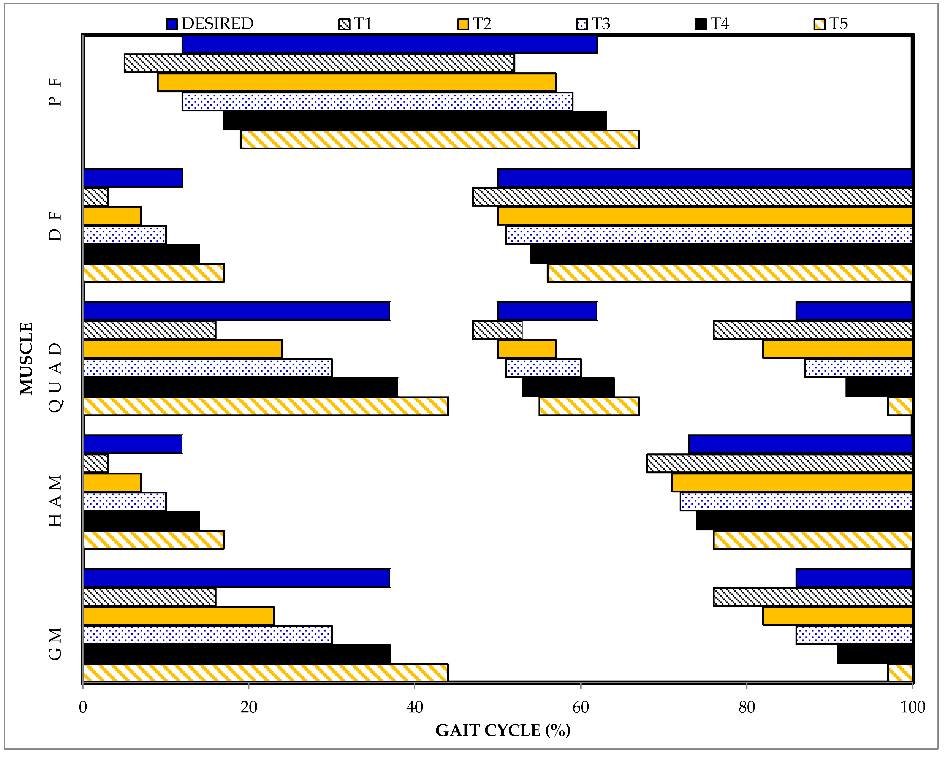
| Muscle Group | Start Time (% GC) | Stop Time (% GC) | |||||||||||
|---|---|---|---|---|---|---|---|---|---|---|---|---|---|
| Desired | T1 | T2 | T3 | T4 | T5 | Desired | T1 | T2 | T3 | T4 | T5 | ||
| G | Avg | 86 | 76 | 82 | 86 | 91 | 97 | 37 | 16 | 23 | 30 | 37 | 44 |
| SD | 1 | 3 | 4 | 4 | 4 | 4 | 2 | 4 | 5 | 4 | 5 | 4 | |
| Max | 91 | 85 | 92 | 97 | 100 | 9* | 43 | 25 | 39 | 40 | 53 | 56 | |
| Min | 81 | 67 | 71 | 75 | 81 | 86 | 33 | 4 | 13 | 18 | 25 | 31 | |
| H | Avg | 73 | 68 | 71 | 72 | 74 | 76 | 12 | 3 | 7 | 10 | 14 | 17 |
| SD | 2 | 4 | 5 | 5 | 4 | 5 | 2 | 3 | 4 | 4 | 7 | 4 | |
| Max | 77 | 81 | 86 | 91 | 90 | 90 | 17 | 13 | 17 | 26 | 53 | 29 | |
| Min | 68 | 56 | 59 | 59 | 63 | 63 | 9 | 96 ** | 0 | 2 | 5 | 9 | |
| Q | Avg | 86 | 76 | 82 | 87 | 92 | 97 | 37 | 16 | 24 | 30 | 38 | 44 |
| SD | 1 | 4 | 4 | 4 | 3 | 4 | 2 | 4 | 5 | 4 | 6 | 5 | |
| Max | 91 | 85 | 92 | 97 | 100 | 9 * | 43 | 25 | 40 | 41 | 66 | 66 | |
| Min | 81 | 57 | 72 | 76 | 82 | 86 | 33 | 1 | 13 | 19 | 26 | 32 | |
| Q2 | Avg | 50 | 47 | 50 | 51 | 53 | 55 | 62 | 53 | 57 | 60 | 64 | 67 |
| SD | 2 | 4 | 4 | 4 | 4 | 4 | 2 | 3 | 4 | 4 | 4 | 5 | |
| Max | 55 | 58 | 63 | 64 | 64 | 69 | 68 | 64 | 70 | 73 | 74 | 79 | |
| Min | 44 | 38 | 41 | 41 | 43 | 44 | 56 | 47 | 49 | 51 | 57 | 57 | |
| DF | Avg | 50 | 47 | 50 | 51 | 54 | 56 | 12 | 3 | 7 | 10 | 14 | 17 |
| SD | 2 | 4 | 4 | 4 | 4 | 5 | 2 | 3 | 4 | 4 | 4 | 4 | |
| Max | 55 | 58 | 64 | 64 | 76 | 74 | 17 | 16 | 17 | 27 | 24 | 29 | |
| Min | 44 | 38 | 41 | 41 | 43 | 44 | 9 | 96 ** | 0 | 2 | 6 | 9 | |
| PF | Avg | 12 | 5 | 9 | 12 | 17 | 19 | 62 | 52 | 57 | 59 | 63 | 67 |
| SD | 2 | 3 | 4 | 4 | 7 | 4 | 2 | 3 | 4 | 4 | 4 | 5 | |
| Max | 17 | 13 | 18 | 28 | 55 | 31 | 68 | 64 | 69 | 73 | 73 | 100 | |
| Min | 9 | 0 | 2 | 5 | 7 | 10 | 56 | 47 | 49 | 51 | 57 | 57 | |
| Trigger Condition | ||||||
|---|---|---|---|---|---|---|
| T1 | T2 | T3 | T4 | T5 | ||
| Recall | Mean | 0.635 | 0.753 | 0.820 | 0.846 | 0.795 |
| SD Error | 0.008 | 0.007 | 0.006 | 0.005 | 0.006 | |
| Precision | Mean | 0.767 | 0.888 | 0.928 | 0.916 | 0.830 |
| SD Error | 0.008 | 0.005 | 0.004 | 0.005 | 0.006 | |
| F1 | Mean | 0.716 | 0.808 | 0.866 | 0.878 | 0.819 |
| SD Error | 0.006 | 0.006 | 0.005 | 0.005 | 0.005 | |
© 2019 by the authors. Licensee MDPI, Basel, Switzerland. This article is an open access article distributed under the terms and conditions of the Creative Commons Attribution (CC BY) license (http://creativecommons.org/licenses/by/4.0/).
Share and Cite
Zahradka, N.; Behboodi, A.; Wright, H.; Bodt, B.; Lee, S. Evaluation of Gait Phase Detection Delay Compensation Strategies to Control a Gyroscope-Controlled Functional Electrical Stimulation System During Walking. Sensors 2019, 19, 2471. https://doi.org/10.3390/s19112471
Zahradka N, Behboodi A, Wright H, Bodt B, Lee S. Evaluation of Gait Phase Detection Delay Compensation Strategies to Control a Gyroscope-Controlled Functional Electrical Stimulation System During Walking. Sensors. 2019; 19(11):2471. https://doi.org/10.3390/s19112471
Chicago/Turabian StyleZahradka, Nicole, Ahad Behboodi, Henry Wright, Barry Bodt, and Samuel Lee. 2019. "Evaluation of Gait Phase Detection Delay Compensation Strategies to Control a Gyroscope-Controlled Functional Electrical Stimulation System During Walking" Sensors 19, no. 11: 2471. https://doi.org/10.3390/s19112471