Article

Sensitivity Enhancement in Surface Plasmon Resonance Biochemical Sensor Based on Transition Metal Dichalcogenides/Graphene Heterostructure

1
School of Mechanical Engineering and Electronic Information, China University of Geosciences (Wuhan), Wuhan 430074, China
2
Center of Fiber Technology, School of Electrical and Electronic Engineering, Nanyang Technological University, Singapore 639798, Singapore
*
Author to whom correspondence should be addressed.
Sensors 2018, 18(7), 2056; https://doi.org/10.3390/s18072056
Submission received: 14 May 2018 / Revised: 14 June 2018 / Accepted: 21 June 2018 / Published: 27 June 2018

Abstract

:
In this work, a surface plasmon resonance (SPR) biosensor based on two-dimensional transition metal dichalcogenides (TMDCs) is proposed to improve the biosensor’s sensitivity. In this sensor, different kinds of two-dimensional TMDCs are coated on both surfaces of metal film. By optimizing the structural parameters, the angular sensitivity can reach as high as 315.5 Deg/RIU with 7-layers WS2 and 36 nm Al thin film, which is 3.3 times of the conventional structure based on single Al thin film. We also obtain maximum phase sensitivity (3.85 × 10⁶ Deg/RIU) with bilayer WS2 and 35 nm Al thin film. The phase sensitivity can be further improved by employing Ag and removing air layer. The proposed configuration is of great potential for biochemical sensing.

1. Introduction

Surface plasmon resonance (SPR) is an optical phenomenon which occurs at the metal-dielectric surface. When light reflects at a SPR angle, free electrons on the metal surface can resonate and absorb light energy, consequently leading to a drastic attenuation of reflected light [1,2]. The SPR condition is sensitive to the environment variations and can be utilized as sensors. The biological molecules interactions in the sensing medium are detected by observing the refractive index changes of the sensor region. Due to advantages such as convenient detection, high sensitivity, real-time measurement, SPR sensors have been used to detect and analyze various biological molecules, such as proteins, nucleic acids and viruses, and have a broad prospect in practical applications [3,4,5,6]. Sensitivity is one of the most important aspects for biological sensing in particular, and how to enhance the sensitivity becomes a research hotspot for SPR biosensors.
Recently, 2D materials such as graphene and transition metal dichalcogenides (TMDCs) have are well-known for their use in constructing SPR sensors due to their unique electrical and optical properties [7]. This is because firstly, the high real part of the dielectric constant allows them to help metal absorb light energy [8]. Secondly, some features such as high surface to volume ratio and tunable biocompatibility can help the biosensor obtain sensitivity enhancements [9]. Finally, when coating these materials on the metal film, they can also protect the metal from oxidation as protective layers [10,11]. Based on these advantages, various 2D-material-assisted SPR sensors are proposed and investigated. Graphene has been proposed for the enhancement of the sensitivity [12]. Zeng et al. presented a highly sensitive SPR biosensor based on graphene-MoS2 hybrid nanostructures to enhance its sensitivity [13]. Air layer and graphene sheet for sensitivity enhancement was analyzed in [14]. Other TMDCs like WS2, MoSe2 and WSe2 are combined with silicon to enhance the sensitivity [15]. Wu et al. proposed a SPR biochemical sensor with heterostructures of few-layer BP and 2D materials (graphene/MoS2/WS2/MoSe2/WSe2) [16]. According to the previous work, it is found that the sensor performances are highly related to the structures and functional materials. To further enhance the sensitivity, both of these two aspects should be properly optimized.
In this paper, SPR sensor constructed by TMDCs/metal/TMDCs/graphene heterostructure is used for both angular and phase sensitivity enhancement. By coating different 2D TMDCs (MoS2/MoSe2/WS2/WSe2) at both sides of the metal, the sensitivity of the proposed sensor can be improved by the enhancement of light-material interaction. Angular sensitivity as high as 315.5 Deg/RIU which is nearly 3 times that of conventional configurations can be obtained. Furthermore, the proposed SPR configuration is suitable for phase detection as well and a pronounced phase sensitivity up to 3.85 × 10⁶ Deg/RIU is predicted.

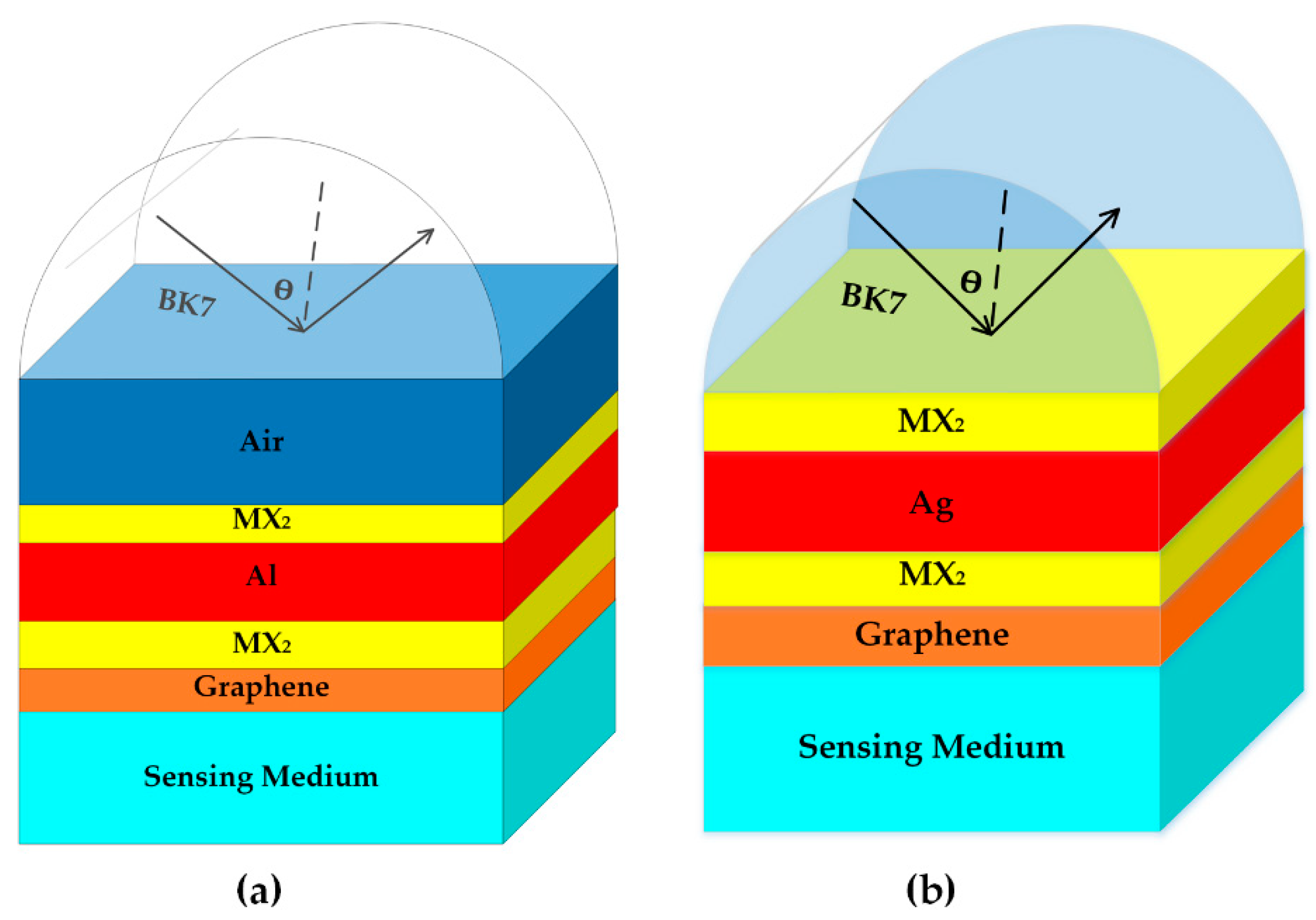
2. Sensor Configuration and Theoretical Model

The schematic diagram of the proposed SPR biosensor is shown in Figure 1a, the configuration contains seven layers and the operation wavelength is 633 nm which is popular for SPR applications [13,14,17]. BK7 glass with refractive index of np = 1.5151 acts as the coupling prism [18]. The refractive index of the air layer is fixed at 1 with thickness of 35 nm. The metal employed in this configuration is Al with dielectric constant of −34.2574 + 0.9108i [19]. Various TMDCs, represented by MX2, are coated at both sides of Al thin film, the thickness and refractive index of TMDCs at 633 nm are shown in Table 1 [20,21]. The graphene layer is coated on the MX2/Al/MX2 hybrid structure as the biomolecular recognition element and the refractive index of graphene is given as [22]:
n G = 3.0 + i C 1 λ 3
where λ is the wavelength and C1 = 5.446 μm−1. The thickness of the monolayer graphene is 0.34 nm. The refractive index of the sensing medium is given as ns = 1.33 + ∆n, where ∆n is the index change of the sensing medium.
In this paper, for the SPR curve calculation and sensing performance analysis, the transfer matrix method (TMM) is employed [23]. In the proposed structure, the thickness, the refractive index, and the dielectric constant of each layer are defined as dk, nk and εk, respectively. The incident angle corresponding to the minimum reflectance is called resonance angle and the angular sensitivity is calculated by probing the spectral shifts of the resonance angle [3] and defined as SA = ∆θres/∆n [24], where ∆θres represents the change of resonance angle. Furthermore, we also discuss phase sensitivity which is defined as Sp = ∆φ/∆n [13], where ∆φ is the differential phase changes corresponding to ∆n.

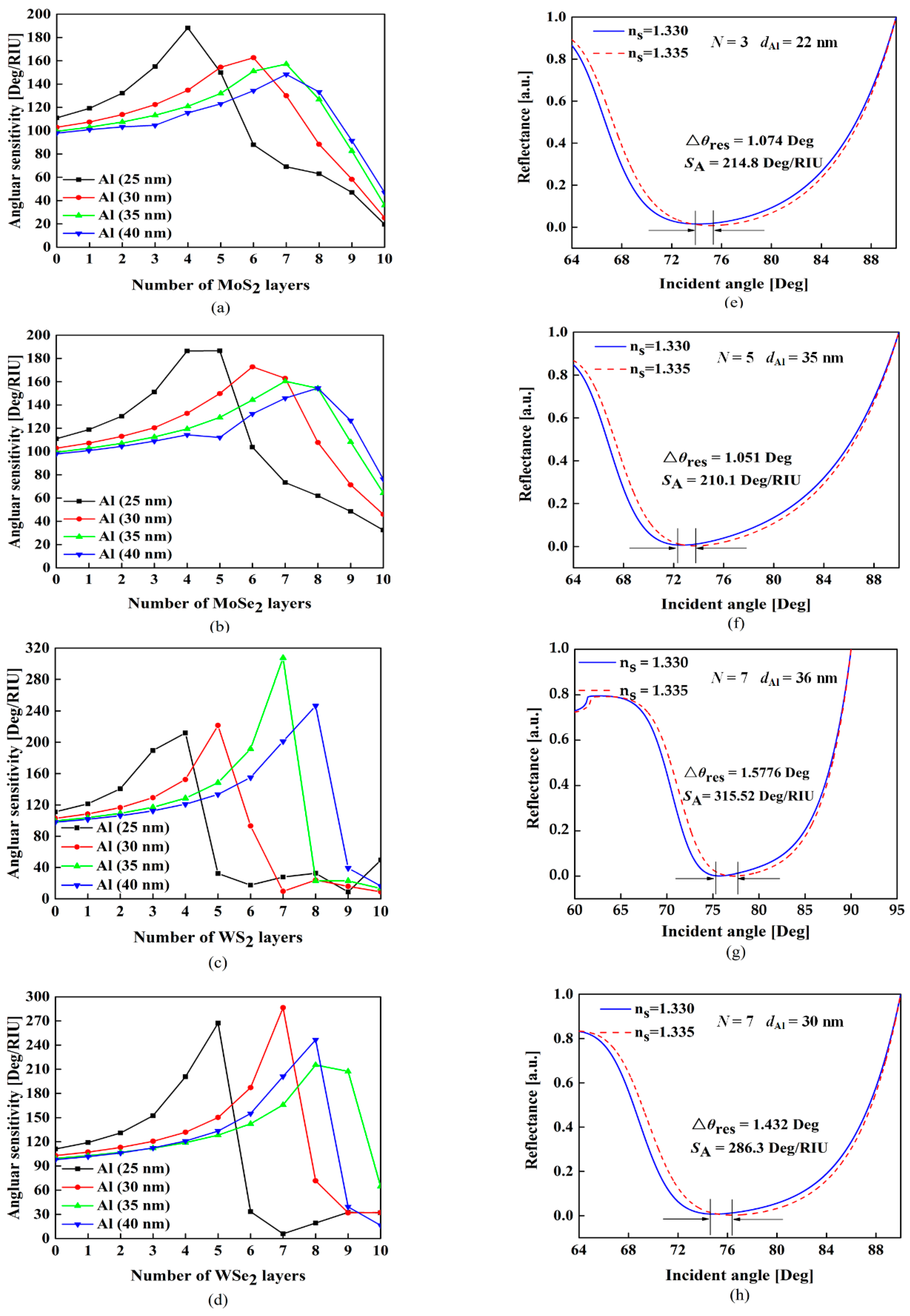
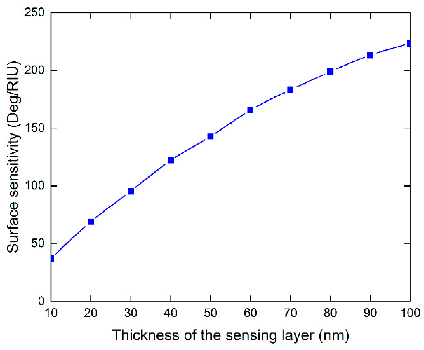
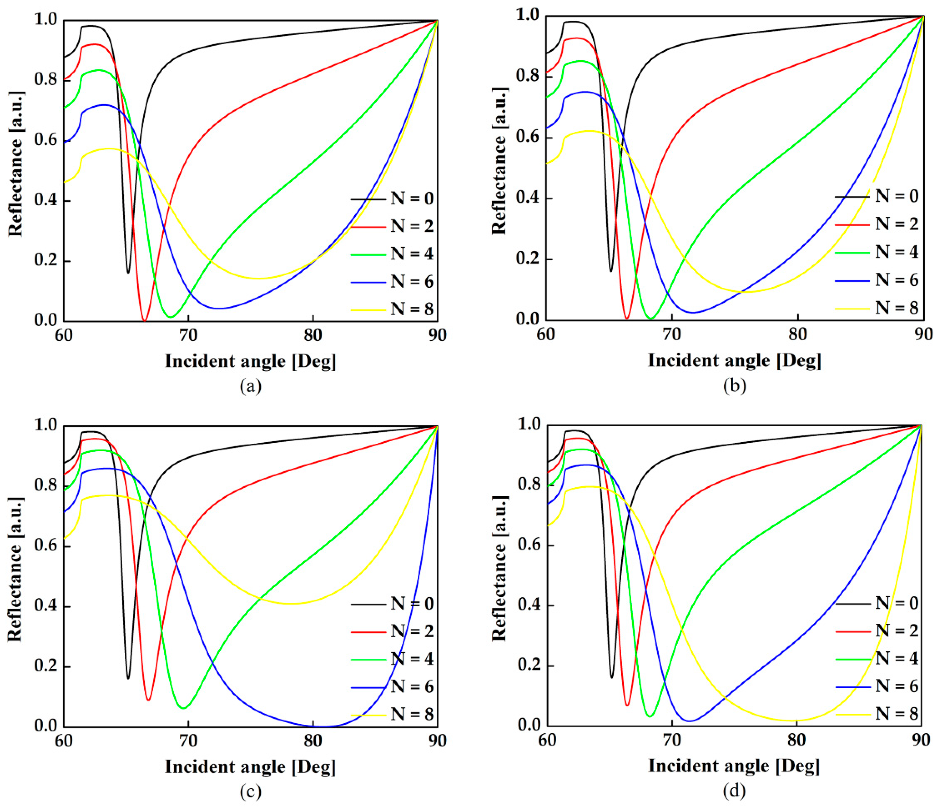
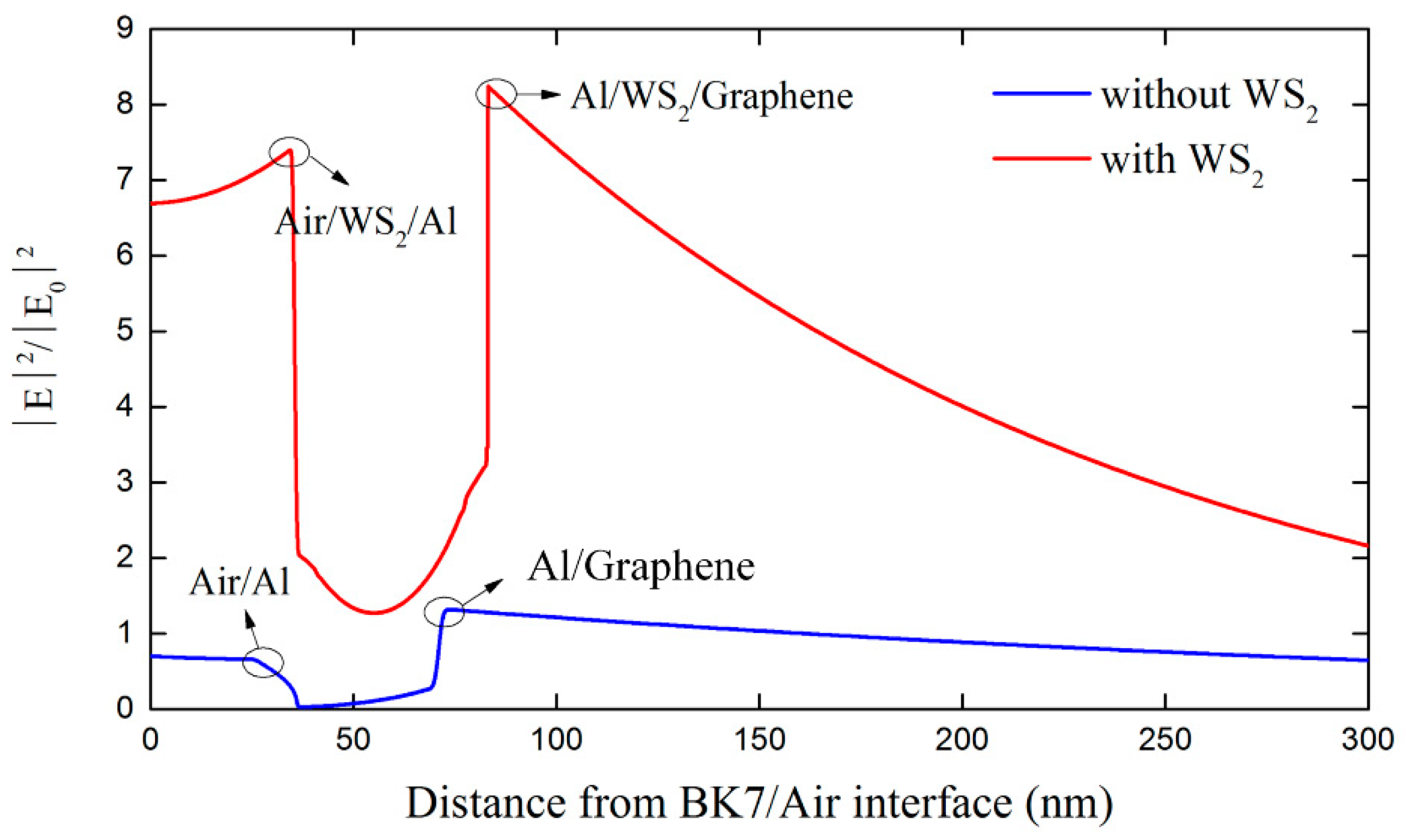
3. Results and Discussions
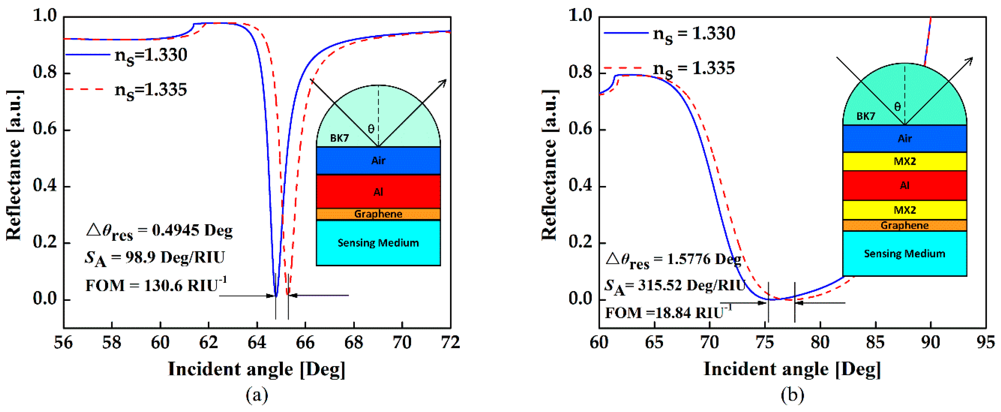
In order to obtain the optimal angular sensitivity for the proposed configuration, we firstly calculate the angular sensitivity with various number of MX2 layers and Al thickness. It should be noted that in this calculation, the MX2 layers at both sides are changed simultaneously. As shown in Figure 2a–d, when the refractive index of sensing medium changes from 1.330 to 1.335 (Δn = 0.005), the SPR curves shows three important features enumerated below.
(1)
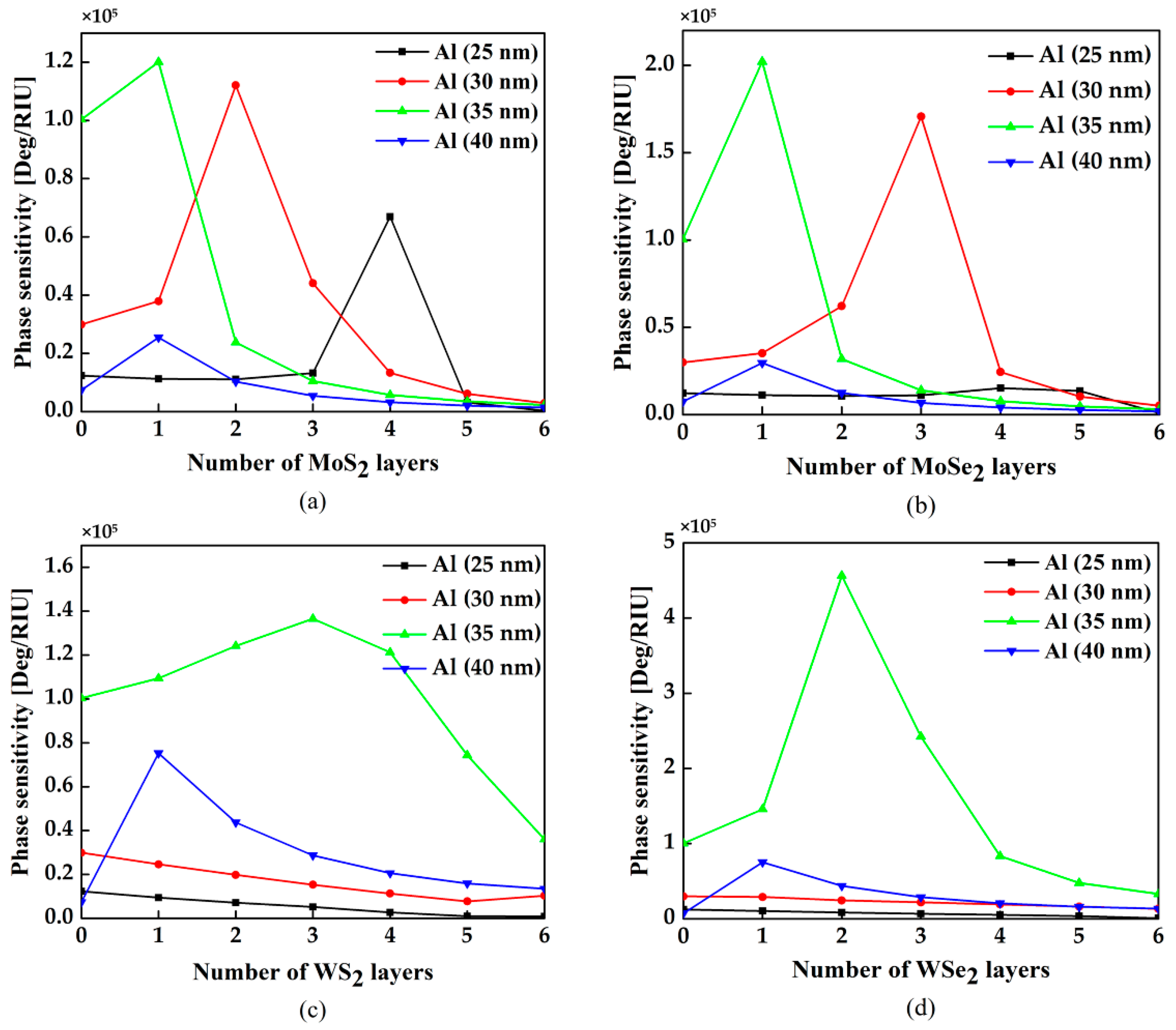
When the thickness of Al thin film is fixed, the sensitivity increases with more MX2 layers mainly due to the enhanced light energy absorption. However, it will decrease rapidly when the number of MX2 layers exceeds the optimal number which is defined as the number of MX2 layers with the highest sensitivity.
(2)
The optimal numbers of MX2 layers will increase when the thickness of Al increases.
(3)
With the same thickness of Al thin film, the enhancement effect offered by different kinds of MX2 are not the same.
It is known that the improvement effect caused by TMDCs is related to their dielectric constants. As illustrated in Table 1, MoS2 has a larger real part of the dielectric constant than others, which means its absorption ability is stronger [15]. Nevertheless, the ability to absorb light is not the only crucial factor to affect sensitivity; electron energy loss related to the imaginary part of the dielectric constant can lead to a counteraction [13]. Comparing to other TMDCs, the WS2 layers have much lower energy loss because of their small imaginary part of the dielectric constant. According to the conditions above, the optimized parameters and the corresponding angular sensitivity for different TMDCs are summarized in Table 2. The highest sensitivities can achieve 214.8 Deg/RIU, 210.1 Deg/RIU, 315.5 Deg/RIU and 286.3 Deg/RIU for the structures containing 3-layer MoS2 with 22 nm Al film, 4-layer MoSe2 with 24 nm Al film, 7-layer WS2 with 36 nm Al film and 7-layer WSe2 with 30 nm Al film, respectively. The corresponding SPR curves are shown in Figure 2e–h.
It should be noted that the intensity of reflection light is very weak at resonance. In order to effectively measure the resonance shift, it is necessary to increase the incident power or adopt detectors with high sensitivity. For example, assuming 10 mW incident light and 0.1 deg angular resolution [25], the reflected power at the SPR dip is estimated to be 6.5 μW and the reflected power variation between the resonance angle and its nearest measurable neighbor is 0.32 μW, which can be easily detected by commercially-available photodiodes at a visible wavelength [26]. Also note that, in our calculation, the sensing medium is set to be homogeneous in order to make fair comparison with previously works [12,13,14,15,16]. In fact, when the SPR biosensors are used for detecting cells with size of several microns [27], a homogeneous sensing medium layer is reasonable. To further investigate the surface sensitivity, the index change caused by the sensing target is applied with finite thickness. Under the optimal condition for WS2, the sensitivities with different sensing layer thickness are shown in Figure 3. It is shown that the surface sensitivity increases with thicker sensing layer. Even with 10 nm thickness, the surface sensitivity can be still as high as 37 Deg/RIU.
To further illustrate the contribution of MX2 on sensitivity, we have drawn the variation of the reflectance with incident angle varying from 60 Deg to 90 Deg for the different number of layers when the thickness of Al thin film is fixed at 30 nm and ns = 1.3300 in Figure 4a–d. It is indicated that the minimum reflectivity, representing the capability of light energy absorption [15], approaches zero firstly when the number of MX2 layers increase, which means the contribution of light energy absorption exceeds the electron energy loss. Meanwhile, the full width at half maximum (FWHM) becomes broader caused by electron energy loss of MX2 layers [13]. With the further increase of the MX2 layers, the FWHM keeps getting broader and the minimum reflectivity begins to diverge from zero. This is because the SPR process must satisfy the energy conservation T + R + A = 1, where T, R and A denote the transmission, reflection and absorption, respectively. Under SPR condition, since the total internal reflection is fulfilled, the transmission T is close to 0. When the number of TMDCs layers is insufficient, the absorbed light energy is not able to promote a strong SPR excitation. Therefore, increasing the TMDCs layers can enhance the light absorption, resulting in higher sensitivity. In this condition, the absorption A is enhanced while the reflection R is reduced. However, the absorption enhancement will be saturated due to the electron energy loss when further adding TMDCs layers. In this condition, the absorption A is degraded while the reflection R is increased.
The sensitivity of conventional structure based on single Al thin film is not high enough since the metallic layer cannot absorb enough light energy to excite a strong SPR. However, TMDCs has a larger real part of dielectric constant, which benefits SPR excitation [15]. Figure 5 shows the configuration and reflectance curves for the conventional SPR sensors and the proposed one with optimal parameters. Prominent sensitivity improvement up to 3.3 times can be observed in the WS2-assisted configuration. Figure 6 plots the electric field distributions in these two structures. There is a stronger field enhancement in the WS2-assisted structure compared to the conventional one, which further verifies the positive contribution provided by the TMDCs. In addition to sensitivity, figure of merit (FOM) is also one of the important aspects that affects the sensing performance. According to our calculation, the structure without TMDCs demonstrates the highest FOM. This is because the energy loss induced by the TMDCs will broaden FWHM. As we know, this phenomenon is also reported in other works of TMDCs-based SPR sensors [12,13,14,15,16]. Therefore, introducing TMDCs into the SPR sensor will contribute to sensitivity enhancement but not FOM enhancement.
Besides angular sensitivity, the differential phase change between p-polarized and s-polarized reflective wave is another approach to detect the analyte [28,29,30]. The variation of the phase sensitivity with respect to the different number of MX2 and thickness are showed in Figure 7a–d. In the structure showing in Figure 1a, we can obtain the highest sensitivity of 1.12 × 10⁵ Deg/RIU for bilayer MoS2 with 30 nm Al film, 2.02 × 10⁵ Deg/RIU for monolayer MoSe2 with 35 nm Al film, 1.37 × 10⁵ Deg/RIU for 3-layer WS2 with 35 nm Al film and 4.56 × 10⁵ Deg/RIU for bilayer WSe2 with 35 nm Al film, respectively. Comparing with angular sensitivity, the optimal number of MX2 layers for phase sensitivity is less.
To further improve the phase sensitivity, we propose another SPR biosensor based on Kretschmann configuration, as shown in Figure 1b. In this sensor, Ag is used to replace Al and the air layer is removed. The optimal conditions of Ag thickness and MX2 layers are summarized in Table 3. The best phase sensitivity as high as 3.85 × 10⁶ Deg/RIU can be achieved with monolayer WS2 and 46 nm Ag film.
For comparison, the performances of previously reported 2D-material-assisted SPR sensors are summarized in Table 4. Significant enhancements on both angular sensitivity and phase sensitivity can be obtained in the proposed sensors.

4. Conclusions

In this paper, SPR biosensors by using 2D TMDCs are proposed to enhance the sensitivity. In such sensors, the functional materials are coated on both sides of the metal layer and the impacts of material type, layer number, and metal thickness on the sensing performance are investigated and analyzed in detail. The results show that the angular sensitivity and phase sensitivity can reach as high as 315.5 Deg/RIU with 7-layers WS2 and 3.85 × 10⁶ Deg/RIU with 1-layers WS2, respectively. The proposed configuration can be promising a candidate for high performance biosensing.

Author Contributions

Conceptualization was designed by T.H.; methodology, validation, and formal analysis were carried out by X.Z., X.W., P.H., J.P., and Y.W.; investigation and data curation were carried out by T.H., P.S.P., and Z.C.; writing-original draft preparation was redacted by X.Z.; writing-review & editing was performed by T.H., and X.Z.

Funding

This work was supported by the National Natural Science Foundation of China (61605179), and the Fundamental Research Funds for the Central Universities, China University of Geosciences (Wuhan) (162301132703 and G1323511794).

Conflicts of Interest

The authors declare no conflicts of interest.

References

  1. Otto, A. Excitation of nonradiative surface plasma waves in silver by method of frustrated total reflection. Zeitschrift für Physik a Hadrons and Nuclei 1968, 216, 398–410. [Google Scholar] [CrossRef]
  2. Kretschmann, E.; Raether, H. Notizen: Radiative decay of nonradiative surface plasmons excited by light. Zeitschrift für Naturforschung A 1968, 23, 2135–2136. [Google Scholar] [CrossRef]
  3. Ahn, H.; Song, H.; Choi, J.R.; Kim, K. Localized Surface Plasmon Resonance Sensor Using Double-Metal-Complex Nanostructures and a Review of Recent Approaches. Sensors 2018, 18, 98. [Google Scholar] [CrossRef] [PubMed]
  4. Zeng, S.; Baillargeat, D.; Ho, H.P.; Yong, K.T. Nanomaterials enhanced surface plasmon resonance for biological and chemical sensing applications. Chem. Soc. Rev. 2014, 43, 3426–3452. [Google Scholar] [CrossRef] [PubMed]
  5. Zeng, S.; Sreekanth, K.V.; Shang, J.; Yu, T.; Chen, C.K.; Yin, F.; Baillargeat, D.; Coquet, P.; Ho, H.P.; Kabashin, A.V.; et al. Graphene–Gold Metasurface Architectures for Ultrasensitive Plasmonic Biosensing. Adv. Mater. 2015, 27, 6163–6169. [Google Scholar] [CrossRef] [PubMed]
  6. Wang, G.; Wang, C.; Yang, R.; Liu, W.; Sun, S. A Sensitive and Stable Surface Plasmon Resonance Sensor Based on Monolayer Protected Silver Film. Sensors 2017, 17, 2777. [Google Scholar] [CrossRef] [PubMed]
  7. Maurya, J.B.; François, A.; Prajapati, Y.K. Two-Dimensional Layered Nanomaterial-Based One-Dimensional Photonic Crystal Refractive Index Sensor. Sensors 2018, 18, 857. [Google Scholar] [CrossRef] [PubMed]
  8. Kooyman, R.P.H. Handbook of surface plasmon resonance. R. Soc. Chem. 2008, 2, 15–34. [Google Scholar]
  9. Zhu, Y.; Murali, S.; Cai, W.; Li, X.; Suk, J.W.; Potts, J.R.; Ruoff, R.S. Graphene and Graphene Oxide: Synthesis, Properties, and Applications. Adv. Mater. 2010, 22, 3906–3924. [Google Scholar] [CrossRef] [PubMed]
  10. Homola, J. Surface plasmon resonance sensors for detection of chemical and biological species. Chem. Rev. 2008, 108, 462–493. [Google Scholar] [CrossRef] [PubMed]
  11. Zhang, N.; Humbert, G.; Gong, T.; Shum, P.P.; Li, K.; Auguste, J.L.; Wu, Z.; Hu, J.; Feng, L.; Dinh, Q.X.; et al. Side-channel photonic crystal fiber for surface enhanced Raman scattering sensing. Sens. Actuators B Chem. 2016, 233, 195–201. [Google Scholar] [CrossRef]
  12. Verma, R.; Gupta, B.D.; Jha, R. Sensitivity enhancement of a surface plasmon resonance based on biomolecules sensor using graphene and silicon layers. Sens. Actuators B Chem. 2011, 160, 623–631. [Google Scholar] [CrossRef]
  13. Zeng, S.; Hu, S.; Xia, J.; Anderson, T.; Dinh, X.Q.; Meng, X.M.; Coquet, P.; Yong, K.T. Graphene-MoS2 hybrid nanostructures enhanced surface plasmon resonance biosensors. Sens. Actuators B Chem. 2015, 207, 801–810. [Google Scholar] [CrossRef]
  14. Wu, L.; Jia, Y.; Jiang, L.; Guo, J.; Dai, X.; Xiang, Y.; Fan, D. Sensitivity improved SPR biosensor based on the MoS2/graphene-aluminum hybrid structure. J. Lightwave Technol. 2016, 35, 82–87. [Google Scholar] [CrossRef]
  15. Ouyang, Q.; Zeng, S.; Dinh, X.Q.; Coquet, P.; Yong, K.T. Sensitivity enhancement of MoS2 nanosheet based surface Plasmon resonance biosensor. Procedia Eng. 2016, 140, 134–139. [Google Scholar] [CrossRef]
  16. Wu, L.; Guo, J.; Wang, Q.; Lu, S.; Dai, X.; Xiang, Y.; Fan, D. Sensitivity enhancement by using few-layer black phosphorus-graphene/TMDCs heterostructure in surface plasmon resonance biochemical sensor. Sens. Actuators B. Chem. 2017, 249, 542–548. [Google Scholar] [CrossRef]
  17. Khageswar, S.; Kumar, M.S.; Kumar, G.P. He-Ne laser (632.8 nm) pre-irradiation gives protection against DNA damage induced by a near-infrared trapping beam. J. Biophotonics 2009, 2, 140–144. [Google Scholar]
  18. Sreekanth, K.V.; Zeng, S.; Yong, K.T.; Yu, T. Sensitivity enhanced biosensor using graphene-based one-dimensional photonic crystal. Sens. Actuators B. Chem. 2013, 182, 424–428. [Google Scholar] [CrossRef]
  19. Jha, R.; Sharma, A.K. Chalcogenide glass prism based SPR sensor with Ag-Au bimetallic nanoparticle alloy in infrared wavelength region. J. Opt. Pure Appl. Opt. 2009, 11, 045502. [Google Scholar] [CrossRef]
  20. Li, Y.; Chernikov, A.; Zhang, X.; Rigosi, A.; Hill, H.M.; van der Zande, A.M.; Chenet, D.A.; Shih, E.M.; Hone, J.; Heinz, T.F. Measurement of the optical dielectric function of monolayer transition-metal dichalcogenides: MoS2, MoSe2, WS2, and WSe2. Phys. Rev. B 2014, 90, 205–422. [Google Scholar] [CrossRef]
  21. Liu, H.L. Optical properties of monolayer transition metal dichalcogenides probed by spectroscopic ellipsometry. Appl. Phys. Lett. 2014, 105, 201905. [Google Scholar] [CrossRef] [Green Version]
  22. Bruna, M.; Borini, S. Optical constants of graphene layers in the visible range. Appl. Phys. Lett. 2009, 94, 031901. [Google Scholar] [CrossRef]
  23. Wu, L.; Ling, Z.; Jiang, L.; Guo, J.; Dai, X.; Xiang, Y.; Fan, D. Long-Range Surface Plasmon with Graphene for Enhancing the Sensitivity and Detection Accuracy of Biosensor. IEEE Photonics J. 2016, 8, 1–9. [Google Scholar] [CrossRef]
  24. Maharana, P.K.; Jha, R.; Palei, S. Sensitivity enhancement by air mediated graphene multilayer based surface plasmon resonance biosensor for near infrared. Sens. Actuators B Chem. 2014, 190, 494–501. [Google Scholar] [CrossRef]
  25. Sreekanth, K.V.; Alapan, Y.; ElKabbash, M.; Wen, A.M.; Ilker, E.; Hinczewski, M.; Gurkan, U.A.; Steinmetz, N.F.; Strangi, G. Enhancing the Angular Sensitivity of Plasmonic Sensors Using Hyperbolic Metamaterials. Adv. Opt. Mater. 2016, 4, 1767–1772. [Google Scholar] [CrossRef] [PubMed] [Green Version]
  26. Corcoran, B.; Monat, C.; Grillet, C.; Moss, D.J.; Eggleton, B.J.; White, T.P.; O'Faolain, L.; Krauss, T.F. Green light emission in silicon through slow-light enhanced third-harmonic generation in photonic-crystal waveguidese. Nat. Photonics 2009, 3, 206–210. [Google Scholar] [CrossRef]
  27. Song, C.L.; Jin, T.; Yan, R.P.; Qi, W.Z.; Huang, T.Y.; Ding, H.F.; Tan, S.H.; Nguyen, N.T.; Xi, L. Opto-acousto-fluidic microscopy for three-dimensional label-free detection of droplets and cells in microchannels. Lab Chip 2018, 9, 1267–1390. [Google Scholar] [CrossRef] [PubMed]
  28. Zeng, S.; Yu, X.; Law, W.C.; Zhang, Y.; Hu, R.; Dinh, X.Q.; Ho, H.P.; Yong, K.T. Size dependence of Au NP-enhanced surface plasmon resonance based on differential phase measurement. Sens. Actuators B Chem. 2013, 176, 1128–1133. [Google Scholar] [CrossRef]
  29. Raether, H. Surface plasmons on smooth and rough surfaces and on gratings. Springer Tracts Mod. Phys. 1983, 111, 354401–373633. [Google Scholar]
  30. Wong, C.L.; Chua, M.; Mittman, H.; Choo, L.X.; Lim, H.Q.; Olivo, M. A Phase-Intensity Surface Plasmon Resonance Biosensor for Avian Influenza A (H5N1) Detection. Sensors 2017, 17, 2363. [Google Scholar] [CrossRef] [PubMed]
Figure 1. Schematic diagram of the SPR biosensor for (a) angle-sensitivity enhancement and (b) phase-sensitivity enhancement.
Sensors 18 02056 g001
Figure 2. (ad) Variation of the angular sensitivity as a function of the number of MX2 layers N with various thickness of Al thin film dAl and (eh) the reflection spectra with different TMDCs materials under optimal structures.
Sensors 18 02056 g002
Figure 3. The change in surface sensitivity as the function of the thickness of biomolecules on the surface.
Sensors 18 02056 g003
Figure 4. Variation of the reflectance respect to the different number of (a) MoS2, (b) WS2, (c) MoSe2 and (d) WSe2 layers with the thickness of Al thin film is fixed at 30 nm and the refractive index of sensing medium ∆n = 1.3300.
Sensors 18 02056 g004
Figure 5. Variation of the reflectance with respect to the incident angle for (a) the conventional biochemical sensor based on single Al film, and (b) the proposed configuration based on optimal angle sensitivity.
Sensors 18 02056 g005
Figure 6. The field distributions along the direction perpendicular to the prism for the proposed configuration with seven WS2 layers and the conventional configuration without WS2 layers.
Sensors 18 02056 g006
Figure 7. Variation of the angular sensitivity as a function of number of MX2 layers with various thickness of Al thin film, (a) MoS2, (b) MoSe2, (c) WS2, and (d) WSe2.
Sensors 18 02056 g007
Table 1. The thickness, refractive index and optical constants of different MX2 at λ = 633 nm.
Type of TMDCThickness of Monolayer (nm)Refractive IndexDielectric Constant
MoS20.655.0805 + 1.1723i24.4368 + 11.9121i
MoSe20.704.6226 + 1.0063i20.3560 + 9.3039i
WS20.804.8937 + 0.3124i23.8511 + 3.0578i
WSe20.704.5501 + 0.4332i20.5156 + 3.9423i
Table 2. Optimized values of thickness of gold and the number of MoS2 layers with corresponding change in angular sensitivity.
Type of TMDCOptimal Thickness of Al (nm)Optimal Number of TMDC LayersAngular Sensitivity (Δn = 0.005)
MoS2223214.8 Deg/RIU
MoSe2244210.1 Deg/RIU
WS2367315.5 Deg/RIU
WSe2307286.3 Deg/RIU
Table 3. Variation of the angular sensitivity as a function of number of MX2 layers with various thickness of Al thin film.
Type of TMDCOptimal Thickness of Al (nm)Optimal Number of TMDC LayersAngular Sensitivity (Δn = 0.005)
MoS24026.32 × 105 Deg/RIU
MoSe24021.54 × 105 Deg/RIU
WS24613.85 × 106 Deg/RIU
WSe24414.57 × 105 Deg/RIU
Table 4. Comparison with the formerly reported 2D-material-assisted SPR sensors.
2D MaterialMetalAngular SensitivityPhase SensitivityReferences
GrapheneAu134.6 Deg/RIU-[12]
Graphene and MoS2Al190.4 Deg/RIU-[13]
Graphene and MoS2Au-8.19 × 10⁴ Deg/RIU[14]
WS2Au155.7 Deg/RIU-[15]
WSe2Au-1.20 × 10⁶ Deg/RIU[15]
BP and TMDCs/grapheneAg279.0 Deg/RIU6.75 × 103 Deg/RIU[16]
WS2 and grapheneAl315.5 Deg/RIU-This work
WS2 and grapheneAg-3.85 × 10⁶ Deg/RIUThis work

