
A Naked Eye Refractive Index Sensor with a Visible Multiple Peak Metamaterial Absorber

Abstract
:
1. Introduction
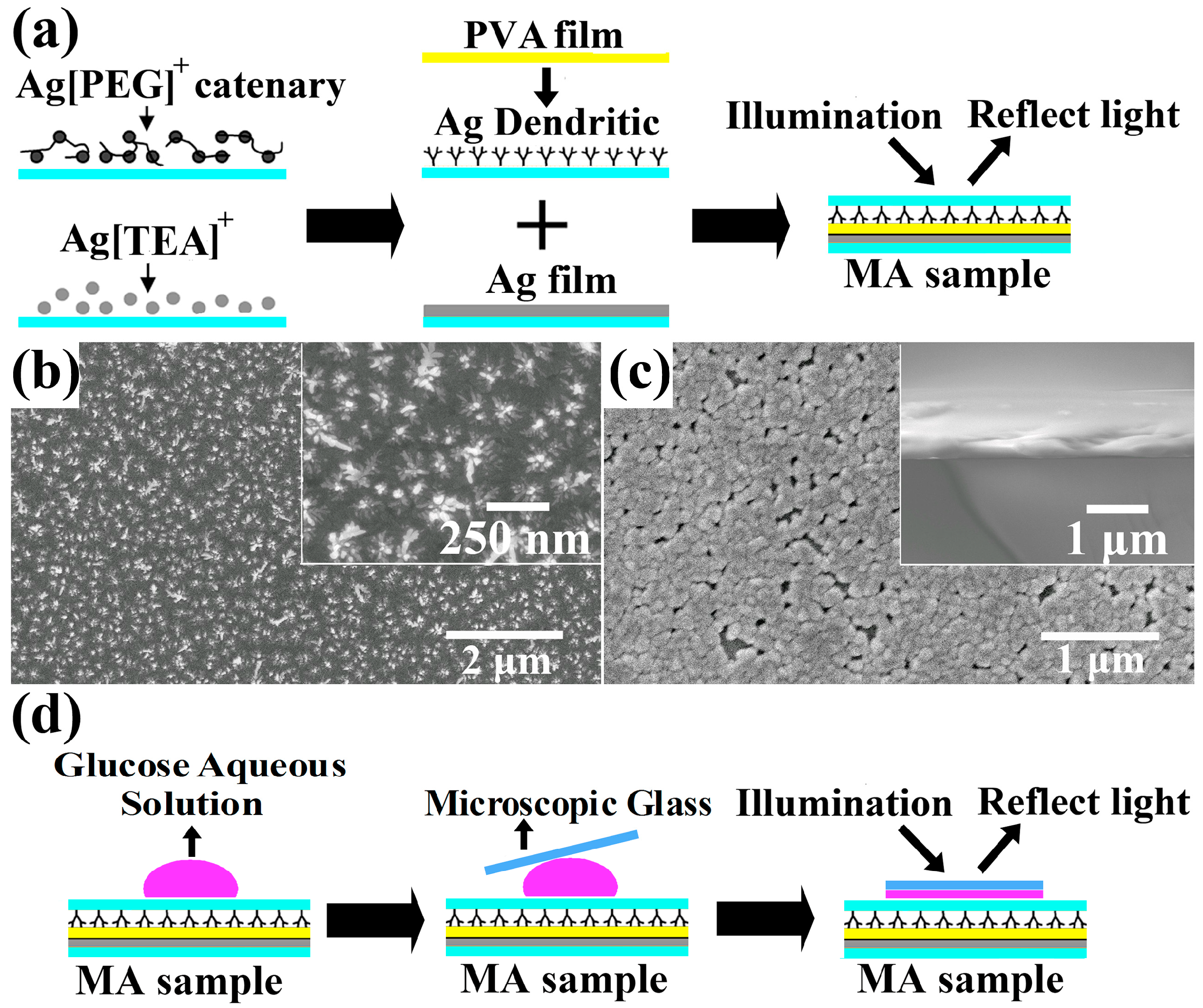
2. Experimental

3. Results and Discussion
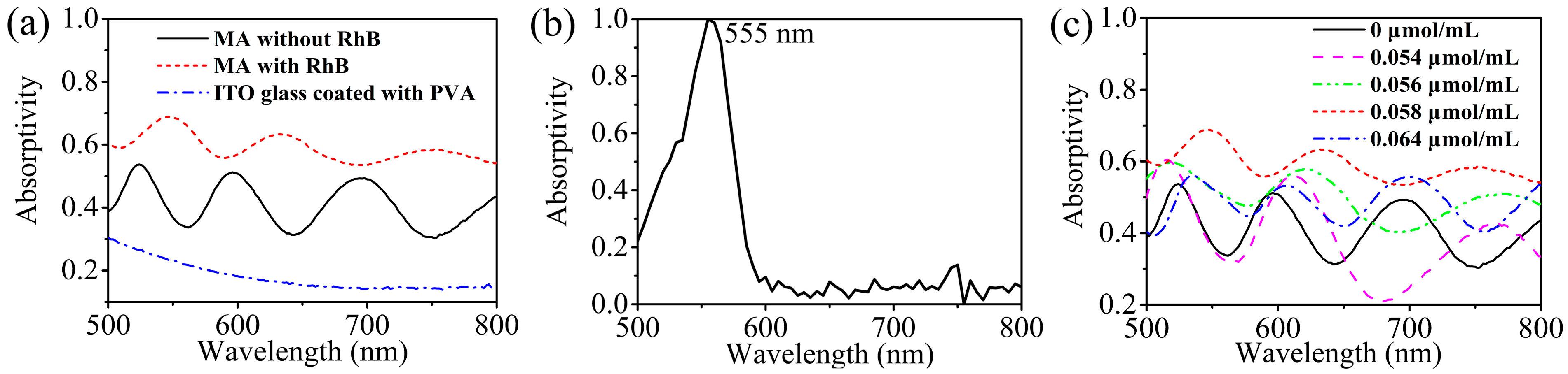
3.1. Measurements of Absorption Properties of MA Samples


| Concentration (μmol/mL) | The Magnitudes/Locations of Absorption Peaks (nm) | ||
|---|---|---|---|
| 0 | 0.537/524 | 0.513/596 | 0.502/698 |
| 0.054 | 0.608/516 | 0.564/612 | 0.430/768 |
| 0.056 | 0.600/518 | 0.584/620 | 0.516/770 |
| 0.058 | 0.692/546 | 0.641/632 | 0.596/752 |
| 0.064 | 0.560/536 | 0.538/606 | 0.506/700 |
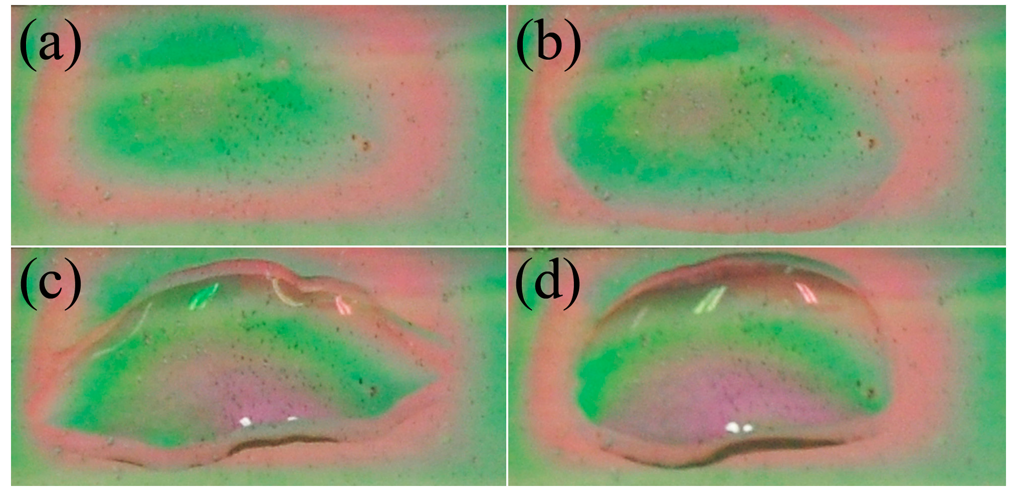
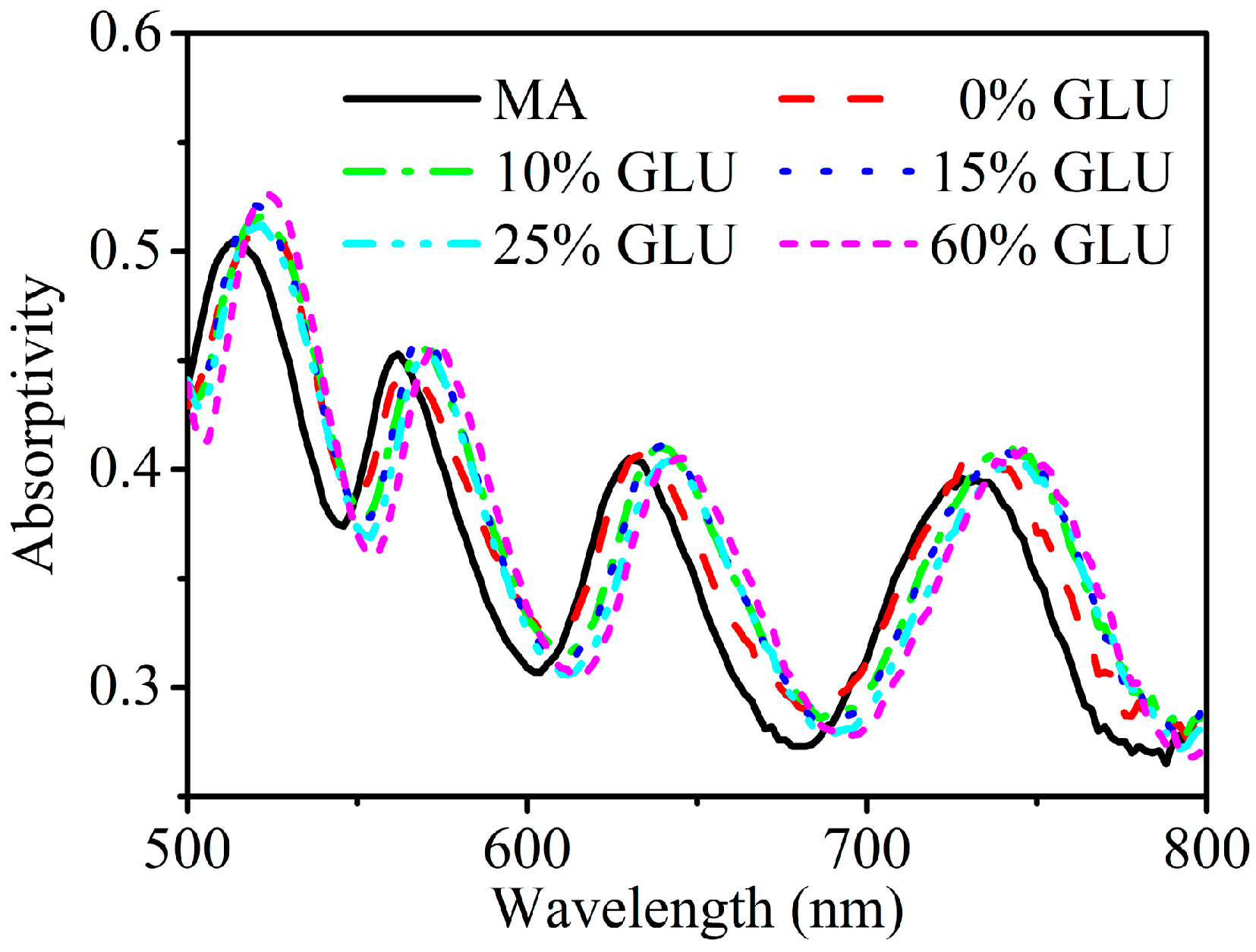
3.2. Measurements of the Sensibility of MA to Variations of the Refractive Index for Glucose Aqueous Solutions

4. Conclusions
Acknowledgments
Author Contributions
Conflicts of Interest
References
- Veselago, V.G. The electrodynamics of substances with simultaneously negative values of ε and μ. Sov. Phys. Usp. 1968, 10, 509–514. [Google Scholar]
- Pendry, J.B. Negative refraction makes a perfect lens. Phys. Rev. Lett. 2000, 85, 3966. [Google Scholar] [CrossRef] [PubMed]
- Zhao, X.P. Bottom-up fabrication methods of optical metamaterials. J. Mater. Chem. 2012, 22, 9439–9449. [Google Scholar] [CrossRef]
- Schurig, D.; Mock, J.J.; Justice, B.J.; Cummer, S.A.; Pendry, J.B.; Starr, A.F.; Smith, D.R. Metamaterial electromagnetic cloak at microwave frequencies. Science 2006, 314, 977–980. [Google Scholar] [CrossRef] [PubMed]
- Tsakmakidis, K.L.; Boardman, A.D.; Hess, O. “Trapped rainbow” storage of light in metamaterials. Nature 2007, 450, 397–401. [Google Scholar] [CrossRef] [PubMed]
- Zhao, X.P.; Luo, W.; Huang, J.X.; Fu, Q.H.; Song, K.; Cheng, X.C.; Luo, C.R. Trapped rainbow effect in visible light left-handed heterostructures. Appl. Phys. Lett. 2009, 95, 071111. [Google Scholar] [CrossRef]
- Lee, J.-H.; Moon, H.-S.; Jang, I.-S.; Choi, J.-S.; Yook, J.-G.; Jung, H.-I. A planar split-ring resonator-based microwave biosensor for label-free detection of biomolecules. Sens. Actuators B Chem. 2012, 169, 26–31. [Google Scholar] [CrossRef]
- Ruffato, G.; Pasqualotto, E.; Sonato, A.; Zacco, G.; Silvestri, D.; Morpurgo, M.; Toni, A.D.; Romanato, F. Implementation and testing of a compact and high-resolution sensing device based on grating-coupled surface plasmon resonance with polarization modulation. Sens. Actuators B Chem. 2013, 185, 179–187. [Google Scholar] [CrossRef]
- Landy, N.I.; Sajuyigbe, S.; Mock, J.J.; Smith, D.R.; Padilla, W.J. Perfect metamaterial absorber. Phys. Rev. Lett. 2008, 100, 207402. [Google Scholar] [CrossRef] [PubMed]
- Liu, N.; Mesch, M.; Weiss, T.; Hentschel, M.; Giessen, H. Infrared perfect absorber and its application as plasmonic sensor. Nano Lett. 2010, 10, 2342–2348. [Google Scholar] [CrossRef] [PubMed]
- Cui, Y.X.; Fung, K.H.; Xu, J.; Ma, H.J.; Jin, Y.; He, S.L.; Fang, N.X. Ultrabroadband light absorption by a sawtooth anisotropic metamaterial slab. Nano Lett. 2012, 12, 1443–1447. [Google Scholar] [CrossRef] [PubMed] [Green Version]
- Kats, M.A.; Sharma, D.; Lin, J.; Genevet, P.; Blanchard, R.; Yang, Z.; Qazilbash, M.M.; Basov, D.N.; Ramanathan, S.; Capasso, F. Ultra-thin perfect absorber employing a tunable phase change material. Appl. Phys. Lett. 2012, 101, 221101. [Google Scholar] [CrossRef]
- Liu, X.M.; Zhao, Q.; Lan, C.W.; Zhou, J. Isotropic Mie resonance-based metamaterial perfect absorber. Appl. Phys. Lett. 2013, 103, 031910. [Google Scholar] [CrossRef]
- Liu, X.L.; Starr, T.; Starr, A.F.; Padilla, W.J. Infrared spatial and frequency selective metamaterial with near-unity absorbance. Phys. Rev. Lett. 2010, 104, 207403. [Google Scholar] [CrossRef] [PubMed]
- Ma, Y.; Chen, Q.; Grant, J.; Saha, S.C.; Khalid, A.; Cumming, D.R.S. A terahertz polarization insensitive dual band metamaterial absorber. Opt. Lett. 2011, 36, 945–947. [Google Scholar] [CrossRef] [PubMed]
- Grant, J.; Ma, Y.; Saha, S.; Lok, L.B.; Khalid, A.; Cumming, D.R.S. Polarization insensitive terahertz metamaterial absorber. Opt. Lett. 2011, 36, 1524–1526. [Google Scholar] [CrossRef] [PubMed]
- Shen, X.P.; Yang, Y.; Zang, Y.Z.; Gu, J.Q.; Han, J.G.; Zhang, W.L.; Cui, T.J. Triple-band terahertz metamaterial absorber: Design, experiment, and physical interpretation. Appl. Phys. Lett. 2012, 101, 154102. [Google Scholar] [CrossRef]
- Xiao, S.M.; Drachev, V.P.; Kildishev, A.V.; Ni, X.J.; Chettiar, U.K.; Yuan, H.K.; Shalaev, V.M. Loss-free and active optical negative-index metamaterials. Nature 2012, 466, 735–740. [Google Scholar] [CrossRef]
- Zhu, W.R.; Rukhlenko, I.D.; Premaratne, M. Light amplification in zero-index metamaterial with gain inserts. Appl. Phys. Lett. 2012, 101, 031907. [Google Scholar] [CrossRef]
- Fang, A.A.; Huang, Z.X.; Koschny, T.; Soukoulis, C.M. Overcoming the losses of a split ring resonator array with gain. Opt. Express 2011, 19, 12688–12699. [Google Scholar] [CrossRef] [PubMed]
- Liu, B.Q.; Zhao, X.P.; Zhu, W.R.; Luo, W.; Cheng, X.C. Multiple pass-band optical left-handed metamaterials based on random dendritic cells. Adv. Funct. Mater. 2008, 18, 3523–3528. [Google Scholar] [CrossRef]
- Zhu, W.R.; Zhao, X.P. Metamaterial absorber with random dendritic cells. EPJ. Appl. Phys. 2010, 50, 21101. [Google Scholar] [CrossRef]
- Zhu, W.R.; Zhao, X.P. Metamaterial absorber with dendritic cells at infrared frequencies. J. Opt. Soc. Am. B 2009, 26, 2382–2385. [Google Scholar] [CrossRef]
- Li, S.; Gong, B.Y.; Cao, D.; Pan, Z.Z.; Luo, C.R.; Zhao, X.P. A green-light gain-assisted metamaterial fabricated by self-assembled electrochemical deposition. Appl. Phys. Lett. 2013, 103, 181910. [Google Scholar] [CrossRef]
- Nunes, P.S.; Mortensen, N.A.; Kutter, J.P.; Mogensen, K.B. Refractive index sensor based on a 1D photonic crystal in a microfluidic channel. Sensors 2010, 10, 2348–2358. [Google Scholar] [CrossRef] [PubMed]
- Liu, Y.Z.; Salemink, H.W.M. All-optical on-chip sensor for high refractive index sensing in photonic crystals. Europhys. Lett. 2014, 107, 34008. [Google Scholar] [CrossRef]
- Song, Z.Y.; Feng, G.Y.; Zhang, T. Accurate measurement of the refractive index D-Glucose solution at various concentrations at different temperature. Chin. J. Lasers 2014, 41, 1208008. [Google Scholar] [CrossRef]
- Wang, X.N.; Luo, C.R.; Hong, G.; Zhao, X.P. Metamaterial optical refractive index sensor detected by the naked eye. Appl. Phys. Lett. 2013, 102, 091902. [Google Scholar] [CrossRef]
© 2015 by the authors; licensee MDPI, Basel, Switzerland. This article is an open access article distributed under the terms and conditions of the Creative Commons Attribution license (http://creativecommons.org/licenses/by/4.0/).
Share and Cite
Ma, H.; Song, K.; Zhou, L.; Zhao, X. A Naked Eye Refractive Index Sensor with a Visible Multiple Peak Metamaterial Absorber. Sensors 2015, 15, 7454-7461. https://doi.org/10.3390/s150407454
Ma H, Song K, Zhou L, Zhao X. A Naked Eye Refractive Index Sensor with a Visible Multiple Peak Metamaterial Absorber. Sensors. 2015; 15(4):7454-7461. https://doi.org/10.3390/s150407454
Chicago/Turabian StyleMa, Heli, Kun Song, Liang Zhou, and Xiaopeng Zhao. 2015. "A Naked Eye Refractive Index Sensor with a Visible Multiple Peak Metamaterial Absorber" Sensors 15, no. 4: 7454-7461. https://doi.org/10.3390/s150407454