1. Introduction
Metabolites are intermediate or final products of a metabolic pathway [
1]. Their sensing is of great interest for the food industry to characterize nutritional aspects [
2], whereas in medicine, metabolites are indicators of the human status of health, providing information about possible diseases or the monitoring of therapy [
3,
4]. Actually, the real popularization of electrochemical sensors is standing out from the other sensing methods (e.g., mass spectrometry [
5] and optical techniques [
6,
7]). In particular, electrochemical enzyme-based biosensors showed great potentialities for the sensing of metabolites due to their high selectivity for the presence of an active site, and their capability to accommodate just one (in the case of oxidases [
8,
9,
10]) or a family (in the case of cytochromes [
11,
12]) of metabolites. However, enzymes cause a series of problems, e.g., interference by ascorbic and uric acids, two molecules greatly present in the human body and that oxidate at the same potential of oxidases [
13]. Moreover, there are not matching enzymes for all the metabolites of interest, as in the case of adenosine triphosphate (ATP).
In our previous works, we proposed a new electrochemical assay for the sensing of metabolites based on different supramolecular cyclodextrin complexes [
14]. We synthesized a new type of ferrocene (Fc) electroactive probe modified by the phenylboronic acid group (4-Fc-PB), and we combined it with two different cyclodextrins: natural β-cyclodextrin (β-CD) for the sensing of fructose [
15], and β-CD modified by a dipicolylamine group (dpa-
p-HB-β-CDs) for the sensing of ATP [
16]. In such a way, the different supramolecular complexes possess specific binding groups that act as lariats, grasping the desired target-molecule near the microenvironment constituted by probe/CD over the course of the electrochemical measurement and guaranteeing the sensing of the metabolite with very high selectivity.
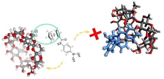
There have essentially been two requirements for the choice of ferrocene as an electroactive probe for the electrochemical sensing of metabolites: the need for a molecule that is easily included in the cyclodextrins cavity and at the same time a molecule that is easy to chemically modify by specific binding groups (i.e., phenylboronic acid) for the interaction with the metabolites. In this sense, the ferrocene moiety has been shown to satisfy both of these requirements.
In order to obtain a detailed description on the origin of a receptor selectivity at a molecular level, Nuclear Magnetic Resonance (NMR) spectroscopy represents a powerful and faceted tool that is particularly useful for the deep characterization of cyclodextrins inclusion complexes [
17,
18], formed with both hydrophilic and lipophilic target molecules [
19,
20,
21,
22].
Herein, we report for the first time a study about the inclusion phenomena between the 4-Fc-PB electroactive probe with β-CD and with dpa-p-HB-β-CDs by using different NMR analyses, to shed light on the stereochemical features underlining the formation of electroactive probe/cyclodextrin supramolecular complexes. In particular, the paper focuses on the calculation of the binding constant of the 4-Fc-PB/β-CD supramolecular complex, on the host/guest interactions involved, and on the elucidation about the origin of a drift in the time observed during the control experiments of the electrochemical measurements for the 4-Fc-PB/dpa-p-HB-β-CD supramolecular complex.
2. Results and Discussion
2.1. Characterization of the Binding Partners
Before investigating the 4-Fc-PB/CDs interaction, a complete characterization of both derivatized ferrocene (4-Fc-PB) and derivatized 3-NH
2-β-CD (dpa-
p-HB-β-CD) was a necessary step. In the
1H NMR spectrum of 4-Fc-PB, the aromatic protons of the phenyl-boronic acid were easily recognized at 7.43 ppm (H
c) and 7.18 ppm (H
b), whereas the protons belonging to the ferrocene derivatized ring were distinguished in the spectral region 4.0-5.0 ppm (
Figure 1, H
e 4.78 ppm, H
f 4.43, and H
g 4.18 ppm).
In the case of dpa-
p-HB-β-CD, the signals of its covalently linked aromatic moiety were fully assigned by using
1H NMR spectrum analysis (
Figure 2) and 2D COSY map (
Figure S1). The anomeric protons of dpa-
p-HB-β-CD (H
1) produced a cluster of different peaks at 4.7–5.1 ppm, indicating effective derivatization. Moreover, the comparison between the proton unit of the dpa-
p-HB-β-CD aromatic ligand (1H, ~1.0) and the integrated area of the anomeric protons (7H, ~6.8) agree well with a single derivatization on C
3-NH
2, as expected. Respect to pure β-CD, in which its symmetrical toroidal geometry produces a unique and well resolved set of signals, derivatized dpa-
p-HB-β-CD showed the existence of conformational distortions and an unsymmetrical chemical environment. As a consequence, the derivatization produced a complicated ensemble of splitting and overlapping signals in the region of dpa-
p-HB-β-CD ring protons (H
2–H
6), which made the task of obtaining information on the stereochemistry of 4-Fc-PB/dpa-
p-HB-β-CD inclusion difficult.
2.2. 1H NMR Investigations on 4-Fc-PB/β-CDs Supramolecular Complex
The calculation of the binding constant of a ferrocene (Fc) probe interacting with a CD cavity is challenging enough. One of the most typically used techniques for the determination of binding constant is UV-Visible spectroscopy [
23]. However, the spectrophotometric method is difficult to apply to the case of Fc since this molecule presents very small spectral variations [
24]. Attempts to evaluate the Fc/β-CD binding constant have been determined as 2.2 × 10
3 M
−1 using molecular dynamic calculations [
25,
26,
27], while solubility measurements based on the vapor-circulation technique have calculated the association constant of Fc with β-CD as 1.7 × 10
4 M
−1 [
28] in water. In this context, it is also commonly accepted that ferrocene prefers an axial orientation over an orthogonal orientation for its inclusion inside the β-CD cavity (
Figure 3) [
25].
The first step of our investigation was firstly to verify the inclusion of the 4-Fc-PB probe in natural β-CDs and secondarily to evaluate the correspondent binding constant by 1H NMR studies. Such an analysis turned out to be necessary in our case since we hypothesized that the phenylboronic group added to the Fc moiety causes a stoichiometric bulk, reducing the binding constant of the inclusion reported in the literature.
A preliminary titration was carried out to exclude any self-association phenomena of 4-Fc-PB within the concentration range used for the association constant determination (0.1–10 mM), which could generate spurious chemical shift variations. As a matter of fact, no shift of 4-Fc-PB proton peaks was observed over 3 orders of magnitude of concentration variations (
Figure S2); therefore, it was possible to solely attribute variation in the chemicals shifts of the 4-Fc-PB probe to the interaction with β-CD. For that,
1H NMR titration was carried out in the presence of a fixed concentration of β-CD (5 mM), and chemical shifts of different proton peaks related to both ferrocene and β-CD were collected (
Figures S3 and S4). The proton shifts of both the H
g proton of the 4-Fc-PB probe and the H
3 protons of the internal cavity of natural β-CDs confirmed the inclusion. Moreover, by fitting the chemical shift variations using a 1:1 model (
Figure S5), it was possible to estimate the association constant as
Ka = 385 ± 9 M
−1 using H
g protons and
Ka = 765 ± 8 M
−1 using H
3CD, with a sufficient accordance between the two values (and both were inferior to Fc/β-CD K
a ≈ 10
3 M
−1 [
25,
26,
27]). This fact can be explained due to the presence of the phenylboronic acid group, which creates additional steric hindrance.
In the case of 4-Fc-PB, a phenylboronic acid moiety is conjugated to one pentadienyl ring of Fc. Since β-CD possesses a hydrophobic cavity capable of accommodating the aromatic portions of a wide variety of compounds, another important point to investigate was the eventual inclusion of the PB moiety into β-CD. In this regard, it is noteworthy that the signals of the protons Hc and Hd of 4-Fc-PB did not change over the titration in the presence of β-CD, hinting that the phenylboronic acid portion is likely not involved in the interaction.
To gain deeper insight into the stereochemistry of the inclusion, a 2D NOESY map has been recorded and carefully analysed (
Figure 4). The non-involvement of the PB fragment in the β-CD inclusion was confirmed once again since no cross-peaks were detected between the protons H
b/H
c and any of H
nβ-CD. This result clearly indicates that the sensing fragment of 4-Fc-PB remained free to exert its activity during the electrochemical analysis. Conversely, NOE effects were detected between H
f/H
g and H
6/6′β-CD protons, indicating an interaction between the ferrocenyl moiety of 4-Fc-PB and the lower rim of CD, with a stronger NOE effect between H
g and H
6/6′β-CD with respect to H
f and H
6/6′β-CD. This is in line with the most sensible 4-Fc-PB protons observed along the NMR titration. However, in the specific case of medium-sized complexes (~1–2 kDa), weak or even null NOE values are due to an unfavourable correlation time (ωτ
c ~1, where ω is the Larmor angular frequency and τ
c is the molecular rotational correlation time of the complex) [
29]. To exclude any undetected cross-peaks due to this eventuality (MW of 4-Fc-PB + β-CD = 1.48 kDa), a 2D ROESY map of the same 4-Fc-PB/β-CD mixture was also made, and both 2D NOESY and ROESY showed the same results (
Figure 4 vs.
Figure S6).
2.3. Characterization of 4-Fc-PB/dpa-p-HB-β-CDs Supramolecular Complex
The description of 4-Fc-PB/β-CD helped in the better rationalization of the more complicated 4-Fc-PB/dpa-
p-HB-β-CD system. As demonstrated in ref. [
16], the presence of the dipicolylamine group is of primary importance for the sensing of ATP since it entraps Zn
2+ ion, creating an electrostatic bonding with the three phosphate groups of ATP. However, the complex 4-Fc-PB/dpa-
p-HB-β-CD developed a time drift during the performance of the control experiments [
16] (
Figure S7), on the contrary to the case of 4-Fc-PB/β-CD used for the sensing of fructose [
15] (
Figure S8). The origin of the time drift was identified in the possibility of intramolecular self-inclusion due to the different degrees of freedom of movement of its lateral arm since dpa-
p-HB-β-CD is derivatized with a dipicolylamine group. Actually, such a phenomenon was effectively hypothesized by a 3D modelling (
Figure S9). A 2D NOESY map confirmed the hypothesis, highlighting the interaction between protons H
h’, H
j’, and H
k’ belonging to the outer pyridinyl ring with the internal protons H
3β-CD and H
5β-CD of the cyclodextrin cavity (
Figure 5). In principle, the occurrence of the combination of intra- and intermolecular inclusions is not to be excluded.
It is reasonable to think that the 4-Fc-PB probe inclusion is not stable over time because the CD cavity is partially occupied by the dipicolylamine group. The limited inclusion of 4-Fc-PB inside dpa-p-HB-β-CD with respect to β-CD causes its faster degradation over time, resulting in the time drift observed.
The crowdedness of the 4-Fc-PB/dpa-
p-HB-β-CD spectra and the effect arising from the self-association of the cyclodextrin did not allow one to evaluate the association constant by
1H NMR titration. However, the complexation shifts (Δδ
CS = δ
mixture−δ
pure, Hz) analysis of the 4-Fc-PB/dpa-
p-HB-β-CD mixture showed that the most affected protons of 4-Fc-PB were those belonging to the ferrocene moiety (H
f/H
g Δδ
CS = −0.03 ppm vs. H
b/H
c = 0.00–0.01 ppm,
Figure S10). Protons belonging to the amino-boronic fragment remained almost unperturbed, suggesting that, in this case too, the inclusion occurs from the cyclopentadienyl rings, leaving the electroactive fragment almost free. Signals produced by the aromatic protons of CD underwent minor variations (δ
CS = 0.01–0.02 ppm).
Since the ultimate application of the 4-Fc-PB/dpa-
p-HB-β-CDs supramolecular complex is in the sensing of ATP, the effect of the presence of ATP was also observed in the
1H NMR spectrum (
Figure S11). The signal broadening of the two Fc aromatic doublets (H
b and H
c) was detected only in the copresence of ATP and dpa-
p-HB-β-CDs, hinting at a synergic interaction, most likely exerted through cis-
diol bonding with the ribose part of ATP and mediated by the presence of the CD. This result supports the detection mechanism introduced in ref. [
16], where specific groups (i.e., phenyl-boronic moiety) grasp the desired target molecule, while the role of the dipicolylamine group was elucidated by electrochemical experiments since, in the absence of the dipicolylamine group, the ATP was impossible to detect [
16].
3. Materials and Methods
Ferrocene carboxylic acid and 3A-amino-3A-deoxy-(2AS, 3AS)-β-CD (NH2-β-CD) were purchased from the Tokyo Chemical Industry Co., Ltd. (Tokyo, Japan) and used without further purification. Natural β-CDs and sodium hydroxide were obtained as special-grade reagents from Fujifilm Wako Pure Chemical Industries Ltd. (Osaka, Japan). Deuterium oxide (D2O), methanol, sodium carbonate, and dimethylsulfoxide-d6 (DMSO-d6) were purchased from Kanto Chemical Co., Inc. (Tokyo, Japan). All other organic solvents, monosaccharides, and reagents were commercially available with guaranteed grade and used as received. Water was doubly distilled and deionized by a Milli-Q water system (WG222, Yamato Scientific Co., Ltd., Tokyo, Japan and Milli-Q Advantage A10, Burlington, MA, USA) before use.
Proton nuclear magnetic resonance (1H NMR), 2D COSY (homonuclear COrrelation SpectroscopY), Nuclear Overhauser Effect SpectroscopY (NOESY), and Rotating Frame Overhauser Enhancement SpectroscopY (ROESY) spectra were measured with a JNM-ECX500 (JEOL Ltd., Tokyo, Japan) at 300 K operating at 500 MHz for 1H. pH values were recorded using a Horiba F-52 pH meter (Horiba, Ltd., Kyoto, Japan). Differential pulse voltammetry (DPV) measurements were performed by Model 440A potentiostat by ALS CH Instruments Inc. (Austin, TX, USA) using an undivided three-electrode cell in Argon.
All the
1H NMR measurements were performed by diluting 4-Fc-PB and CDs in DMSO-d
6/D
2O (1:10 v/v), in the presence of Na
2CO
3 (10 mM), to simulate, as much as possible, the same conditions of the electrochemical experiments. For both the NOESY and ROESY studies, the sample has been prepared as follows: 5 mM of 4-Fc-PB probe and 5 mM of natural β-CDs (or dpa-
p-HB-β-CDs) have been diluted in DMSO-d
6/D
2O (1:10 v/v), in the presence of Na
2CO
3 (10 mM), to simulate, as much as possible, the same conditions of the electrochemical experiments. NMR data processing for the determination of the association constant was carried out by DynaFit software ver. 4.09.012, using a 1:1 stoichiometry binding model [
30]. 3D modelling was drawn using Avogadro 1.2.0.
4. Conclusions
Within this work, a detailed NMR investigation on inclusion phenomena between a 4-Fc-PB electroactive probe with β-CD and with dpa-p-HB-β-CD has been reported, shedding light on the stereochemical features of the complexes. In particular, the calculation of the binding constant of the 4-Fc-PB/β-CD supramolecular complex through 1H NMR titration analysis has been carried out. The association constant was inferior to the Fc/β-CD because the presence of the bulky phenylboronic acid group of 4-Fc-PB acts as hindrance added to the Fc moiety. Nonetheless, the 2D NMR studies (NOESY and ROESY) showed that the phenylboronic acid portion is not involved in the inclusion and thus is capable of exerting its electrochemical activity. It was possible to further elucidate the origin of a drift in the time observed during the control experiments of the electrochemical measurements for the 4-Fc-PB and dpa-p-HB-β-CD supramolecular complex. The 2D NOESY map confirmed that this drift is mainly caused by the self-inclusion of the dipicolylamine group in the CD cavity, hampering the proper inclusion of the electrochemical probe and exposing it to degradation over time. However, complexation shifts analysis of the 4 Fc-PB/dpa-p-HB-β-CD mixture showed that the most affected protons of 4 Fc-PB were those belonging to the ferrocene moiety, while those of the amino-boronic fragment remained almost unperturbed, suggesting that, in this case too, the inclusion occurs in a similar modality. Finally, the synergistic interaction of the phenylboronic moiety with ATP only in the co-presence of dpa-p-HB-β-CD has also been observed.
The obtained results are of primary importance for the future evaluation of other electroactive moieties as a valid alternative to the 4-Fc-PB probe. Promising applications are related to the chemical modification of other kinds of electroactive probes by the same specific binding groups discussed here (the phenylboronic acid and dipicolylamine groups). If the synthesis of these molecules will easily succeed, and by assuming that they will show a binding constant higher than the one reported for 4-Fc-PB probe in this work, we hypothesize that the electrochemical sensing of metabolites will be improved as consequence as well.
Author Contributions
Conceptualization, M.A.C.; methodology, A.C., M.A.C., T.H. (Takeshi Hashimoto) and T.H. (Takashi Hayashita); software, A.C.; validation, M.A.C. and A.C.; formal analysis, A.C., T.H. (Takeshi Hashimoto) and T.H. (Takashi Hayashita); investigation, M.A.C.; resources, M.A.C.; data curation, A.C.; writing—original draft preparation, A.C. and M.A.C.; writing—review and editing, A.C., M.A.C., T.H. (Takeshi Hashimoto) and T.H. (Takashi Hayashita); visualization, A.C.; supervision, T.H. (Takeshi Hashimoto) and T.H. (Takashi Hayashita); project administration, T.H. (Takashi Hayashita); funding acquisition, T.H. (Takashi Hayashita). All authors have read and agreed to the published version of the manuscript.
Funding
This work was supported by JSPS KAKENHI Grant Numbers JP18K05180 and JP20H02772.
Institutional Review Board Statement
Not applicable.
Informed Consent Statement
Not applicable.
Data Availability Statement
Acknowledgments
The Sophia Alumni Entrepreneurs Club Scholarship is gratefully acknowledged for the generous financial support.
Conflicts of Interest
The authors declare no conflict of interest.
References
- Drew, S.W.; Demain, A.L. Effect of Primary Metabolites on Secondary Metabolism. Annu. Rev. Microbiol. 1977, 31, 343–356. [Google Scholar]
- Ayyub, O.B.; Ibrahim, M.B.; Briber, R.M.; Kofinas, P. Self-Assembled Block Copolymer Photonic Crystal for Selective Fructose Detection. Biosens. Bioelectron. 2013, 46, 124–129. [Google Scholar] [CrossRef]
- Herman, M.A.; Kahn, B.B. Glucose Transport and Sensing in the Maintenance of Glucose Homeostasis and Metabolic Harmony. J. Clin. Investig. 2006, 116, 1767–1775. [Google Scholar] [CrossRef] [PubMed] [Green Version]
- Omidfar, K.; Ahmadi, A.; Syedmoradi, L.; Khoshfetrat, S.M.; Larijani, B. Point-of-Care Biosensors in Medicine: A Brief Overview of Our Achievements in This Field Based on the Conducted Research in EMRI (Endocrinology and Metabolism Research Institute of Tehran University of Medical Sciences) over the Past Fourteen Years. J. Diabetes Metab. Disord. 2020, 28, 1–5. [Google Scholar] [CrossRef] [PubMed]
- Wu, Y.; An, Q.; Li, D.; Kang, L.; Zhou, C.; Zhang, J.; Pan, C. Multi-Residue Analytical Method Development and Risk Assessment of 56 Pesticides and Their Metabolites in Tea by Chromatography Tandem Mass Spectroscopy. Food Chem. 2022, 375, 131819. [Google Scholar] [CrossRef] [PubMed]
- Luis, G.P.; Granda, M.; Badía, R.; Díaz-García, M.E. Selective Fluorescent Chemosensor for Fructose. Analyst 1998, 123, 155–158. [Google Scholar] [CrossRef]
- Abu-Lehia, I.H. A Simple and Rapid Colorimetric Method for Lactose Determination in Milk. Food Chem. 1987, 24, 233–240. [Google Scholar] [CrossRef]
- Boero, C.; Casulli, M.A.; Olivo, J.; Foglia, L.; Orso, E.; Mazza, M.; Carrara, S.; De Micheli, G. Design, Development, and Validation of an in-Situ Biosensor Array for Metabolite Monitoring of Cell Cultures. Biosens. Bioelectron. 2014, 61, 251–259. [Google Scholar] [CrossRef] [Green Version]
- Olivo, J.; Foglia, L.; Casulli, M.A.; Boero, C.; Carrara, S.; De Micheli, G. Glucose and Lactate Monitoring in Cell Cultures with a Wireless Android Interface. In Proceedings of the IEEE 2014 Biomedical Circuits and Systems Conference, BioCAS 2014, Lausanne, Switzerland, 22–24 October 2014. [Google Scholar]
- Boero, C.; Casulli, M.A.; Olivo, J.; Foglia, L.; Carrara, S.; De Micheli, G. Live Demonstration: In-Situ Biosensors Array for Cell Culture Monitoring. In Proceedings of the IEEE 2014 Biomedical Circuits and Systems Conference, BioCAS 2014, Lausanne, Switzerland, 22–24 October 2014. [Google Scholar]
- Cavallini, A.; de Micheli, G.; Carrara, S. Comparison of Three Methods of Biocompatible Multi-Walled Carbon Nanotubes Confinement for the Development of Implantable Amperometric Adenosine-5′-Triphosphate Biosensors. Sens. Lett. 2011, 9, 1838–1844. [Google Scholar] [CrossRef]
- Baj-Rossi, C.; de Micheli, G.; Carrara, S. Electrochemical Detection of Anti-Breast-Cancer Agents in Human Serum by Cytochrome P450-Coated Carbon Nanotubes. Sensors 2012, 12, 6520–6537. [Google Scholar] [CrossRef]
- Palmisano, F. An Interference-Free Biosensor Based on Glucose Oxidase Electrochemically Immobilized in a Non-Conducting Poly (Pyrrole) Film for Continuous Subcutaneous Monitoring of Glucose through Microdialysis Sampling. Biosens. Bioelectron. 1993, 8, 393–399. [Google Scholar] [CrossRef]
- Casulli, M.A.; Taurino, I.; Carrara, S.; Hayashita, T. Integration Methods of Cyclodextrins on Gold and Carbon Electrodes for Electrochemical Sensors. C—J. Carbon Res. 2019, 5, 78. [Google Scholar] [CrossRef] [Green Version]
- Casulli, M.A.; Taurino, I.; Hashimoto, T.; Carrara, S.; Hayashita, T. Electrochemical Assay for Extremely Selective Recognition of Fructose Based on 4-Ferrocene-Phenylboronic Acid Probe and β-Cyclodextrins Supramolecular Complex. Small 2020, 16, 2003359. [Google Scholar] [CrossRef]
- Casulli, M.A.; Taurino, I.; Hashimoto, T.; Carrara, S.; Hayashita, T. Electrochemical Sensing of Adenosin Triphosphate by Specific Binding to Dipicolylamine Group in Cyclodextrin Supramolecular Complex. ACS Appl. Bio Mater. 2021, 4, 3041–3045. [Google Scholar] [CrossRef]
- Schneider, H.-J.; Hacket, F.; Rüdiger, V.; Ikeda, H. NMR Studies of Cyclodextrins and Cyclodextrin Complexes. Chem. Rev. 1998, 98, 1755–1785. [Google Scholar] [CrossRef]
- Kfoury, M.; Landy, D.; Fourmentin, S. Characterization of Cyclodextrin/Volatile Inclusion Complexes: A Review. Molecules 2018, 23, 1204. [Google Scholar] [CrossRef] [Green Version]
- Cesari, A.; Recchimurzo, A.; Fabiano, A.; Balzano, F.; Rossi, N.; Migone, C.; Uccello-Barretta, G.; Zambito, Y.; Piras, A.M. Improvement of Peptide Affinity and Stability by Complexing to Cyclodextrin-Grafted Ammonium Chitosan. Polymers 2020, 12, 474. [Google Scholar] [CrossRef] [Green Version]
- Cesari, A.; Balzano, F.; Barretta, G.U.; Recchimurzo, A. Hydrolysis and Enantiodiscrimination of (R)- and(S)-Oxazepam Hemisuccinate by Methylatedβ-Cyclodextrins: An NMR Investigation. Molecules 2021, 26, 6347. [Google Scholar] [CrossRef]
- Cesari, A.; Piras, A.M.; Zambito, Y.; Uccello Barretta, G.; Balzano, F. 2-Methyl-β-Cyclodextrin Grafted Ammonium Chitosan: Synergistic Effects of Cyclodextrin Host and Polymer Backbone in the Interaction with Amphiphilic Prednisolone Phosphate Salt as Revealed by NMR Spectroscopy. Int. J. Pharm. 2020, 587, 119698. [Google Scholar] [CrossRef]
- Cesari, A.; Uccello Barretta, G.; Kirschner, K.N.; Pappalardo, M.; Basile, L.; Guccione, S.; Russotto, C.; Lauro, M.R.; Cavaliere, F.; Balzano, F. Interaction of Natural Flavonoid Eriocitrin with β-Cyclodextrin and Hydroxypropyl-β-Cyclodextrin: An NMR and Molecular Dynamics Investigation. New J. Chem. 2020, 44, 16431–16441. [Google Scholar] [CrossRef]
- Schalley, C. (Ed.) Analytical Methods in Supramolecular Chemistry; Wiley-VCH: Weinheim, Germany, 2007; ISBN 9783527315055. [Google Scholar]
- Takeuchi, M.; Mizuno, T.; Shinkai, S.; Shirakami, S.; Itoh, T. Chirality Sensing of Saccharides Using a Boronic Acid-Appended Chiral Ferrocene Derivative. Tetrahedron Asymmetry 2000, 11, 3311–3322. [Google Scholar] [CrossRef]
- Osa, T.; Davies, J.E.; Atwood, J.L. (Eds.) Clathrate Compounds, Molecular Inclusion Phenomena, and Cyclodextrins; Reidel Publishing Company: Dordrecht, The Netherlands; Boston, MA, USA; Lancaster, UK, 1984; ISBN 9789401088725. [Google Scholar]
- Matsue, T.; Evans, D.H.; Osa, T.; Kobayashi, N. Electron-Transfer Reactions Associated with Host-Guest Complexation. Oxidation of Ferrocenecarboxylic Acid in the Presence of β-Cyclodextrin. J. Am. Chem. Soc. 1985, 107, 3411–3417. [Google Scholar] [CrossRef]
- Matsue, T.; Suda, M.; Uchida, I.; Kato, T.; Akiba, U.; Osa, T. Electrocatalytic Oxidation of NADH by Ferrocene Derivatives and the Influence of Cyclodextrin Complexation. J. Electroanal. Chem. 1987, 234, 163–173. [Google Scholar] [CrossRef]
- Wu, J.-S.; Toda, K.; Tanaka, A.; Sanemasa, I. Association Constants of Ferrocene with Cyclodextrins in Aqueous Medium Determined by Solubility Measurements of Ferrocene. Bull. Chem. Soc. Jpn. 1998, 71, 1615–1618. [Google Scholar] [CrossRef]
- Neuhaus, D. Nuclear Overhauser Effect. In Encyclopedia of Magnetic Resonance; John Wiley & Sons, Ltd.: Chichester, UK, 2011. [Google Scholar]
- Kuzmič, P. Program DYNAFIT for the Analysis of Enzyme Kinetic Data: Application to HIV Proteinase. Anal. Biochem. 1996, 237, 260–273. [Google Scholar] [CrossRef]
| Publisher’s Note: MDPI stays neutral with regard to jurisdictional claims in published maps and institutional affiliations. |
© 2022 by the authors. Licensee MDPI, Basel, Switzerland. This article is an open access article distributed under the terms and conditions of the Creative Commons Attribution (CC BY) license (https://creativecommons.org/licenses/by/4.0/).


