Article

Pathway Analysis of Selected Circulating miRNAs in Plasma of Breast Cancer Patients: A Preliminary Study

1
Biomedical Centre Martin, Jessenius Faculty of Medicine, Comenius University in Bratislava, 03601 Martin, Slovakia
2
Department of Obstetrics and Gynaecology, Jessenius Faculty of Medicine, Comenius University in Bratislava, Martin University Hospital, 03659 Martin, Slovakia
3
Department of Medical Biology, Jessenius Faculty of Medicine, Comenius University in Bratislava, 03601 Martin, Slovakia
4
Department of Pathological Anatomy, Jessenius Faculty of Medicine, Comenius University in Bratislava, Martin University Hospital, 03659 Martin, Slovakia
5
Department of Gynaecologic Oncology, The Norwegian Radium Hospital, Oslo University Hospital, 0379 Oslo, Norway
6
OBGY Health & Care, Ltd., 01001 Zilina, Slovakia
*
Author to whom correspondence should be addressed.
Int. J. Mol. Sci. 2020, 21(19), 7288; https://doi.org/10.3390/ijms21197288
Submission received: 31 August 2020 / Revised: 25 September 2020 / Accepted: 29 September 2020 / Published: 2 October 2020

Abstract

:
MicroRNAs in the circulation of breast cancer (BC) patients have great potential for the early diagnosis, treatment and monitoring of breast cancer. The aim of this preliminary study was to obtain the expression profile of selected miRNAs in the plasma of BC patients that could discriminate BC patients from healthy volunteers and may be useful in early detection of BC. Significantly deregulated miRNAs were evaluated by pathway analysis with the prediction of potential miRNA targets. The study enrolled plasma samples from 65 BC patients and 34 healthy volunteers. Selected miRNAs were screened in pilot testing by the real-time PCR (qPCR) method, and the most appropriate reference genes were selected for normalisation by the geNorm algorithm. In the final testing, we detected miR-99a, miR-130a, miR-484 and miR-1260a (p < 0.05) as significantly up-regulated in the plasma of BC patients. Kyoto Encyclopaedia of Genes and Genomes (KEGG) pathway analysis revealed that all significantly deregulated miRNAs are involved in the Hippo and Transforming Growth Factor-beta (TGF-beta) signalling pathways. Our study confirmed a different profile of selected circulating miRNAs in the plasma of BC patients with an emphasis on some critical points in the analysis process.

1. Introduction

Breast cancer is the leading gynaecologic cancer disease worldwide with an estimated incidence of 46.3 age-standardised rate (ASR) and estimated mortality of 13.0 ASR per 100,000 women [1]. In 28 countries of the European Union (EU), a total of 404,920 new BC cases were estimated in 2018, which represents 29.2% of all cancers in women in the EU. A higher incidence of BC is seen in developed countries [2], which according to one theory, could be associated with higher socioeconomic status and better availability and accuracy of screening examinations (mammography, ultrasound examination and magnetic resonance imaging) and life expectancy, as the risk for BC increases after 50 years of life [3].
Breast cancer is heterogeneous disease comprising phenotypically diverse tumours categorised into molecular subtypes based on the overexpression of receptors for the hormones oestrogen (ER) or progesterone (PR), or for the human epidermal growth factor receptor 2 (HER2). According to the St. Galen Consensus 2011, the molecular subtypes of BC are identified as luminal A (ER+/PR+/HER2-/low Ki-67), luminal B (ER+/PR+/HER2-/+/high Ki-67), HER2-overexpression (ER-/PR-/HER2+) and triple-negative/TNBC (ER-/PR-/HER2-). Molecular subtypes of BC are important in the definition of therapy indications, as systematic treatment recommendations, risk and predictive factors should be considered [4].
It is known that hereditary breast cancer represents 5–10% of all BC cases [5]. Other cases are accompanied with risk factors such as early age at menarche and later age at menopause, late age at first birth, nulliparity and fewer children, oral contraceptives and hormone therapy, alcohol consumption, greater BMI and body fat distribution. Physical activity and breastfeeding have a protective effect on the occurrence of BC [6,7]. However, the dual effect of pregnancy and breastfeeding after 35 years may increase the risk of BC [8].
Early detection of BC and appropriate adjuvant therapy contribute to reducing the number of deaths from BC. Currently, many states have established a national screening program of BC, which includes breast self-examination and clinical examination and a gold standard imaging method such as mammography at age-defined intervals [9]. However, mammography is not suitable for pregnant women; moreover, in women younger than 40 years, the dense breast parenchyma can produce false positive and false negative results. A modern procedure currently in the research phase involves the detection of candidate cancer-specific biomarkers non-invasively, in patients’ body fluids. One such method is the detection of microRNAs in the patient’s circulation and this seems to be promising in the improvement of BC screening and in the treatment and monitoring of the disease [10].
MicroRNAs (miRNAs) are a class of short non-coding RNAs 21–23 ribonucleotides in length, with the ability to control protein translation and/or influence the stability of messenger RNA (mRNA). MicroRNAs are key regulators involved in the homeostasis of multiple biological systems such as organ function as well as pathobiological processes, for example cancer and aging [11]. It is still unclear if miRNAs are secreted passively or actively by cells, or if cell-free miRNAs in the circulation are specific or random because of their expression heterogeneity between cancer types, tumour stage, treatment outcome and survival [12].
The aim of this preliminary study was to detect miRNA expression in the plasma of breast cancer patients in order to discriminate BC patients from healthy volunteers; these miRNAs could be useful in the early detection of BC. To control for the role of significantly deregulated miRNAs in the circulation of BC patients, we performed pathway analysis with the prediction of miRNA targets and gene ontology annotations.

2. Results

2.1. Patient Characteristics

A total of 99 women of the Caucasian race were recruited in the study, including 65 patients with breast cancer (BC) and 34 normal (control) patients. The patient characteristics are summarised in Table 1. The mean age was 56 years (interquartile range IQR; 47, 68) in BC patients and 41 years (IQR; 28, 46.5) in control patients. Of the BC patients, 40% (21/65) presented with ductal cancer, 15% (8/65) with lobular invasive cancer and 45% (24/65) with invasive cancer with no specific type. The composition based on the molecular subtypes of BC was as follows: 63% (33/65) patients were classified as luminal A, 25% (13/65) patients were luminal B, 7.7% (4/65) patients were HER2 positive and 3.8% (2/65) were triple-negative cancers (TNC). The cohort of BC patients was composed of 14% (8/65) grade 1, 53% (30/65) grade 2 and 33% (19/65) grade 3.

2.2. MicroRNA Profiling in Plasma of Normal and Breast Cancer Patients

Based on our literature review [10] and our yet unpublished data on breast cancer tissues and circulating miRNA expression, five references [13,14,15,16] and seven target miRNAs [17,18] detectable in plasma were chosen to examine as the most representative biomarkers in plasma of BC patients and control subjects. The process of miRNA profiling consisted of two phases: pilot testing (Phase 1) and final testing (Phase 2). The scheme of the study design is displayed in Figure 1. The quality of total RNA extraction and reverse transcription was checked using synthetic spike-ins. Overall, 65 patients with breast cancer and 34 healthy control subjects without any history of malignancy were tested in both experiments.

2.2.1. Phase 1: Pilot Testing of miRNA Biomarkers

MicroRNA biomarkers were chosen based on a panel of 179 miRNAs detectable in plasma in Exiqon’s database built on total RNA extracted from human blood plasma or serum. Pilot testing was performed on 15 BC and 15 control samples, each sample was analysed in triplicate by miRCURY Locked Nucleic Acid (LNA) miRNA assays using a LightCycler 480 (Roche) and the second derivative method for cycle quantification value (Cq) calculation. Secondary data analysis was performed in the GeneGlobe data analysis tool with the geNorm normalisation method [19] on pre-defined reference miRNAs only. From five analysed miRNAs, miR-520d-5p and miR-1228-3p had no measured expression in plasma samples. The remaining three reference miRNAs had a good stability factors (miR-16-5p–0.108; miR-103a-3p–0.189, miR-191-5p–0.168); nevertheless, the Cq arithmetic mean of miR-191-5p expression in the control and BC groups was higher than 30 cycles and was not suitable as a reference gene. The Cq data were compared to miR-16-5p and miR-103a-3p that were chosen as feasible reference genes for the study. Pilot testing revealed that miR-484 was significantly up-regulated in BC samples (p<0.05) when compared to controls (Table 2).

2.2.2. Phase 2: Final Testing

Profiling of the selected miRNAs was performed in the remaining samples. In the final testing by the GeneGlobe data analysis tool, all BC (n = 65) and control (n = 34) samples were analysed by the same methodology as in pilot testing. The miRNAs miR-191-5p, miR-520d-5p and miR-1228-3p were excluded from the analysis. The average arithmetic means of miR-16-5p (stability factor 0.1164) and miR-103a-3p (stability factor 0.21) was 25.63 and 25.29 in the control and cancer groups, respectively. The final testing confirmed up- or downregulation in seven miRNAs with no significant differences in the plasma of BC patients (Table 2).

2.3. Identification of Deregulated miRNA by the ddCt Method

Input data from the GeneGlobe data analysis tool were computed using the standard delta–delta Ct method (ddCt) with miR-16-5p and miR-103a-3p as references. Fold change was calculated, and the homogeneity of data was visualised. Up- or downregulation of miRNAs was confirmed in 100% of analysed samples when compared to the GeneGlobe final experiment (Table 2). The delta–delta Ct method identified miR-99a-5p, miR-130a-3p, miR-484 and miR-1260a as significantly up-regulated in BC patients when compared to control samples.
The causality of all miRNAs and histopathological parameters was tested by robust ANOVA. However, no significant associations were found. The table with p-values and dotplots with overlaid medians in cases with weak significance are listed in Table S1.

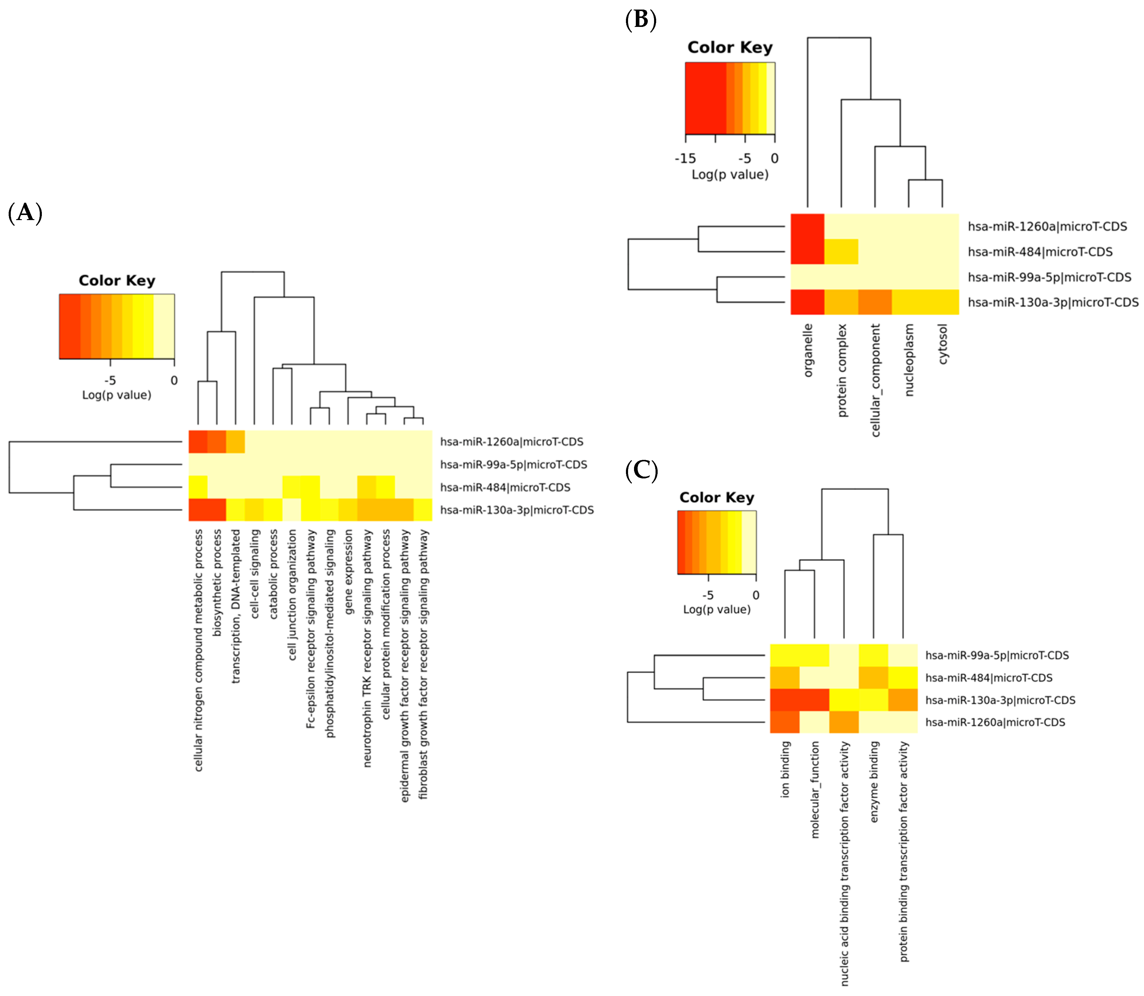
2.4. Signal and Functional Pathway Analysis

The most differentially expressed miRNAs were analysed by KEGG pathway analysis using the DIANA-mirPath v3.0 online web analysis tool to investigate the mechanism of their function and to analyse their target genes. All four miRNAs were significantly enriched in pathways involving the Hippo signalling pathway (hsa04390) and TGF-beta signalling pathway (hsa04350).
MicroRNAs miR-130a-3p, miR-484 and miR-1260a were enriched in the phosphatidylinositol signalling system (hsa04070) and oestrogen signalling pathway (hsa04915). MicroRNAs miR-130a-3p, miR-484 and miR-99a were enriched in the glioma (hsa05214) and mammalian target of rapamycin (mTOR) signalling pathway (hsa04150). The genes affected by the deregulation of miRNAs are presented in Table 3.
The Gene Ontology (GO) analysis was useful to investigate biological function in subcategories such as biological process, cellular component and molecular function corresponding to the target genes regulated by the up-regulated miRNAs. The GO annotation results for the gene union of the target genes of up-regulated miRNAs in breast cancer are listed in Table 4. A more detailed description of the GO categories and corresponding miRNAs is displayed in the heatmap (Figure 2).

3. Discussion

The preliminary study targeted differentially expressed miRNAs in the circulation of breast cancer patients compared to healthy volunteers. Breast cancer is a heterogeneous disease with different cell populations within the same tumour [20], and a better understanding is needed regarding breast cancer molecular pathways leading to the development of new therapeutic strategies that have an advantage in the prognosis and diagnosis of BC. A novel approach is based on the fact that tumour cells and/or tumour nucleic acids including miRNAs are released into the circulation and might serve as potential diagnostic markers for tumour development, metastasis and response to treatment [21].
In the technical part of the study, we wanted to overcome some critical points in the steps such as RNA extraction from plasma, reverse transcription, real-time PCR (qPCR) and data analysis. The process of RNA extraction and reverse transcription was controlled by synthetic spike-ins with different concentrations to monitor the presence of inhibitors and sample quality. In qPCR, we used the second derivative method for Cq calculation that has been described as being more reliable and appropriate when a large number of assays are analysed on multiple plates [22]. This procedure allowed us to obtain uniform Cq values without marked variation that could bias the statistical analysis.
We analysed five potentially reference miRNAs that have been described as the most useful in the normalisation of qPCR data. Therefore, the geNorm algorithm in pilot testing was used to find the most stable reference miRNAs with Cq below 30 cycles. Normalisation to more than one reference gene produces more reliable results and reduces the bias due to the differences between samples in the efficiency of miRNA extraction as well as in reverse transcription and amplification. Inadequate choice of reference genes also produces misleading results [23].
In the selection of the most appropriate reference genes, miR-520d-5p and miR-1228-3p were recommended as the most stable and useful in the spectrum of clinical material, such as exosomes, plasma and tissues [24]. However, the study revealed no expression of the mentioned genes in the plasma of either healthy or cancer patients. MicroRNA-191-5p was also excluded after pilot testing because control and BC patients had a Cq higher than 30 cycles; under these conditions, the variability in the amount of miRNA increases and the results may not be representative [25]. The most stable reference miRNAs (miR-16-5p and miR-103a-3p) were also verified in the final testing of control and BC patient plasma. The difference between the control and BC groups was 0.34 cycles in the arithmetic mean of both reference miRNAs. Another approach allows for normalisation to non-human miRNA, using an external control like cel-miR-39, or other small nuclear RNAs longer than 100 nucleotides. However, adding external control hardly balances the amount among samples, and longer RNAs are secreted and protected in the circulation differently to miRNAs [12].
MicroRNA-16 and miR-103a-3p are generally used as references, although some studies have reported a significant downregulation of miR-16-5p in the plasma and/or serum of BC as well as TNBC patients [26]. Ng et al. (2013) do not support the use of miR-16-5p for normalisation because of its elevated expression in the plasma of breast cancer patients [27]. In another study, miR-16-5p was identified as the most consistent reference miRNA between different subtypes of BC tissue [28] and in the plasma of cancer patients [29]. In a study by Chan et al. (2013), the authors identified miR-103a and miR-191 as the most stably expressed miRNAs in the serum of BC patients [30], although serum was identified to contain a higher concentration of miRNA [31]. Increased expression miR-103a-3p together with miR-320a, miR-361-5p and miR-21-5p, were chosen through the Cancer Genome Atlas as the most clinically relevant miRNAs [32]. High expression of the miR-103/107 family is associated with metastasis and a poor prognosis. Induction of epithelial to mesenchymal transition (EMT) via the downregulation of miR-200 has also been reported to be associated with the miR-103/107 family at the cellular level [33]. On the contrary, miR-103 and miR-107 were used as references in a study on the pathologic response to preoperative chemotherapy in TNBC core biopsies [34]. Serum expression of miR-24 and miR-103a was identified as a valuable biomarker for distinguishing atypical hyperplasia and early-stage of breast cancer [35]. Conversely, Normfinder and the geNorm algorithm have found stable expression of miR-103a together with other miRNAs in multiple diseases, e.g., in patients with Parkinson’s disease [36] and renal cell carcinoma [37]. In this study, we identified four significantly up-regulated (miR-99a, miR-130a, miR-484 and miR-1260a) miRNAs in the plasma of breast cancer patients.
MicroRNA-99a has a tumour suppressive role in the mammalian target of rapamycin (mTOR) signalling pathway [38]. It is downregulated in human cancers, which leads to the upregulation of mTOR and insulin-like growth factor receptor type 1 (IGF1R) in cancer tissue [39]. Upregulation of mTOR and fibroblast growth factor receptor 3 (FGFR3) inhibits tumour growth activated by tyrosine-protein kinase cellular sarcoma (c-SRC) [40]. The overexpression of miR-99a inhibits BC cell proliferation, migration and invasion in vivo by targeting FGFR3 and thus might have potential as a therapeutic target [41,42]. mTOR belongs to the PhosphoInositide 3-Kinase (PI3K)-related kinase family (PIKK), and miR-99a has a role in the PI3K/ protein kinase B (Akt) signalling pathway in endometrial cancer through a dual inhibitory effect. Therefore, miR-99a might have potential as an miR-99a mimic in cancer therapy [43]. A study on data of breast cancer tissues and serum samples that validated findings in clinical samples identified mir-99a together with miR-21-3p and miR-21-5p as the most eligible biomarkers for the early identification of breast cancer [44]
MicroRNA-130a expression is aberrant in various types of cancer, including breast cancer, mirroring its different roles as an oncogene or tumour suppressor. Studies on cell cultures and mice showed that miR-130a-3p acts as an onco-miR targeting Phosphatase and Tensin homolog (PTEN) and driving malignant cell survival and growth in the tumour [45]. Another study identifying the relationship between miR-130a and PTEN expression showed that downregulation of PTEN activates the PI3K/Akt/ endothelial Nitric Oxide SynthaseeNOS signalling pathway in the inflammatory response after human coronary artery endothelial cell (HCAEC) injury [46]. Another way of driving cell proliferation and migration has been described through the overexpression miR-130a and suppressing TGFBR2 expression in gastric cancer. Thus, miR-130a expression directly influences the TGF-β signalling pathway and is linked to EMT markers [47]. Plasma expression of miR-130a in BC patients varies according to HER2 status and lymph node positivity [48] and together with cluster miR-17-92, miR-22 and miR-29a/c can differentiate between TNBC and luminal A [49]. On the contrary, exosomal miR-130a-3p expression has a suppressive effect on breast cancer stem cell proliferation, invasion and migration via the downregulation of Ras superfamily of small GTPases-RAB5B; decreased miR-130a-3p expression is associated with lymph node metastasis and advanced tumor stage [50]. MiR-130a is involved in multiple signalling pathways regulating drug susceptibility [51], and studies have shown a heterogeneous pattern of miR-130a leverage.
We identified miR-484 as significantly upregulated (fold change (FC) = 1.22, p = 0.0002) in the plasma of BC patients. Similarly, Zaero et al. (2014) found higher expression of miR-484 in the serum (fold change–FC = 1.6, p = 0.0026) of early breast cancer patients. In accordance with these authors, we did not find any correlation between histopathological parameters and miR-484 expression [52]. In a study on ovarian cancer cells, the authors ascertained that miR-484, miR-642 and miR-217 are able to predict chemoresistance in ovarian cancer. Analysis of miR-484 revealed that it is involved in the regulation of angiogenic factors and its overexpression downregulates VEGFB and VEGFR2 expression; different levels of miR-484 may reflect the response to therapy [53]. High expression of miR-484 in the serum of patients with non-small cell lung cancer (NSCLC) was positively correlated with histological grade, lymph node metastasis, distant metastasis, clinical stage and poor overall survival [54]. In contrast, miR-484 is recommended as a suitable endogenous control for miRNA experiments [14,55].
Little is known about miR-1260a expression. Some studies have identified miR-1260a as an immune-related miRNA that has an overlapping seed sequence with miR-1260b. Robust miR-1260a/1260b expression in maternal breast milk may impart immune protection on the infant gut [56]. Expression of miR-1260a has been described as bring downregulated in TNBC tissue [57] and high levels of miR-1260a in the serum of metastatic BC patients is associated with a poor prognosis [18].
While the expression of miR-130a and miR-484 was in harmony with published results, miR-99a and miR-1260a were detected in the opposite pattern in the circulation. This discrepant expression is difficult to explain at this time; it could be due to the multiple origins of circulating miRNAs that do not originate only in the tumour environment [58]. It is also difficult to draw a clear conclusion from studies that have examined the expression of circulating miRNAs due to the different methods used to determine expression, i.e., microarray, next generation sequencing or qPCR. There are also differences regarding the qPCR system and what type of chemistry was used (TaqMan probes or SYBRgreen and a melt curve). Another critical point is the data analysis and algorithm to obtain the Cq value. The selection of the most suitable reference miRNA affects the subsequent analysis, and the most suitable references in one study may not be applicable in other studies. Consequently, the results of some studies are incomparable and is very difficult to come to a conclusion regarding these critical points since no precise guidelines for standardisation are available.
In our study, we identified some miRNAs (miR-99a, miR-130a, miR-484 and miR-1260a) that were significantly up-regulated in the plasma of breast cancer patients. At first glance, the fold change was not distinct from the control group. However, the input data are relatively uniform, as we followed several recommendations [23,25,59]. The most important differentially expressed miRNAs were analysed for pathways where they are significantly involved in the development of breast cancer, whether binding sites on target genes are experimentally confirmed or predicted via algorithms. The verification of true miRNA binding to specific target genes was not possible in this study.
A preliminary study has limitations, e.g., small sample size included in the BC and control cohorts and the small subgroups in the evaluation of histopathological parameters and miRNA expression. In subsequent studies, we plan to analyse the miRNA profile of BC tissues and to examine the entire image of circulating miRNAs in the plasma of BC and control cohort by the microarray method.
In subsequent studies, it will be necessary to evaluate circulating miRNA expression in more samples with a standardised and accepted methodology to obtain a broader view on the complex state of BC patients. Accurate information on miRNA expression in the plasma of cancer patients may help with a personalised approach and lead to the development of novel targeted therapy strategies. The level of circulating miRNA expression is further influenced by many factors that are related to the physiological state of the organism, and may even change during the day. The simple idea of early detection of miRNAs in a patient’s circulation then becomes a complex task in which many difficulties have to be overcome.

4. Materials and Methods

4.1. Patient Recruitment, Blood Collection and Plasma Preparation

All breast cancer patients were recruited at the Clinic of Obstetrics and Gynaecology, University Hospital Martin, Jessenius Faculty of Medicine, Comenius University in Martin and signed the informed consent form for inclusion into the study. The study was approved by the institutional Ethics Committee in accordance with the Declaration of Helsinki. The cohort included 65 breast cancer patients and 34 healthy volunteers. Breast cancer (BC) patients were selected according to the following criteria: (a) all patients were female; (b) all patients were diagnosed with a defined clinical stage; (c) disease was confirmed by routine histopathology; (d) none of the patients had another cancer or disease that may affect the miRNA plasma profile and e) none of the patients underwent preoperative radiotherapy or adjuvant chemotherapy. The healthy cohort was predominantly composed of female faculty staff and female volunteers who wanted to participate in the study with no history of malignant disease and no inflammatory conditions. All blood samples were collected in EthyleneDiamineTetraAcetic acid (EDTA)-anticoagulant tubes and processed as soon as possible, no later than 4 h after collection. The short-term storage and transport temperature was 4 °C. Plasma was separated from blood using two-step centrifugation (3000 revolution per minute/RPM for 10 min and 14,000 RPM for 10 min to minimise or remove the cell debris, residual platelets, microparticles, etc.). The level of haemolysis in plasma samples was evaluated by visual inspection and the measurement of oxyhaemoglobin absorbance at 414 nm. Clear plasma was stored at −80 °C until analysis.

4.2. RNA Extraction, Reverse Transcription of RNA and Quality Control

Circulating RNA was extracted from 200 μL of plasma using the miRNeasy Serum/Plasma Advanced kit (Qiagen, Hilden, Germany) according to the manufacturer’s recommended protocol. Total circulating RNA, including the miRNA fraction, was resuspended in 20 μL of RNase-free water. One microliter of total RNA was then reversely transcribed into complementary DNA (cDNA) in a 10 μL reaction using the miRCURY LNA RT kit (Qiagen) according to the manufacturer’s recommendations. The process of RNA extraction and cDNA synthesis was monitored through synthetic spike-ins (RNA spike-in kit, Qiagen) added in both steps according to recommendations of the manufacturer.

4.3. Real-Time Quantitative Polymerase Chain Reaction (qPCR)

Based on the literature review, specific miRNAs were selected with potentially stable plasma expression (reference miRNAs, e.g., miR-16-5p, miR-103a-3p, miR-191-5p, miR-1228-3p, miR-520d-5p) and target miRNAs (miR-10b-5p, miR-99a-5p, miR-130a-3p, miR-342-3p, miR-484, miR-486-5p, miR-1260a) [10,17,52,60] with elevated expression in the plasma of cancer patients. The expression level of selected target and reference miRNAs was determined by the miRCURY LNA miRNA PCR system (Qiagen), and the quality of samples was tested by synthetic spike-in oligonucleotides (RNA Spike-in kit, Qiagen). All protocols for miRNA quantification were performed using a Bravo liquid handling station (Agilent, Santa Clara, CA, USA) and run on a LightCycler 480 instrument (Roche Diagnostics GmbH, Mannheim, Germany) with rapid analysis on LC480 instrument software by the second derivative method for Cq calculation. Secondary analysis of Cq values was performed in the GeneGlobe web tool (Qiagen), where isolation and transcription quality controls were evaluated, a stabilisation factor for reference miRNAs was calculated and data were normalised. These factors were taken into account when calculating the change in expression between the control group and the group of breast cancer patients with the corresponding p-value.

4.4. Statistical Analysis and Bioinformatics

miRNA expression and fold change (FC) were computed using the standard formula [61]. The data were visualised by a boxplot overlaid with a swarmplot. The null hypothesis that the population median FC is equal to 1 was tested by the Wilcoxon test. The null hypothesis that the population median FC is the same for two levels of a categorical clinical parameter (e.g., ER) was tested by the Wilcoxon two-sample test. The analogous null hypothesis for a categorical clinical parameter with more than two levels (e.g., grade), was tested by robust ANOVA. Findings with a p-value below 0.05 were considered statistically significant. The data analyses were performed in R [62] ver. 3.5.2, using the libraries beeswarm [63], robustbase [64] and the WRS2 package [65].

4.5. Signalling and Functional Analysis of Differentially Expressed miRNAs

The most statistically significant miRNAs were analysed using online software to assess the miRNA regulatory roles and identify controlled pathways (mirPath v.3) from the Kyoto Encyclopaedia of Genes and Genomes (KEGG) based on the functional pathway enrichment analysis, as well as multiple segments of Gene Ontology analysis (GO) in Homo sapiens [66]. The specific analysis tool was used to carry out the combination of the available in silico predicted targets from DIANA-micro T-CDS with high quality experimentally supported interactions. A p-value of <0.05 was considered statistically significant, and the Micro T threshold was set at 0.8. In the KEGG analysis, we prefer the gene union and in GO analysis, the category union and intersection for merging the results with false discovery rate (FDR) correction.

Supplementary Materials

Supplementary materials can be found at https://www.mdpi.com/1422-0067/21/19/7288/s1.

Author Contributions

Conceptualisation, P.Z. and Z.D.; methodology, V.H., Z.K., M.B. and M.K.; software, M.G., V.H. and Z.K.; validation, V.H. and Z.K.; formal analysis, V.H., Z.K. and M.G.; investigation, V.H., Z.K., E.K., Z.L. and P.Z.; resources, D.B., D.D., I.S., M.J., K.Z., M.S., A.L., E.K. and Z.L.; data curation, Z.D., E.K., Z.L., V.H., Z.K., M.B. and M.K.; writing—original draft preparation, V.H.; writing—review and editing, V.H., Z.K., E.K., Z.D. and M.S.; supervision, Z.D. and P.Z.; project administration, Z.D.; funding acquisition P.Z. All authors have read and agreed to the published version of the manuscript.

Funding

This study was funded by the Slovak Research and Development Agency under contract no. APVV-16-0021 and is the result of the project implementation: “Centre of excellence for research in personalised therapy (CEVYPET)”, ITMS: 26220120053 supported by the Operational Programme Research and Innovation and “Integrative strategy in development of personalised medicine of selected malignant tumours and its impact on quality of life”, ITMS: 313011V446, projects co-financed by the ERDF.

Acknowledgments

The authors wish to thank Zora Lasabova and the Department of Molecular Biology and Genomics, Jessenius Faculty of Medicine in Martin, Comenius University in Bratislava for technical support.

Conflicts of Interest

The authors declare no conflict of interest.

References

  1. Ferlay, J.; Colombet, M.; Soerjomataram, I.; Mathers, C.; Parkin, D.M.; Piñeros, M.; Znaor, A.; Bray, F. Estimating the global cancer incidence and mortality in 2018: GLOBOCAN sources and methods. Int. J. Cancer 2019, 144, 1941–1953. [Google Scholar] [CrossRef] [PubMed] [Green Version]
  2. Dafni, U.; Tsourti, Z.; Alatsathianos, I. Breast Cancer Statistics in the European Union: Incidence and Survival across European Countries. Breast Care 2019, 14, 344–353. [Google Scholar] [CrossRef] [PubMed]
  3. Fuller, M.S.; Lee, C.I.; Elmore, J.G. Breast Cancer Screening: An Evidence-Based Update. Med. Clin. N. Am. 2015, 99, 451–468. [Google Scholar] [CrossRef] [PubMed] [Green Version]
  4. Goldhirsch, A.; Wood, W.C.; Coates, A.S.; Gelber, R.D.; Thürlimann, B.; Senn, H.-J. Strategies for subtypes—dealing with the diversity of breast cancer: Highlights of the St Gallen International Expert Consensus on the Primary Therapy of Early Breast Cancer 2011. Ann. Oncol. 2011, 22, 1736–1747. [Google Scholar] [CrossRef]
  5. Gadzicki, D.; Evans, D.G.; Harris, H.; Julian-Reynier, C.; Nippert, I.; Schmidtke, J.; Tibben, A.; van Asperen, C.J.; Schlegelberger, B. Genetic testing for familial/hereditary breast cancer—Comparison of guidelines and recommendations from the UK, France, the Netherlands and Germany. J. Community Genet. 2011, 2, 53–69. [Google Scholar] [CrossRef] [Green Version]
  6. Rudolph, A.; Song, M.; Brook, M.N.; Milne, R.L.; Mavaddat, N.; Michailidou, K.; Bolla, M.K.; Wang, Q.; Dennis, J.; Wilcox, A.N.; et al. Joint associations of a polygenic risk score and environmental risk factors for breast cancer in the Breast Cancer Association Consortium. Int. J. Epidemiol. 2018, 47, 526–536. [Google Scholar] [CrossRef] [Green Version]
  7. Chang-Claude, J.; Eby, N.; Kiechle, M.; Bastert, G.; Becher, H. Breastfeeding and breast cancer risk by age 50 among women in Germany. Cancer Causes Control CCC 2000, 11, 687–695. [Google Scholar] [CrossRef]
  8. Horn, J.; Vatten, L.J. Reproductive and hormonal risk factors of breast cancer: A historical perspective. Int. J. Womens Health 2017, 9, 265–272. [Google Scholar] [CrossRef] [Green Version]
  9. McDonald, K.-A.; Young, J. Breast Cancer Screening Modalities. In Preventive Oncology for the Gynecologist; Mehta, S., Singla, A., Eds.; Springer: Singapore, 2019; pp. 353–366. ISBN 9789811334382. [Google Scholar]
  10. Zubor, P.; Kubatka, P.; Kajo, K.; Dankova, Z.; Polacek, H.; Bielik, T.; Kudela, E.; Samec, M.; Liskova, A.; Vlcakova, D.; et al. Why the Gold Standard Approach by Mammography Demands Extension by Multiomics? Application of Liquid Biopsy miRNA Profiles to Breast Cancer Disease Management. Int. J. Mol. Sci. 2019, 20, 2878. [Google Scholar] [CrossRef] [Green Version]
  11. Tran, T.H.; Montano, M.A. Chapter 1—MicroRNAs: Mirrors of Health and Disease. In Translating MicroRNAs to the Clinic; Laurence, J., Ed.; Academic Press: Boston, MA, USA, 2017; pp. 1–15. ISBN 978-0-12-800553-8. [Google Scholar]
  12. Cui, M.; Wang, H.; Yao, X.; Zhang, D.; Xie, Y.; Cui, R.; Zhang, X. Circulating MicroRNAs in Cancer: Potential and Challenge. Front. Genet. 2019, 10. [Google Scholar] [CrossRef] [Green Version]
  13. Li, M.; Zhou, Y.; Xia, T.; Zhou, X.; Huang, Z.; Zhang, H.; Zhu, W.; Ding, Q.; Wang, S. Circulating microRNAs from the miR-106a–363 cluster on chromosome X as novel diagnostic biomarkers for breast cancer. Breast Cancer Res. Treat. 2018, 170, 257–270. [Google Scholar] [CrossRef] [PubMed]
  14. Rice, J.; Roberts, H.; Rai, S.N.; Galandiuk, S. Housekeeping genes for studies of plasma microRNA: A need for more precise standardization. Surgery 2015, 158, 1345–1351. [Google Scholar] [CrossRef] [PubMed]
  15. Mitchell, P.S.; Parkin, R.K.; Kroh, E.M.; Fritz, B.R.; Wyman, S.K.; Pogosova-Agadjanyan, E.L.; Peterson, A.; Noteboom, J.; O’Briant, K.C.; Allen, A.; et al. Circulating microRNAs as stable blood-based markers for cancer detection. Proc. Natl. Acad. Sci. USA. 2008, 105, 10513–10518. [Google Scholar] [CrossRef] [PubMed] [Green Version]
  16. Hu, J.; Wang, Z.; Liao, B.-Y.; Yu, L.; Gao, X.; Lu, S.; Wang, S.; Dai, Z.; Zhang, X.; Chen, Q.; et al. Human miR-1228 as a stable endogenous control for the quantification of circulating microRNAs in cancer patients. Int. J. Cancer 2014, 135, 1187–1194. [Google Scholar] [CrossRef]
  17. Zografos, E.; Zagouri, F.; Kalapanida, D.; Zakopoulou, R.; Kyriazoglou, A.; Apostolidou, K.; Gazouli, M.; Dimopoulos, M.-A. Prognostic role of microRNAs in breast cancer: A systematic review. Oncotarget 2019, 10, 7156–7178. [Google Scholar] [CrossRef] [PubMed] [Green Version]
  18. Madhavan, D.; Peng, C.; Wallwiener, M.; Zucknick, M.; Nees, J.; Schott, S.; Rudolph, A.; Riethdorf, S.; Trumpp, A.; Pantel, K.; et al. Circulating miRNAs with prognostic value in metastatic breast cancer and for early detection of metastasis. Carcinogenesis 2016, 37, 461–470. [Google Scholar] [CrossRef] [PubMed] [Green Version]
  19. Vandesompele, J.; De Preter, K.; Pattyn, F.; Poppe, B.; Van Roy, N.; De Paepe, A.; Speleman, F. Accurate normalization of real-time quantitative RT-PCR data by geometric averaging of multiple internal control genes. Genome Biol. 2002, 3, research0034.1–research0034.11. [Google Scholar] [CrossRef] [Green Version]
  20. Turashvili, G.; Brogi, E. Tumor Heterogeneity in Breast Cancer. Front. Med. 2017, 4, 227. [Google Scholar] [CrossRef] [Green Version]
  21. Pantel, K.; Alix-Panabières, C. Liquid biopsy: Potential and challenges. Mol. Oncol. 2016, 10, 371–373. [Google Scholar] [CrossRef] [Green Version]
  22. Luu-The, V.; Paquet, N.; Calvo, E.; Cumps, J. Improved real-time RT-PCR method for high-throughput measurements using second derivative calculation and double correction. BioTechniques 2005, 38, 287–293. [Google Scholar] [CrossRef]
  23. Schwarzenbach, H.; da Silva, A.M.; Calin, G.; Pantel, K. Data Normalization Strategies for MicroRNA Quantification. Clin. Chem. 2015, 61, 1333–1342. [Google Scholar] [CrossRef] [PubMed]
  24. Danese, E.; Minicozzi, A.M.; Benati, M.; Paviati, E.; Lima-Oliveira, G.; Gusella, M.; Pasini, F.; Salvagno, G.L.; Montagnana, M.; Lippi, G. Reference miRNAs for colorectal cancer: Analysis and verification of current data. Sci. Rep. 2017, 7. [Google Scholar] [CrossRef] [PubMed]
  25. Taylor, S.C.; Nadeau, K.; Abbasi, M.; Lachance, C.; Nguyen, M.; Fenrich, J. The Ultimate qPCR Experiment: Producing Publication Quality, Reproducible Data the First Time. Trends Biotechnol. 2019, 37, 761–774. [Google Scholar] [CrossRef] [PubMed] [Green Version]
  26. Shin, V.Y.; Siu, J.M.; Cheuk, I.; Ng, E.K.O.; Kwong, A. Circulating cell-free miRNAs as biomarker for triple-negative breast cancer. Br. J. Cancer 2015, 112, 1751–1759. [Google Scholar] [CrossRef] [PubMed]
  27. Ng, E.K.O.; Li, R.; Shin, V.Y.; Jin, H.C.; Leung, C.P.H.; Ma, E.S.K.; Pang, R.; Chua, D.; Chu, K.-M.; Law, W.L.; et al. Circulating microRNAs as Specific Biomarkers for Breast Cancer Detection. PLoS ONE 2013, 8, e53141. [Google Scholar] [CrossRef] [Green Version]
  28. Rinnerthaler, G.; Hackl, H.; Gampenrieder, S.P.; Hamacher, F.; Hufnagl, C.; Hauser-Kronberger, C.; Zehentmayr, F.; Fastner, G.; Sedlmayer, F.; Mlineritsch, B.; et al. miR-16-5p Is a Stably-Expressed Housekeeping MicroRNA in Breast Cancer Tissues from Primary Tumors and from Metastatic Sites. Int. J. Mol. Sci. 2016, 17, 156. [Google Scholar] [CrossRef]
  29. Sourvinou, I.S.; Markou, A.; Lianidou, E.S. Quantification of Circulating miRNAs in Plasma: Effect of Preanalytical and Analytical Parameters on Their Isolation and Stability. J. Mol. Diagn. 2013, 15, 827–834. [Google Scholar] [CrossRef]
  30. Chan, M.; Liaw, C.S.; Ji, S.M.; Tan, H.H.; Wong, C.Y.; Thike, A.A.; Tan, P.H.; Ho, G.H.; Lee, A.S.-G. Identification of Circulating MicroRNA Signatures for Breast Cancer Detection. Clin. Cancer Res. 2013, 19, 4477–4487. [Google Scholar] [CrossRef] [Green Version]
  31. Graveel, C.R.; Calderone, H.M.; Westerhuis, J.J.; Winn, M.E.; Sempere, L.F. Critical analysis of the potential for microRNA biomarkers in breast cancer management. Breast Cancer Targets Ther. 2015, 7, 59–79. [Google Scholar] [CrossRef] [Green Version]
  32. Chang, J.T.-H.; Wang, F.; Chapin, W.; Huang, R.S. Identification of MicroRNAs as Breast Cancer Prognosis Markers through the Cancer Genome Atlas. PLoS ONE 2016, 11. [Google Scholar] [CrossRef] [Green Version]
  33. Martello, G.; Rosato, A.; Ferrari, F.; Manfrin, A.; Cordenonsi, M.; Dupont, S.; Enzo, E.; Guzzardo, V.; Rondina, M.; Spruce, T.; et al. A MicroRNA Targeting Dicer for Metastasis Control. Cell 2010, 141, 1195–1207. [Google Scholar] [CrossRef] [PubMed] [Green Version]
  34. Kolacinska, A.; Morawiec, J.; Fendler, W.; Malachowska, B.; Morawiec, Z.; Szemraj, J.; Pawlowska, Z.; Chowdhury, D.; Choi, Y.E.; Kubiak, R.; et al. Association of microRNAs and pathologic response to preoperative chemotherapy in triple negative breast cancer: Preliminary report. Mol. Biol. Rep. 2014, 41, 2851–2857. [Google Scholar] [CrossRef] [PubMed] [Green Version]
  35. An, X.; Quan, H.; Lv, J.; Meng, L.; Wang, C.; Yu, Z.; Han, J. Serum microRNA as potential biomarker to detect breast atypical hyperplasia and early-stage breast cancer. Future Oncol. Lond. Engl. 2018, 14, 3145–3161. [Google Scholar] [CrossRef] [PubMed]
  36. Martins, M.; Rosa, A.; Guedes, L.C.; Fonseca, B.V.; Gotovac, K.; Violante, S.; Mestre, T.; Coelho, M.; Rosa, M.M.; Martin, E.R.; et al. Convergence of miRNA Expression Profiling, α-Synuclein Interacton and GWAS in Parkinson’s Disease. PLoS ONE 2011, 6. [Google Scholar] [CrossRef] [PubMed]
  37. Wotschofsky, Z.; Meyer, H.-A.; Jung, M.; Fendler, A.; Wagner, I.; Stephan, C.; Busch, J.; Erbersdobler, A.; Disch, A.C.; Mollenkopf, H.-J.; et al. Reference genes for the relative quantification of microRNAs in renal cell carcinomas and their metastases. Anal. Biochem. 2011, 417, 233–241. [Google Scholar] [CrossRef] [PubMed]
  38. Hu, Y.; Zhu, Q.; Tang, L. MiR-99a Antitumor Activity in Human Breast Cancer Cells through Targeting of mTOR Expression. PLoS ONE 2014, 9. [Google Scholar] [CrossRef]
  39. Torres, A.; Torres, K.; Pesci, A.; Ceccaroni, M.; Paszkowski, T.; Cassandrini, P.; Zamboni, G.; Maciejewski, R. Deregulation of miR-100, miR-99a and miR-199b in tissues and plasma coexists with increased expression of mTOR kinase in endometrioid endometrial carcinoma. BMC Cancer 2012, 12, 369. [Google Scholar] [CrossRef] [Green Version]
  40. Oneyama, C.; Ikeda, J.; Okuzaki, D.; Suzuki, K.; Kanou, T.; Shintani, Y.; Morii, E.; Okumura, M.; Aozasa, K.; Okada, M. MicroRNA-mediated downregulation of mTOR/FGFR3 controls tumor growth induced by Src-related oncogenic pathways. Oncogene 2011, 30, 3489–3501. [Google Scholar] [CrossRef] [Green Version]
  41. Long, X.; Shi, Y.; Ye, P.; Guo, J.; Zhou, Q.; Tang, Y. MicroRNA-99a Suppresses Breast Cancer Progression by Targeting FGFR3. Front. Oncol. 2020, 9. [Google Scholar] [CrossRef] [Green Version]
  42. Yang, Z.; Han, Y.; Cheng, K.; Zhang, G.; Wang, X. miR-99a directly targets the mTOR signalling pathway in breast cancer side population cells. Cell Prolif. 2014, 47, 587–595. [Google Scholar] [CrossRef]
  43. Li, Y.; Zhang, Z.; Zhang, X.; Lin, Y.; Luo, T.; Xiao, Z.; Zhou, Q. A dual PI3K/AKT/mTOR signaling inhibitor miR-99a suppresses endometrial carcinoma. Am. J. Transl. Res. 2016, 8, 719–731. [Google Scholar] [PubMed]
  44. Yu, X.; Liang, J.; Xu, J.; Li, X.; Xing, S.; Li, H.; Liu, W.; Liu, D.; Xu, J.; Huang, L.; et al. Identification and Validation of Circulating MicroRNA Signatures for Breast Cancer Early Detection Based on Large Scale Tissue-Derived Data. J. Breast Cancer 2018, 21, 363–370. [Google Scholar] [CrossRef] [PubMed]
  45. Wei, H.; Cui, R.; Bahr, J.; Zanesi, N.; Luo, Z.; Meng, W.; Liang, G.; Croce, C.M. miR-130a deregulates PTEN and stimulates tumor growth. Cancer Res. 2017, 77, 6168–6178. [Google Scholar] [CrossRef] [PubMed] [Green Version]
  46. Song, C.-L.; Liu, B.; Shi, Y.-F.; Liu, N.; Yan, Y.-Y.; Zhang, J.-C.; Xue, X.; Wang, J.-P.; Zhao, Z.; Liu, J.-G.; et al. MicroRNA-130a alleviates human coronary artery endothelial cell injury and inflammatory responses by targeting PTEN via activating PI3K/Akt/eNOS signaling pathway. Oncotarget 2016, 7, 71922–71936. [Google Scholar] [CrossRef]
  47. Duan, J.; Zhang, H.; Qu, Y.; Deng, T.; Huang, D.; Liu, R.; Zhang, L.; Bai, M.; Zhou, L.; Ying, G.; et al. Onco-miR-130 promotes cell proliferation and migration by targeting TGFβR2 in gastric cancer. Oncotarget 2016, 7, 44522–44533. [Google Scholar] [CrossRef] [Green Version]
  48. Stückrath, I.; Rack, B.; Janni, W.; Jäger, B.; Pantel, K.; Schwarzenbach, H. Aberrant plasma levels of circulating miR-16, miR-107, miR-130a and miR-146a are associated with lymph node metastasis and receptor status of breast cancer patients. Oncotarget 2015, 6, 13387–13401. [Google Scholar] [CrossRef]
  49. Braicu, C.; Raduly, L.; Morar-Bolba, G.; Cojocneanu, R.; Jurj, A.; Pop, L.-A.; Pileczki, V.; Ciocan, C.; Moldovan, A.; Irimie, A.; et al. Aberrant miRNAs expressed in HER-2 negative breast cancers patient. J. Exp. Clin. Cancer Res. 2018, 37, 257. [Google Scholar] [CrossRef] [Green Version]
  50. Kong, X.; Zhang, J.; Li, J.; Shao, J.; Fang, L. MiR-130a-3p inhibits migration and invasion by regulating RAB5B in human breast cancer stem cell-like cells. Biochem. Biophys. Res. Commun. 2018, 501, 486–493. [Google Scholar] [CrossRef]
  51. Zhang, H.; Jiang, L.; Sun, D.; Li, J.; Ji, Z. The role of miR-130a in cancer. Breast Cancer 2017, 24, 521–527. [Google Scholar] [CrossRef]
  52. Zearo, S.; Kim, E.; Zhu, Y.; Zhao, J.T.; Sidhu, S.B.; Robinson, B.G.; Soon, P.S. MicroRNA-484 is more highly expressed in serum of early breast cancer patients compared to healthy volunteers. BMC Cancer 2014, 14, 200. [Google Scholar] [CrossRef]
  53. Vecchione, A.; Belletti, B.; Lovat, F.; Volinia, S.; Chiappetta, G.; Giglio, S.; Sonego, M.; Cirombella, R.; Onesti, E.C.; Pellegrini, P.; et al. A microRNA signature defines chemoresistance in ovarian cancer through modulation of angiogenesis. Proc. Natl. Acad. Sci. USA. 2013, 110, 9845–9850. [Google Scholar] [CrossRef] [PubMed] [Green Version]
  54. Zhuang, Z.; Sun, C.; Gong, H. High serum miR-484 expression is associated with the diagnosis and prognosis of patients with non-small cell lung cancer. Exp. Ther. Med. 2019, 18, 4095–4102. [Google Scholar] [CrossRef] [Green Version]
  55. Rzepiel, A.; Kutszegi, N.; Gézsi, A.; Sági, J.C.; Egyed, B.; Péter, G.; Butz, H.; Nyírő, G.; Müller, J.; Kovács, G.T.; et al. Circulating microRNAs as minimal residual disease biomarkers in childhood acute lymphoblastic leukemia. J. Transl. Med. 2019, 17, 372. [Google Scholar] [CrossRef] [PubMed]
  56. Carney, M.C.; Tarasiuk, A.; DiAngelo, S.L.; Silveyra, P.; Podany, A.; Birch, L.L.; Paul, I.M.; Kelleher, S.; Hicks, S.D. Metabolism-related microRNAs in maternal breast milk are influenced by premature delivery. Pediatr. Res. 2017, 82, 226–236. [Google Scholar] [CrossRef] [PubMed]
  57. Sugita, B.M.; Pereira, S.R.; de Almeida, R.C.; Gill, M.; Mahajan, A.; Duttargi, A.; Kirolikar, S.; Fadda, P.; de Lima, R.S.; Urban, C.A.; et al. Integrated copy number and miRNA expression analysis in triple negative breast cancer of Latin American patients. Oncotarget 2019, 10, 6184–6203. [Google Scholar] [CrossRef] [Green Version]
  58. Turchinovich, A.; Burwinkel, B. Distinct AGO1 and AGO2 associated miRNA profiles in human cells and blood plasma. RNA Biol. 2012, 9, 1066–1075. [Google Scholar] [CrossRef] [Green Version]
  59. Bustin, S.A.; Benes, V.; Garson, J.A.; Hellemans, J.; Huggett, J.; Kubista, M.; Mueller, R.; Nolan, T.; Pfaffl, M.W.; Shipley, G.L.; et al. The MIQE Guidelines: Minimum Information for Publication of Quantitative Real-Time PCR Experiments. Clin. Chem. 2009, 55, 611–622. [Google Scholar] [CrossRef] [Green Version]
  60. McGuire, A.; Brown, J.A.L.; Kerin, M.J. Metastatic breast cancer: The potential of miRNA for diagnosis and treatment monitoring. Cancer Metastasis Rev. 2015, 34, 145–155. [Google Scholar] [CrossRef] [Green Version]
  61. Livak, K.J.; Schmittgen, T.D. Analysis of Relative Gene Expression Data Using Real-Time Quantitative PCR and the 2−ΔΔCT Method. Methods 2001, 25, 402–408. [Google Scholar] [CrossRef]
  62. R: The R Project for Statistical Computing. Available online: https://www.r-project.org/ (accessed on 13 July 2020).
  63. Eklund, A. Beeswarm: The Bee Swarm Plot, an Alternative to Stripchart version 0.2.3 from CRAN. Available online: https://rdrr.io/cran/beeswarm/ (accessed on 13 July 2020).
  64. Maechler, M.; Rousseeuw, P.; Croux, C.; Todorov, V.; Ruckstuhl, A.; Salibian-Barrera, M.; Verbeke, T.; Koller, M.; Conceicao, E.; diPalma, M.A. Robustbase: Basic Robust Statistics. R package version 0.93-6. Available online: http://robustbase.r-forge.r-project.org/ (accessed on 13 July 2020).
  65. Mair, P.; Wilcox, R. Robust statistical methods in R using the WRS2 package. Behav. Res. Methods 2020, 52, 464–488. [Google Scholar] [CrossRef]
  66. Vlachos, I.S.; Zagganas, K.; Paraskevopoulou, M.D.; Georgakilas, G.; Karagkouni, D.; Vergoulis, T.; Dalamagas, T.; Hatzigeorgiou, A.G. DIANA-miRPath v3.0: Deciphering microRNA function with experimental support. Nucleic Acids Res. 2015, 43, W460–W466. [Google Scholar] [CrossRef] [PubMed]
Figure 1. The scheme of study design.
Ijms 21 07288 g001
Figure 2. Heatmaps of the Gene Ontology (GO) analysis. GO enrichment of putative targets was performed for four significantly up-regulated miRNAs at three levels: (A) biological process, (B) cellular component and (C) molecular function. Different colours represent different levels of pathway term enrichment in the miRNA target genes. Red indicates a higher degree of enrichment.
Ijms 21 07288 g002
Table 1. Clinicopathological data of the included patients.
Characteristics Number of Patients (%)
All patients 65 (100%)
AgeMedian (IQR)56 (47,68)
Missing5
HistologyDuctal21 (40%)
Lobular8 (15%)
Invasive cancer, NST24 (45%)
Missing12
SubtypeLuminal A33 (63%)
Luminal B13 (25%)
HER24 (7.7%)
TNC2 (3.8%)
Missing13
Grade18 (14%)
230 (53%)
319 (33%)
Missing8
ER statusPositive48 (87%)
Negative7 (13%)
Missing10
PR statusPositive43 (78%)
Negative12 (22%)
Missing10
HER2Positive9 (18%)
Negative41 (82%)
Missing15
Tumour sizeTIS4 (6.9%)
<20 mm38 (66%)
20 mm–50 mm13 (22%)
>50 mm3 (5.2%)
Missing7
Nodal statusNegative27 (56%)
N117 (35%)
N24 (8.3%)
Missing17
LVIPositive28 (51%)
Negative27 (49%)
Missing10
PNIPositive11 (20%)
Negative44 (80%)
Missing10
ki67≤15%34 (65%)
>15%18 (35%)
Missing 13
Abbreviations: IQR, interquartile range; NST, no specific type; TNC, triple-negative cancer; ER, oestrogen; PR, progesterone; TIS, tumour in situ; mm, millimetre; LVI, lymphovascular invasion; PNI, perineural invasion.
Table 2. Fold change data analysis of tested circulating miRNAs in the plasma of cancer patients compared to the control group obtained in the Wilcoxon test. Significant values are displayed in bold.
miRNA IDGeneGlobe Data Analysis ToolddCt Method
Pilot TestingFinal Testing
FCReg.p-valueFCReg.p-valueFCReg.p-value
miR-10b1.240.170.860.880.910.68
miR-160.950.450.950.401-1
miR-99a1.270.221.060.441.10.03
miR-103a1.060.371.050.621-1
miR-130a0.940.911.010.341.33<0.01
miR-1911.180.13------
miR-3420.670.050.760.370.760.35
miR-4841.350.011.140.361.22<0.01
miR-4860.900.660.920.340.890.37
miR-520d1.430.31------
miR-12281.430.31------
miR-1260a0.820.581.030.121.2<0.01
Arrows represent up- or downregulation in breast cancer (BC) cases when compared to controls. Abbreviations: ID, identification; ddCt, delta–delta cycle threshold; FC, fold change; Reg., regulation.
Table 3. Kyoto Encyclopaedia of Genes and Genomes (KEGG) analysis of the significantly up-regulated miRNAs in the plasma of BC patients.
KEGG PathwayPathway IDp-ValuemiRNAsTarget Genes
Prion diseaseshsa050203.17 × 10−22miR-130a-3pPNRP
Hippo signalling pathwayhsa043900.007miR-130a-3pWNT2B, FRMD6, TGFB2, TGFBR2, BMPR2, PPP1CB
miR-484YAP1, WNT2B, WWC1, TP53BP2, CDH1, AXIN2, DLG2, TCF7, FGF1
miR-99a-5pFDZ5, FDZ8, SMAD7
miR-1260aDLG2
Phosphatidylinositol signalling systemhsa040700.0082miR-130a-3pCDS1, DGKE, CALM2, PLCB1, PIKFYVE, PIK3C2A, PTEN, PLCB4, DGKH
miR-484CALM1, PLCZ1, PIKFYVE, PIK3CD
miR-1260aPIP4K2C, DGKH
Oestrogen signalling pathwayhsa049150.0136miR-484CREB3L3, CALM1, PIK3CD, GRM1, KCNJ5
miR-130a-3pESR1, ADCY1, SOS2, CALM2, PLCB1, KCNJ6, HSPA8, PLCB4
miR-1260aADCY1, ATF6B, KCNJ6, SP1
Gliomahsa052140.0136miR-484CALM1, PIK3CD, PDGFA
miR-130a-3pSOS2, E2F2, TGFA, CALM2, IGF1, CDKN1A, PTEN
miR-99a-5pMTOR
TGF-beta signalling pathwayhsa043500.0389miR-484ACVR1B, PITX2
miR-130a-3pINHBB, SMURF2, INHBA, ACVR1, SKP1, ZFYVE9, SMAD5, TGFB2, TGFBR2, BMPR2
miR-1260aSKP1, SP1
miR-99a-5pSMAD7
Glycosaminoglycan biosynthesis—heparan sulphate/heparinhsa005340.0399miR-1260aEXT2, HS3ST2
miR-99a-5pHS3ST3B1, HS3ST2
mTOR signalling pathwayhsa041500.0399miR-99a-5pMTOR
miR-130a-3pTSC1, RRAGD, PRKAA2, PRKAA1, IGF1, EIF4E2, PTEN, ULK2
miR-484RPS6KA1, PIK3CD
Table 4. Gene Ontology (GO) annotation results of the target genes of up-regulated microRNAs in breast cancer samples.
GO Termp-ValueTarget Gene Account
Biological process
GO:0034641: cellular nitrogen compound metabolic process1.11 × 10−16387
GO:0009058: biosynthetic process4.86 × 10−14241
GO:0048011: neurotrophin TRK receptor signalling pathway 8.09 × 10−631
GO:0006464: cellular protein modification process 1.64 × 10−5151
GO:0007173: epidermal growth factor receptor signalling pathway 0.000217
GO:0006351: transcription, DNA-templated0.0007154
GO:0038095: Fc-epsilon receptor signalling pathway0.001520
GO:0010467: gene expression 0.003525
GO:0007267: cell-cell signalling0.018832
GO:0009056: catabolic process 0.025064
GO:0048015: phosphatidylinositol-mediated signalling0.031510
GO:0034330: cell junction organisation0.047111
GO:0008543: fibroblast growth factor receptor signalling pathway0.049711
Cellular component
GO:0043226: organelle<1 × 10−325772
GO:0043234: protein complex3.68 × 10−6239
GO:0005575: cellular component0.00018496
GO:0005829: cytosol0.0066796
GO:0005654: nucleoplasm0.0076547
Molecular function
GO:0043167: ion binding<1 × 10−325Top of Form
488 Bottom of Form
GO:0003674: molecular function1.374 × 10−7Top of Form
515 Bottom of Form
GO:0001071: nucleic acid binding transcription factor activity3.503 × 10−6Top of Form
74 Bottom of Form
GO:0000988: protein binding transcription factor activity1.861 × 10−5Top of Form
50 Bottom of Form
GO:0019899: enzyme binding5.208 × 10−5Top of Form
98 Bottom of Form

