Robustness of Clonogenic Assays as a Biomarker for Cancer Cell Radiosensitivity

 ,
,
Abstract
:
1. Introduction
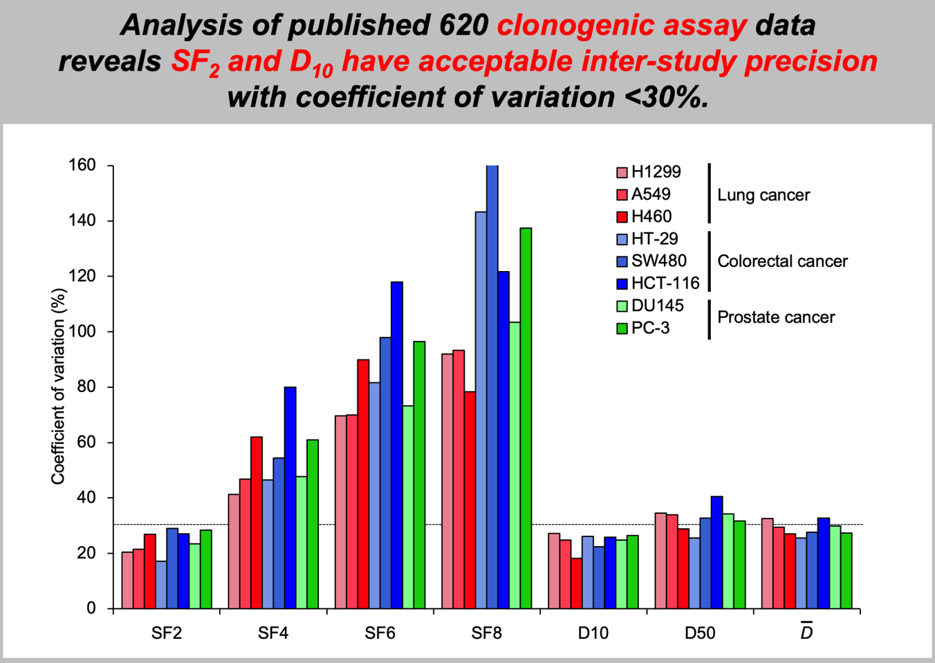
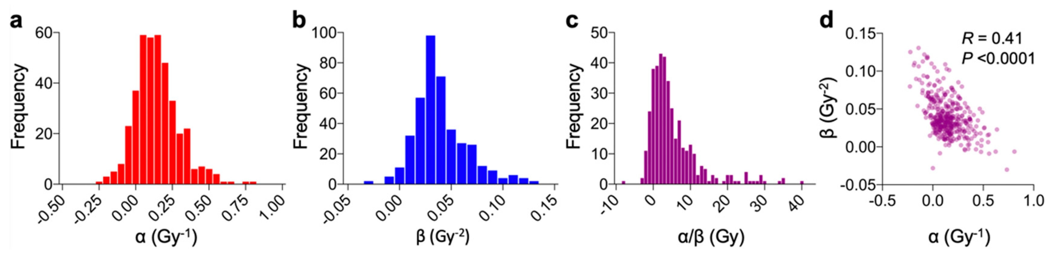
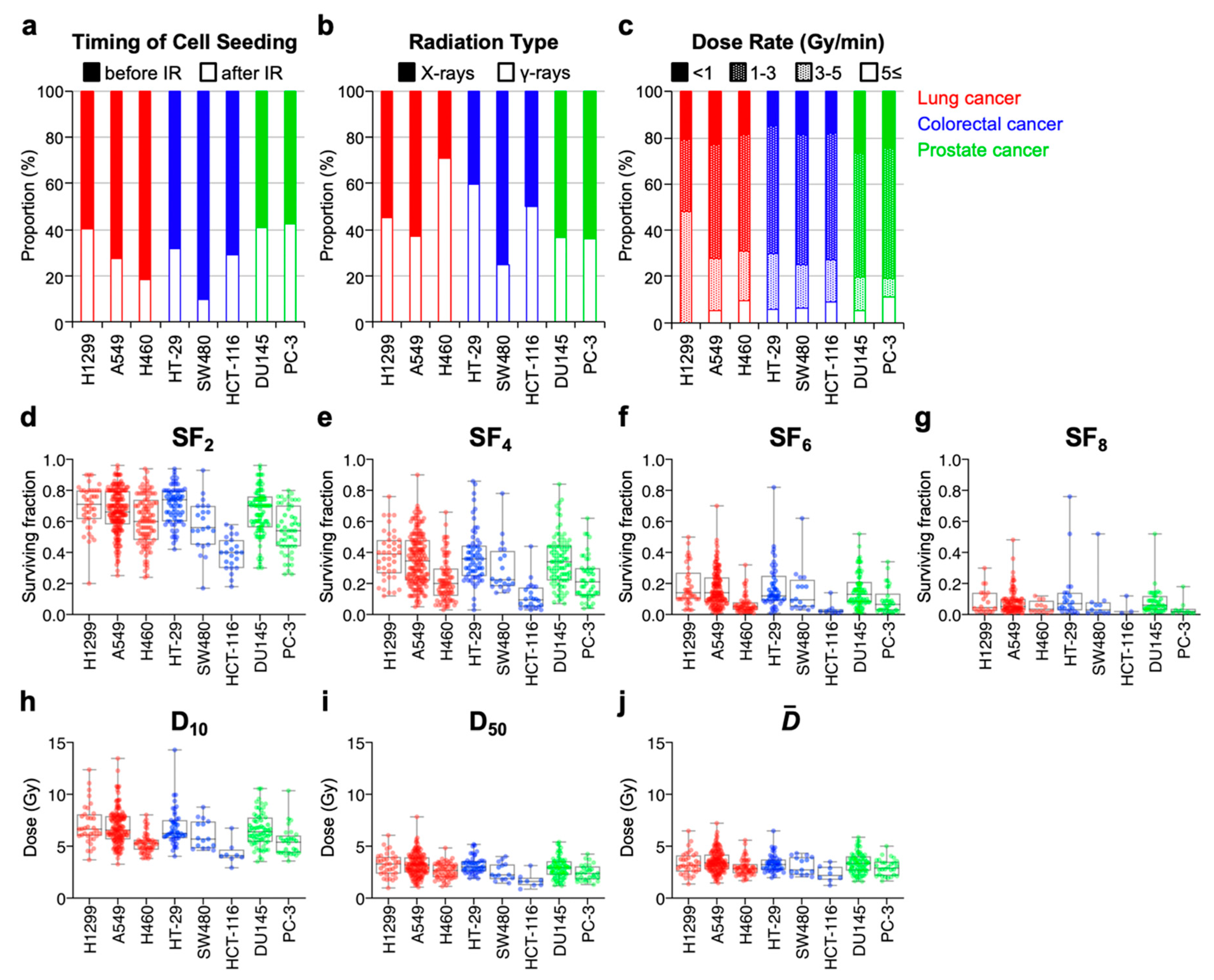
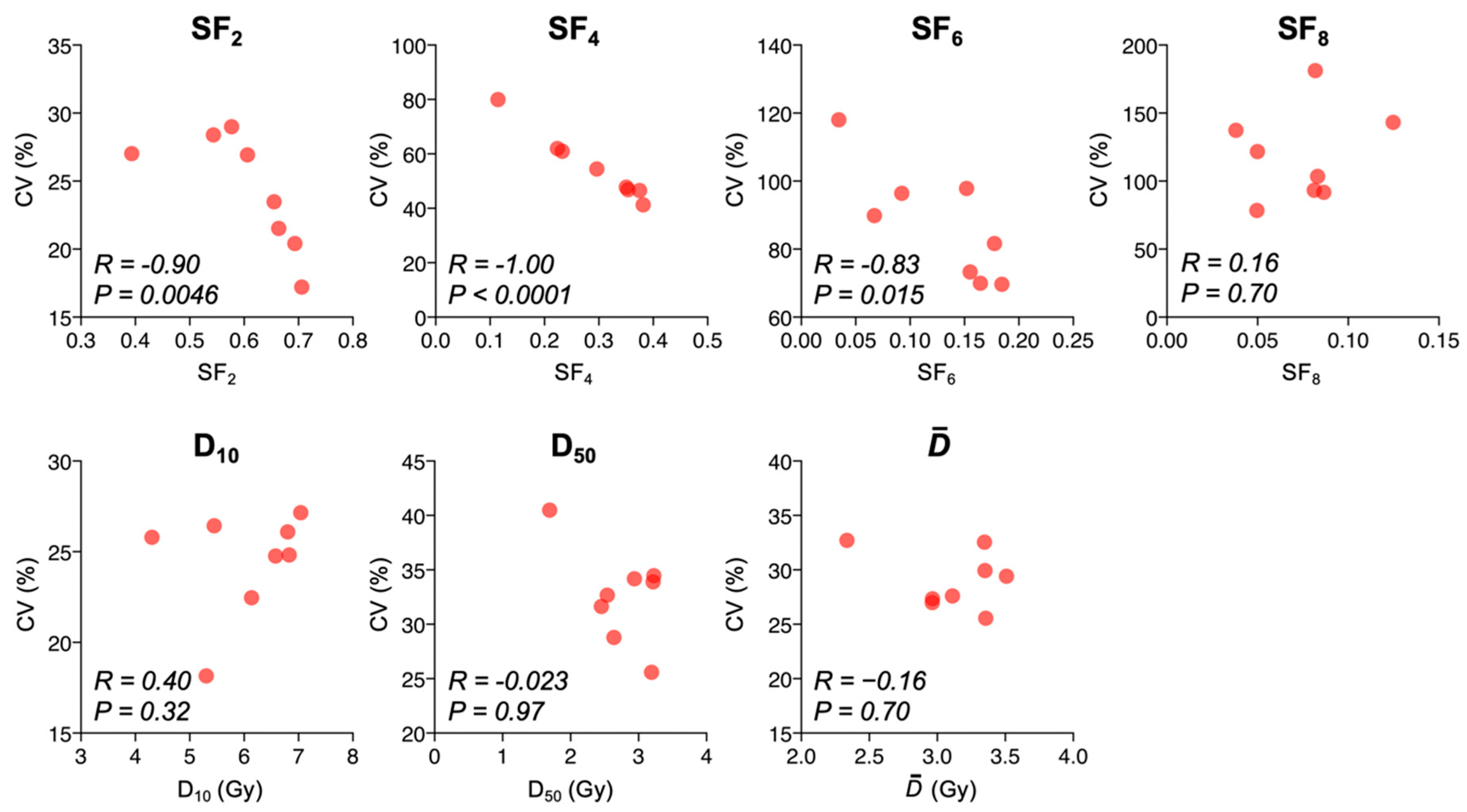
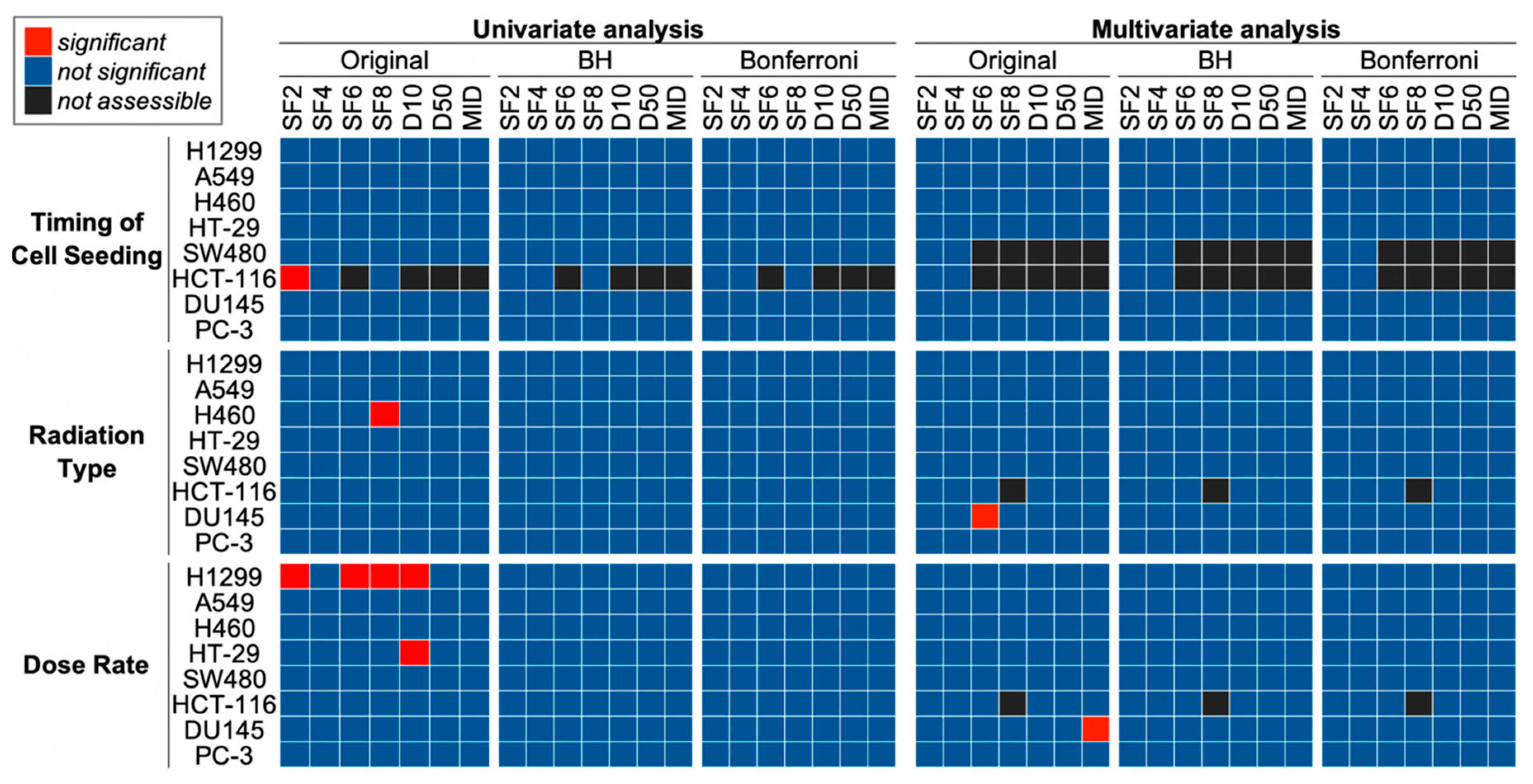
2. Results
3. Discussion
4. Materials and Methods
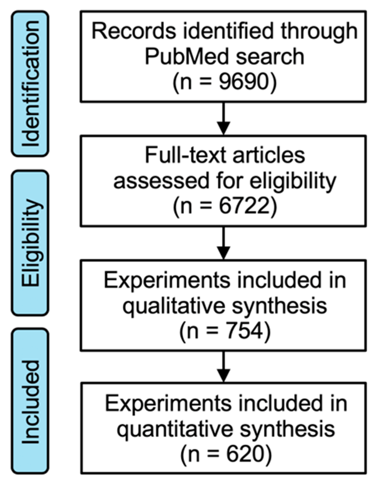
4.1. Literature Search
4.2. Acquisition of Clonogenic Assay Data
4.3. Statistical Analysis
5. Recommendations
5.1. Protocol
5.2. Cell Lines
5.3. Experimental Settings
5.4. Control Treatment
5.5. Integration of Clonogenic Survival Data
Supplementary Materials
Author Contributions
Funding
Conflicts of Interest
Abbreviations
| CCLE | Cancer Cell Line Encyclopedia |
| CV | coefficient of variation |
| mean inactivation dose | |
| LQ model | linear-quadratic model |
| FDA | Food and Drug Administration |
References
- Hinkson, I.V.; Davidsen, T.M.; Klemm, J.D.; Kerlavage, A.R.; Kibbe, W.A. A comprehensive infrastructure for big data in cancer research: Accelerating cancer research and precision medicine. Front. Cell Dev. Biol. 2017, 5, 83. [Google Scholar] [CrossRef] [PubMed]
- Barretina, J.; Caponigro, G.; Stransky, N.; Venkatesan, K.; Margolin, A.A.; Kim, S.; Wilson, C.J.; Lehár, J.; Kryukov, G.V.; Sonkin, D.; et al. The Cancer Cell Line Encyclopedia enables predictive modelling of anticancer drug sensitivity. Nature 2012, 483, 603–607. [Google Scholar] [CrossRef] [PubMed]
- Catalogue of Somatic Mutations in Cancer. Available online: http://cancer.sanger.ac.uk/cosmic (accessed on 24 June 2019).
- Pavlopoulou, A.; Bagos, P.G.; Koutsandrea, V.; Georgakilas, A.G. Molecular determinants of radiosensitivity in normal and tumor tissue: A bioinformatic approach. Cancer Lett. 2017, 403, 37–47. [Google Scholar] [CrossRef] [PubMed]
- Steel, G.G. End-points for measuring radiation effects on tumours. In Basic Clinical Radiobiology, 3rd ed.; Steel, G.G., Ed.; CRC Press: Boca Raton, FL, USA, 2002; pp. 184–186. [Google Scholar]
- Franken, N.A.P.; Rodermond, H.M.; Stap, J.; Haveman, J.; van Bree, C. Clonogenic assay of cells in vitro. Nat. Protoc. 2006, 1, 2315–2319. [Google Scholar] [CrossRef] [PubMed]
- Fertil, B.; Malaise, E.P. Inherent cellular radiosensitivity as a basic concept for human tumor radiotherapy. Int. J. Radiat. Oncol. Biol. Phys. 1981, 7, 621–629. [Google Scholar] [CrossRef]
- Deacon, J.; Peckham, M.J.; Steel, G.G. The radioresponsiveness of human tumours and the initial slope of the cell survival curve. Radiother Oncol. 1984, 2, 317–323. [Google Scholar] [CrossRef]
- Fertil, B.; Malaise, E.P. Intrinsic radiosensitivity of human cell lines is correlated with radioresponsiveness of human tumors: Analysis of 101 published survival curves. Int. J. Radiat. Oncol. Biol. Phys. 1985, 11, 1699–1707. [Google Scholar] [CrossRef]
- Fertil, B.; Dertinger, H.; Courdi, A.; Malaise, E.P. Mean inactivation dose: A useful concept for intercomparison of human cell survival curves. Radiat. Res. 1984, 99, 73–84. [Google Scholar] [CrossRef] [PubMed]
- Nuryadi, E.; Permata, T.B.M.; Komatsu, S.; Oike, T.; Nakano, T. Inter-assay precision of clonogenic assays for radiosensitivity in cancer cell line A. Oncotarget 2018, 9, 13706–13712. [Google Scholar] [CrossRef]
- Friedrich, T.; Scholz, U.; Elsässer, T.; Durante, M.; Scholz, M. Systematic analysis of RBE and related quantities using a database of cell survival experiments with ion beam irradiation. J. Radiat Res. 2013, 54, 494–514. [Google Scholar] [CrossRef]
- Want, E.J.; Masson, P.; Michopoulos, F.; Wilson, I.D.; Theodoridis, G.; Plumb, R.S.; Shockcor, J.; Loftus, N.; Holmes, E.; Nicholson, J.K. Global metabolic profiling of animal and human tissues via UPLC-MS. Nat. Protoc. 2013, 8, 17–32. [Google Scholar] [CrossRef] [PubMed]
- Want, E.J.; Wilson, I.D.; Gika, H.; Theodoridis, G.; Plumb, R.S.; Shockcor, J.; Holmes, E.; Nicholson, J.K. Global metabolic profiling procedures for urine using UPLC–MS. Nat. Protoc. 2010, 5, 1005–1018. [Google Scholar] [CrossRef] [PubMed]
- Steel, G.G. The radiosensitivity of human tumour cells. In Basic Clinical Radiobiology, 3rd ed.; Steel, G.G., Ed.; CRC Press: Boca Raton, FL, USA, 2002; pp. 188–190. [Google Scholar]
- Eschrich, S.A.; Pramana, J.; Zhang, H.; Zhao, H.; Boulware, D.; Lee, J.H.; Bloom, G.; Rocha-Lima, C.; Kelley, S.; Calvin, D.P.; et al. A gene expression model of intrinsic tumor radiosensitivity: Prediction of response and prognosis after chemoradiation. Int. J. Radiat. Oncol. Biol. Phys. 2009, 75, 489–496. [Google Scholar] [CrossRef] [PubMed]
- Kim, H.S.; Kim, S.C.; Kim, S.J.; Park, C.H.; Jeung, H.C.; Kim, Y.B.; Ahn, J.B.; Chung, H.C.; Rha, S.Y. Identification of a radiosensitivity signature using integrative metaanalysis of published microarray data for NCI-60 cancer cells. BMC Genomics 2012, 13, 348. [Google Scholar] [CrossRef] [PubMed]
- Steel, G.G. Experimental tumor systems. In Basic Clinical Radiobiology, 3rd ed.; Steel, G.G., Ed.; CRC Press: Boca Raton, FL, USA, 2002; pp. 182–184. [Google Scholar]
- Fasterius, E.; Al-Khalili, S.C. Analysis of public RNA-sequencing data reveals biological consequences of genetic heterogeneity in cell line populations. Sci. Rep. 2018, 8, 11226. [Google Scholar] [CrossRef]
- Hall, E.J.; Giaccia, A.J. Predictive assays. In Radiobiology for the Radiologist, 5th ed.; Hall, E.J., Giaccia, A.J., Eds.; Lippincott Williams & Wilkins: Philadelphia, PA, USA, 2000; pp. 419–431. [Google Scholar]
- Humphrey, R.M.; Sinclair, W.K. The relative biological effectiveness of 22-Mevp X-rays cobalt-60 gamma rays, and 200 Kvcp X-rays. VIII. determined by the growth capacity of tumor cells in vitro. Radiat Res. 1963, 20, 593–599. [Google Scholar] [CrossRef]
- Winzel, E.; van der Merwe, E.J.; Groenewald, W.; Pistorius, S.; Slabbert, J.P.; Robinson, L.; Böhm, L. The relative biological effectiveness of 100 kV X-rays determined by the V-79 cell colony assay. S. Afr. Med. J. 1987, 71, 693–695. [Google Scholar]
- Siegel, R.L.; Miller, K.D.; Jemal, A. Cancer statistics. CA 2018, 68, 7–30. [Google Scholar]
- Peduzzi, P.; Concato, J.; Kemper, E.; Holford, T.R.; Feinstein, A.R. A simulation study of the number of events per variable in logistic regression analysis. J. Clin. Epidemiol. 1996, 49, 1373–1379. [Google Scholar] [CrossRef]
- Vittinghoff, E.; McCulloch, C.E. Relaxing the rule of ten events per variable in logistic and Cox regression. Am. J. Epidemiol. 2007, 165, 710–718. [Google Scholar] [CrossRef]
- Oike, T.; Ogiwara, H.; Torikai, K.; Nakano, T.; Yokota, J.; Kohno, T. Garcinol, a histone acetyltransferase inhibitor, radiosensitizes cancer cells by inhibiting non-homologous end joining. Int. J. Radiat Oncol Biol Phys. 2012, 84, 815–821. [Google Scholar] [CrossRef] [PubMed]
- Kanda, Y. Investigation of the freely available easy-to-use software “EZR” for medical statistics. Bone Marrow Transplant. 2013, 48, 452–458. [Google Scholar] [CrossRef] [PubMed]
- Anakura, M.; Nachankar, A.; Kobayashi, D.; Amornwichet, N.; Hirota, Y.; Shibata, A.; Oike, T.; Nakano, T. Radiosensitivity differences between EGFR mutant and wild-type lung cancer cells are larger at lower doses. Int. J. Mol. Sci. 2019, 20, 3635. [Google Scholar] [CrossRef] [PubMed]


© 2019 by the authors. Licensee MDPI, Basel, Switzerland. This article is an open access article distributed under the terms and conditions of the Creative Commons Attribution (CC BY) license (http://creativecommons.org/licenses/by/4.0/).
Share and Cite
Matsui, T.; Nuryadi, E.; Komatsu, S.; Hirota, Y.; Shibata, A.; Oike, T.; Nakano, T. Robustness of Clonogenic Assays as a Biomarker for Cancer Cell Radiosensitivity. Int. J. Mol. Sci. 2019, 20, 4148. https://doi.org/10.3390/ijms20174148
Matsui T, Nuryadi E, Komatsu S, Hirota Y, Shibata A, Oike T, Nakano T. Robustness of Clonogenic Assays as a Biomarker for Cancer Cell Radiosensitivity. International Journal of Molecular Sciences. 2019; 20(17):4148. https://doi.org/10.3390/ijms20174148
Chicago/Turabian StyleMatsui, Toshiaki, Endang Nuryadi, Shuichiro Komatsu, Yuka Hirota, Atsushi Shibata, Takahiro Oike, and Takashi Nakano. 2019. "Robustness of Clonogenic Assays as a Biomarker for Cancer Cell Radiosensitivity" International Journal of Molecular Sciences 20, no. 17: 4148. https://doi.org/10.3390/ijms20174148
APA StyleMatsui, T., Nuryadi, E., Komatsu, S., Hirota, Y., Shibata, A., Oike, T., & Nakano, T. (2019). Robustness of Clonogenic Assays as a Biomarker for Cancer Cell Radiosensitivity. International Journal of Molecular Sciences, 20(17), 4148. https://doi.org/10.3390/ijms20174148