Article

Fine Mapping and Candidate Gene Prediction for White Immature Fruit Skin in Cucumber (Cucumis sativus L.)

1
Key Laboratory of Horticultural Plant Biology, Ministry of Education, College of Horticulture and Forestry, Huazhong Agricultural University, Wuhan 430070, China
2
Zhejiang Wuwangnong Academy of Seeds Science Co., Ltd., Hangzhou 310020, China
3
Key Laboratory of Horticultural Plant Biology, Ministry of Education, College of Plant Science and Technology, Huazhong Agricultural University, Wuhan 430070, China
*
Author to whom correspondence should be addressed.
Int. J. Mol. Sci. 2018, 19(5), 1493; https://doi.org/10.3390/ijms19051493
Submission received: 5 April 2018 / Revised: 7 May 2018 / Accepted: 8 May 2018 / Published: 17 May 2018
(This article belongs to the Section Molecular Plant Sciences)

Abstract

:
In this study, a single recessive gene (designated w0) was identified to control the white immature fruit color. Genetic mapping with simple sequence repeats (SSR) markers located the w0 gene in the distal region of cucumber chromosome 3 (Chr.3). Fine mapping was then conducted using the method of draft genome scaffold-assisted chromosome walking with 7304 F2 individuals, which allowed for the assignment of the gene locus to a 100.3 kb genomic DNA region with two flanking markers, Q138 and Q193. Thirteen candidate genes were predicted in the 100.3 kb region. Quantitative real-time polymerase chain reaction (qRT-PCR) analysis showed that the expression of the Csa3G904140 gene, which encodes a two-component response regulator-like protein, was much higher in the immature fruit skin of the green parental line (Q1) than in the white parental line (H4). A coding sequence analysis suggested that a single-base insertion occurred at the ninth exon, resulting in a frameshift mutation in Csa3G904140 of H4, and the mutation was consistent with the phenotype in 17 green/white germplasms. Therefore, Csa3G904140 was taken as the likely candidate gene controlling the immature fruit color of cultivated cucumber. This study will contribute to the cloning of candidate genes and the development of white cucumber cultivars using marker-assisted breeding.

1. Introduction

Cucumber, Cucumis sativus L. (2n = 2x = 14), an important cucurbitaceous crop, is one of the top ten vegetables in the world. For most horticultural crops including cucumber, immature fruit skin color is a highly important external quality trait that influences consumer choice [1]. Since traditional breeding takes a long time to produce results, studying the inheritance of fruit skin color will facilitate marker-assisted selection and promote economic value.
There are many studies carried on pericarp color in horticultural plants. The genetic model of fruit color in bitter gourd was found to be a quantitative model through generation mean analysis; green is the dominant trait to white [2]. Gene mapping of pericarp color in wax gourd showed that the trait was controlled by one single gene; dark green color was dominant over yellow color, and the locus was located on chromosome 5 [3]. The fruit color of sweet pepper at the marketable mature stage was controlled by different nuclear genes: green vs. yellow was controlled by one completely dominant gene, yellow vs. white was controlled by two duplicate dominant genes, and green vs. white had differences in three genes [4]. The fruit color of a tomato consisted of the pericarp color and flesh color, while the genetics between them were completely independent [5]; studies showed that the pericarp color was controlled by one pair of nuclear genes, where transparent color was recessive to yellow color [6]. A quantitative trait locus (QTL) approach suggests that PavMYB10 could be the major determinant of fruit skin in sweet cherries [7]. In 2016, PavMYB10 was confirmed to be a reliable DNA molecular marker to select fruit skin color in sweet cherries [8].
The inheritance of pericarp color in cucumber has also been studied. Fine mapping of the pleiotropic locus B for orange mature fruit skin color in cucumber was conducted and identified a 50 kb region containing an R2R3-MYB transcription factor [9]. Studies on the uniform immature fruit color trait have shown that the mottled fruit color phenotype (U) was dominant over the uniform fruit color phenotype (u), and a single gene was responsible for phenotype segregation [10,11]. The u gene was fine mapped between the newly developed codominant SSR markers SSR10 and SSR27 at genetic distances of 0.8 and 0.5 cM, respectively [1]. The w0 white immature fruit skin color gene was located on Chr.3 at approximately 1150 kb in cucumber [12]. Wang et al. [13] studied the genetics of the white immature fruit color trait in cucumber and identified the molecular marker SSR15312, which was located on Chr.3. A premature stop codon, due to a single-base insertion, was found to be responsible for white immature fruit color in cucumber; the sole candidate gene, associated with chloroplast development and chlorophyll biosynthesis, was named APRR2 [14].
In this study, genetic analysis of different F2 and backcross populations in cucumber revealed that one single dominant gene was associated with white immature fruit color in cucumber, and the locus was mapped to Chr.3. Subsequently, 1655 homozygotes derived from the 7304 F2 individuals from the cross of the Q1 × H4 hybrid were used for fine mapping of the white immature fruit color gene. The gene was mapped to a 100.3 kb region between markers Q138 and Q193 at a genetic distance of 0.03 and 0.03 cM, respectively. Quantitative real-time PCR analysis showed that the expression of the Csa3G904140 gene was much higher in the immature fruit of Q1 than in H4. Furthermore, there was a frameshift mutation in Csa3G904140. Therefore, Csa3G904140 was taken as one likely candidate gene. These findings will facilitate marker-assisted selection of the white immature fruit color trait in cucumber breeding and the cloning of the white immature fruit color gene.

2. Results

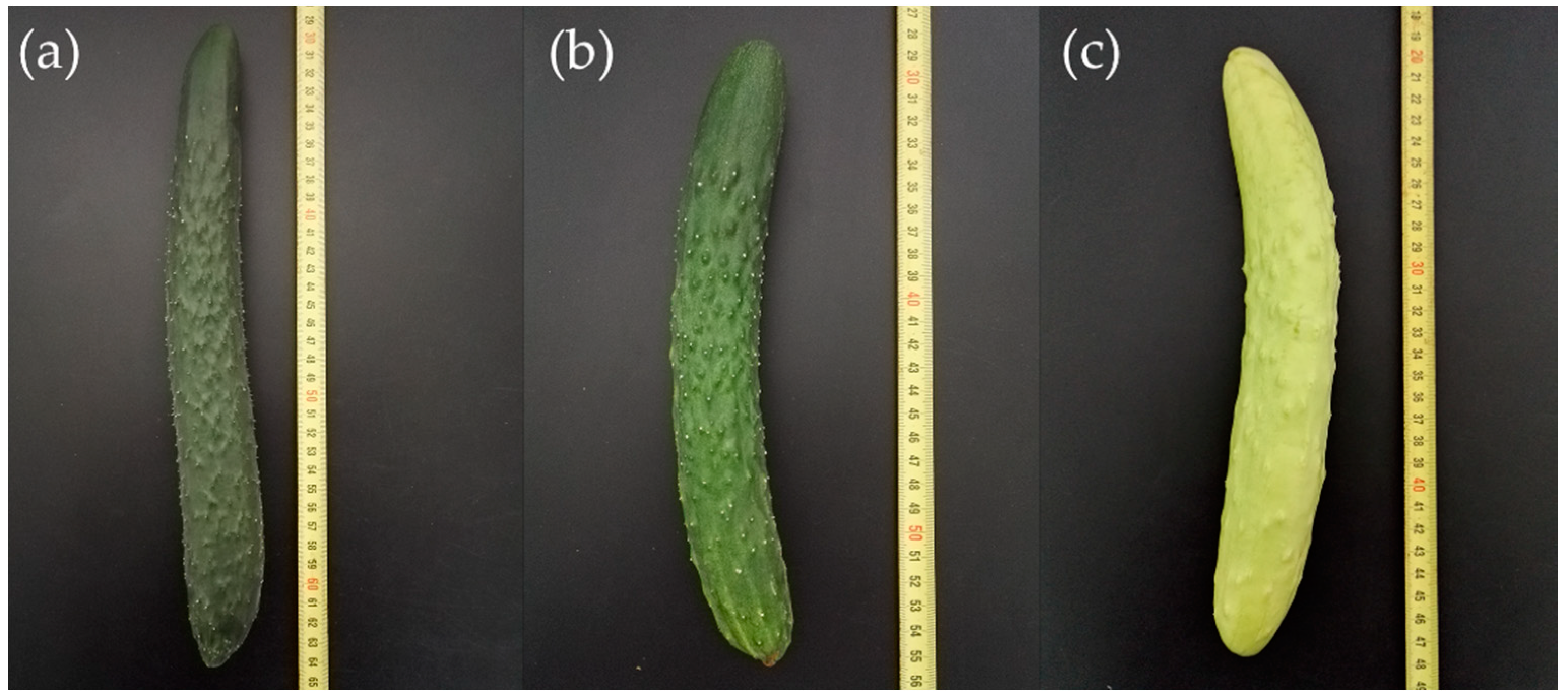
2.1. Genetic Analysis of the Immature Fruit Skin Color Trait

The skin color of two parental inbred lines and their offspring is shown in Figure 1, and the fruit skin color of the F1 cross (H4 × Q1) was green without segregation, revealing the recessive nature of the mutation. Genetic analysis of the immature fruit skin color trait is shown in Table 1. The F2 population derived from the crossing combination P1 × P2 segregated for 115 plants with green immature fruit skin and 29 plants with white immature fruit skin, which fitted an expected segregation ratio of 3:1 (χ2 = 1.56, p = 0.211). None of the BCP1 individuals from a backcross between F1 and P1 were white. For the backcross of F1 with P2, 65 plants had green fruits and 55 plants had white fruits in the BCP2 population, showing a segregation ratio of 1:1 (χ2 = 0.68, p = 0.411). These results indicated that the white immature fruit skin color in Q1 is controlled by a single recessive nuclear gene named w0.

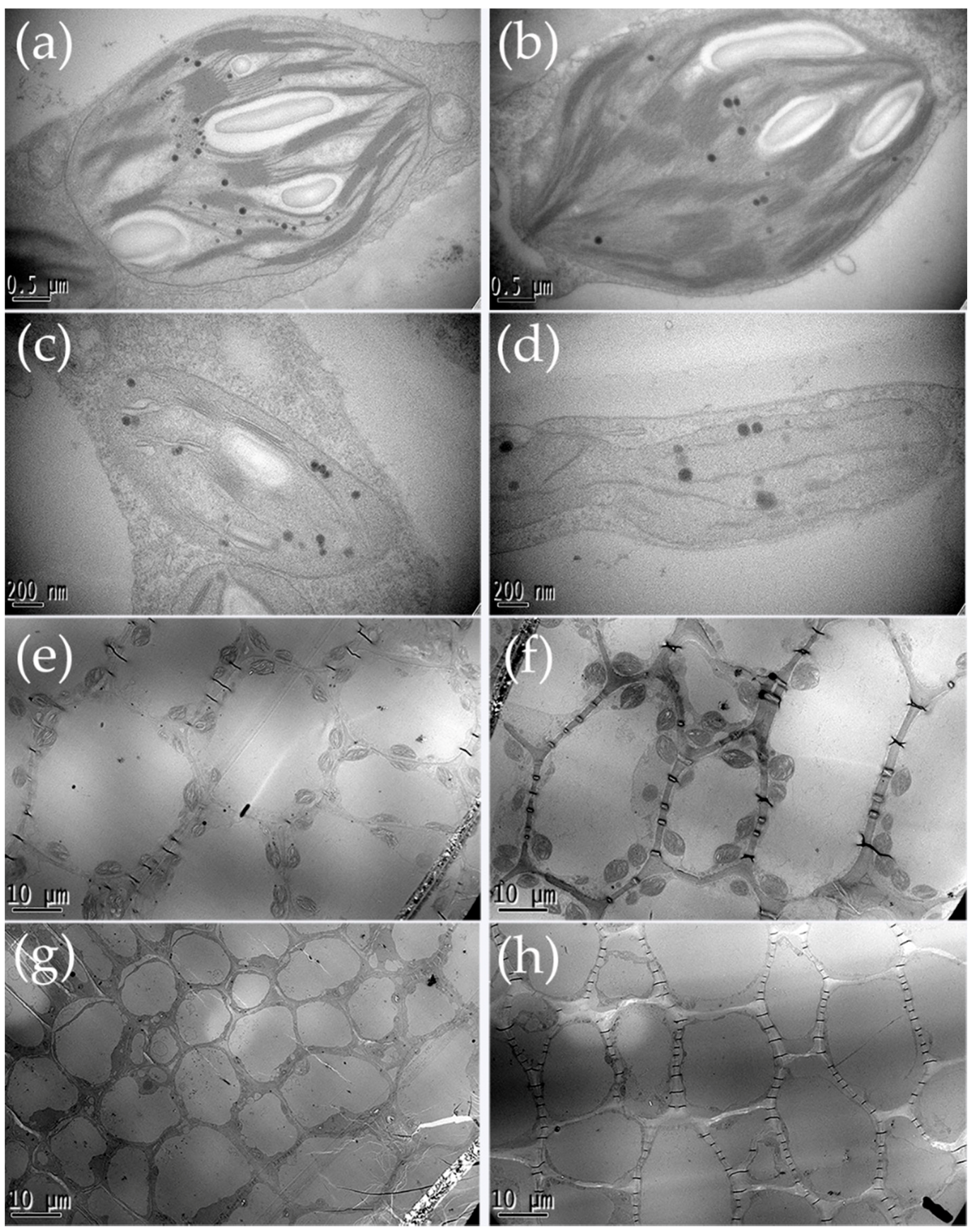
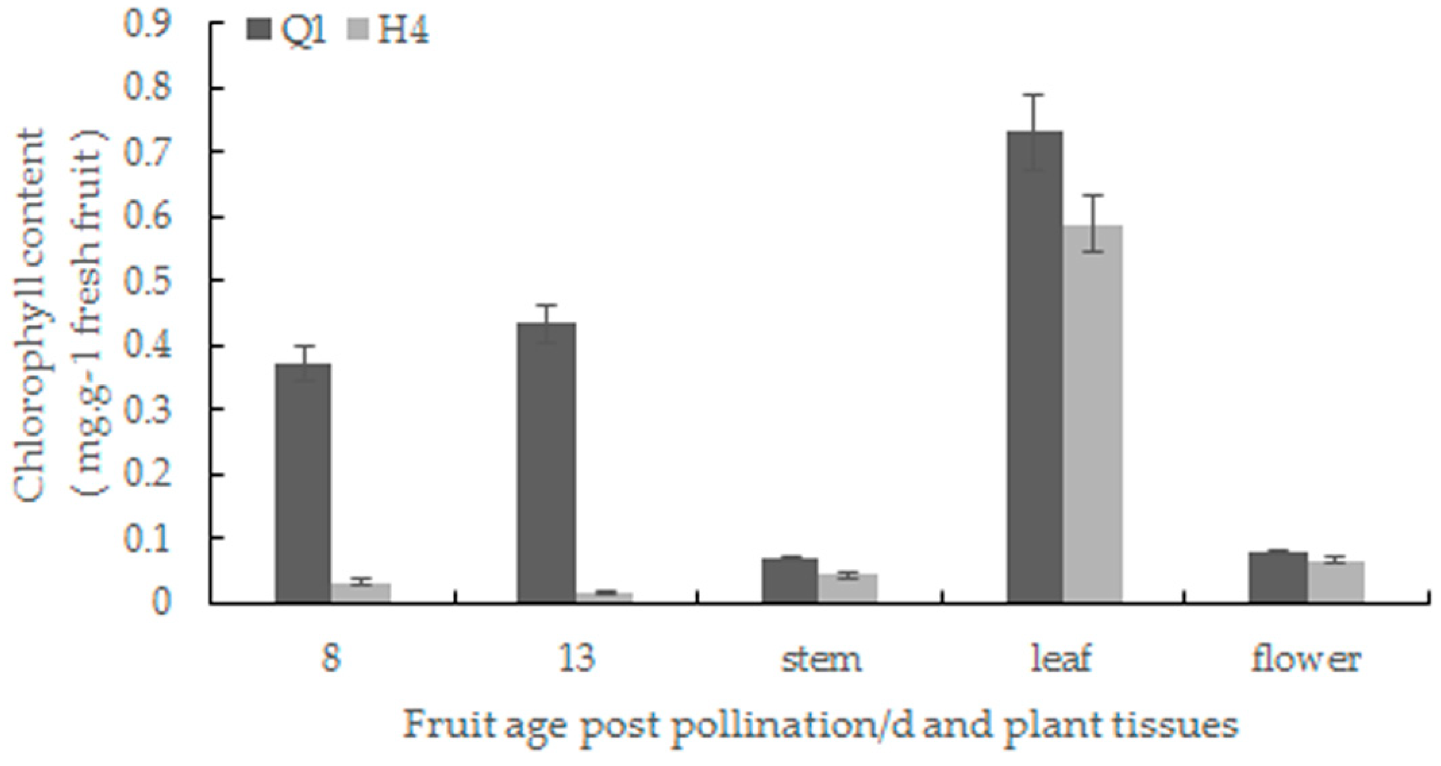
2.2. Chlorophyll Content Determination and Chloroplast Observation

High levels of chlorophyll were detected in the pericarp of 8- and 13-day-old fruit, stems, leaves and flowers in the green parental line compared to the white parental line, and the significant difference was found in the pericarp of fruit (Figure 2). Next, transmission electron microscopy was used to observe the number and ultrastructure of chloroplasts in the pericarp of the two parental lines. The chloroplast of H4 exhibited extensive internal vacuolization and premature senescence. The grana thylakoid of H4 lacked starch grains, and the internal structure of the plastid was fuzzy. The total number of plastoglobuli was decreased in H4 by comparing with Q1 (Figure 3a–d). Moreover, the chloroplast number and size of H4 were fewer and smaller than Q1 (Figure 3e–h).

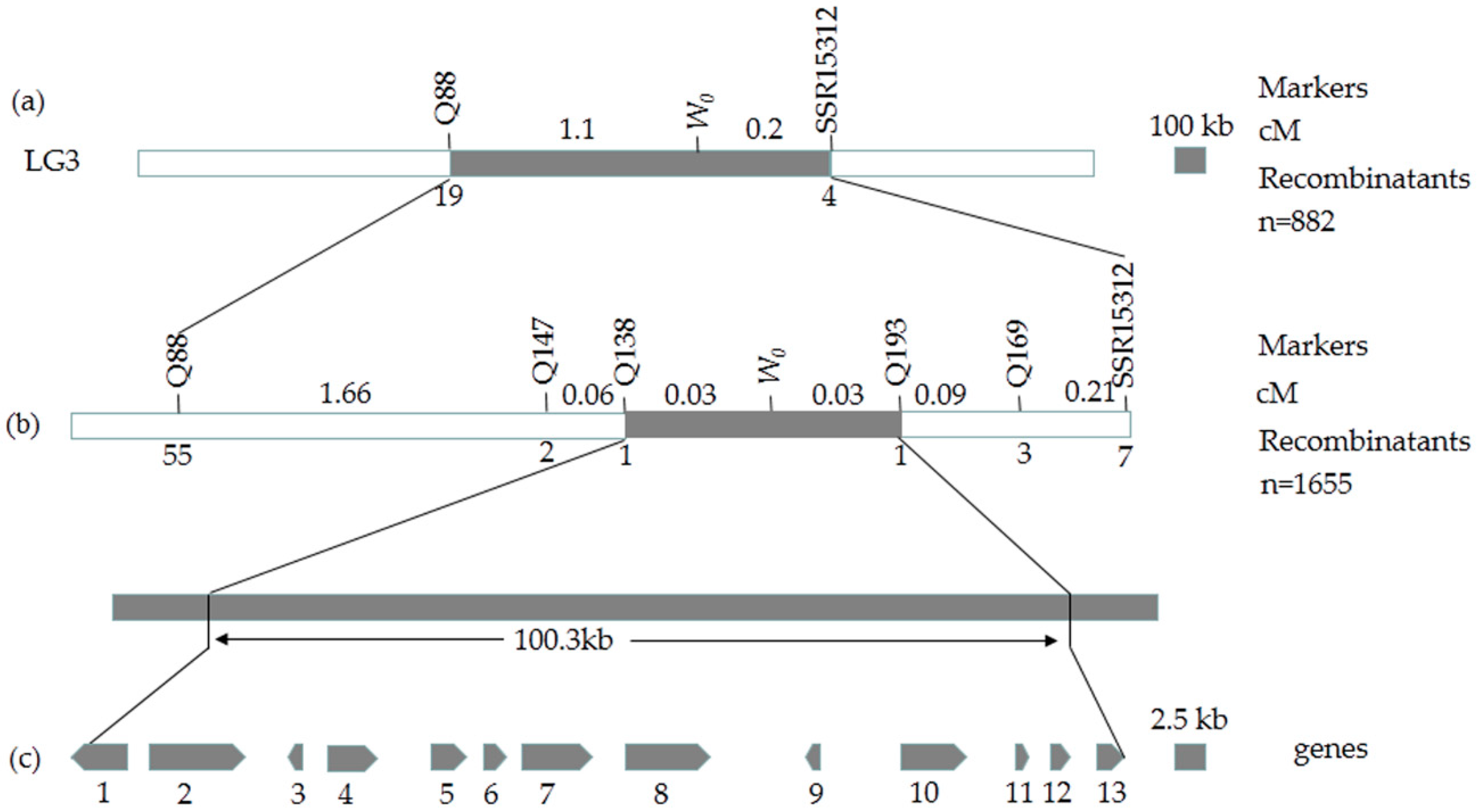
2.3. Preliminary Mapping of the White Immature Fruit Skin Color Trait

To determine the chromosomal location of the w0 gene, 170 pairs of SSR primers (Table S1) were selected from the published 995 pairs of SSR markers in cucumber [15] at an interval of 3.5 cM. One polymorphic marker, SSR15312, was identified through bulked segregant analysis (BSA).
The SSR15312 was then used to genotype 144 F2 individuals, and this marker was found to be closely linked with fruit skin color. Moreover, the linkage between the marker and the phenotype was also detected in 120 progenies of the BCP1 population and 72 progenies of the BCP2 population. These data proved that the marker SSR15312 was closely linked to the target gene. Ren et al. [15] reported that SSR15312 is located on Chr.3, suggesting that the w0 gene is mapped to Chr.3.

2.4. Fine Mapping of the White Immature Fruit Skin Color Trait

To fine map this gene, an F1 population of H4 × Q1 was self-pollinated to produce an F2 population, 175 pairs of cleaved amplified polymorphic sequences (CAPS) markers (Table S2) were developed with 3916 F2 individuals. Among them, four pairs of polymorphic markers, Q88, Q138, Q139, and Q4-2, were identified and genotyped in 882 white plants in the F2 population. Q88 was at the left side of SSR15312 with 19 recombinants, while SSR15312 had 4 recombinants. In addition, the markers, Q138, Q139 and Q4-2, co-segregated with the target gene (Figure 4a). Then, the F2 population was enlarged to 7304 individuals, another 63 pairs of CAPS markers (Table S2) were developed, including three new polymorphic markers, Q147, Q193, and Q169, were developed.
Linkage analysis of these eight pairs of polymorphic markers with the w0 gene of cucumber using 1655 white plants in the F2 population verified the recombination rate to calculate the genetic distance. The results indicated that there was still no recombination between Q139, Q4-2, Q142 and the white immature fruit skin color gene, while two different recombinants were detected between the two closest flanking markers Q138 and Q193 at a genetic distance of 0.03 cM (Figure 4b).
The two closest flanking markers, Q138 and Q193, were developed from the Csa3G904060 gene and the Csa3G910680 gene. A basic local alignment search tool (BLAST) of the cucumber genome sequence was used (http://cucurbitgenomics.org/blast) to determine that two genes are located at positions 39193398 (the first base of the 5′-terminus sequence) and 39293751 (the last base of the 3′-terminus sequence) on Chr.3, respectively; the physical distance between them is 100.3 kb, which contains 13 candidate genes (Figure 4c).

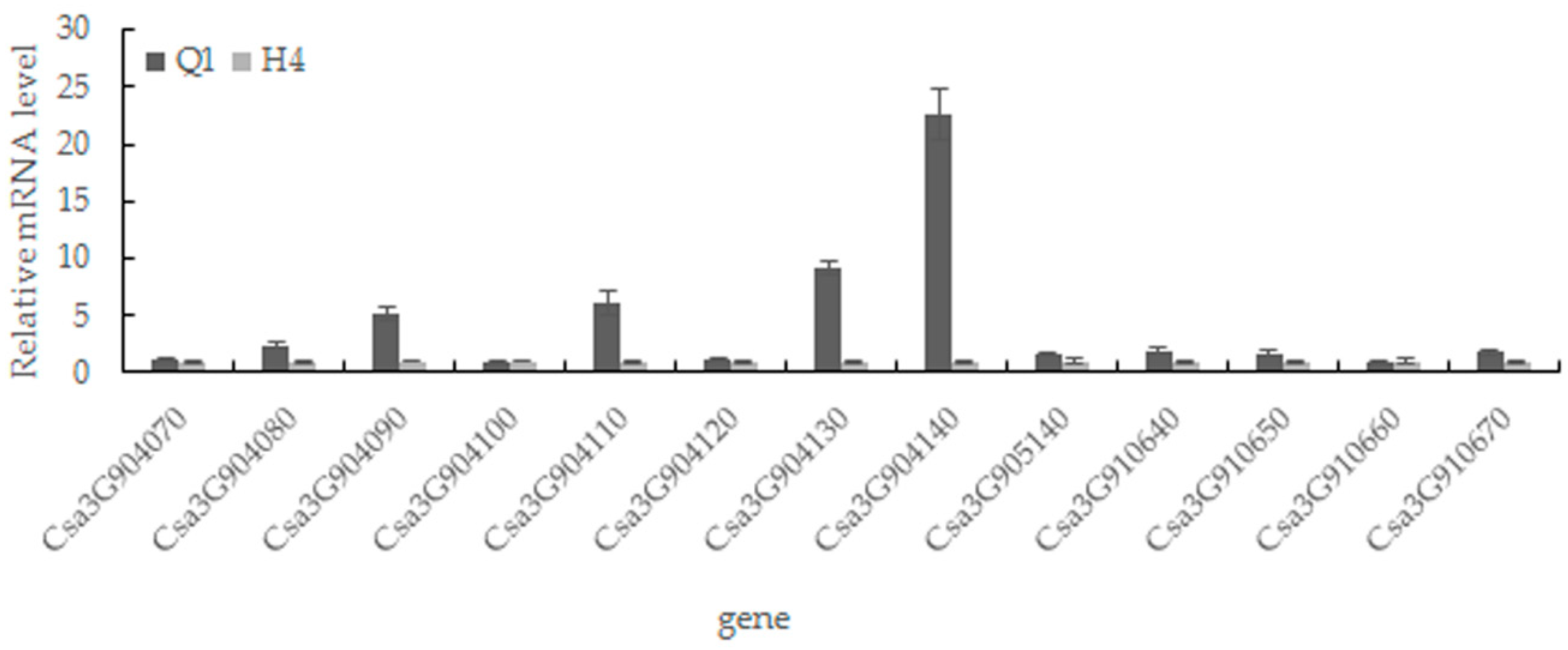
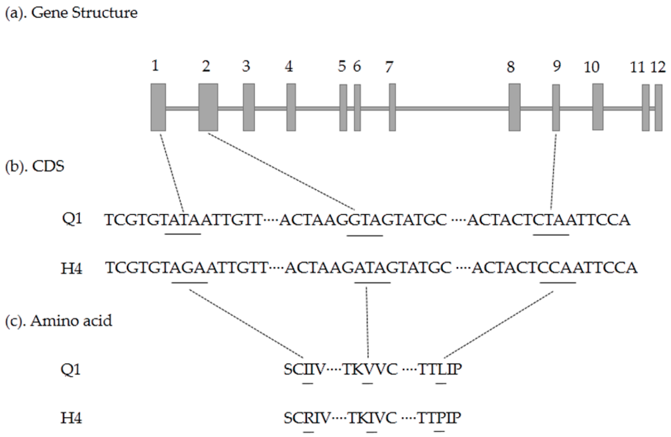
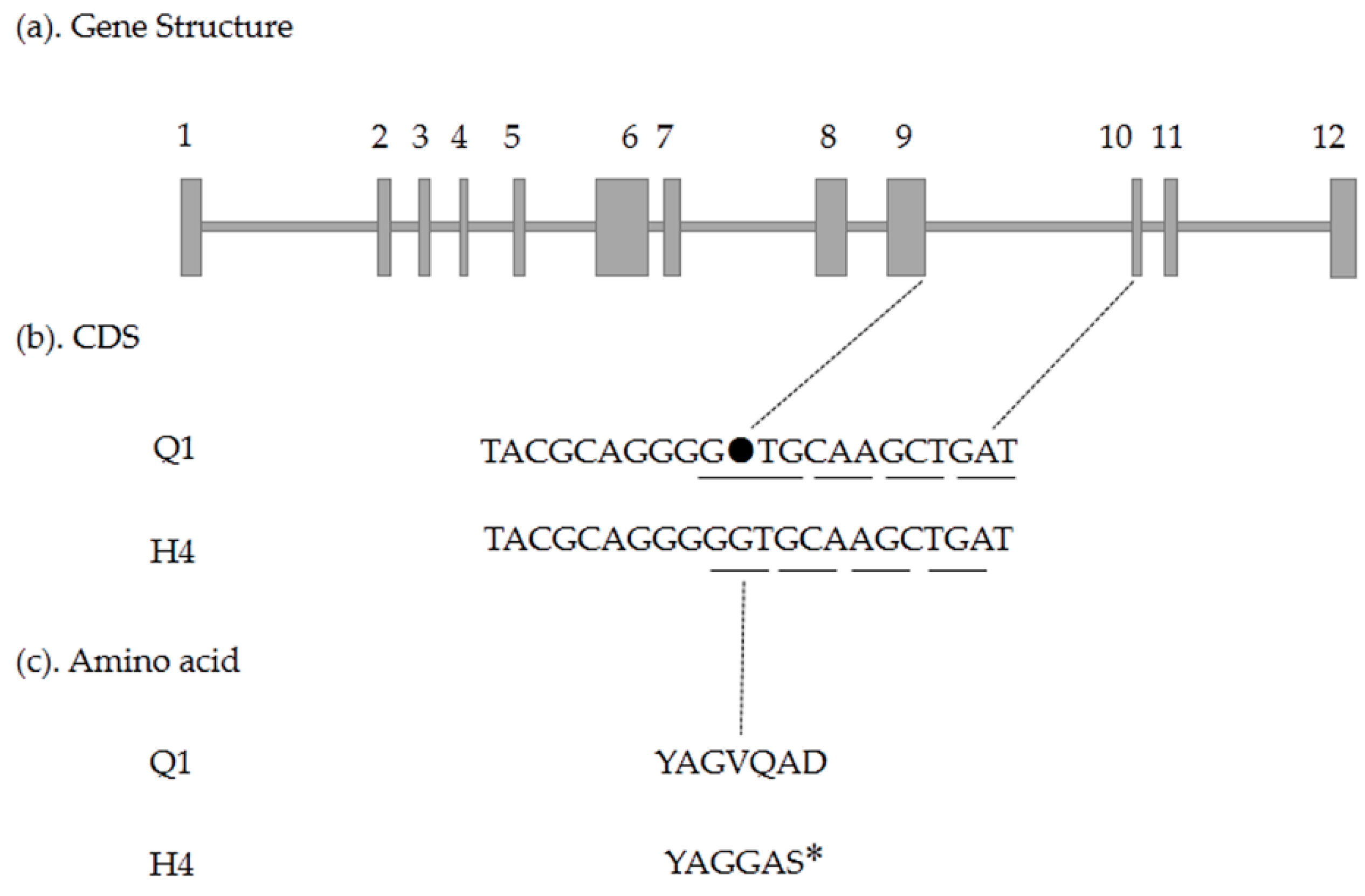
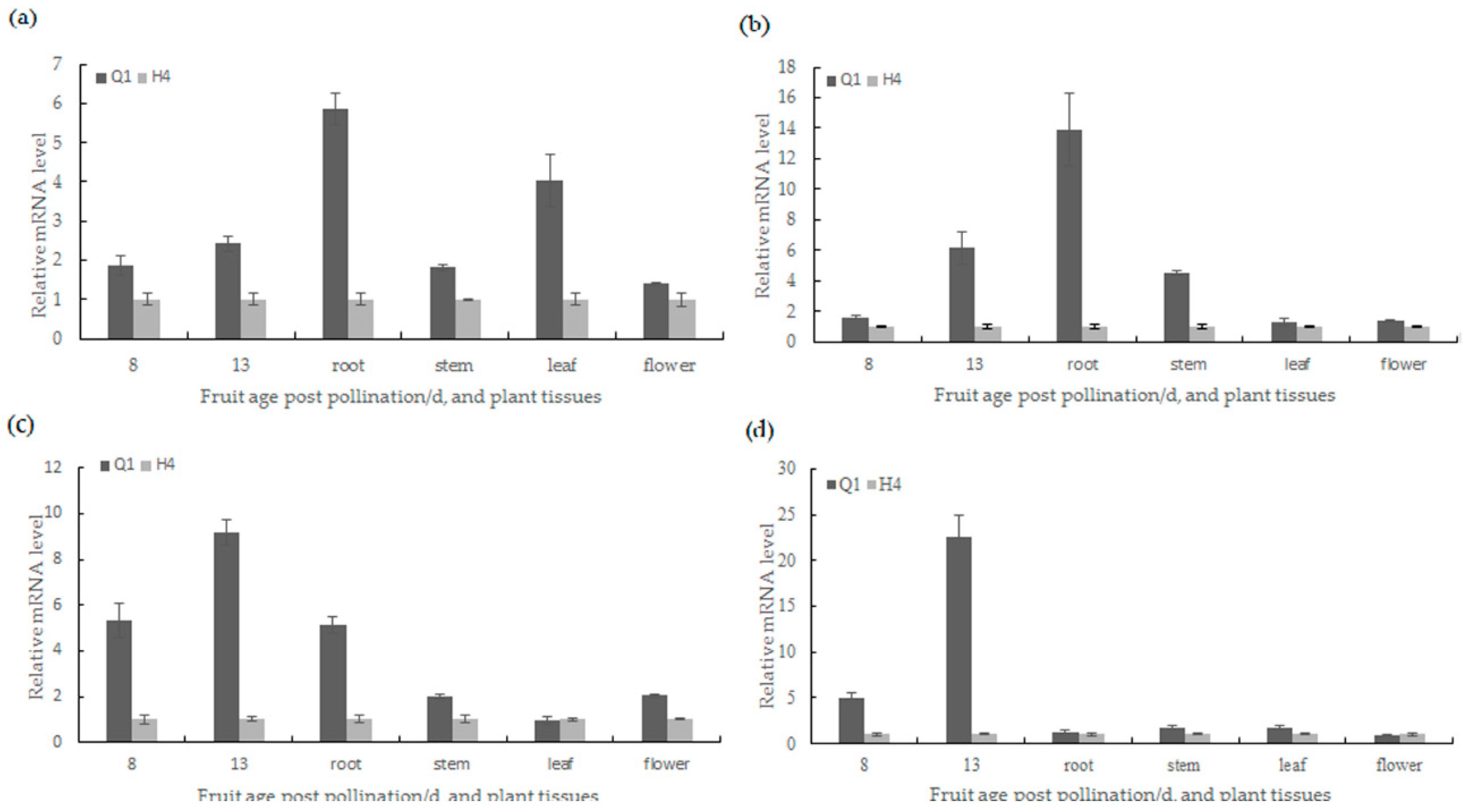
2.5. Candidate Gene Prediction and Gene Expression Analysis

The function of all 13 candidate genes was predicted by Cucurbit Genomics Database (http://cucurbitgenomics.org/search/genome/2) (Table 2). Then, qRT-PCR analysis (primer sequences are available in Table S3) was performed to test whether the gene expression was altered between the two parents. As shown in Figure 5, the expression levels of the Csa3G904130 and Csa3G904140 were 9.2-fold and 22.6-fold higher in Q1 than H4, respectively. The other two genes (Csa3G904080 and Csa3G904110) were 2.4-fold and 6.2-fold higher in Q1 than H4 in expression quantity. The results of the tissue expression revealed that Csa3G904080 was highly expressed in root and leaf; Csa3G904110 was highly expressed in pericarp, root and stem; Csa3G904130 was highly expressed in pericarp and root; While Csa3G904140 was highly expressed in the pericarp of fruit rather than other tissues (Figure 6).
To analyze gene sequences of these four candidate genes, we designed primers (Table S4) to amplify the entire coding sequence and performed TA cloning. By comparing candidate gene sequences between the two parents, three single nucleotide polymorphism (SNP) mutations in exon were detected within the Csa3G904080 gene, which led to three amino acid changes (I83R, V316I, and L1328P) (Figure 7); there was one single-nucleotide insertion resulting in a premature stop codon within the Csa3G904140 gene (Figure 8), and two same sense mutations within the Csa3G904110 gene. The cDNA sequence of Csa3G904130 in Q1 was the same as that of H4. In addition, the mutation in the Csa3G904140 gene was consistent with the phenotype of 17 green/white germplasms (Tables S5 and S6), while the mutations in the Csa3G904080 gene were not consistent with the phenotype of 17 green/white germplasms (Table S7). Furthermore, the marker Q142 associated with the Csa3G904140 gene cosegregated with white fruit skin color. Therefore, Csa3G904140 was likely to be the candidate gene controlling the white fruit skin color in cucumber.

3. Discussion

The white immature fruit skin color is one of the most valuable external quality traits in cucumber, which may directly affect a consumer’s choice of cucumber fruit. Therefore, it is valuable to identify the genes or loci associated with white skin color. In this study, a recessive gene w0 was identified to control white fruit skin color in cultivated cucumber, consistent with the results reported in previous studies [13]. Map-based cloning is a primary strategy that has been used to identify important trait genes in many organisms [16,17]. However, cucumber cultivars have a low genetic diversity, which increases the difficulty in the use of map-based cloning in cucumber [18]. Whole genome sequencing of the cucumber has enabled large-scale development of molecular markers for this crop [19]. Therefore, based on the published cucumber genome sequence (http://cucurbitgenomics.org/organism/2), markers were developed. By the stepwise increase of the mapping population sizes and taking advantage of draft genome assemblies, we identified the w0 gene to a 100.3 kb region containing 13 candidate genes, which laid a solid foundation for gene cloning.
To ensure the accuracy of the result, we used the cucumber genome browser, version 2 (http://cucurbitgenomics.org/JBrowse/) and BLAST of the National Center for Biotechnology Information (NCBI) (http://blast.ncbi.nlm.nih.gov/Blast.cgi) to predict candidate genes in the target region. Consequently, a total of 13 candidate genes were identified in the region between the two markers Q138 and Q193. The results from sequence alignment analysis showed that Csa3G904080 gene sequence between Q1 and H4 existed three amino acid mutations, but the mutations were not consistent with the phenotype of 17 green/white germplasms. In addition, the gene expression of Csa3G904080 was higher in root and leaf than in fruit skin, and the gene function is related to photosynthesis. Thus, it was unlikely that Csa3G904080 was responsible for the white pigmentation. Csa3G904110 and Csa3G904130 had a high relative expression level in fruit skin, however, Csa3G904110 had a more high expression level in the root and Csa3G904130 also had high expression in the root, and these two genes had no amino acid mutation between Q1 and H4. For another nine candidate genes, the relative expression level in fruit skin was too low. While the Csa3G904140 gene contained one single-base insertion between the two parental inbred lines, and the mutation was consistent with the phenotype of 17 green/white germplasms. Moreover, a 22-fold increase in the expression of Csa3G904140 was found in the green-skinned parental line Q1. Therefore, the Csa3G904140 gene was likely to be the candidate gene controlling immature fruit skin colors of cultivated cucumber.
The chloroplast is a half-autonomous organelle that commonly exists in land plants, algae and protists and is responsible for photosynthesis. A search for conserved domains against NCBI CDD (http://www.ncbi.nlm.nih.gov/structure/cdd/wrpsb.cgi) revealed that Csa3G904140 contained a MYB-like DNA-binding domain. This DNA-binding domain is primarily responsible for the color of fruit, many of which also contain a transcriptional activation domain [20,21]. Studies have shown that nuclear Sig genes—encoded sigma factors—play a key role in controlling RNA polymerase binding to promoters in chloroplasts [22]. Nuclear-encoded proteins greatly influence chloroplast RNA editing, post-transcriptional processing and the maintenance of RNA stability [23]. Subcellular localization of the Csa3G904140 protein was predicted using Plant-PLoc (http://www.csbio.sjtu.edu.cn/bioinf/plant/#) to show its presence in the nucleus.
Pan et al. reported that an APRR2 gene increased the pigment content and the levels of chlorophyll in immature fruit [24]. Moreover, APRR1, which is related to APRR2, was thought to be involved in plastid development and ripening [24,25]. A single candidate gene, APRR2, was defined to control white immature fruit skin color in cucumber by delimiting the physical interval to an 8.2 kb region [14]. The NCBI annotation of Csa3G904140 was predicted to be Cucumis sativus two-component response regulator-like APRR2. In our study, the chlorophyll content was much higher in the green compared to the white lines, and microscopy revealed that the chloroplast number and size of H4 was less and smaller than Q1. This gene, Csa3G904140, seemed to be the candidate gene for the proposed gene locus conditioning immature fruit skin colors of cultivated cucumber. Whether this w0 gene was the same as in a previous report [14] requires further study.

4. Materials and Methods

4.1. Plant Materials and Genetic Mapping Population

A green-skinned cucumber inbred line Q1 and a white-skinned line H4 were used as the male and female parent, respectively, to generate one F1 population, one F2 population and two BC1 populations, including BCP1 and BCP2. Their skin colors were uniform, and both lines were from our research group. In these 17 green/white cucumber lines, some were obtained from the National Vegetable Germplasm Resources Intermediate Library of the National Crop Germplasm Resource Platform—Vegetable Germplasm Resources sub-platform (China), and other germplasms were provided by our research group.
Genomic DNA of all individuals was isolated from young leaves using a modified Hexadecyltrimethy Ammoniun Bromide (CTAB) method described by Porebski et al. [26]. Six DNA samples of green and white skin phenotypes were bulked and used to identify markers linked to the color gene; the method was described by Michelmore et al. [27]. All experiments were conducted during the growing seasons between 2012 and 2017 in the National Vegetable Improvement Center and the State Key Laboratory of Horticultural Plant of Huazhong Agricultural University.

4.2. Chlorophyll Content Determination and Chloroplast Observation

Pericarp samples from 8- and 13-day-old fruits after pollination and other tissues (roots, stems, leaves and flowers) were excised and extracted with 96% alcohol in the dark. After 24 h, the extracted pigments were immediately spectrophotometrically determined at specific absorption coefficients using the method described by Wellburn et al. [28].
To observe the chloroplast ultrastructure of the pericarp, tissues from 8- and 13-day-old cucumber fruit (after pollination) were excised with a sterile razor blade and fixed immediately in 3.5% (v/v) glutaraldehyde solution. Chloroplast observations were conducted with a H7650 microscope (HITACHI, Tokyo, Japan).

4.3. Design of SSR and CAPS Markers

Sequences of the SSR primers were derived from the Cucurbit Genomics Database (http://cucurbitgenomics.org/). A total of 170 pairs of SSR primers distributed evenly on seven chromosomes were synthesized by Sangon Biotech Co., Ltd. (Shanghai, China).
PCR amplification for SSR analysis was carried out in a 10 μL volume containing 1 μL (50 ng/μL) template DNA, 1 μL primers (10 μM), 5 μL 2× Taq PCR Master Mix and 3 μL ddH2O. The reaction conditions were as follows: 94 °C for 5 min, followed by 35 cycles at 94 °C for 30 s, 56 °C for 30 s, and 72 °C for 30 s, with a final extension at 72 °C for 10 min. The amplicons were separated on 9% denatured polyacrylamide gels [29].
CAPS primers were developed using Primer 3.0 (http://bioinfo.ut.ee/primer3-0.4.0/) according to the cleavage sites of genome sequence (http://cucurbitgenomics.org/organism/2) in the candidate region, and synthesized by Sangon Biotech Co., Ltd. (Shanghai, China). The PCR for CAPS was also carried out in a 10 μL mixture, similar to that for SSR markers. The reaction conditions were as follows: 95 °C for 4 min, followed by 30 cycles at 94 °C for 30 s, 56 °C for 30 s, and 72 °C for 1 min, and a final extension at 72 °C for 7 min. The amplicons were separated on 9% denatured polyacrylamide gels. Non-polymorphic amplicons were digested with restriction endonucleases. A 10 μL digestion system included 4 μL PCR product, 0.1 μL enzyme, 1 μL buffer, and 4.9 μL ddH2O. The digestion process was adjusted according to the manufacturer’s recommendations for the corresponding enzyme. Polymorphic markers were used to genotype the population.

4.4. Gene Prediction and Expression Analysis

Two software tools, the cucumber genome browser, version 2 (http://cucurbitgenomics.org/JBrowse/) and BLAST of NCBI (http://blast.ncbi.nlm.nih.gov/Blast.cgi), were applied to predict candidate genes.
Quantitative real-time PCR was used to analyze the expression of the candidate gene in the two parents. Pericarp samples from 8-day-old and 13-day-old fruit after pollination and other tissues, including roots, stems, leaves, and flowers, were excised, and RNA was isolated using a modified Trizol method described by Cheng [30]. First-strand cDNA was synthesized with a Prime Script™ RT reagent Kit with gDNA Eraser (Perfect Real Time) (TaKaRa, Tokyo, Japan) according to the manufacturer’s instructions. The 18sRNA gene was used as the reference, and the primer sequences are shown as follows.
  • 18sRNA-F: AGAAACGGCTACCACATC
  • 18sRNA-R: CCAAGGTCCAACTACGAG
Using a 96-well plate and SYBR Green-based dye, the qRT-PCR reaction system contained 1 μL template cDNA (100 ng/μL), 5 μL 2× SYBR Green PCR Master Mix (TaKaRa, Tokyo, Japan), and 0.4 μL forward and reverse primers (10 μM), with water added to a total reaction of 10 μL. The qRT-PCR was performed using a CFX96 Real-Time PCR Detection System (Bio-Rad, Hercules, CA, USA). The values from triplicate reactions were averaged, and the relative expression level was measured by means of 2−ΔΔCt [31].

4.5. Candidate Gene Cloning and Sequencing

TA cloning was used to analyze the sequence of the candidate gene in the two parents. RNA was isolated using a modified Trizol method as described previously [30]. First-strand cDNA was synthesized with a Prime Script™ II 1st Strand cDNA Synthesis Kit (TaKaRa, Tokyo, Japan) according to the manufacturer’s instructions. Primers were designed to amplify the entire coding sequence. PCR amplification was carried out according to the user manual with I-5™ 2× High-Fidelity Master Mix (MCLAB, San Francisco, CA, USA). The amplicon was separated on 1% agarose gels and extracted with a TaKaRa MiniBEST Agarose Gel DNA Extraction Kit Ver.4.0 (TaKaRa, Tokyo, Japan) according to the manufacturer’s instructions. The purified DNA product was then subjected to a “+A base” reaction using a kit (ZOMANBIO, Beijing, China), ligated to the pMD18-T vector with a pMD™18-T Vector Cloning Kit (TaKaRa, Tokyo, Japan), and inserted into DH5α chemically competent cell (Weidibio, Shanghai, China) according to the manufacturer’s recommendations. Finally, three correct monoclonal spots were sequenced by Sangon Biotech Co., Ltd. (Shanghai, China), and the sequences of the two parents were compared using the DNAMAN program.

Supplementary Materials

Supplementary materials can be found at https://www.mdpi.com/1422-0067/19/5/1493/s1.

Author Contributions

Zheng-Jie Wang, Yue-Jin Xu and Jun-Hui Xia conceived and designed the experiments; Hong-Yu Tang, Xu Dong, Jian-Ke Wang and Fei Xie performed the experiments; Hong-Yu Tang, Xu Dong and Yu Zhang analyzed the data; Zheng-Jie Wang contributed reagents/materials/analysis tools; Hong-Yu Tang and Xuan Yao wrote the paper.

Funding

This research was supported by the National Key Research and Development Program of China (2017YFD010803), the China Agriculture Research System (CARS-24-A-06) and the Fundamental Research Funds for the Central Universities (2662016PY106).

Acknowledgments

The authors are grateful to Xuan Yao for critical reading of the manuscript and the anonymous reviewers for their constructive comments for revisions.

Conflicts of Interest

The authors declare no conflicts of interest.

References

  1. Yang, X.Q.; Li, Y.; Zhang, W.W.; He, H.L.; Pan, J.S.; Cai, R. Fine mapping of the uniform immature fruit color gene u in cucumber (Cucumis sativus L.). Euphytica 2014, 196, 341–348. [Google Scholar] [CrossRef]
  2. Huang, H.Y.; Hsieh, C.H. Genetic research on fruit color traits of the bitter gourd (Momordica charantia L.). Hortic. J. 2017, 86, 238–243. [Google Scholar] [CrossRef]
  3. Jiang, B.; Liu, W.R.; Xie, D.S.; Peng, Q.W.; He, X.M.; Lin, Y.E.; Liang, Z.J. High-density genetic map construction and gene mapping of pericarp color in wax gourd using specific-locus amplified fragment (SLAF) sequencing. BMC Genom. 2015, 16. [Google Scholar] [CrossRef] [PubMed]
  4. Xue, L.B.; Wu, X.X.; Chen, J.L. Study on heredity of fruit colors of sweet pepper. Acta Hortic. Sin. 2005, 32, 513–515. [Google Scholar]
  5. Wu, L.; Liu, J.Y.; Liang, Y. Inheritance on fruit color character between green and red of tomato. Acta Hortic. Sin. 2016, 43, 674–682. [Google Scholar]
  6. Jin, F.M.; Xue, J.; Jia, Y.H.; Liu, Z.Q. Research advance of tomato fruit color related gene. Tianjin Agric. Sci. 2006, 12, 3–6. [Google Scholar]
  7. Sooriyapathirana, S.S.; Khan, A.; Sebolt, A.M.; Wang, D.C.; Bushakra, J.M.; Lin-Wang, K.; Allan, A.C.; Gardiner, S.E.; Chagne, D.; Iezzoni, A.F. QTL analysis and candidate gene mapping for skin and flesh color in sweet cherry fruit (Prunus avium L.). Tree Genet. Genom. 2010, 6, 821–832. [Google Scholar] [CrossRef]
  8. Jin, W.M.; Wang, H.; Li, M.F.; Wang, J.; Yang, Y.; Zhang, X.M.; Yan, G.H.; Zhang, H.; Liu, J.S.; Zhang, K.C. The R2R3 MYB transcription factor PavMYB10.1 involves in anthocyanin biosynthesis and determines fruit skin colour in sweet cherry (Prunus avium L.). Plant Biotechnol. J. 2016, 14, 2120–2133. [Google Scholar] [CrossRef] [PubMed]
  9. Li, Y.H.; Wen, C.L.; Weng, Y.Q. Fine mapping of the pleiotropic locus B for black spine and orange mature fruit color in cucumber identifies a 50 kb region containing a R2R3-MYB transcription factor. Theor. Appl. Genet. 2013, 126, 2187–2196. [Google Scholar] [CrossRef] [PubMed]
  10. Yuan, X.J.; Pan, J.S.; Cai, R.; Guan, Y.; Liu, L.Z.; Zhang, W.W.; Li, Z.; He, H.L.; Zhang, C.; Si, L.T.; et al. Genetic mapping and QTL analysis of fruit and flower related traits in cucumber (Cucumis sativus L.) using recombinant inbred lines. Euphytica 2008, 164, 473–491. [Google Scholar] [CrossRef]
  11. Miao, H.; Zhang, S.P.; Wang, X.W.; Zhang, Z.H.; Li, M.; Mu, S.Q.; Cheng, Z.C.; Zhang, R.W.; Huang, S.W.; Xie, B.Y.; et al. A linkage map of cultivated cucumber (Cucumis sativus L.) with 248 microsatellite marker loci and seven genes for horticulturally important traits. Euphytica 2011, 182, 167–176. [Google Scholar] [CrossRef]
  12. Dong, S.Y.; Miao, H.; Zhang, S.P.; Liu, M.M.; Wang, Y.; Gu, X.F. Genetic analysis and gene mapping of white fruit skin in cucumber (Cucumis sativus L.). Acta Bot. Boreal. Occident. Sin. 2012, 11, 2177–2181. [Google Scholar]
  13. Wang, J.K.; Fang, X.X.; Li, X.H.; Chen, Y.; Wan, Z.J.; Xu, Y.J. Genetic study on immature fruit color of cucumber. Acta Hortic. Sin. 2013, 128, 479–486. [Google Scholar]
  14. Liu, H.; Jiao, J.Q.; Liang, X.J.; Liu, J.; Meng, H.W.; Chen, S.X.; Li, Y.H.; Cheng, Z.H. Map-based cloning, identification and characterization of the w gene controlling white immature fruit color in cucumber (Cucumis sativus L.). Theor. Appl. Genet. 2016, 129, 1247–1256. [Google Scholar] [CrossRef] [PubMed]
  15. Ren, Y.; Zhang, Z.H.; Liu, J.H.; Staub, J.E.; Han, Y.H.; Cheng, Z.C.; Li, X.F.; Lu, J.Y.; Miao, H.; Kang, H.X.; et al. Integrated genetic and cytogenetic map of the cucumber genome. PLoS ONE 2009, 4, e5795. [Google Scholar] [CrossRef] [PubMed]
  16. He, J.P.; Ke, L.P.; Hong, D.F.; Xie, Y.Z.; Wang, G.C.; Liu, P.W.; Yang, G.S. Fine mapping of a recessive genic male sterility gene (Bnms3) in rapeseed (Brassica napus) with AFLP- and Arabidopsis-derived PCR markers. Theor. Appl. Genet. 2008, 117, 11–18. [Google Scholar] [CrossRef] [PubMed]
  17. Li, S.C.; Liu, M.W.; Wang, S.Q.; Deng, Q.M.; Zheng, A.P.; Liu, H.N.; Wang, L.X.; Zhu, J.; Li, P. Fine mapping of a dominant minute-grain gene, Mi3, in rice. Mol. Breed. 2012, 30, 1045–1051. [Google Scholar] [CrossRef]
  18. Dijkhuizen, A.; Kennard, W.C.; Havey, M.J.; Staub, J.E. RFLP variation and genetic relationships in cultivated cucumber. Euphytica 1996, 90, 79–87. [Google Scholar]
  19. Cavagnaro, P.F.; Senalik, D.A.; Yang, L.M.; Simon, P.W.; Harkins, T.T.; Kodira, C.D.; Huang, S.W.; Weng, Y.Q. Genome-wide characterization of simple sequence repeats in cucumber (Cucumis sativus L.). BMC Genom. 2010, 11, 569. [Google Scholar] [CrossRef] [PubMed]
  20. Baranowskij, N.; Frohberg, C.; Prat, S.; Willmitzer, L. A novel DNA binding protein with homology to Myb oncoproteins containing only one repeat can function as a transcriptional activator. EMBO J. 1994, 13, 5383–5392. [Google Scholar] [PubMed]
  21. Allan, A.C.; Hellens, R.P.; Laing, W.A. MYB transcription factors that colour our fruit. Trends Plant Sci. 2008, 13, 99–102. [Google Scholar] [CrossRef] [PubMed]
  22. Lysenko, E.A. Plant sigma factors and their role in plastid transcription. Plant Cell Rep. 2007, 26, 845–859. [Google Scholar] [CrossRef] [PubMed]
  23. Vaistij, F.E.; Goldschmidt-Clermont, M.; Wostrikoff, K.; Rochaix, J.-D. Stability determinants in the chloroplast psbB/T/H mRNAs of Chlamydomonas reinhardtii. Plant J. 2000, 21, 469–482. [Google Scholar] [CrossRef] [PubMed]
  24. Pan, Y.; Bradley, G.; Pyke, K.; Ball, G.; Lu, C.G.; Fray, R.; Marshall, A.; Jayasuta, S.; Baxter, C.; van Wijk, R.; et al. Network inference analysis identifies an APRR2-like gene linked to pigment accumulation in tomato and pepper fruits. Plant Physiol. 2013, 161, 1476–1485. [Google Scholar] [CrossRef] [PubMed]
  25. Makino, S.; Kiba, T.; Imamura, A.; Hanaki, N.; Nakamura, A.; Suzuki, T.; Taniguchi, M.; Ueguchi, C.; Sugiyama, T.; Mizuno, T. Genes encoding pseudo-response regulators: Insight into His-to-Asp phosphorelay and circadian rhythm in Arabidopsis thaliana. Plant Cell Physiol. 2000, 41, 791–803. [Google Scholar] [CrossRef] [PubMed]
  26. Porebski, S.; Bailey, L.G.; Baum, B.R. Modification of a CTAB DNA extraction protocol for plants containing high polysaccharide and polyphenol components. Plant Mol. Biol. Rep. 1997, 15, 8–15. [Google Scholar] [CrossRef]
  27. Michelmore, R.W.; Paran, I.; Kesseli, R.V. Identification of markers linked to disease-resistance genes by bulked segregant analysis: a rapid method to detect markers in specific genomic regions by using segregating populations. Proc. Natl. Acad. Sci. USA 1991, 88, 9828–9832. [Google Scholar] [CrossRef] [PubMed]
  28. Wellburn, A.R.; Lichtenthaler, H. Formulae and program to determine total carotenoids and chlorophylls a and b of leaf extracts in different solvents. Adv. Photosynth. Res. 1984, 2, 9–12. [Google Scholar]
  29. Zhang, W.W.; He, H.L.; Guan, Y.; Du, H.; Yuan, L.H.; Li, Z.; Yao, D.Q.; Pan, J.S.; Cai, R. Identification and mapping of molecular markers linked to the tuberculate fruit gene in the cucumber (Cucumis sativus L.). Theor. Appl. Genet. 2010, 120, 645–654. [Google Scholar] [CrossRef] [PubMed]
  30. Cheng, S.H.; Seemann, J.R. Extraction and purification of RNA from plant tissue enriched in polysaccharides. Methods Mol. Biol. 1998, 86, 27–32. [Google Scholar] [PubMed]
  31. Livak, K.J.; Schmittgen, T.D. Analysis of relative gene expression data using real-time quantitative PCR and the 2−ΔΔCt method. Methods 2001, 25, 402–408. [Google Scholar] [CrossRef] [PubMed]
Figure 1. Immature fruit skin color of the parental lines and their progeny: (a) A green-skinned cucumber inbred line Q1 (P1); (b) A green-skinned F1 population of H4 × Q1; (c) A white-skinned line H4 (P2).
Ijms 19 01493 g001
Figure 2. Chlorophyll content analysis in tissues between two parental inbred lines.
Ijms 19 01493 g002
Figure 3. Epidermis chloroplast analysis of Q1 and H4 under microscopy: (a) epidermis chloroplast ultrastructure of Q1 in 8-day-old after pollination; (b) epidermis chloroplast ultrastructure of Q1 in 13-day-old after pollination; (c) epidermis chloroplast ultrastructure of H4 in 8-day-old after pollination; (d) epidermis chloroplast ultrastructure of H4 in 13-day-old after pollination; (e) epidermis chloroplast number and size of Q1 in 8-day-old after pollination; (f) epidermis chloroplast number and size of Q1 in 13-day-old after pollination; (g) epidermis chloroplast number and size of H4 in 8-day-old after pollination; (h) epidermis chloroplast number and size of H4 in 13-day-old after pollination.
Ijms 19 01493 g003
Figure 4. Sketch map of the fruit skin color gene and candidate gene analysis: (a) Linkage map constructed using 882 F2 recessive individuals. The w0 gene was mapped to the region between markers Q88 and SSR15312; (b) Fine mapping of the w0 gene locus. The w0 gene was localized to the region between the flanking makers Q138 and Q193 using 1655 recessive individuals; (c) The annotated gene in the candidate region of the w0 gene locus.
Ijms 19 01493 g004
Figure 5. Quantitative real-time PCR analysis of the candidate gene in Q1 and H4. The data are presented as the average values of three replicates.
Ijms 19 01493 g005
Figure 6. Quantitative real-time PCR analysis of candidate gene in tissues between Q1 and H4: (a) relative mRNA levels of the Csa3G904080 gene in different tissues; (b) relative mRNA levels of the Csa3G904110 gene in different tissues; (c) relative mRNA levels of the Csa3G904130 gene in different tissues; (d) relative mRNA levels of the Csa3G904140 gene in different tissues.
Ijms 19 01493 g006
Figure 7. Comparison analysis of the cDNA sequence and amino acid sequence of the Csa3G904080 gene between Q1 and H4; a 3-bp mutation resulted in three amino acid mutations: (a) gene structure of Csa3G904080, including twelve exons and eleven introns; (b) coding sequence analysis of three SNP mutations; (c) three amino acids mutation due to three SNP mutations.
Ijms 19 01493 g007
Figure 8. Comparison analysis of the cDNA sequence of Csa3G904140 gene between Q1 and H4; the insertion lead to a premature stop codon: (a) gene structure of Csa3G904140, including 12 exons and 11 introns; (b) coding sequence analysis of a single-nucleotide insertion; (c) a frameshift result in a premature stop codon; * delegates the stop of protein translation.
Ijms 19 01493 g008
Table 1. Segregation of immature fruit color in the three populations of cucumber.
PopulationObservationExpected Separation Ratioχ2p Value
GreenWhite
BCP1 a12001:0--
BCP2 b65551:10.680.411
F2 c115293:11.560.211
a BCP1 = F1 (H4 × Q1) × Q1; b BCP2 = F1 (H4 × Q1) × H4; c F2 population was derived from the self-pollination of F1 (H4 × Q1).
Table 2. The Cucurbit Genomics Database description of all 13 candidate genes.
Gene IDCucurbit Genomics Database Description
Csa3G904070Putative peptide/nitrate transporter; contains IPR000109 (Proton-dependent oligopeptide transporter family), IPR016196 (Major facilitator superfamily domain, general substrate transporter)
Csa3G904080Pyruvate kinase; contains IPR001697 (Pyruvate kinase)
Csa3G904090Unknown protein; contains IPR008502 (Prolamin-like domain)
Csa3G904100Ribose-phosphate pyrophosphokinase; contains IPR005946 (Ribose-phosphate diphosphokinase)
Csa3G904110Tobamovirus multiplication 2B
Csa3G904120Peroxidase; contains IPR010255 (Haem peroxidase)
Csa3G904130Tetraspanin family protein; contains IPR018499 (Tetraspanin/Peripherin)
Csa3G904140Two-component response regulator-like protein; contains IPR009057 (Homeodomain-like), IPR011006 (CheY-like superfamily)
Csa3G905140Unknown protein
Csa3G910640Unknown protein; contains IPR018996 (Inner nuclear membrane protein MAN1)
Csa3G910650Allyl alcohol dehydrogenase-like protein
Csa3G910660DnaJ homolog subfamily C member; contains IPR009057 (Homeodomain-like)
Csa3G910670Expansin L; contains IPR007117 (Expansin, cellulose-binding-like domain), IPR014733 (Barwin-like endoglucanase)

