Article

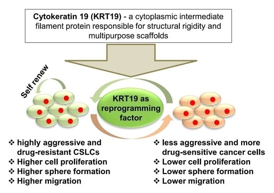
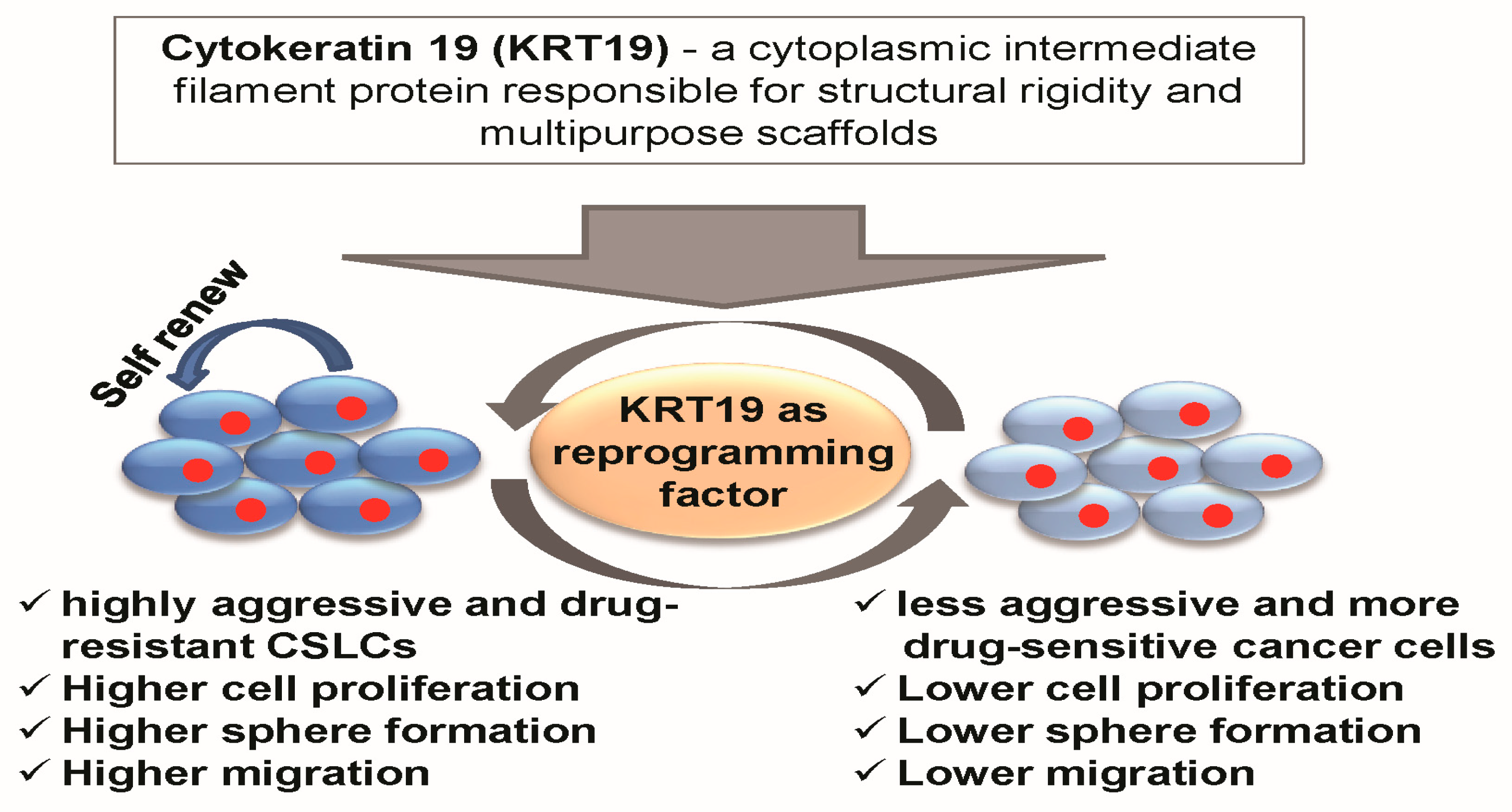
Cytokeratin 19 (KRT19) has a Role in the Reprogramming of Cancer Stem Cell-Like Cells to Less Aggressive and More Drug-Sensitive Cells

Department of Stem Cell and Regenerative Biotechnology, Incurable Disease Animal Model & Stem Cell Institute (IDASI), Konkuk University, Seoul 05029, Korea
*
Author to whom correspondence should be addressed.
Int. J. Mol. Sci. 2018, 19(5), 1423; https://doi.org/10.3390/ijms19051423
Submission received: 15 March 2018 / Revised: 4 May 2018 / Accepted: 7 May 2018 / Published: 9 May 2018
(This article belongs to the Special Issue Cell Reprogramming)

Abstract

:
Cytokeratin 19 (KRT19) is a cytoplasmic intermediate filament protein, which is responsible for structural rigidity and multipurpose scaffolds. In several cancers, KRT19 is overexpressed and may play a crucial role in tumorigenic transformation. In our previous study, we revealed the role of KRT19 as signaling component which mediated Wnt/NOTCH crosstalk through NUMB transcription in breast cancer. Here, we investigated the function of KRT19 in cancer reprogramming and drug resistance in breast cancer cells. We found that expression of KRT19 was attenuated in several patients-derived breast cancer tissues and patients with a low expression of KRT19 were significantly correlated with poor prognosis in breast cancer patients. Consistently, highly aggressive and drug-resistant breast cancer patient-derived cancer stem cell-like cells (konkuk university-cancer stem cell-like cell (KU-CSLCs)) displayed higher expression of cancer stem cell (CSC) markers, including ALDH1, CXCR4, and CD133, but a much lower expression of KRT19 than that is seen in highly aggressive triple negative breast cancer MDA-MB231 cells. Moreover, we revealed that the knockdown of KRT19 in MDA-MB231 cells led to an enhancement of cancer properties, such as cell proliferation, sphere formation, migration, and drug resistance, while the overexpression of KRT19 in KU-CSLCs resulted in the significant attenuation of cancer properties. KRT19 regulated cancer stem cell reprogramming by modulating the expression of cancer stem cell markers (ALDH1, CXCR4, and CD133), as well as the phosphorylation of Src and GSK3β (Tyr216). Therefore, our data may imply that the modulation of KRT19 expression could be involved in cancer stem cell reprogramming and drug sensitivity, which might have clinical implications for cancer or cancer stem cell treatment.

Graphical Abstract

1. Introduction

Heterogeneity at the cellular and molecular level is a prominent feature of most tumor types, including breast cancer [1]. Most breast cancer cases occur in the United States of America (USA) and it is the second most prevalent cancer worldwide [2]. Diverse self-renewing cell subpopulations have been observed in primary tumors and they were shown to be genetically and phenotypically heterogeneous, possibly augmenting a number of cancer growth patterns [1]. Heterogeneity of cancer is potentially controlled by the cellular microenvironment, as well as genetic and epigenetic factors [3]. Nowadays, stem cell research becomes the hot topic to the researchers because stem cells have the ability to self-renew and differentiate into various kinds of cells [4]. Current research suggests that genetic reprogramming may play a potential role in stem/cancer stem cell biology, by sustaining the pluripotency of stem cells (SCs) and increasing their differentiation into mature cell populations [5,6]. Studies have shown that dysplastic growth could be induced by the ectopic expression of stemness marker genes in adult epithelial tissues [7,8]. In 2006, Takahashi and Yamanaka showed for the first time that pluripotent stem (PS) cells could be generated from mouse embryonic fibroblasts by ectopic expression of transcription factors (TFs), called Yamanaka factors, such as OCT4, C-MYC, KLF4, and SOX2 [9]. Beside these Yamanaka factors, currently, there are around 25 TFs that are reported to be expressed in SCs. Of them, most of the TFs are suppressed in normal somatic cells, but are abnormally expressed in cancer cells [10,11], suggesting that the ectopic expression of stemness marker genes may cause the generation of abnormal cancer stem cells (CSCs). For instance, several stemness markers, such as OCT4, C-MYC, KLF4, NANOG, SALL4, and SOX2 are reported to be highly expressed in various cancers and associated with poor clinical outcomes of patients [12,13,14,15,16,17,18,19,20,21,22]. Recently, another study demonstrated that early termination of reprogramming could generate cells possessing a number of stemness signatures, but was unsuccessful in transforming into induced pluripotent stem cells (iPSC) and in ending originate neoplasia, such as Wilms tumor [23]. Recent advances in stem cell biology have explored the presence of cancer stem cell-like cells (CSLCs) in several cancers, such as breast, brain, colon, leukemia, and prostate cancers [24,25,26,27,28]. CSCs are a subtype of cells that have the capacity to self-renew, produce a heterogeneous subset of cancer cells, and initiate tumor generation [29,30]. These findings may suggest a possible relationship between embryonic stem cells (ESCs)/iPSC reprogramming and tumor generation.
KRT19 (Cytokeratin 19) is known to be the smallest (40 kDa) member of the acidic type I cytokeratin family proteins (KRTs) and may play a potential role in tumor detection by reverse transcriptase polymerase chain reaction (RT-PCR) in the bone marrow, lymph nodes, and peripheral blood of patients with breast cancer [31,32]. It has a highly preserved α-helical central domain and lack of C-terminal non-helical tail domain; α-helical central domain is very important for intermediate filament formation [33]. As a cytoplasmic intermediate filament protein, KRT19 could be responsible for structural rigidity and multipurpose scaffolds, as well as being a marker of epithelial cells and tissues [34]. It is also known that KRTs may interact with several signal transduction molecules, such as adaptors, effectors, kinases, and receptors, which may regulate signaling pathways and mediate cell apoptosis, cell cycle arrest, invasion, and metastasis [34,35]. Recent studies have revealed that KRT19 is crucially involved in the cancer stemness of hepatocellular carcinoma (HCC) [36]. Moreover, a recent study showed that HCC progression could be regulated through PDGFRα-laminin B1-keratin 19 cascade and this cascade could drive early recurrence, microvascular invasion, and metastasis in HCC [37,38,39]. Furthermore, it is demonstrated that KRT19 transcription could be increased through HER2/ERK/SP1 signaling pathway, in a consequence, KRT19 translocated to HER2 receptor then bound and stabilized the HER2 activation in breast and lung cancer [40,41]. In addition, KRT19 is normally expressed in the stem cell region of the hair follicle [42,43], and it is overexpressed in various radio-resistant solid tumors including colon and intestine progenitor/stem cells [44]. However, KRT19 shows discrepant relationships with both breast carcinoma and chemotherapy resistance [40,45,46,47]. Our previous study demonstrated that KRT19 could attenuate ALDH1high/CXCR4high/CD133high breast cancer stem cell-like cells (CSLCs) properties by regulating NUMB-mediated wnt/notch signaling crosstalk [46]. Moreover, KRT19 is reported to be a tumor suppressor gene in breast cancer, which regulates the nuclear translocation of EGR1 to the PTEN promoter [47]. KRT19 was also found to be a regulator of p38-MAPK/XBP-1 signaling cascade-mediated endoplasmic reticulum stress in breast cancer [48]. However, the roles that are involved in KRT19-driven breast cancer stem cell-like cell reprogramming and drug sensitivity are yet to be elucidated.
Herein, we have aimed to distinctly identify the role of KRT19 in cancer stem cell reprogramming. As KRT19 is involved in cancer regulation through signaling pathway, as we reported somewhere [46]. Here, we have hypothesized that KRT19 may have cancer regulating role through the reprogramming of cancer stem cell by partially modulating stemness, metastasis, and drug-resistant properties. Therefore, we investigated the role of KRT19 in cancer stem cell reprogramming and drug sensitivity by the overexpression and knockdown of KRT19 in KU-CSLCs (konkuk university-cancer stem cell-like cell) and MDA-MB231 cells. We found that the knockdown or overexpression of KRT19 in MDA-MB231 or KU-CSLCs cells modulated cancer reprogramming and drug sensitivity, as compared to normal MDA-MB231 or KU-CSLCs. These modulations may be partially due to the alteration of cancer stem cell markers (ALDH1, CXCR4, and CD133) expression as well as the phosphorylation of Src and GSK3β (Tyr216). Our result thus implies that KRT19 has the potential for targeting highly drug-resistant CSCs and for reprogramming to less aggressive phenotypes, which may be useful for the treatment of drug-resistant breast CSCs.

2. Results

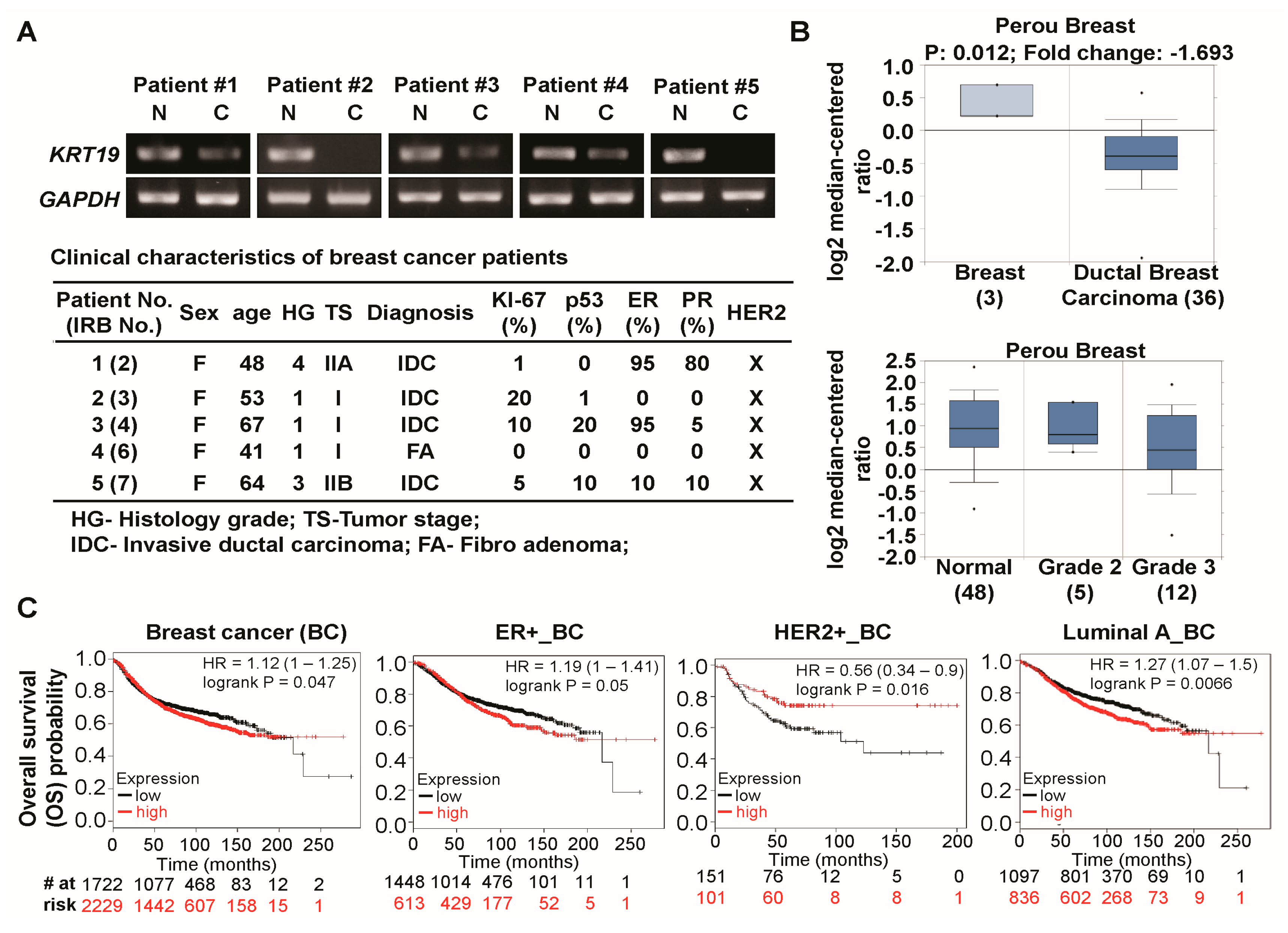
2.1. Expression of Cytokeratin 19 (KRT19) Is Downregulated in Several Breast Cancer Patients and Correlated with Breast Cancer Prognosis

As KRT19 expressed in various cancer types [36,37,38,39,40,41,44,45,46,47], we checked the expression level of KRT19 in five paired breast cancer tissue and adjacent normal breast tissues using reverse transcriptase-polymerase chain reaction (RT-PCR) analysis. However, our RT-PCR results showed that the expression of KRT19 was attenuated in breast cancer tissue (C) when compared to their normal (N) counterparts (Figure 1A, upper panel); the clinical characterization of breast cancer patients was overviewed in Figure 1A, lower panel. We then checked the KRT19 expression in breast cancer using Oncomine database. The KRT19 was significantly downregulated in ductal breast cancer tissue as compared to the normal tissue (Figure 1B, upper panel) [49], although KRT19 expression was upregulated in other breast cancer studies (Oncomine). Moreover, KRT19 expression downregulated in more aggressive breast cancer tissue than normal and less aggressive tissue (Figure 1B, lower panel) [49]. Next, we aimed to investigate the relationship between KRT19 expression and the patient’s clinical outcomes using the Kaplan-Meier plotter. We found that patients with low expression of KRT19 were significantly correlated with poor prognosis in breast cancer (BC), ER+_, HER2+_, and luminal A_BC patients (Figure 1C), suggesting that KRT19 is downregulated in breast cancer and is significantly correlated with poor breast cancer prognosis.

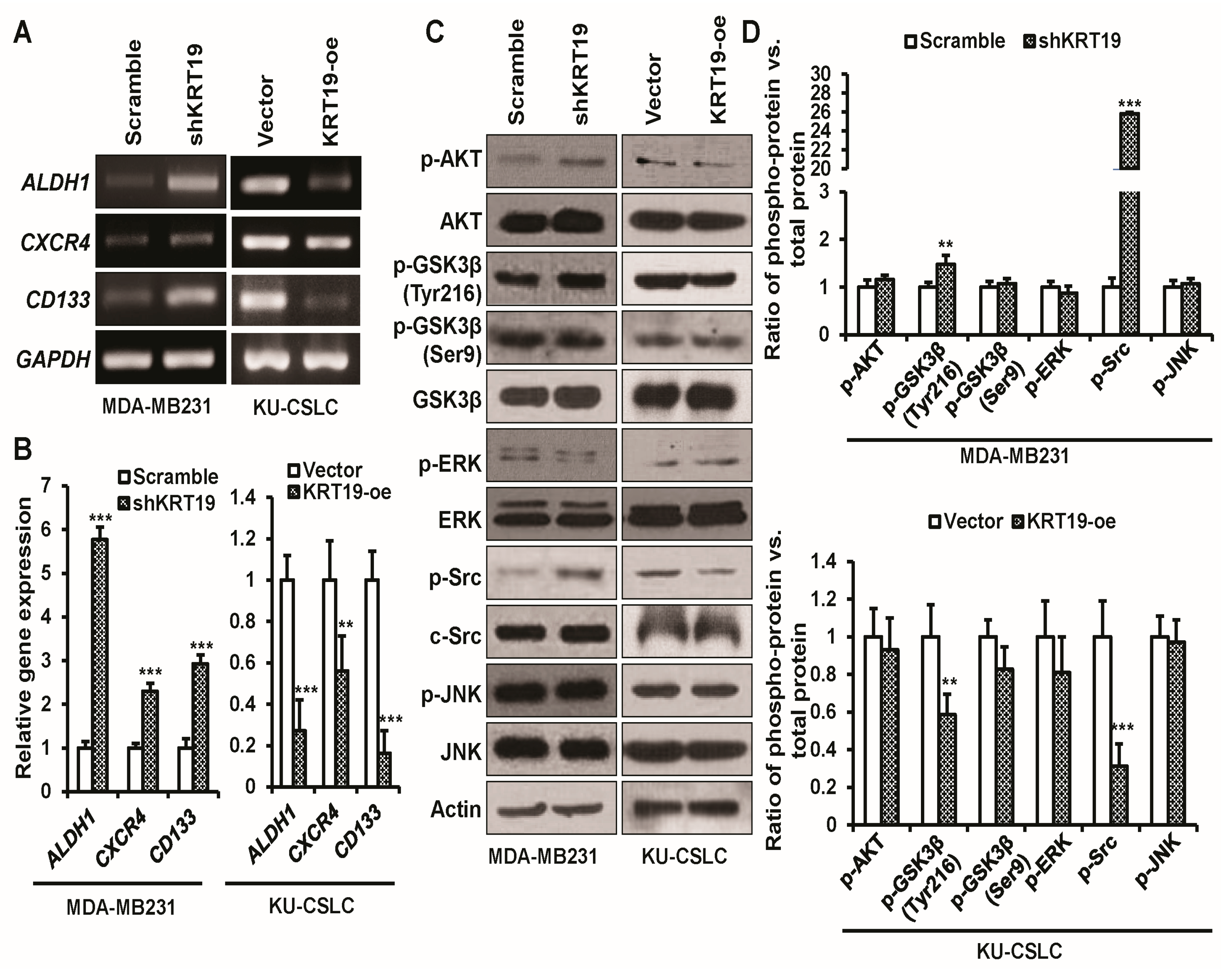
2.2. Chemo-Treated Breast Cancer Patient-Derived KU-CSLCs Show High Expression of ALDH1, CXCR4, and CD133, and More Aggressive Cancer Phenotypes

It is known that the overexpression of ALDH1, CXCR4, and CD133 produces cells with an aggressive and highly drug-resistant cancer stem cell-like phenotype [50,51]. Our previous study reported that the expression of KRT19 was upregulated in various breast cancer cell lines (i.e., MDA-MB231, MCF7) and was downregulated in highly aggressive cancer stem-like cells [46]. In our present study, we have also confirmed the expression pattern of KRT19 in MDA-MB231 and highly aggressive and drug-resistant breast cancer patient-derived cancer stem cell-like KU-CSLC (konkuk University-cancer stem cell-like cell) cells, using RT-PCR and western blot (WB) analysis (Figure 2A). To characterize chemo-treated breast cancer patient-derived KU-CSLCs, a triple negative breast cancer cell line, MDA-MB231, and KU-CSLC cells, were analyzed for the expression of ALDH1, CXCR4, and CD133 while using RT-PCR. The data indicated that the expression of ALDH1, CXCR4, and CD133 was significantly upregulated in KU-CSLC cells (Figure 2B). We then checked the expression of stemness marker genes (NANOG, OCT4, and SOX2), EMT markers (N-cadherin and E-cadherin), and the tumor suppressor gene P53, using qRT-PCR analysis. The KU-CSLC cells showed a significant upregulation of stemness marker genes (NANOG, OCT4, and SOX2) and N-cadherin, while a downregulation pattern of E-cadherin and P53 was observed in KU-CSLC cells when compared to MDA-MB231 cells (Figure 2C). The colony formation efficiency was significantly higher in KU-CSLC cells as compared to MDA-MB231 cells (Figure 2D). Afterward, the in vivo tumorigenic efficiency was investigated for both MDA-MB231 and KU-CSLCs. To verify in vivo tumorigenic efficiency, we used five-month old female SCID mice and subcutaneously implanted a range of cell numbers, 100, 1000, and 10,000, of both MDA-MB231 and KU-CSLC cells into the lower flanks of the mice. The tumor generation and growth in mice were monitored and mice were sacrificed four weeks post tumor formation. The results showed that for KU-CSLC cells, even 100 cells were enough to significantly induce tumor growth when compared to MDA-MB231 cells, which required 10,000 cells (Figure 2E). Concurrently, tumor weights from the KU-CSLC cells were significantly heavier than those resulting from MDA-MB231 cells (Figure 2F). These results suggest that chemo-treated breast cancer patient-derived KU-CSLC cells are more aggressive and drug-resistant than the aggressive triple negative MDA-MB231 cells.

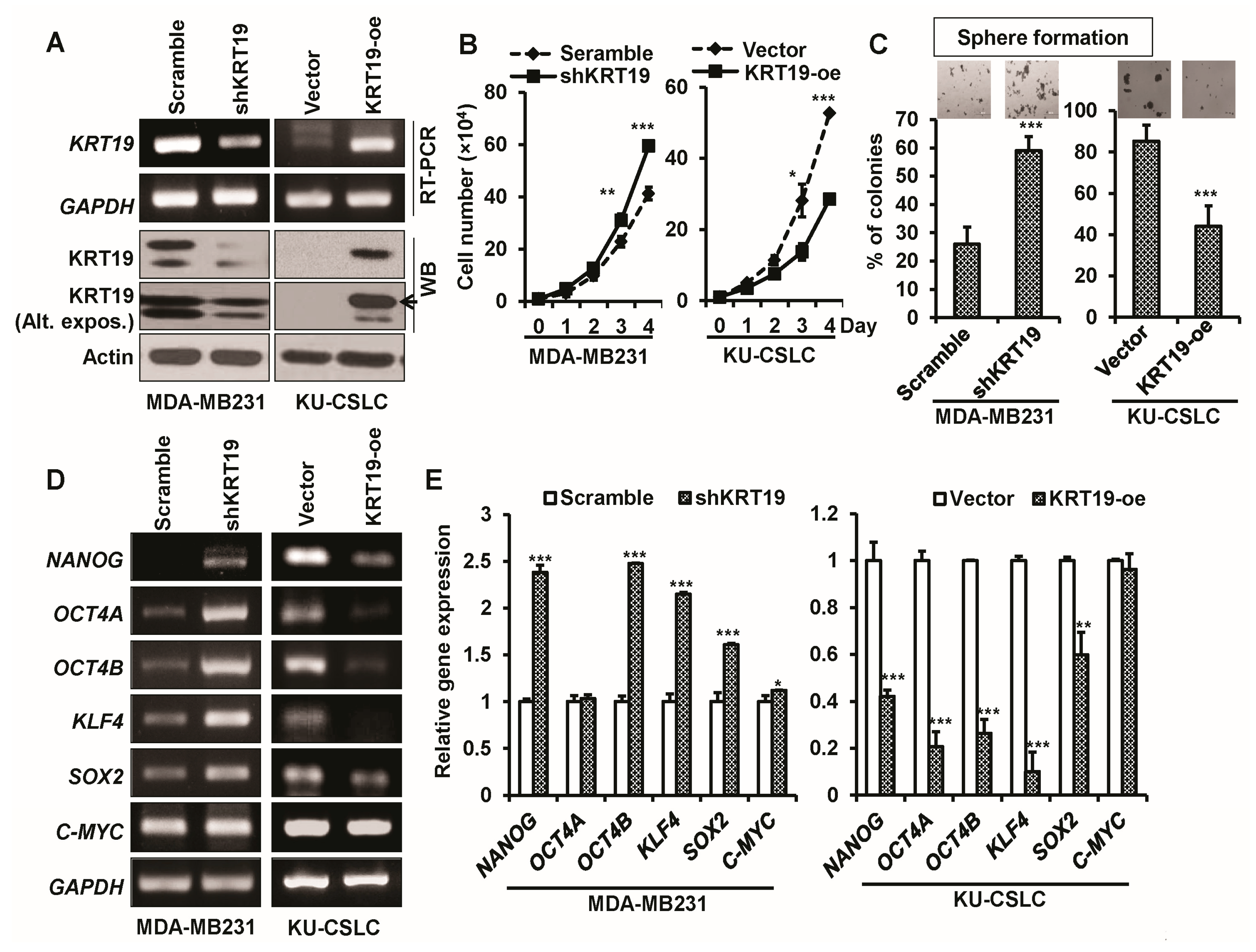
2.3. Knockdown or Overexpression of KRT19 Regulates Cell Proliferation and Sphere Formation in MDA-MB231 and KU-CSLC Cells

As KRT19 was upregulated in MDA-MB231 and downregulated in KU-CSLC cells, we next knocked down the expression of KRT19 by using a KRT19 specific shRNA (shKRT19) and overexpressed KRT19 (KRT19-oe) in KU-CSLC cells to investigate the function of KRT19 in breast cancer. The knockdown and overexpression of KRT19 were confirmed by RT-PCR and WB analysis (Figure 3A). Cell proliferation analysis demonstrated that cell proliferation was increased in shKRT19 infected MDA-MB231 cells and decreased in the KRT19 overexpressing KU-CSLC cells, when compared to their control counterparts (Figure 3B). To further explore the role of KRT19 on cell growth, we investigated the sphere formation ability using a sphere-forming assay. We observed a significantly increased number of the spheres in MDA-MB231 cells that were infected with the shKRT19 virus, as compared to the scramble, whereas a reduced sphere forming ability was seen in KRT19 overexpressing KU-CSLC cells, when compared with those infected with the vector (Figure 3C). A subsequent RT-PCR analysis for stemness marker genes was performed, with the results showing that the expression of stemness markers (NANOG, OCT4B, KLF4, SOX2, and C-MYC) was significantly increased in shKRT19 virus-infected MDA-MB231 cells and downregulated in KRT19 overexpressing KU-CSLC cells, as compared to scramble or vector infected MDA-MB231 or KU-CSLC cells, respectively (Figure 3D,E), suggesting that KRT19 may regulate cell growth and sphere formation ability in breast cancer.

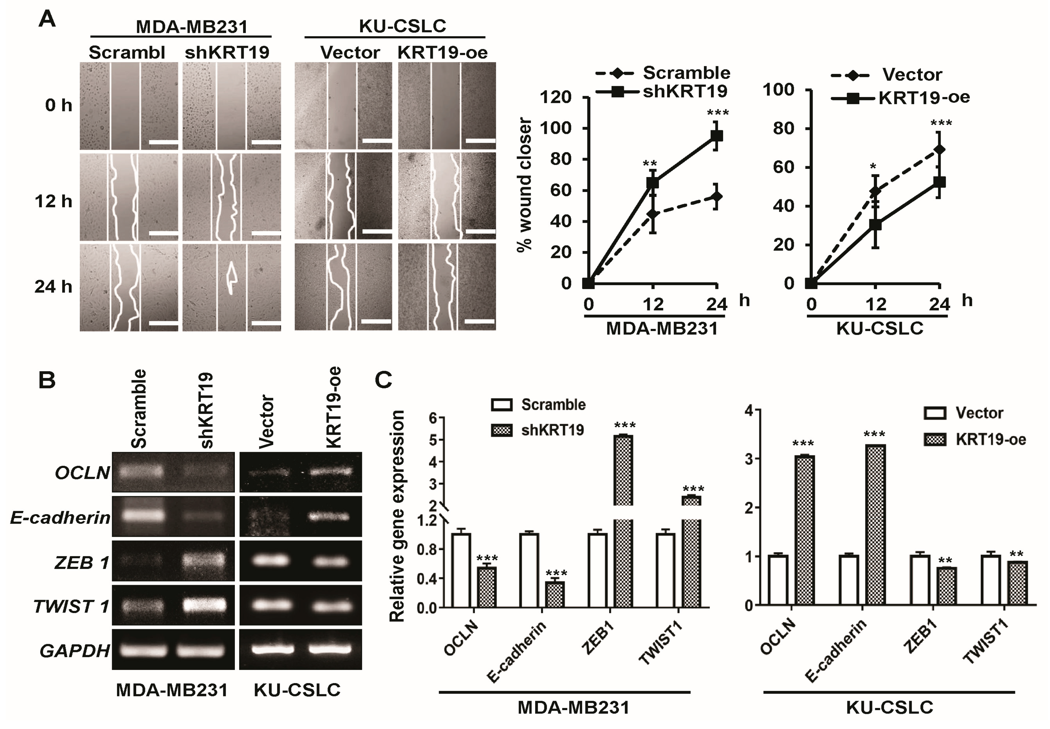
2.4. Silencing or Upregulation of KRT19 Regulates Cell Migration in MDA-MB231 and KU-CSLC Cells

To explore the function of KRT19 on cell migration in breast cancer, we first performed a wound healing cell migration analysis. The results showed that cell migration during wound healing was increased upon KRT19 knockdown in MDA-MB231 cells, while a reduction in cell migration was observed in KRT19-overexpressing KU-CSLC cells, when compared to those of scramble- or vector-expressing MDA-MB231 or KU-CSLC cells, respectively (Figure 4A). Afterwards, an RT-PCR analysis was performed to assess the expression of EMT marker genes, with the results displaying that MET (mesenchymal to epithelial transition) markers (OCLN and E-cadherin) were downregulated and EMT markers (ZEB1 and TWIST1) were upregulated in KRT19-silencing MDA-MB231 cells, whereas the opposite result was observed in KRT19-overexpressing KU-CSLC cells as compared to their control precursors (Figure 4B,C). This suggested that the silencing or overexpression of KRT19 could regulate cell migration in MDA-MB231 or KU-CSLC cells.

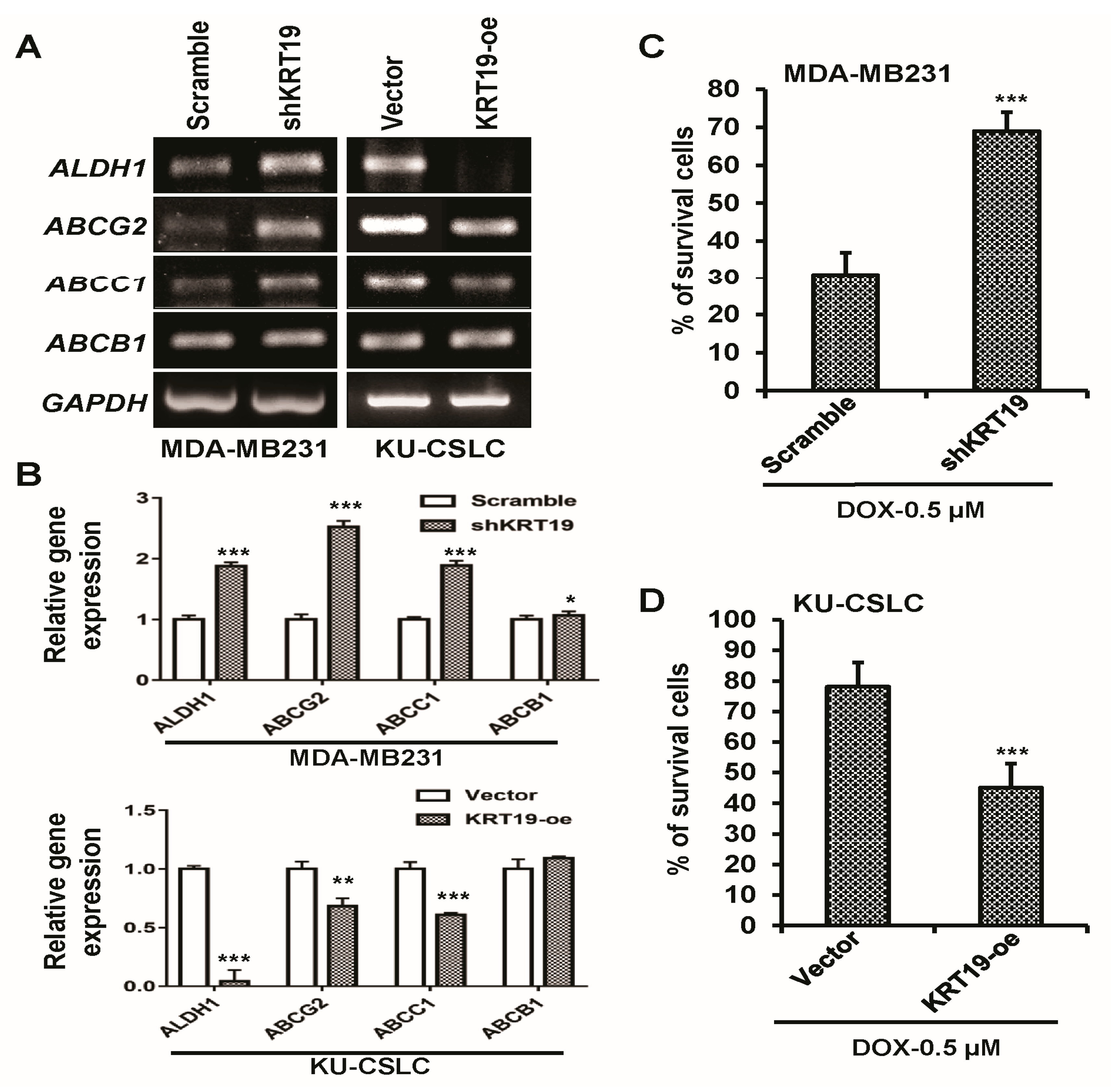
2.5. KRT19 Can Regulate Drug Resistance Capacity in Breast Cancer Cells

To further examine the function of KRT19 on drug sensitivity, we examined the expression of drug-resistance marker genes using RT-PCR analysis. The results showed that the knockdown of KRT19 significantly upregulated the expression of drug-resistance markers (ALDH1, ABCG2, ABCC1, and ABCB1), as compared to those of the control shRNA-infected MDA-MB231 cells (Figure 5A,B). We then checked the cell survival in the presence of several doses of doxorubicin (DOX), which is a well-known anti-cancer drug, in both MDA-MB231 and KU-CSLC cells, to assess drug resistance capability (Supplementary Figure S1). A specific dose (0.5 µM) was chosen for further study. The cell survival results in the presence of DOX (0.5 µM) showed that silencing of KRT19 significantly augmented cell survival, compared to those of scramble-infected MDA-MB231 cells (Figure 5C). We then assessed the expression of drug-resistance marker genes during KRT19 overexpression in KU-CSLCs. The RT-PCR analysis of drug-resistance markers revealed a significant downregulation in KRT19 overexpressing KU-CSLCs, when compared to their normal counterparts (Figure 5A,B). Subsequently, we checked drug-resistance capability in the presence of DOX, by overexpressing KRT19 in highly aggressive drug-resistant KU-CSLC cells. To determine drug-resistance capabilities, cell survival analysis was performed in the presence of DOX (0.5 µM) in both vector- and KRT19-overexpressing KU-CSLC cells. Cell survival was significantly suppressed in KRT19-overexpressing cells as compared to those of vector-infected KU-CSLC cells (Figure 5C), implying that KRT19 may regulate drug-sensitivity in highly drug-resistant breast cancer stem cell-like cells.

2.6. KRT19 Regulates Cancer Stem Cell Reprogramming through p-GSK3β (Tyr216) and p-Src Signaling Pathway

As the KU-CSLC showed high ALDH1, CXCR4, and CD133 gene expression, and low KRT19 expression, we assessed the effect of KRT19 overexpression or knockdown in KU-CSLC or MDA-MB231 cells. The RT-PCR analysis revealed that the overexpression or knockdown of KRT19 could modulate ALDH1, CXCR4, and CD133 gene expression in KU-CSLC or MDA-MB231 cells (Figure 6A,B). We next aimed to explore the underlying mechanism by which KRT19 regulates cancer reprogramming. Although KRT19 regulates EGR1/PTEN/AKT, β-catenin/NUMB/NOTCH, and HER2/ERK/SP1 cascades [29,46,47], we focused on the AKT, GSK3β, ERK, Src, and JNK signaling pathway in this study, as these signaling pathways are crucially involved in cancer progression [52,53,54,55,56]. The WB analysis demonstrated that the phosphorylation of p-GSK3β (Tyr216) and p-Src levels were increased significantly upon the knockdown or overexpression of KRT19 in MDA-MB231 or KU-CSLC cells, while the expression of p-AKT, p-GSK3β (Ser9), p-ERK, and p-JNK was unchanged (Figure 6C,D), which is suggested that KRT19 could regulate the cancer stem cell reprogramming by partially modulating the phosphorylation of GSK3β (Tyr216) and Src in breast cancer or cancer stem cells.

3. Discussion

CSCs (cancer stem cells) share several characteristics with typical stem cells, such as self-renewal and their undifferentiated and differentiated states. CSCs have been reported to originate from cancer cells with non–stemness properties, via a reprogramming mechanism that is very similar to that which is involved in iPSC (induced pluripotent stem cells) generation [23]. In addition, several key transcription factors, including NANOG, OCT4, KLF4, and SOX2, which induced iPSC and maintained the stemness properties of embryonic and somatic stem cells, have also been highly expressed in CSCs [8,57,58]. A number of recent studies have reported that cancer could be developed by the early termination of the reprogramming process in numerous tissues, through altered genetic and epigenetic regulation [7,8,23]. Thus, it could be inferred that teratoma formation from iPSCs is strongly associated with premature reprogramming, which is also important for the development of CSCs. In our current study, we have reported a relationship between KRT19 (cytokeratin 19) expression with CSC reprogramming, and drug sensitivity in breast cancer and cancer stem cell-like cells. Moreover, we discovered that KRT19 regulates CSC reprogramming and drug sensitivity in breast cancer and cancer stem cell-like cells, through mediating stemness (NANOG, OCT4, KLF4, and SOX2) and drug-resistant (ALDH1, ABCG2, ABCC1, and ABCB1) marker expression. Concurrently, we showed that KRT19 regulates breast cancer cell migration and EMT (epithelial to mesenchymal transition)-responsive gene expression. However, further study may elucidate the function of KRT19 in key regulatory pathways upon cancer reprogramming and drug sensitivity in various cancer stem cells.
KRT19, a cytokeratin protein, is reported to be differentially expressed in several cancers. It is highly expressed in breast, colon, liver, and intestine cancer, and it is associated with poor clinical outcomes in patients [36,40,44,45]. KRT19 expression, however, is also known to be negatively correlated with cancer progression in breast cancer stem cell-like cells and cell lines [46,47,48]. So far, the function of KRT19 in breast cancer still remains to be revealed. In this study, we found the downregulated KRT19 expression pattern in breast cancer tissue when compared to the adjacent normal tissue, which is an agreement with Oncomine database. Oncomine search, a freely accessible database, could make the research easier to correlate the experimental data with published genome-wide RNAseq data [59,60]. The KRT19 expression is reported to be correlated with cancer progression and prognosis [36,44,45,46,47]. Our data also evidences that KRT19 expression is significantly associated with clinical outcomes in breast cancer. In this current study, we also used patient-derived KU-CSLC cells, which have high drug-resistance, stemness, tumorigenic, and aggressive cancer stem cell-like cell properties, and a commercially available aggressive MDA-MB231 cell line to identify the role of KRT19 in cancer reprogramming and drug sensitivity. Our results also demonstrated that the expression of KRT19 was very low in highly aggressive KU-CSLC cells, when compared to those of MDA-MB231, which we previously reported on [46]. In this study, we further demonstrated that the knockdown of KRT19 in MDA-MB231 cells significantly induced cell proliferation and sphere formation, suggesting that KRT19 is a potent cancer suppressor gene. Moreover, the silencing of KRT19 augmented cell migration and drug-resistance of MDA-MB231, which means that the low expression of KRT19 produced an aggressive phenotype in cancer cells, as previously reported [40,46,47].
In addition, we demonstrated that the expression of ALDH1, CXCR4, and CD133 was higher in KU-CSLCs than in the aggressive MDA-MB231 cell line. It is also reported that ALDH1, CXCR4, and CD133 are well-known markers of highly aggressive, drug-resistant, invasive, and cancer stem cell phenotypes [50,51], suggesting that our KU-CSLCs are highly aggressive and drug-resistant cancer stem cell-like cells. Recent studies indicate that the aberrant expression of ALDH1 contributes to cancer stem cell transformation from less aggressive cancer with the association of CD133 [61,62]. Moreover, CD133+ and CXCR4+ are reported to involve in cancer stem cell phenotype in various cancer types [63,64,65]. Thus, targeting ALDH1, CRCR4, and CD133 could provide us a possibility to target cancer stem cell population. We have demonstrated that KRT19 overexpression inhibited aggressiveness, including abnormal cell proliferation, sphere formation, migration, and drug resistance in KU-CSLCs. Moreover, key transcription factors comprising OCT4, KLF4, NANOG, and SOX2, which enhanced the iPSC generation and maintained the self-renewal properties in embryonic and somatic stem cells, have also been highly expressed in CSCs [8,57,58]. Previous studies also demonstrated that these transcription factors involved in cancer progression and associated with clinical outcomes [12,13,14,15,16,17,20,21,22]. Our data showed a corroborative evidence that overexpression/knockdown of KRT19 significantly altered the expression of OCT4, KLF4, NANOG, and SOX2 in KU-CSLC or MDA-MB231 cells. This implies that through OCT4, KLF4, NANOG, and SOX2 alteration, KRT19 regulates cancer stem cell reprogramming efficiency in KU-CSLCs.
Cancer metastasis is one of the crucial factors to develop cancer stem cell phenotype [66]. It is noted that stemness markers expression also regulates the metastasis properties in cancer cells [67,68,69,70]. Targeting cancer stem cell metastasis through the regulation of stemness markers would be the worthy solution in cancer treatment. Moreover, it is reported that OCT4 expression mediates cancer stem cell reprogramming in CD133+ lung cancer cells [71]. It also mediated drug resistance in prostate cancer. Other stem cell markers, in addition, contributed to cancer stem cell recurrence and drug resistance [72,73,74,75]. Our confirmative data showed that overexpression/knockdown of KRT19 significantly regulated the expression of EMT [76] and drug-resistance [77] markers in both KU-CSLC or MDA-MB231 cells, which may partially due to the regulation of stemness markers expression and imply a partial regulation of cancer phenotypes and sensitivity to potent anti-cancer drugs.
The pathologic reprogramming procedure in cancer is reported to be critical in regulating the CSC induction and cancer reappearance [78,79]. In our data, KRT19 not only regulates cancer stemness, metastasis, and drug resistance, but it also modulates the expression of stemness, metastatic, and drug-resistant markers. This phenomenon could mediate through the signaling pathways. It is reported that KRT19 regulates several signaling cascades, including PDGFRα/laminin B1/keratin 19, HER2/ERK/SP1, WNT/NUMB/NOTCH, EGR1/PTEN/AKT, etc. [37,40,41,46,47]. In this study, we have reported that KRT19 regulated the phosphorylation of Src and GSK3β at Tyr216, while the other signaling proteins, such as p-AKT, p-ERK, and p-JNK were unregulated. Src is reported to support ALDH1+ CSC phenotype and reprogramming in various cancer types, including breast cancer [80]. Moreover, Src is also involved in the regulation of invasion and migration in several cancer types [81,82] and the ablation of Src could alternate the CSC phenotype [83], which is corroborated with our data. Previous studies also reported that dysregulation of Src and GSK3β signaling could mediate cancer progression, metastasis, drug resistance, and ultimately transform into cancer stem cell phenotypes [83,84,85,86]. Factually, alterations in GSK-3 activity have been explored due to the changes in GSK-3α and GSK-3β phosphorylation at Ser21 and Ser9, respectively, by Akt activation [87]. But, the activation of GSK-3α and GSK-3β depends on the phosphorylation at Tyr-279 and Tyr-216 [88,89]. Moreover, a recent in vitro and in vivo study showed that targeting Src-mediated GSK3β phosphorylation at Tyr216 could inhibit prostate cancer progression [86]. Src is also essential for the wound healing and fibrotic events where p-GSK3β (Tyr216) played an important role in fibrotic events mean that Src kinase may modulate the GSK3β activity [86], which might be in agreement with our study that KRT19 mediated cancer stem cell reprogramming might be through the regulation of Src/GSK3β phosphorylation at Tyr216 in breast cancer. Hence, the function of KRT19 in cancer stem cell reprogramming and the drug-resistance pathway is remarkable (Figure 7), and it would be interesting to explore the underlying mechanisms in detail.

4. Materials and Methods

4.1. Human Tissue Samples

Human breast tumor and normal tissue samples were collected from the Breast Cancer Center of Konkuk University Hospital with Institutional Review Board (IRB, KUH 1020003) approval. Breast tumor and normal tissues were collected from neoadjuvant chemotherapy (four cycles of preoperative Doxorubicin and cyclophosphamide)-treated stage III patients. The occupied tissues were rinsed with PBS (phosphate-buffered saline) and were incubated with type IV collagenase (Sigma-Aldrich, St. Louis, MO, USA) for 20 min at room temperature (RT). A single-cell suspension was prepared by filtering the supernatant through a 100 μm cell strainer (BD Bioscience, San Jose, CA, USA) and was cultured in DMEM (Dulbecco’s Modified Eagle’s medium; Sigma-Aldrich, Saint Louis, MO, USA) supplemented with 10% FBS (fetal bovine serum; GE Healthcare Hyclone, Pittsburgh, PA, USA) and 1% P/S (100 U/mL penicillin/streptomycin; GE Healthcare Hyclone) at 37 °C with 5% CO2. Unless stated otherwise, cells were cultured on the adherent tissue culture plates (Nunc). Cancer (C) and normal (N) tissue samples from five patients were used in the study.

4.2. Bioinformatics Analysis

Oncomine database (www.oncomine.org/) was used to obtain the KRT19 expression in breast cancer tissue and their normal tissues [59,60]. The fold change of KRT19 mRNA expression in breast cancer tissue when compared to their normal counterparts was obtained as the parameters of threshold p-value: 1 × 10⁻⁴; fold change: 2; and, gene ranking in the top 10%.
Kaplan–Meier curves were obtained using the KMplot program (http://kmplot.com/analysis/) [90]. To analyze the clinical outcomes of KRT19 gene, the patient samples were split into two groups, according to the median gene expression of the probe of KRT19 (201650_at). The log-rank p-value and the hazard ratio with 95% confidence intervals was also calculated.

4.3. Cell Culture

MDA-MB231, a human breast cancer cell line, was acquired from ATCC (American Type Culture Collection, Manassas, VA, USA). KU-CSLCs (konkuk University-cancer stem cell-like cell; CD133high/CXCR4high/ALDH1high) were collected from breast tumor tissue specimens, which were derived from chemo-treated patients at the Breast Cancer Center, Konkuk University Hospital, Seoul. The study was performed with the consent of the Institutional Review Board (IRB, KUH 1020003). Cells (MDA-MB231 and KU-CSLCs) were cultured in either RPMI (Roswell Park Memorial Institute) 1640 or high glucose-DMEM (Sigma-Aldrich) that was supplemented with 10% FBS (Hyclone) and 1% penicillin/streptomycin (Hyclone) at 5% CO2 and 37 °C in a humidified incubator. Short tandem repeat (STR) profiling was used for cell authentication and cells were tested by BioMycoX® Mycoplasma PCR Detection Kit for mycoplasma contamination detection (Cat. No. CS-D-25) (Cellsafe, Yeongtong-Gu, Suwon, Republic of Korea).

4.4. Total RNA Extraction and Reverse Transcriptase-PCR (RT-PCR) Analyses

For total RNA isolation, cells were harvested using a total RNA extraction kit (iNtRON Biotechnology, Jungwon-Gu, Seongnam-Si, Gyeonggi-do, Republic of Korea) by following the manufacturer’s instructions. The concentration of total RNA was quantified using a Nanodrop (ND1000) (Nanodrop Technologies Inc., Wilmington, DE, USA). Afterward, cDNA was synthesized using 1–2 μg of total RNA with M-MLV reverse transcriptase (RTase) (Promega, Madison, WI, USA), following the manufacturer’s instructions.

4.5. Polymerase Chain Reaction (PCR) and Quantitative Real-Time-PCR (qRT-PCR)

After the cDNA synthesis, PCR analysis was carried out using rTaq Plus 5X PCR master mix (Elpis biotech, Seo-gu, Daejeon, Republic of Korea), following the manufacturer’s instructions. The primer sequences that were used in this study are listed in Table 1. The PCR products were run on 1.5% agarose gels and imaged. The density of the PCR band was analyzed using ImageJ software. Quantitative real-time-PCR was performed using a thermal cycler (PTC-200) with a Chromo4 optical detector (MJ Research/BioRad, Hercules, CA, USA) using Fast 2X SYBR Green Master Mix (Applied Biosystems, Stockholm, Sweden). The housekeeping gene GAPDH was used as loading control, as described previously [46,91].

4.6. In Vivo Tumorigenicity Analysis

For in vivo tumorigenicity analysis, five-month old female SCID (severe combined immunodeficiency) mice were used [92], which were obtained from Korean Research Institute of Bioscience and Biotechnology (KRIBB) (Republic of Korea). For generating the tumor, MDA-MB231 or KU-CSLCs cells (102, 103, or 104 cells) were inoculated subcutaneously into the lower flanks of the mice. The tumor formation and growth of the mice were monitored, and the mice were sacrificed at four weeks post-tumor generation. Subsequent tumor volumes and weights were calculated, as described previously [93]. During the study, mice were allocated into two groups, non-randomly, based on their body weight. Each group contained at least five mice. The mice were omitted if they died or did not generate any tumor. The investigator was blinded during the study for the allocation of the group. According to the institutional guidelines, the mice were cared for and treated with permission (KU16003).

4.7. Knockdown and Overexpression of KRT19

For short hairpin RNA (shRNA) knockdown assay, sense and antisense oligonucleotides for control (scramble), and KRT19 (shKRT19), as described in our previous report [46], containing BamHI and EcoRI restriction sites, were annealed using annealing buffer [100 mM NaCl, Tris-EDTA (TE) buffer (pH 8) (Sigma-Aldrich)]. Afterward, the annealed double-stranded oligonucleotides were cloned into the pGreenPuro lentiviral shRNA expression vector (System Biosciences, Mountain View, CA, USA) to construct shKRT19 or Scramble vectors.
For the overexpression of KRT19, first, its complete coding sequence (CDS) was cloned into the pGEM®-T Easy vector (Promega) using the T vector primers, which were designed by the primer3 program (http://primer3.ut.ee/). The complete CDS without the stop codon was further cloned into the pCDH-EF1-MCS-T2A-copGFP (pCDH) lentiviral vector (System Biosciences) containing XbaI and EcoRI restriction sites, to construct the KRT19-oe vector, with the empty pCDH vector used as a control (vector). The primers that were used for the T Easy vector and pCDH vector clone were described in our previous report [46].
For lentivirus production, human HEK293T cells were transfected with viral packaging plasmids (i.e., RRE and REV) and the specific lentiviral expression vector using the calcium phosphate standard transfection protocol. Viruses were collected and were concentrated using an ultracentrifuge (Optima L-90K, Beckman Coulter Inc., Brea, CA, USA) at 48 h post incubation, at 5% CO2 and 37 °C. Either scrambled or shKRT19 lentiviruses were then infected to MDA-MB231 cells, while KU-CSLCs were infected with either control (vector) or KRT19-overexpressing (KRT19-oe) lentiviruses and were incubated overnight at 5% CO2 and 37 °C. Afterward, the virus-containing media was changed and cells were maintained with normal growth media. To produce stably expressing cells, ~8.0 × 10⁸ IU/mL viral particles were incubated with cells. To calculate virus titer, we used the following equation, titer in IU/mL = [(number cells at starting time) × (dilution factor) × (percent of infection)/(volume of virus solution added, expressed in mL)], as described previously [94]. The <95% virus-infected GFP-expressing cells were used for the future study, and knockdown or overexpression was confirmed by RT-PCR and WB analyses.

4.8. Western Blotting (WB) Analysis

For WB analysis, cells were harvested using cell lysis buffer [100 mM Tris-HCl (pH 7.5), 1% Triton X-100 (Sigma-Aldrich), 10 mM NaCl, 10% glycerol (Amresco, Solon, OH, USA), 50 mM sodium fluoride (Sigma-Aldrich), 1 mM sodium orthovanadate (Sigma-Aldrich), 1 mM phenylmethylsulfonyl fluoride (PMSF; Sigma-Aldrich), and 1 mM p-nitrophenyl phosphate (Sigma-Aldrich)], and the lysates were centrifuged at 13,000 rpm for 15 min at 4 °C. For protein quantification, Bradford protein assay reagent (BioRad) was used, and the quantified proteins were separated either 10 or 12% sodium dodecyl sulfate-polyacrylamide gel electrophoresis (SDS-PAGE). The separated proteins were transferred onto nitrocellulose membranes (Bio-Rad, Philadelphia, PA, USA). Afterward, the membranes were stained with 1× Ponceau staining for 5 min, and then blocked with 5% skimmed milk in Tris-buffered saline (TBS) for 1 h. The membranes were then incubated with primary antibodies against KRT19 (SC-53258), AKT (SC-1619), phospho (p)-AKT (SC-16646-R), GSK3β (SC-9166), p-GSK3β-Ser9 (SC-11757), ERK (SC-153), p-ERK (SC-7383), c-Src (SC-18), JNK (SC-7345), p-JNK (SC-6254), actin (SC-1616) (Santa Cruz Biotechnology, Dallas, TX, USA) p-Src (2101S) (Cell signaling, Danvers, MA, USA), and p-GSK3β-Tyr216 (05-413) (Merck Millipore, Burlington, MA, USA), overnight at 4 °C on a shaker. After overnight incubation, the membranes were washed with TBST for 30 min and were then incubated with appropriate second antibodies, including anti-mouse (SC-2005), -goat (SC-2020), or -rabbit (SC-2004) IgGs tagged with horseradish peroxidase (HRP) for 2 h at room temperature (Santa Cruz Biotechnology). After washing the membranes, protein expressions were detected on X-ray films using an enhanced chemiluminescence (ECL) kit (Amersham Bioscience, Piscataway, NJ, USA), as described previously [46,91,95]. To detect the expression of KRT19, we exposed the X-ray films for alternative time points (Alt. expos.) on the membranes, which are shown in Figure 3A.

4.9. Cell Proliferation Analysis

For cell proliferation analysis, cells (2 × 10⁴ cells/well) were cultured in 12-well dishes. Cells were cultured for four days; every 24 h, the cells were trypsinized and enumerated three times using a hemocytometer, as described previously [46,96].

4.10. Sphere Formation Assay

For the analysis of sphere formation, Cells (4 × 10⁴ cells/well) were cultured in 35 mm non-coated dishes with SM (sphere-formation medium: serum-free DMEM/F12 supplemented with 10 μg/mL insulin (Invitrogen, Carlsbad, CA, USA), 0.4% bovine serum albumin (BSA; Sigma-Aldrich), B27-supplement (1:50; Invitrogen), and 20 ng/mL epidermal growth factor (EGF; Sigma-Aldrich)). After five days of culture, Spheres from MDA-MB-231 and KU-CSLCs were collected and stained with crystal violet (Sigma-Aldrich). The stained spheres were photographed using a Nikon Eclipse TE2000-U microscope (Nikon Instruments Inc., Melville, NY, USA) prior to colony enumerating, as described previously [46].

4.11. Wound-Healing/Migration Assay

For wound-healing/migration analysis, 1 × 10⁶ cells were seeded in 60-mm cell culture dishes and were grown to ~90% confluence. Prior to the analysis, the cells were treated with mitomycin C (10 µg/mL) for 3–4 h at 5% CO2 and 37 °C, to prevent cell growth. After that, the cell monolayer was scratched with a 200 µL pipette tip to create a wound. Subsequently, the cells were washed three times with 1× PBS and images of the wound were taken using a Nikon Eclipse TE2000-U microscope (Nikon Instruments Inc.) at the indicated time-points (0, 12, and 24 h), as described previously [46,97].

4.12. Drug-Resistance Assay

For the analysis of drug-resistance, cells (1 × 10⁵ cells/well) were cultured in 12-well cell culture dishes and were cultured overnight at 5% CO2 and 37 °C. Afterward, cells were incubated with various doses of doxorubicin (DOX; 0.01, 0.1, 0.25, 0.5, and 1 µM, respectively) and were cultured for another 48 h. After 48 h incubation, cells were counted or medium was exchanged with a fresh medium containing EZ-Cytox (Daeil Lab Service, Seoul, Korea) and was incubated for an additional 3–4 h at 37 °C in an atmosphere of 5% CO2. The absorbance was measured at 450 nm by using a microplate reader Bio-Rad x-Mark™ spectrophotometer (Bio-Rad). The results were represented as a percentage (%) of surviving cells, as described previously [46,98].

4.13. Statistical Analyses

For statistical analysis, all of the experiments were independently undertaken at least three times. Data were presented as the mean ± standard deviation (SD) using Microsoft Excel 2010 software and statistical analyses were performed by ANOVA with a Bonferroni adjustment for comparing treated vs. control group by GraphPad InStat V. 3.0 software (GraphPad, San Diego, CA, USA). Results were considered to be statistically significant for p-values < 0.05.

5. Conclusions

In conclusion, our current study has revealed that there is an inverse relationship between the expression of KRT19 and breast cancer progression. We showed that KRT19 was downregulated in highly aggressive patient-derived KU-CSLCs, and that the overexpression of KRT19 could inhibit aggressiveness, including cell proliferation, sphere formation, migration, and drug resistance (overviewed in Figure 7), by suppressing the expression of the genes associated with these phenotypes. Moreover, after the knockdown of KRT19 in relatively high KRT19 expressing cells, MDA-MB231 induces cell proliferation, sphere formation, migration, and drug resistance and their responsive gene expression. In addition, we demonstrated that the overexpression or knockdown of KRT19 regulated cancer stem cell reprogramming by modulating the expression of cancer stem cell markers (ALDH1, CXCR4, and CD133) as well as the level of p-Src and p-GSK3β (Tyr216). Thus, our data suggest that KRT19 might function as a promising tumor suppressor gene for targeting highly aggressive patient-derived ALDH1high, CXCR4high, and CD133high breast cancer stem cell-like cells, and could enable cancer cell reprogramming to less aggressive and more drug-sensitive cells.

Supplementary Materials

Supplementary materials can be found at https://www.mdpi.com/1422-0067/19/5/1423/s1.

Author Contributions

S.K.S. came up with the idea and participated in the design, performed the experiments, analyzed the data, and wrote the manuscript. K.K., G.-M.Y., and H.Y.C. partially participated in performing the experiments, analyzed the data. S.-G.C. designed the experiments, reorganized the data, and wrote and edited the manuscript. All authors reviewed and approved the manuscript.

Acknowledgments

This work was supported by grants from the National Research Foundation (NRF) funded by the Korean government (2017M3A9C6029562, 2015R1A5A1009701, and 2013M3A9D3045880) and supported by the KU Research Professor Program of Konkuk University.

Conflicts of Interest

The authors declare no conflict of interest.

Abbreviations

KRT19cytokeratin 19
iPSCinduced pluripotent stem cell
PSpluripotent stem
CSCcancer stem cell
KU-CSLCkonkuk University-cancer stem cell-like cell
KRT19-oekeratin 19-overexpression
RT-PCRreverse transcriptase polymerase chain reaction
TFTranscription factor
qRT-PCRquantitative real-time-PCR
shRNAshort hairpin RNA
shKRT19short hairpin keratin19
SCIDsevere combined immunodeficiency
WBwestern blot
EMTepithelial to mesenchymal transition
METmesenchymal to epithelial transition
DOXdoxorubicin
STRshort tandem repeat
CDScoding sequence
SDS-PAGEsodium dodecyl sulfate-polyacrylamide gel electrophoresis
TBStris-buffered saline
PBSphosphate-buffered saline
TBSTtris-buffered saline Tween 20
HRPhorseradish peroxidase
ECLenhanced chemiluminescence

References

  1. Almendro, V.; Marusyk, A.; Polyak, K. Cellular Heterogeneity and Molecular Evolution in Cancer. Annu. Rev. Pathol. Mech. 2013, 8, 277–302. [Google Scholar] [CrossRef] [PubMed]
  2. Ferlay, J.; Soerjomataram, I.; Dikshit, R.; Eser, S.; Mathers, C.; Rebelo, M.; Parkin, D.M.; Forman, D.; Bray, F. Cancer incidence and mortality worldwide: Sources, methods and major patterns in GLOBOCAN 2012. Int. J. Cancer 2015, 136, E359–E386. [Google Scholar] [CrossRef] [PubMed]
  3. You, J.S.; Jones, P.A. Cancer genetics and epigenetics: Two sides of the same coin? Cancer Cell 2012, 22, 9–20. [Google Scholar] [CrossRef] [PubMed]
  4. Reya, T.; Morrison, S.J.; Clarke, M.F.; Weissman, I.L. Stem cells, cancer, and cancer stem cells. Nature 2001, 414, 105–111. [Google Scholar] [CrossRef] [PubMed]
  5. Chestkov, I.V.; Khomyakova, E.A.; Vasilieva, E.A.; Lagarkova, M.A.; Kiselev, S.L. Molecular barriers to processes of genetic reprogramming and cell transformation. Biochemistry 2014, 79, 1297–1307. [Google Scholar] [CrossRef] [PubMed]
  6. Bogomazova, A.N.; Vassina, E.M.; Kiselev, S.I.; Lagarkova, M.A.; Lebedeva, O.S.; Nekrasov, E.D.; Panova, A.V.; Philonenko, E.S.; Khomyakova, E.A.; Tskhovrebova, L.V.; et al. Genetic Cell Reprogramming: A New Technology for Basic Research and Applied Usage. Genetika 2015, 51, 466–478. [Google Scholar] [CrossRef] [PubMed]
  7. Hochedlinger, K.; Yamada, Y.; Beard, C.; Jaenisch, R. Ectopic expression of Oct-4 blocks progenitor-cell differentiation and causes dysplasia in epithelial tissues. Cell 2005, 121, 465–477. [Google Scholar] [CrossRef] [PubMed]
  8. Chiou, S.H.; Wang, M.L.; Chou, Y.T.; Chen, C.J.; Hong, C.F.; Hsieh, W.J.; Chang, H.T.; Chen, Y.S.; Lin, T.W.; Hsu, H.S.; et al. Coexpression of Oct4 and Nanog Enhances Malignancy in Lung Adenocarcinoma by Inducing Cancer Stem Cell-Like Properties and Epithelial-Mesenchymal Transdifferentiation. Cancer Res. 2010, 70, 10433–10444. [Google Scholar] [CrossRef] [PubMed]
  9. Takahashi, K.; Yamanaka, S. Induction of pluripotent stem cells from mouse embryonic and adult fibroblast cultures by defined factors. Cell 2006, 126, 663–676. [Google Scholar] [CrossRef] [PubMed] [Green Version]
  10. Monk, M.; Holding, C. Human embryonic genes re-expressed in cancer cells. Oncogene 2001, 20, 8085–8091. [Google Scholar] [CrossRef] [PubMed]
  11. Zhao, W.; Ji, X.; Zhang, F.; Li, L.; Ma, L. Embryonic Stem Cell Markers. Molecules 2012, 17, 6196. [Google Scholar] [CrossRef] [PubMed]
  12. Schoenhals, M.; Kassambara, A.; De Vos, J.; Hose, D.; Moreaux, J.; Klein, B. Embryonic stem cell markers expression in cancers. Biochem. Biophs. Res. Commun. 2009, 383, 157–162. [Google Scholar] [CrossRef] [PubMed]
  13. Gillis, A.J.M.; Stoop, H.; Biermann, K.; van Gurp, R.J.; Swartzman, E.; Cribbes, S.; Ferlinz, A.; Shannon, M.; Oosterhuis, J.W.; Looijenga, L.H.J. Expression and interdependencies of pluripotency factors LIN28, OCT3/4, NANOG and SOX2 in human testicular germ cells and tumours of the testis. Int. J. Androl. 2011, 34, E160–E174. [Google Scholar] [CrossRef] [PubMed]
  14. Forghanifard, M.M.; Khales, S.A.; Javdani-Mallak, A.; Rad, A.; Farshchian, M.; Abbaszadegan, M.R. Stemness state regulators SALL4 and SOX2 are involved in progression and invasiveness of esophageal squamous cell carcinoma. Med. Oncol. 2014, 31, 922. [Google Scholar] [CrossRef] [PubMed]
  15. Sodja, E.; Rijavec, M.; Koren, A.; Sadikov, A.; Korosec, P.; Cufer, T. The prognostic value of whole blood SOX2, NANOG and OCT4 mRNA expression in advanced small-cell lung cancer. Radiol. Oncol. 2016, 50, 188–196. [Google Scholar] [CrossRef] [PubMed]
  16. Tai, S.K.; Yang, M.H.; Chang, S.Y.; Chang, Y.C.; Li, W.Y.; Tsai, T.L.; Wang, Y.F.; Chu, P.Y.; Hsieh, S.L. Persistent Kruppel-like factor 4 expression predicts progression and poor prognosis of head and neck squamous cell carcinoma. Cancer Sci. 2011, 102, 895–902. [Google Scholar] [CrossRef] [PubMed]
  17. Lin, C.Y.; Loven, J.; Rahl, P.B.; Paranal, R.M.; Burge, C.B.; Bradner, J.E.; Lee, T.I.; Young, R.A. Transcriptional Amplification in Tumor Cells with Elevated c-Myc. Cell 2012, 151, 56–67. [Google Scholar] [CrossRef] [PubMed]
  18. Xu, K.; Zhu, Z.; Zeng, F. Expression and significance of Oct4 in bladder cancer. J. Huazhong Univ. Sci. Technol. Med. Sci. 2007, 27, 675–677. [Google Scholar] [CrossRef] [PubMed]
  19. Hatefi, N.; Nouraee, N.; Parvin, M.; Ziaee, S.A.M.; Mowla, S.J. Evaluating the Expression of Oct4 as a Prognostic Tumor Marker in Bladder Cancer. Iran. J. Basic Med. Sci. 2012, 15, 1154–1161. [Google Scholar] [PubMed]
  20. De Resende, M.F.; Chinen, L.T.D.; Vieira, S.; Jampietro, J.; da Fonseca, F.P.; Vassallo, J.; Campos, L.C.; Guimares, G.C.; Soares, F.A.; Rocha, R.M. Prognostication of OCT4 isoform expression in prostate cancer. Tumor Biol. 2013, 34, 2665–2673. [Google Scholar] [CrossRef] [PubMed]
  21. Rodini, C.O.; Suzuki, D.E.; Saba-Silva, N.; Cappellano, A.; de Souza, J.E.S.; Cavalheiro, S.; Toledo, S.R.C.; Okamoto, O.K. Expression analysis of stem cell-related genes reveal OCT4 as a predictor of poor clinical outcome in medulloblastoma. J. Neuro-Oncol. 2012, 106, 71–79. [Google Scholar] [CrossRef] [PubMed]
  22. Li, C.G.; Yan, Y.; Ji, W.D.; Bao, L.L.; Qian, H.H.; Chen, L.; Wu, M.C.; Chen, H.Z.; Li, Z.G.; Su, C.Q. OCT4 Positively Regulates Survivin Expression to Promote Cancer Cell Proliferation and Leads to Poor Prognosis in Esophageal Squamous Cell Carcinoma. PLoS ONE 2012, 7, e49693. [Google Scholar] [CrossRef] [PubMed]
  23. Ohnishi, K.; Semi, K.; Yamamoto, T.; Shimizu, M.; Tanaka, A.; Mitsunaga, K.; Okita, K.; Osafune, K.; Arioka, Y.; Maeda, T.; et al. Premature Termination of Reprogramming In Vivo Leads to Cancer Development through Altered Epigenetic Regulation. Cell 2014, 156, 663–677. [Google Scholar] [CrossRef] [PubMed]
  24. Al-Hajj, M.; Wicha, M.S.; Benito-Hernandez, A.; Morrison, S.J.; Clarke, M.F. Prospective identification of tumorigenic breast cancer cells. Proc. Natl. Acad. Sci. USA 2003, 100, 3983–3988. [Google Scholar] [CrossRef] [PubMed]
  25. Singh, S.K.; Clarke, I.D.; Terasaki, M.; Bonn, V.E.; Hawkins, C.; Squire, J.; Dirks, P.B. Identification of a cancer stem cell in human brain tumors. Cancer Res. 2003, 63, 5821–5828. [Google Scholar] [PubMed]
  26. Ricci-Vitiani, L.; Lombardi, D.G.; Pilozzi, E.; Biffoni, M.; Todaro, M.; Peschle, C.; De Maria, R. Identification and expansion of human colon-cancer-initiating cells. Nature 2007, 445, 111–115. [Google Scholar] [CrossRef] [PubMed]
  27. Bonnet, D.; Dick, J.E. Human acute myeloid leukemia is organized as a hierarchy that originates from a primitive hematopoietic cell. Nat. Med. 1997, 3, 730–737. [Google Scholar] [CrossRef] [PubMed]
  28. Collins, A.T.; Berry, P.A.; Hyde, C.; Stower, M.J.; Maitland, N.J. Prospective identification of tumorigenic prostate cancer stem cells. Cancer Res. 2005, 65, 10946–10951. [Google Scholar] [CrossRef] [PubMed]
  29. Clarke, M.F.; Dick, J.E.; Dirks, P.B.; Eaves, C.J.; Jamieson, C.H.; Jones, D.L.; Visvader, J.; Weissman, I.L.; Wahl, G.M. Cancer stem cells––Perspectives on current status and future directions: AACR Workshop on cancer stem cells. Cancer Res. 2006, 66, 9339–9344. [Google Scholar] [CrossRef] [PubMed]
  30. Pardal, R.; Clarke, M.F.; Morrison, S.J. Applying the principles of stem-cell biology to cancer. Nat. Rev. Cancer 2003, 3, 895–902. [Google Scholar] [CrossRef] [PubMed]
  31. Ignatiadis, M.; Xenidis, N.; Perraki, M.; Apostolaki, S.; Politaki, E.; Kafousi, M.; Stathopoulos, E.N.; Stathopoulou, A.; Lianidou, E.; Chlouverakis, G. Different prognostic value of cytokeratin-19 mRNA positive circulating tumor cells according to estrogen receptor and HER2 status in early-stage breast cancer. J. Clin. Oncol. 2007, 25, 5194–5202. [Google Scholar] [CrossRef] [PubMed]
  32. Bozionellou, V.; Mavroudis, D.; Perraki, M.; Papadopoulos, S.; Apostolaki, S.; Stathopoulos, E.; Stathopoulou, A.; Lianidou, E.; Georgoulias, V. Trastuzumab administration can effectively target chemotherapy-resistant cytokeratin-19 messenger RNA–positive tumor cells in the peripheral blood and bone marrow of patients with breast cancer. Clin. Cancer Res. 2004, 10, 8185–8194. [Google Scholar] [CrossRef] [PubMed]
  33. Fradette, J.; Germain, L.; Seshaiah, P.; Coulombe, P.A. The type I keratin 19 possesses distinct and context-dependent assembly properties. J. Biol. Chem. 1998, 273, 35176–35184. [Google Scholar] [CrossRef] [PubMed]
  34. Coulombe, P.A.; Wong, P. Cytoplasmic intermediate filaments revealed as dynamic and multipurpose scaffolds. Nat. Cell Biol. 2004, 6, 699–706. [Google Scholar] [CrossRef] [PubMed]
  35. Hendrix, M.J.C.; Seftor, E.A.; Chu, Y.W.; Trevor, K.T.; Seftor, R.E.B. Role of intermediate filaments in migration, invasion and metastasis. Cancer Metast. Rev. 1996, 15, 507–525. [Google Scholar] [CrossRef]
  36. Kawai, T.; Yasuchika, K.; Ishii, T.; Katayama, H.; Yoshitoshi, E.Y.; Ogiso, S.; Kita, S.; Yasuda, K.; Fukumitsu, K.; Mizumoto, M.; et al. Keratin 19, a Cancer Stem Cell Marker in Human Hepatocellular Carcinoma. Clin. Cancer Res. 2015, 21, 3081–3091. [Google Scholar] [CrossRef] [PubMed]
  37. Govaere, O.; Petz, M.; Wouters, J.; Vandewynckel, Y.-P.; Scott, E.J.; Topal, B.; Nevens, F.; Verslype, C.; Anstee, Q.M.; Van Vlierberghe, H. The PDGFRα-laminin B1-keratin 19 cascade drives tumor progression at the invasive front of human hepatocellular carcinoma. Oncogene 2017, 36, 6605–6616. [Google Scholar] [CrossRef] [PubMed]
  38. Govaere, O.; Komuta, M.; Berkers, J.; Spee, B.; Janssen, C.; de Luca, F.; Katoonizadeh, A.; Wouters, J.; van Kempen, L.C.; Durnez, A.; et al. Keratin 19: A key role player in the invasion of human hepatocellular carcinomas. Gut 2014, 63, 674–685. [Google Scholar] [CrossRef] [PubMed]
  39. Kim, H.; Choi, G.H.; Na, D.C.; Ahn, E.Y.; Kim, G.I.; Lee, J.E.; Cho, J.Y.; Yoo, J.E.; Choi, J.S.; Park, Y.N. Human hepatocellular carcinomas with “Stemness”-related marker expression: Keratin 19 expression and a poor prognosis. Hepatology 2011, 54, 1707–1717. [Google Scholar] [CrossRef] [PubMed]
  40. Ju, J.H.; Oh, S.; Lee, K.M.; Yang, W.; Nam, K.S.; Moon, H.G.; Noh, D.Y.; Kim, C.G.; Park, G.; Park, J.B.; et al. Cytokeratin19 induced by HER2/ERK binds and stabilizes HER2 on cell membranes. Cell Death Differ. 2015, 22, 665–676. [Google Scholar] [CrossRef] [PubMed]
  41. Ohtsuka, T.; Sakaguchi, M.; Yamamoto, H.; Tomida, S.; Takata, K.; Shien, K.; Hashida, S.; Miyata-Takata, T.; Watanabe, M.; Suzawa, K.; et al. Interaction of cytokeratin 19 head domain and HER2 in the cytoplasm leads to activation of HER2-Erk pathway. Sci. Rep. 2016, 6, 39557. [Google Scholar] [CrossRef] [PubMed]
  42. Lapouge, G.; Youssef, K.K.; Vokaer, B.; Achouri, Y.; Michaux, C.; Sotiropoulou, P.A.; Blanpain, C. Identifying the cellular origin of squamous skin tumors. Proc. Natl. Acad. Sci. USA 2011, 108, 7431–7436. [Google Scholar] [CrossRef] [PubMed]
  43. Means, A.L.; Xu, Y.W.; Zhao, A.Z.; Ray, K.C.; Gu, G.Q. A CK19(CreERT) knockin mouse line allows for conditional DNA recombination in epithelial cells in multiple endodermal organs. Genesis 2008, 46, 318–323. [Google Scholar] [CrossRef] [PubMed]
  44. Asfaha, S.; Hayakawa, Y.; Muley, A.; Stokes, S.; Graham, T.A.; Ericksen, R.E.; Westphalen, C.B.; von Burstin, J.; Mastracci, T.L.; Worthley, D.L.; et al. Krt19(+)/Lgr5(-) Cells Are Radioresistant Cancer-Initiating Stem Cells in the Colon and Intestine. Cell Stem Cell 2015, 16, 627–638. [Google Scholar] [CrossRef] [PubMed]
  45. Kabir, N.N.; Ronnstrand, L.; Kazi, J.U. Keratin 19 expression correlates with poor prognosis in breast cancer. Mol. Biol. Rep. 2014, 41, 7729–7735. [Google Scholar] [CrossRef] [PubMed]
  46. Saha, S.; Choi, H.; Kim, B.; Dayem, A.; Yang, G.; Kim, K.; Yin, Y.; Cho, S. KRT19 directly interacts with β-catenin/RAC1 complex to regulate NUMB-dependent NOTCH signaling pathway and breast cancer properties. Oncogene 2017, 36, 332–349. [Google Scholar] [CrossRef] [PubMed]
  47. Ju, J.H.; Yang, W.; Lee, K.M.; Oh, S.; Nam, K.; Shim, S.; Shin, S.Y.; Gye, M.C.; Chu, I.S.; Shin, I. Regulation of Cell Proliferation and Migration by Keratin19-Induced Nuclear Import of Early Growth Response-1 in Breast Cancer Cells. Clin. Cancer Res. 2013, 19, 4335–4346. [Google Scholar] [CrossRef] [PubMed]
  48. Bambang, I.F.; Lu, D.; Li, H.P.; Chiu, L.L.; Lau, Q.C.; Koay, E.; Zhang, D.H. Cytokeratin 19 regulates endoplasmic reticulum stress and inhibits ERp29 expression via p38 MAPK/XBP-1 signaling in breast cancer cells. Exp. Cell Res. 2009, 315, 1964–1974. [Google Scholar] [CrossRef] [PubMed]
  49. Perou, C.M.; Sorlie, T.; Eisen, M.B.; van de Rijn, M.; Jeffrey, S.S.; Rees, C.A.; Pollack, J.R.; Ross, D.T.; Johnsen, H.; Akslen, L.A.; et al. Molecular portraits of human breast tumours. Nature 2000, 406, 747–752. [Google Scholar] [CrossRef] [PubMed]
  50. Miki, J.; Furusato, B.; Li, H.; Gu, Y.; Takahashi, H.; Egawa, S.; Sesterhenn, I.A.; McLeod, D.G.; Srivastava, S.; Rhim, J.S. Identification of putative stem cell markers, CD133 and CXCR4, in hTERT-immortalized primary nonmalignant and malignant tumor-derived human prostate epithelial cell lines and in prostate cancer specimens. Cancer Res. 2007, 67, 3153–3161. [Google Scholar] [CrossRef] [PubMed]
  51. Douville, J.; Beaulieu, R.; Balicki, D. ALDH1 as a Functional Marker of Cancer Stem and Progenitor Cells. Stem Cells Dev. 2009, 18, 17–25. [Google Scholar] [CrossRef] [PubMed]
  52. Martini, M.; De Santis, M.C.; Braccini, L.; Gulluni, F.; Hirsch, E. PI3K/AKT signaling pathway and cancer: An updated review. Ann. Med. 2014, 46, 372–383. [Google Scholar] [CrossRef] [PubMed]
  53. Zhang, X.; Jiang, G.; Sun, M.; Zhou, H.; Miao, Y.; Liang, M.; Wang, E.; Zhang, Y. Cytosolic THUMPD1 promotes breast cancer cells invasion and metastasis via the AKT-GSK3-Snail pathway. Oncotarget 2017, 8, 13357–13366. [Google Scholar] [CrossRef] [PubMed]
  54. Sun, Y.; Liu, W.Z.; Liu, T.; Feng, X.; Yang, N.; Zhou, H.F. Signaling pathway of MAPK/ERK in cell proliferation, differentiation, migration, senescence and apoptosis. J. Recept. Signal. Transduct. Res. 2015, 35, 600–604. [Google Scholar] [CrossRef] [PubMed]
  55. Kim, R.K.; Cui, Y.H.; Yoo, K.C.; Kim, I.G.; Lee, M.; Choi, Y.H.; Suh, Y.; Lee, S.J. Radiation promotes malignant phenotypes through SRC in breast cancer cells. Cancer Sci. 2015, 106, 78–85. [Google Scholar] [CrossRef] [PubMed]
  56. Malki, A.; Elbayaa, R.Y.; Ashour, H.M.A.; Loffredo, C.A.; Youssef, A.M. Novel thiosemicarbazides induced apoptosis in human MCF-7 breast cancer cells via JNK signaling. J. Enzym. Inhib. Med. Chem. 2015, 30, 786–795. [Google Scholar] [CrossRef] [PubMed]
  57. Yu, F.; Li, J.; Chen, H.; Fu, J.; Ray, S.; Huang, S.; Zheng, H.; Ai, W. Kruppel-like factor 4 (KLF4) is required for maintenance of breast cancer stem cells and for cell migration and invasion. Oncogene 2011, 30, 2161–2172. [Google Scholar] [CrossRef] [PubMed]
  58. Leis, O.; Eguiara, A.; Lopez-Arribillaga, E.; Alberdi, M.J.; Hernandez-Garcia, S.; Elorriaga, K.; Pandiella, A.; Rezola, R.; Martin, A.G. Sox2 expression in breast tumours and activation in breast cancer stem cells. Oncogene 2012, 31, 1354–1365. [Google Scholar] [CrossRef] [PubMed]
  59. Rhodes, D.R.; Yu, J.J.; Shanker, K.; Deshpande, N.; Varambally, R.; Ghosh, D.; Barrette, T.; Pandey, A.; Chinnaiyan, A.M. ONCOMINE: A cancer microarray database and integrated data-mining platform. Neoplasia 2004, 6, 1–6. [Google Scholar] [CrossRef]
  60. Rhodes, D.R.; Kalyana-Sundaram, S.; Mahavisno, V.; Varambally, R.; Yu, J.J.; Briggs, B.B.; Barrette, T.R.; Anstet, M.J.; Kincead-Beal, C.; Kulkarni, P.; et al. Oncomine 3.0: Genes, pathways, and networks in a collection of 18,000 cancer gene expression profiles. Neoplasia 2007, 9, 166–180. [Google Scholar] [CrossRef] [PubMed]
  61. Mansour, S.F.; Atwa, M.M. Clinicopathological Significance of CD133 and ALDH1 Cancer Stem Cell Marker Expression in Invasive Ductal Breast Carcinoma. Asian Pac. J. Cancer Prev. 2015, 16, 7491–7496. [Google Scholar] [CrossRef] [PubMed]
  62. Roudi, R.; Korourian, A.; Shariftabrizi, A.; Madjd, Z. Differential Expression of Cancer Stem Cell Markers ALDH1 and CD133 in Various Lung Cancer Subtypes. Cancer Invest. 2015, 33, 294–302. [Google Scholar] [CrossRef] [PubMed]
  63. Cioffi, M.; D’Alterio, C.; Camerlingo, R.; Tirino, V.; Consales, C.; Riccio, A.; Ierano, C.; Cecere, S.C.; Losito, N.S.; Greggi, S.; et al. Identification of a distinct population of CD133+CXCR4+ cancer stem cells in ovarian cancer. Sci. Rep. 2015, 5, 10357. [Google Scholar] [CrossRef] [PubMed]
  64. Tu, Z.B.; Xie, S.P.; Xiong, M.; Liu, Y.C.; Yang, X.Y.; Tembo, K.M.; Huang, J.; Hu, W.D.; Huang, X.X.; Pan, S.; et al. CXCR4 is involved in CD133-induced EMT in non-small cell lung cancer. Int. J. Oncol. 2017, 50, 505–514. [Google Scholar] [CrossRef] [PubMed]
  65. Sun, Y.; Yoshida, T.; Okabe, M.; Zhou, K.X.; Wang, F.; Soko, C.; Saito, S.; Nikaido, T. Isolation of Stem-Like Cancer Cells in Primary Endometrial Cancer Using Cell Surface Markers CD133 and CXCR4. Transl. Oncol. 2017, 10, 976–987. [Google Scholar] [CrossRef] [PubMed]
  66. Adorno-Cruz, V.; Kibria, G.; Liu, X.; Doherty, M.; Junk, D.J.; Guan, D.; Hubert, C.; Venere, M.; Mulkearns-Hubert, E.; Sinyuk, M.; et al. Cancer stem cells: Targeting the roots of cancer, seeds of metastasis, and sources of therapy resistance. Cancer Res. 2015, 75, 924–929. [Google Scholar] [CrossRef] [PubMed]
  67. Li, C.G.; Zhu, M.L.; Lou, X.L.; Liu, C.Y.; Chen, H.Z.; Lin, X.J.; Ji, W.D.; Li, Z.G.; Su, C.Q. Transcriptional factor OCT4 promotes esophageal cancer metastasis by inducing epithelial-mesenchymal transition through VEGF-C/VEGFR-3 signaling pathway. Oncotarget 2017, 8, 71933–71945. [Google Scholar] [CrossRef] [PubMed]
  68. Liu, K.C.; Xie, F.; Gao, A.D.; Zhang, R.; Zhang, L.; Xiao, Z.W.; Hu, Q.; Huang, W.F.; Huang, Q.J.; Lin, B.S.; et al. SOX2 regulates multiple malignant processes of breast cancer development through the SOX2/miR-181a-5p, miR-30e-5p/TUSC3 axis. Mol. Cancer 2017, 16, 62. [Google Scholar] [CrossRef] [PubMed]
  69. Murgai, M.; Ju, W.; Eason, M.; Kline, J.; Kaplan, R.N. KLF4-dependent perivascular plasticity enhances pre-metastatic niche formation and metastasis. Nat. Med. 2017, 23, 1176–1190. [Google Scholar] [CrossRef] [PubMed]
  70. Guo, T.; Kong, J.; Liu, Y.; Li, Z.; Xia, J.; Zhang, Y.; Zhao, S.; Li, F.; Li, J.; Gu, C. Transcriptional activation of NANOG by YBX1 promotes lung cancer stem-like properties and metastasis. Biochem. Biophys. Res. Commun. 2017, 487, 153–159. [Google Scholar] [CrossRef] [PubMed]
  71. Chen, Y.-C.; Hsu, H.-S.; Chen, Y.-W.; Tsai, T.-H.; How, C.-K.; Wang, C.-Y.; Hung, S.-C.; Chang, Y.-L.; Tsai, M.-L.; Lee, Y.-Y. Oct-4 expression maintained cancer stem-like properties in lung cancer-derived CD133-positive cells. PLoS ONE 2008, 3, e2637. [Google Scholar] [CrossRef] [PubMed]
  72. Saigusa, S.; Tanaka, K.; Toiyama, Y.; Yokoe, T.; Okugawa, Y.; Ioue, Y.; Miki, C.; Kusunoki, M. Correlation of CD133, OCT4, and SOX2 in rectal cancer and their association with distant recurrence after chemoradiotherapy. Ann. Surg. Oncol. 2009, 16, 3488–3498. [Google Scholar] [CrossRef] [PubMed]
  73. Mu, P.; Zhang, Z.; Benelli, M.; Karthaus, W.R.; Hoover, E.; Chen, C.-C.; Wongvipat, J.; Ku, S.-Y.; Gao, D.; Cao, Z. SOX2 promotes lineage plasticity and antiandrogen resistance in TP53-and RB1-deficient prostate cancer. Science 2017, 355, 84–88. [Google Scholar] [CrossRef] [PubMed]
  74. Riz, I.; Hawley, T.S.; Hawley, R.G. KLF4-SQSTM1/p62-associated prosurvival autophagy contributes to carfilzomib resistance in multiple myeloma models. Oncotarget 2015, 6, 14814–14831. [Google Scholar] [CrossRef] [PubMed]
  75. Tsai, L.L.; Yu, C.C.; Chang, Y.C.; Yu, C.H.; Chou, M.Y. Markedly increased Oct4 and Nanog expression correlates with cisplatin resistance in oral squamous cell carcinoma. J. Oral Pathol. Med. 2011, 40, 621–628. [Google Scholar] [CrossRef] [PubMed]
  76. Moreno-Bueno, G.; Portillo, F.; Cano, A. Transcriptional regulation of cell polarity in EMT and cancer. Oncogene 2008, 27, 6958–6969. [Google Scholar] [CrossRef] [PubMed]
  77. De Figueiredo-Pontes, L.L.; Pintao, M.C.T.; Oliveira, L.C.O.; Dalmazzo, L.F.F.; Jacomo, R.H.; Garcia, A.B.; Falcao, R.P.; Rego, E.M. Determination of P-glycoprotein, MDR-related protein 1, breast cancer resistance protein, and lung-resistance protein expression in leukemic stem cells of acute myeloid leukemia. Cytom. B-Clin. Cytom. 2008, 74B, 163–168. [Google Scholar] [CrossRef] [PubMed]
  78. Goding, C.R.; Pei, D.; Lu, X. Cancer: Pathological nuclear reprogramming? Nat. Rev. Cancer 2014, 14, 568–573. [Google Scholar] [CrossRef] [PubMed]
  79. Nuclear reprogramming and the cancer genome. Nat. Genet. 2013, 45, 963.
  80. Kurebayashi, J.; Kanomata, N.; Moriya, T.; Kozuka, Y.; Watanabe, M.; Sonoo, H. Preferential antitumor effect of the Src inhibitor dasatinib associated with a decreased proportion of aldehyde dehydrogenase 1-positive cells in breast cancer cells of the basal B subtype. BMC Cancer 2010, 10, 568. [Google Scholar] [CrossRef] [PubMed]
  81. Bolos, V.; Gasent, J.M.; Lopez-Tarruella, S.; Grande, E. The dual kinase complex FAK-Src as a promising therapeutic target in cancer. OncoTargets Ther. 2010, 3, 83–97. [Google Scholar] [CrossRef]
  82. Orgaz, J.L.; Pandya, P.; Dalmeida, R.; Karagiannis, P.; Sanchez-Laorden, B.; Viros, A.; Albrengues, J.; Nestle, F.O.; Ridley, A.J.; Gaggioli, C.; et al. Diverse matrix metalloproteinase functions regulate cancer amoeboid migration. Nat. Commun. 2014, 5, 4255. [Google Scholar] [CrossRef] [PubMed]
  83. Thakur, R.; Trivedi, R.; Rastogi, N.; Singh, M.; Mishra, D.P. Inhibition of STAT3, FAK and Src mediated signaling reduces cancer stem cell load, tumorigenic potential and metastasis in breast cancer. Sci. Rep. 2015, 5, 10194. [Google Scholar] [CrossRef] [PubMed]
  84. Paladino, D.; Yue, P.; Furuya, H.; Acoba, J.; Rosser, C.J.; Turkson, J. A novel nuclear Src and p300 signaling axis controls migratory and invasive behavior in pancreatic cancer. Oncotarget 2016, 7, 7253–7267. [Google Scholar] [CrossRef] [PubMed]
  85. Ren, H.; Fang, J.; Ding, X.J.; Chen, Q.Y. Role and inhibition of Src signaling in the progression of liver cancer. Open Life Sci. 2016, 11, 513–518. [Google Scholar] [CrossRef]
  86. Goc, A.; Al-Husein, B.; Katsanevas, K.; Steinbach, A.; Lou, U.; Sabbineni, H.; DeRemer, D.L.; Somanath, P.R. Targeting Src-mediated Tyr216 phosphorylation and activation of GSK-3 in prostate cancer cells inhibit prostate cancer progression in vitro and in vivo. Oncotarget 2014, 5, 775–787. [Google Scholar] [CrossRef] [PubMed]
  87. Jope, R.S.; Yuskaitis, C.J.; Beurel, E. Glycogen synthase kinase-3 (GSK3): Inflammation, diseases, and therapeutics. Neurochem. Res. 2007, 32, 577–595. [Google Scholar] [CrossRef] [PubMed]
  88. Hughes, K.; Nikolakaki, E.; Plyte, S.E.; Totty, N.F.; Woodgett, J.R. Modulation of the glycogen synthase kinase-3 family by tyrosine phosphorylation. EMBO J. 1993, 12, 803–808. [Google Scholar] [PubMed]
  89. Kaidanovich-Beilin, O.; Woodgett, J.R. GSK-3: Functional Insights from Cell Biology and Animal Models. Front. Mol. Neurosci. 2011, 4, 40. [Google Scholar] [CrossRef] [PubMed]
  90. Lánczky, A.; Nagy, Á.; Bottai, G.; Munkácsy, G.; Szabó, A.; Santarpia, L.; Győrffy, B. miRpower: A web-tool to validate survival-associated miRNAs utilizing expression data from 2178 breast cancer patients. Breast Cancer Res. Treat. 2016, 160, 439–446. [Google Scholar] [CrossRef] [PubMed]
  91. Dayem, A.A.; Kim, B.; Gurunathan, S.; Choi, H.Y.; Yang, G.; Saha, S.K.; Han, D.; Han, J.; Kim, K.; Kim, J.H.; et al. Biologically synthesized silver nanoparticles induce neuronal differentiation of SH-SY5Y cells via modulation of reactive oxygen species, phosphatases, and kinase signaling pathways. Biotechnol. J. 2014, 9, 934–943. [Google Scholar] [CrossRef] [PubMed]
  92. Nishikawa, S.; Konno, M.; Hamabe, A.; Hasegawa, S.; Kano, Y.; Fukusumi, T.; Satoh, T.; Takiguchi, S.; Mori, M.; Doki, Y.; et al. Surgically resected human tumors reveal the biological significance of the gastric cancer stem cell markers CD44 and CD26. Oncol. Lett. 2015, 9, 2361–2367. [Google Scholar] [CrossRef] [PubMed]
  93. Feldman, J.P.; Goldwasser, R.; Mark, S.; Schwartz, J.; Orion, I. A mathematical model for tumor volume evaluation using two-dimensions. J. Appl. Quant. Methods 2009, 4, 455–462. [Google Scholar]
  94. Kutner, R.H.; Zhang, X.Y.; Reiser, J. Production, concentration and titration of pseudotyped HIV-1-based lentiviral vectors. Nat. Protoc. 2009, 4, 495–505. [Google Scholar] [CrossRef] [PubMed]
  95. Saha, S.K.; Yin, Y.; Kim, K.; Yang, G.M.; Dayem, A.A.; Choi, H.Y.; Cho, S.G. Valproic Acid Induces Endocytosis-Mediated Doxorubicin Internalization and Shows Synergistic Cytotoxic Effects in Hepatocellular Carcinoma Cells. Int. J. Mol. Sci. 2017, 18, 1048. [Google Scholar] [CrossRef] [PubMed]
  96. Moon, S.H.; Kim, D.K.; Cha, Y.; Jeon, I.; Song, J.; Park, K.S. PI3K/Akt and Stat3 signaling regulated by PTEN control of the cancer stem cell population, proliferation and senescence in a glioblastoma cell line. Int. J. Oncol. 2013, 42, 921–928. [Google Scholar] [CrossRef] [PubMed]
  97. Kujawski, M.; Kortylewski, M.; Lee, H.; Herrmann, A.; Kay, H.; Yu, H. Stat3 mediates myeloid cell-dependent tumor angiogenesis in mice. J. Clin. Investig. 2008, 118, 3367–3377. [Google Scholar] [CrossRef] [PubMed]
  98. Hua, G.J.; Liu, Y.P.; Li, X.Y.; Xu, P.R.; Luo, Y.C. Targeting glucose metabolism in chondrosarcoma cells enhances the sensitivity to doxorubicin through the inhibition of lactate dehydrogenase-A. Oncol. Rep. 2014, 31, 2727–2734. [Google Scholar] [CrossRef] [PubMed]
Figure 1. Expression pattern of KRT19 (keratin 19) in breast cancer and its clinical significance. (A) mRNA expression level of KRT19 was analyzed using reverse transcriptase polymerase chain reaction (RT-PCR) in breast cancer tissue (C; n = 5) as compared with adjacent normal breast tissues (N; n = 5). GAPDH was used as loading control (upper panel). Clinical characteristics of breast cancer patients for KRT19 expression (lower panel); (B) Relative levels of KRT19 expression was analyzed in normal and ductal breast carcinomas (p = 0.012) (upper panel) and different tumor grade of ductal breast carcinomas (lower panel) using Oncomine databases. The image was downloaded from the Oncomine datasets provided by Perou et al. [49]; (C) Kaplan–Meier curves for overall survival (OS) probability of patients with breast cancer (BC) (n = 3951); ER+_BC (n = 2061); HER2+_BC (n = 252); and Luminal A_BC (n = 1933) expressing high and low expression levels of KRT19.
Ijms 19 01423 g001
Figure 2. Patient-derived cancer stem-like cells (konkuk university-cancer stem cell-like cell (KU-CSLCs)) show downregulated KRT19 expression and highly aggressive cancer stem cell-like cells (CSLC) properties. (A) mRNA and protein expression levels of KRT19 were analyzed using RT-PCR and western blot (WB) in MDA-MB231 and KU-CSLC cells; (B) mRNA expression levels of ALDH1, CXCR4, and CD133 were analyzed using real-time polymerase chain reaction (qRT-PCR) in MDA-MB231 and KU-CSLC cells. The specific band intensity was measured by the scanning densitometry program in ImageJ and normalized to that of a housekeeping gene, GAPDH. * p < 0.05; *** p < 0.001; (C) mRNA expression levels of stemness (NANOG, OCT4, and SOX2), epithelial-to-mesenchymal (EMT; N- and E-cadherin) markers, and P53 were analyzed using qRT-PCR in MDA-MB231 and KU-CSLC cells. ** p < 0.01; *** p < 0.001; (D) Colony formation assay was analyzed in serum-free media (SFM) using non-coated plates. After five days of culture, colonies from either KU-CSLCs or MDA-MB-231 cells were collected and stained with crystal violet. The stained colonies were enumerated and presented as colony forming cells (×104). *** p < 0.001; (E,F) An indicated number of MDA-MB231 or KU-CSLC cells were injected into SCID mice (n = 5) and after four weeks of post tumor formation, the mice were excised. Tumor volume (mm3) and weight (g) were assessed after the biopsy. *** p < 0.001.
Ijms 19 01423 g002
Figure 3. KRT19 could regulate cancer properties in MDA-MB231 or KU-CSLC cells. (A) mRNA and protein expression levels of KRT19 were analyzed using RT-PCR and WB in the MDA-MB231-scramble, MDA-MB231-shKRT19, KU-CSLC-vector, and KU-CSLC-KRT19-oe (keratin19-overexpression) cells, respectively. Alternative exposure (Alt. expos.) was used for KRT19 protein expression in indicated cells; (B) The effects of KRT19-silencing or -overexpression on cell proliferation were analyzed by cell counting. Cells were enumerated up to four days in the indicated virus-infected cells. * p < 0.05; ** p < 0.01; *** p < 0.001. (C) Cells were cultured in sphere-forming media (SFM) using non-coated plates. The spheres were stained in indicated cells by crystal violet and were counted on day five. The spheres of the crystal violet stained cells were captured by phase contrast microscopy and presented as a percentage (%) of colonies. *** p < 0.001. (D,E) mRNA expression levels of stemness marker genes (NANOG, OCT4A, OCT4B, KLF4, SOX2, and C-MYC) were analyzed in the indicated cells using RT-PCR analysis. The specific band intensity was measured by the scanning densitometry program in ImageJ and normalized to that of a housekeeping gene, GAPDH. * p < 0.05; ** p < 0.01; *** p < 0.001.
Ijms 19 01423 g003
Figure 4. Knockdown or overexpression of KRT19 highly enhances or inhibits cancer wound healing migration ability in MDA-MB231 or KU-CSLC cells. (A) Migration ability of the indicated cells was analyzed using a wound-healing migration assay. The percentage (%) of the enclosure was captured by phase contrast microscopy (left panel) (Scale bar represents 500 μm) and was presented as a percentage (%) of the enclosure (right panel) at the indicated time points. * p < 0.05; ** p < 0.01; *** p < 0.001; (B,C) mRNA expression levels of epithelial and mesenchymal transition (EMT) marker genes (OCLN, E-cadherin, ZEB1, and TWIST1) were analyzed in the indicated cells using RT-PCR analysis. The specific band intensity was measured using the scanning densitometry program in ImageJ and normalized to that of a housekeeping gene, GAPDH. ** p < 0.01; *** p < 0.001.
Ijms 19 01423 g004
Figure 5. Silencing or overexpression of KRT19 strongly induces or attenuates cancer drug-resistance capacity in MDA-MB231 or KU-CSLC cells. (A,B) mRNA expression levels of multidrug-resistant (MDR) marker genes (ALDH1, ABCG2, ABCC1, and ABCB1) were analyzed in the indicated cells using RT-PCR analysis. The specific band intensity was measured using the scanning densitometry program in ImageJ and normalized to that of a housekeeping gene, GAPDH. * p < 0.05; ** p < 0.01; *** p < 0.001; (C,D) The effects of KRT19 silencing or overexpression on drug resistance was quantified by cell enumeration after 48 h of doxorubicin treatment (0.5 µM) in the indicated cells. Data are shown as percentages (%) of survived cells. *** p < 0.001.
Ijms 19 01423 g005
Figure 6. KRT19 regulates cancer stem cell reprogramming through p-GSK3β (Tyr216) and p-Src signaling pathway. (A,B) mRNA expression levels of cancer stem cell markers (ALDH1, CXCR4, and CD133) were analyzed in the indicated cells while using RT-PCR analysis. The specific band intensity was measured using the scanning densitometry program in ImageJ and normalized to that of a housekeeping gene, GAPDH. ** p < 0.01; *** p < 0.001; (C,D) Representative WB analysis of the AKT, GSK3β, ERK, Src, and JNK signaling proteins in indicated cells. The specific band intensity was measured by the scanning densitometry program in ImageJ and normalized to that of a total protein. ** p < 0.01; *** p < 0.001.
Ijms 19 01423 g006
Figure 7. Schematic diagram demonstrating the KRT19-mediated modulation of cancer reprogramming.
Ijms 19 01423 g007
Table 1. Primer sequences for specific genes used for RT-PCR analysis.
Accession No.GeneForward Primer (5′→3′)Reverse Primer (5′→3′)
NM_000689.4ALDH1CTGCTGGCGACAATGGAGTGTCAGCCCAACCTGCACAG
NM_001008540.2CXCR4CGCCACCAACAGTCAGAGAACACAACCACCCACAAGTC
NM_001145852.1CD133GTCACCATTGACTTCTTGGTGCTGTTGTCAGATGGAGTTACGCAGGTTTC
NM_002046.5GAPDHAATCCCATCACCATCTTCCAGCACGATACCAAAGTTGTCATGG
NM_002276.4KRT19GCGAGCTAGAGGTGAAGATCCGGAAGTCATCTGCAGCCA
NM_004827.2ABCG2TTATCCGTGGTGTGTCTGGAGTCCTGCTTGGAAGGCTCTATG
NM_004996.3ABCC1GCCGGTGAAGGTTGTGTACTCTGACGAAGCAGATGTGGAA
NM_001348946.1ABCB1GAGGAAGACATGACCAGGTACTGTCGCATTATAGCATGAA
NM_001285986.1OCT4GTCCCAGGACATCAAAGCTCCTCCAGGTTGCCTCTCACTC
NM_002701.5OCT4ACGTGAAGCTGGAGAAGGAGAAGCTGCAAGGGCCGCAGCTTACACATGTTC
NM_001285987.1OCT4BATGCATGAGTCAGTGAACAGCCACATCGGCCTGTGTATAT
NM_001314052.1KLF4GAACTGACCAGGCACTACCGTTCTGGCAGTGTGGGTCATA
NM_003106.3SOX2ACACCAATCCCATCCACACTGCAAGAAGCCTCTCCTTGAA
NM_024865.3NANOGATACCTCAGCCTCCAGCAGAGCAGGACTGCAGAGATTCCT
NM_002467.4C-MYCCTCGGATTCTCTGCTCTCTCGCCTCTTGACATTCTC
NM_001126118.1P53GCCCAACAACACCAGCTCCTCCTGGGCATCCTTGAGTTCC
XM_017008921.2N-cadherinTGGATGGACCTTATGTTGCTAACACCTGTCTTGGGATCAA
NM_001205255.1OCLNCTTCAGGCAGCCTCGTTACATCCTCCTCCAGCTCATCACA
NM_001317185.1E-cadherinCAG CAC GTA CAC AGC CCT AAACC CAC CTC TAA GGC CAT CT
NM_001323654.1ZEB1GCCAATAAGCAAACGATTCTGTTTGGCTGGATCACTTTCAAG
NM_000474.3TWIST1CTCAGCTACGCCTTCTCGACTGTCCATTTTCTCCTTCTCTG

