Article

The Effect of Bornyl cis-4-Hydroxycinnamate on Melanoma Cell Apoptosis Is Associated with Mitochondrial Dysfunction and Endoplasmic Reticulum Stress

1
Department of Food Science, National Pingtung University of Science and Technology, Pingtung 91202, Taiwan
2
Department of Beauty Science, Meiho University, Pingtung 91202, Taiwan
3
Department of Biological Science and Technology, Meiho University, Pingtung 91202, Taiwan
4
Department of Biological Science and Technology, National Pingtung University of Science and Technology, Pingtung 91201, Taiwan
5
Department of Biotechnology, Kaohsiung Medical University, Kaohsiung 80708, Taiwan
*
Author to whom correspondence should be addressed.
Int. J. Mol. Sci. 2018, 19(5), 1370; https://doi.org/10.3390/ijms19051370
Submission received: 6 April 2018 / Revised: 22 April 2018 / Accepted: 28 April 2018 / Published: 4 May 2018
(This article belongs to the Section Biochemistry)

Abstract

:
Bornyl cis-4-hydroxycinnamate, an active compound isolated from Piper betle stems, was investigated in terms of its effects on A2058 and A375 melanoma cell proliferation and protein expression in this study. We used flow cytometric analysis to examine the early stages of apoptosis induced by bornyl cis-4-hydroxycinnamate in the two melanoma cell lines and employed comparative proteomic analysis to investigate the effects of this compound on protein expression in A375 cells. Master maps generated by PDQuest software from two-dimensional electrophoresis (2-DE) analysis of A375 cells showed that the expression levels of 35 proteins were significantly altered, with 18 proteins upregulated and 17 downregulated. The proteomics study identified several proteins that are involved in mitochondrial dysfunction and endoplasmic reticulum stress (ER stress), in addition to apoptosis-associated proteins, including prohibitin, hypoxia-upregulated protein 1, stress 70 protein, 78 kDa glucose-regulated protein (GRP78), and protein deglycase DJ-1 (protein DJ-1) in melanoma cells exposed to bornyl cis-4-hydroxycinnamate. The treatment also resulted in a marked decline of the mitochondrial membrane potential, in cytochrome C release into the cytosol, in the activation of Bcl-2-associated X protein (Bax), Bcl-2-associated death promoter protein (Bad), caspase-3, and caspase-9, and in the decreased expression of p-Bad, B-cell lymphoma 2 (Bcl-2), Bcl-xl, and induced myeloid leukemia cell differentiation protein-1 (Mcl-1), indicating that apoptosis induced by bornyl cis-4-hydroxycinnamate was mediated by the mitochondria through the caspase-dependent pathway. Also, salubrinal (an eukaryotic initiation factor 2α inhibitor; eIF2α inhibitor) was able to protect the cells from bornyl cis-4-hydroxycinnamate-induced apoptosis. Bornyl cis-4-hydroxycinnamate-related cell death also implied that the protein kinase R-like endoplasmic reticulum kinase (PERK)–eIF2α–ATF4–CHOP signal pathways was activated upon bornyl cis-4-hydroxycinnamate treatment. Altogether, our results support the conclusion that bornyl cis-4-hydroxycinnamate-induced apoptosis in melanoma cells is associated with mechanisms correlated with the activation of caspase cascades, mitochondrial dysfunction, and endoplasmic reticulum stress, and indicate that this molecule has the potential to be developed as a chemotherapeutic agent for human melanoma.

Graphical Abstract

1. Introduction

Melanoma has a relatively low incidence among skin cancers, but also causes the vast majority of skin cancer deaths. In addition, the incidence rates of melanoma worldwide continue to rise rapidly [1]. In 2017, the American Cancer Society estimated that more than 87,000 new melanomas would be diagnosed in the US and approximately 10,000 people would die of melanoma that year [2]. The majority of patients with metastatic melanoma are almost invariably incurable because no effective chemotherapy or immunotherapy are available [3,4,5]. Therefore, currently, surgical resection alone or in combination with other therapies is still the best option to prolong the survival of patients [6,7]. Despite the great numbers of studies and clinical trials that have been performed in recent decades, dacarbazine, the most active single agent against melanoma, can only achieve an approximate 20% objective response rate [8,9], and interleukin (IL)-2 therapy only produces a response rate below 20% [10,11]. Therefore, the development of novel drugs and therapeutic approaches for malignant melanoma is an urgent medical issue.
Piper betle Linn. (Piperaceae), known as the betel vine, is a perennial semi-woody climber with a strong pungent and aromatic flavor that grows widely in Southeast Asia. It is an important medicinal and economical plant in some Asian countries, such as Taiwan and India, and has been proven to exhibit antioxidative, antimicrobial, and anti-hemolytic activities [12,13,14]. Several bioactive constituents such as lignins, polyphenols, alkaloids, steroids, saponins, tannins, and terpenes have been isolated from the leaves and stems of P. betle [15]. Bornyl cis-4-hydroxycinnamate is an active compound isolated from P. betle stem. In this study, we investigated the effects of bornyl cis-4-hydroxycinnamate on the proliferation of melanoma cells and employed a comparative proteomics approach to identify the molecules involved in bornyl cis-4-hydroxycinnamate-induced apoptosis in A2058 and A375 melanoma cells. We aimed to ascertain the underlying mechanism, which may lead to the development of bornyl cis-4-hydroxycinnamate as a new treatment agent or a potential strategy against human melanoma.

2. Results

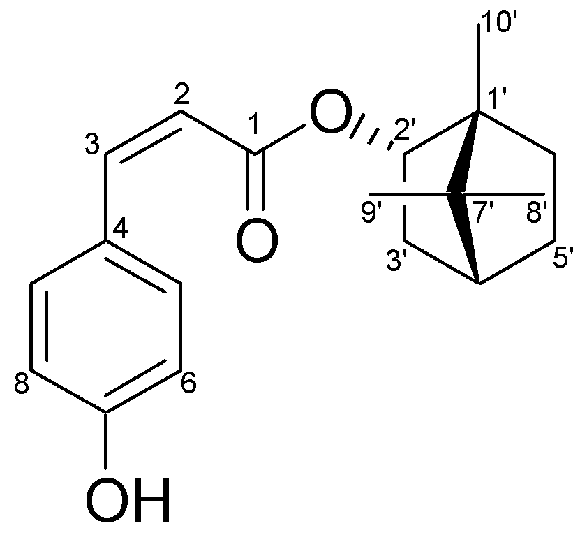
2.1. Characterization of the Constituents of the Ethyl Acetate (EA) Fraction of P. betle Stems

One monoterpene, bornyl cis-4-hydroxycinnamate, was afforded from the ethyl acetate fraction of P. betle stems, and its chemical structure was confirmed by comparing its physical and spectral data (specific rotation, mass spectrometry (MS), and nuclear magnetic resonance (NMR) spectroscopy) with the values described in the literature [16]. Bornyl cis-4-hydroxycinnamate (Figure 1): amorphous white powder; [α]27 D = +16.1° (c 0.5, CHCl3); 1H-NMR (400 MHz, CDCl3) δ 0.84 (3H, s, H-10′), 0.86 (3H, s, H-8′), 0.91 (3H, s, H-9′), 1.03 (1H, dd, J = 13.6, 3.2 Hz, H-3′b), 1.19 (1H, m, H-5′b), 1.24 (1H, m, H-6′b), 1.67 (1H, t, J = 4.4 Hz, H-4′), 1.72 (1H, m, H-5′a), 1.87 (1H, m, H-6′a), 2.41 (1H, m, H-3′a), 4.90 (1H, br d, J = 10 Hz, H-2′), 5.85 (1H, d, J = 12.8 Hz, H-2), 6.74 (2H, d, J = 8.4 Hz, H-6, H-8), 6.85 (1H, d, J = 12.8 Hz, H-3), 7.53 (2H, d, J = 8.8 Hz, H-5, H-9); 13C-NMR (CDCl3) δ: 13.5 (C-10′), 18.7 (C-9′), 19.6 (C-8′), 27.0 (C-6′), 27.9 (C-5′), 36.6 (C-3′), 44.8 (C-4′), 47.7 (C-7′), 48.7 (C-1′), 80.4 (C-2′), 115.2 (C-6, C-8), 117.0 (C-2), 126.8 (C-4), 132.0 (C-5, C-9), 144.0 (C-3), 157.3 (C-7), 168.0 (C-1); infrared spectroscopy (IR) (KBr) νmax: 3370, 1685, 1605, 1515, 1155 cm−1; electron-impact (EI) MS (70 eV) m/z (relative intensity) 300 [M]+ (10), 164 (5), 147 (100), 136 (3), 119 (20), 91 (15), 81 (6), 69 (3), 55 (4). NMR spectroscopy and mass spectrometry (MS).

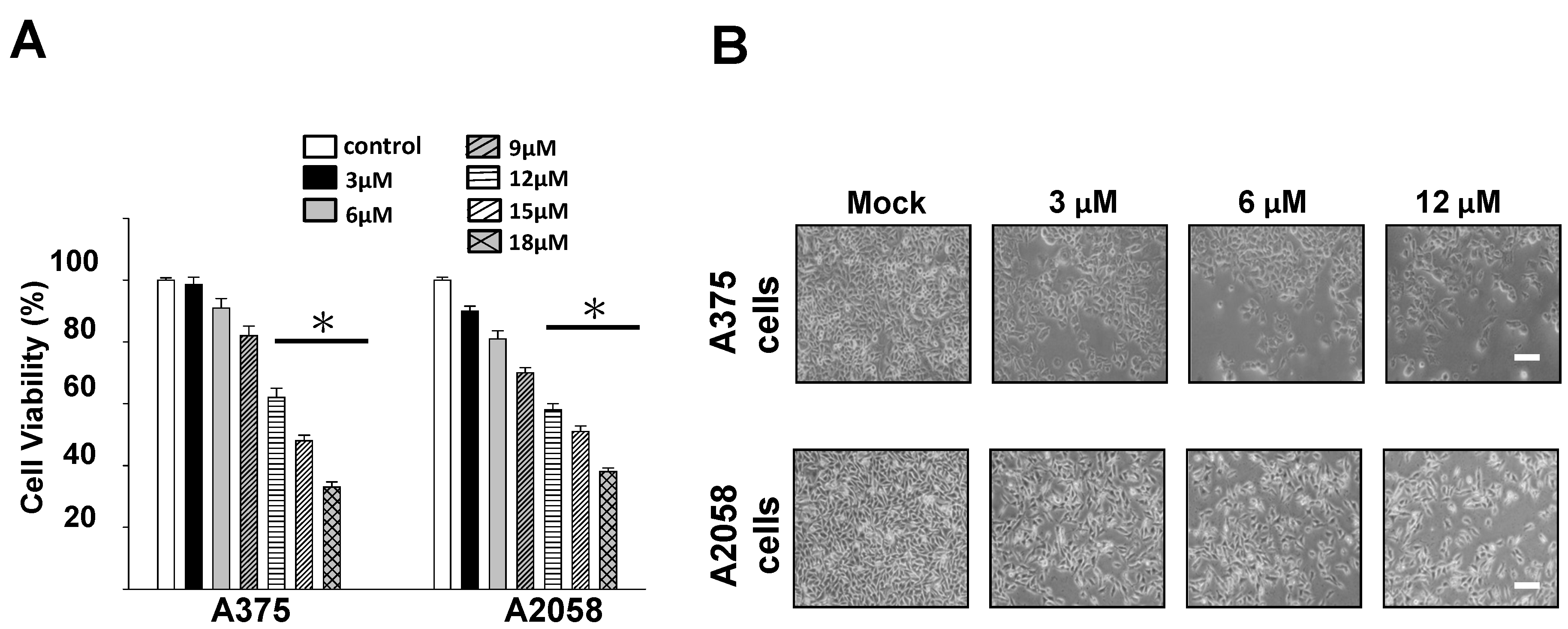
2.2. Cytotoxic and Antiproliferative Effects of Bornyl cis-4-Hydroxycinnamate on Melanoma Cells

To examine the potential cytotoxic effects of bornyl cis-4-hydroxycinnamate on melanoma cells (A2058 and A375 cells), 3-(4,5-Dimethylthiazol-2-yl)-2,5-diphenyltetrazolium bromide (MTT) assays and analyses of the morphological changes were performed. The cytotoxic effect of various concentrations of bornyl cis-4-hydroxycinnamate (3, 6, 9, 12, 15, and 18 µM) on A2058 and A375 melanoma cells was examined, and the results of the MTT assays revealed that bornyl cis-4-hydroxycinnamate reduced the proliferation of the two melanoma cell lines in a concentration-dependent manner (Figure 2A). With 12 µM bornyl cis-4-hydroxycinnamate treatment, the cell number of A2058 and A375 cells was significantly reduced (Figure 2B).

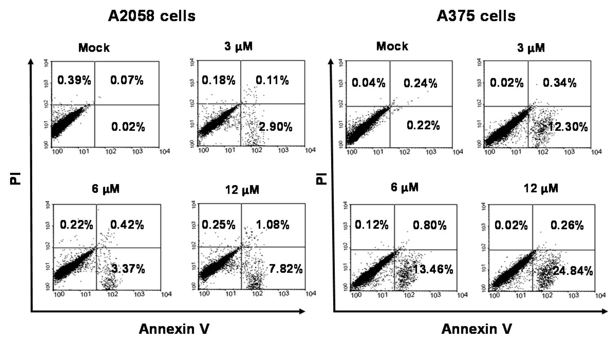
2.3. Apoptosis Is Triggered by Bornyl cis-4-Hydroxycinnamate in Melanoma Cells

Figure 2 shows that bornyl cis-4-hydroxycinnamate inhibited A2058 and A375 cell proliferation in a concentration-dependent manner. Apoptotic assays were then performed to investigate whether bornyl cis-4-hydroxycinnamate also induced melanoma cell apoptosis. After exposure to bornyl cis-4-hydroxycinnamate, flow cytometry analysis of annexin V-fluorescein isothiocyanate (FITC)/propidium iodide (PI) double stained cells showed that the mock treatment and the bornyl cis-4-hydroxycinnamate treatment at 3, 6, and 12 μM led A2058/A375 cells to display early apoptosis, at levels of 0.02%/0.22%, 2.9%/12.3%, 3.37%/13.46%, and 7.82%/24.84%, respectively (Figure 3). These results demonstrated that bornyl cis-4-hydroxycinnamate efficiently induced early apoptosis in A2058 and A375 melanoma cells.

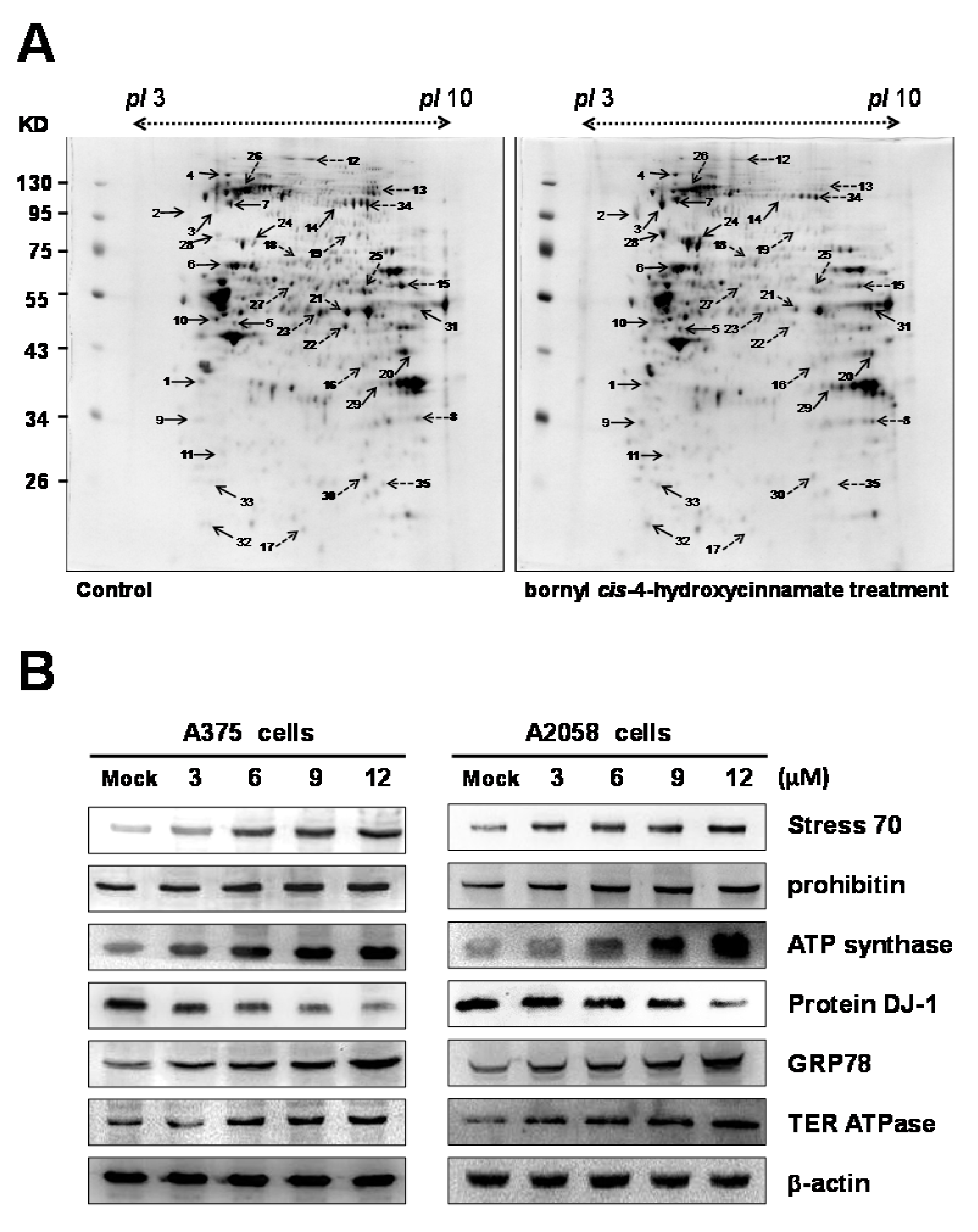
2.4. Proteomic Analysis of Bornyl cis-4-Hydroxycinnamate-Treated A375 Cells

Following 12 μM bornyl cis-4-hydroxycinnamate treatment for 24 h, proteomic analysis was used to identify critical proteins involved in its cellular effect in A375 cells. Two-dimensional electrophoresis (2-DE) analysis with PDQuest image analysis software (Bio-Rad, Hercules, CA, USA) indicated that several proteins exhibited an intensity difference greater than 1.5-fold in the 2-DE maps. After gel digestion, liquid chromatograph (LC)-MS/MS analysis and MASCOT search engine v2.3 (Matrix Science, London, UK) protein identification demonstrated that a total of 35 differential protein spots were significantly altered between the control and the bornyl cis-4-hydroxycinnamate-treated A375 cells. In Table 1, the 35 identified proteins, with their detailed information, including calculated Mw (molecular weight)/pI (isoelectric point), MS/MS matched sequence coverage, MASCOT scores, fold changes (up-/downregulation), cellular location, and protein function, are presented. Among the 35 differentially expressed proteins, 18 were upregulated after bornyl cis-4-hydroxycinnamate treatment, i.e., nucleophosmin, hypoxia-upregulated protein 1, sodium/potassium-transporting ATPase subunit α-1, Hsp90, ATP synthase subunit β, vimentin, transitional endoplasmic reticulum ATPase (TER ATPase), proliferating cell nuclear antigen, tubulin β-4, prohibitin, glycogen phosphorylase, stress 70, fructose-bisphosphate aldolase A, glucose-regulated protein 78 (GRP78), glyceraldehyde-3-phosphate dehydrogenase (GAPDH), RNA-binding protein 8A, and Ran-specific GTPase-activating protein. On the other hand, 17 proteins were downregulated after bornyl cis-4-hydroxycinnamate treatment, i.e., voltage-dependent anion-selective channel protein, clathrin heavy chain 1, C-1-tetrahydrofolate synthase, serine hydroxymethyltransferase, poly(rC)-binding protein 2, protein DJ-1, prelamin-A/C, bifunctional UDP-N-acetylglucosamine 2-epimerase/N-acetylmannosamine kinase, α-enolase, elongation factor thermo unstable (EF-Tu), dihydrolipoyl dehydrogenase, heat-shock protein 105 kDa, tryptophan-tRNA ligase, triosephosphate isomerase, elongation factor 2 and 3-hydroxyacyl-CoA (coenzyme A) dehydrogenase type-2.
Differential proteins, such as stress 70, prohibitin, protein DJ-1, GRP78, ATP synthase, and TER ATPase were found to be associated with the induction of apoptosis and acted against cell proliferation. Western blotting was used to validate the changed profiles of these proteins. Altogether, proteomic analysis of 2-DE data and western blotting were in agreement in terms of the trends of changes of these identified proteins (Figure 4B).

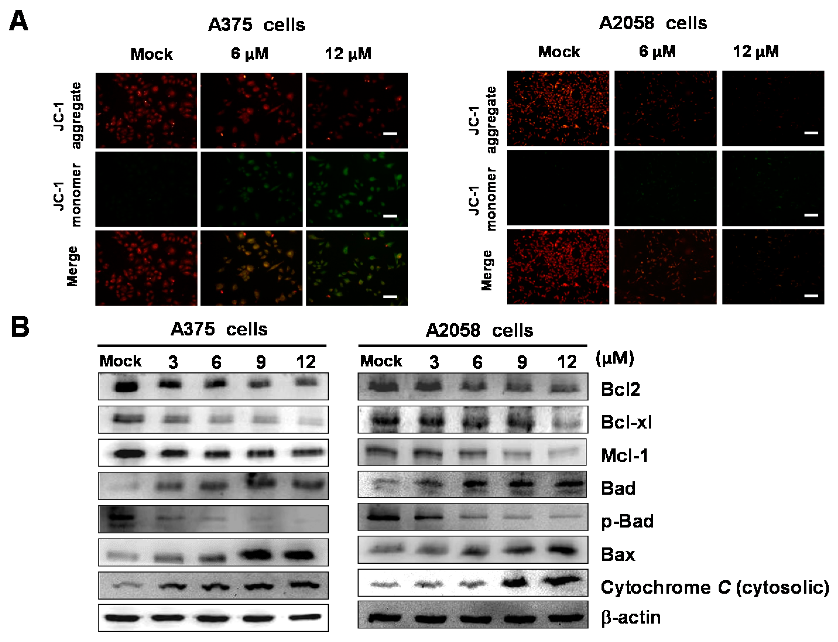
2.5. Bornyl cis-4-Hydroxycinnamate Activates the Apoptosis Pathway through the Induction of Mitochondrial Depolarization

As per the aforementioned results showing that bornyl cis-4-hydroxycinnamate induced apoptosis, we also noted that several mitochondrial-related proteins were identified in the bornyl cis-4-hydroxycinnamate-treated cells from 2-DE analysis, including prohibitin, stress 70, and ATP synthase subunit β. These proteins are known to be involved in energy production. We then employed the tetraethylbenzimidazolylcarbocyanine iodide (JC)-1 dye to measure the loss of mitochondrial membrane potential (Δψm) in A2058 and A375 cells after bornyl cis-4-hydroxycinnamate treatment. Fluorescence microscopy showed that bornyl cis-4-hydroxycinnamate-treated cells had a lower intensity of red fluorescence and a higher intensity of green florescence due to Δψm loss (Figure 5A). The mitochondrial-mediated apoptosis pathway plays an important role in apoptosis. We further analyzed molecules involved in the mitochondrial-mediated apoptosis pathway by examining the expression of related protein markers, including Bcl-2, Bcl-xl, Mcl-1, Bad, p-Bad, Bax, and cytosolic cytochrome C. As shown in Figure 5B, bornyl cis-4-hydroxycinnamate significantly increased the expression levels of Bad, Bax, and cytosolic cytochrome C in A2058 and A375 cells. In contrast, the levels of other marker proteins, such as Bcl-2, Bcl-xl, Mcl-1, and p-Bad, were decreased after bornyl cis-4-hydroxycinnamate treatment.

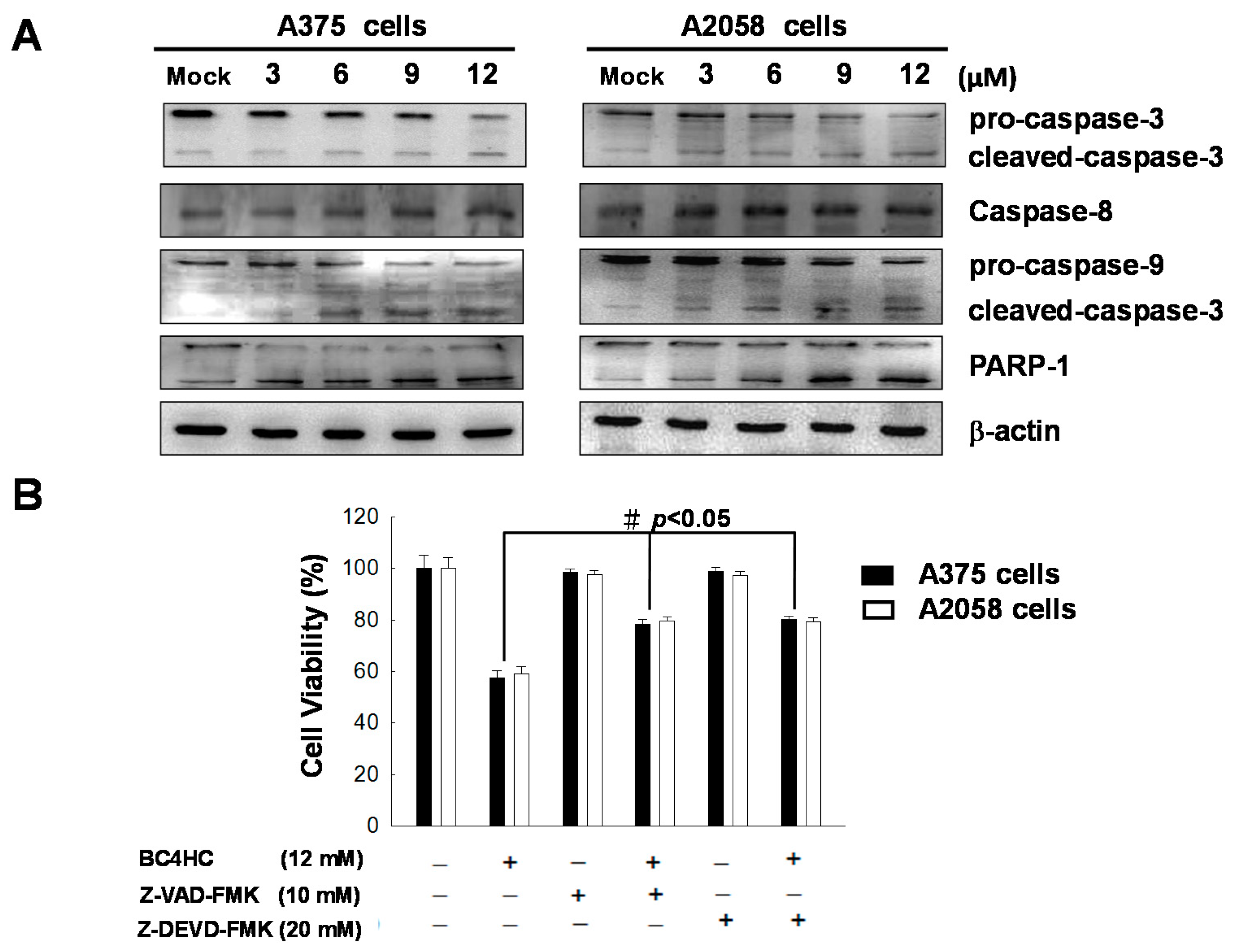
2.6. Bornyl cis-4-Hydroxycinnamate Activates the Caspase-Dependent Pathway Leading to Cell Apoptosis

To confirm the role of caspase in bornyl cis-4-hydroxycinnamate-induced apoptosis, the levels of caspase-3, caspase-8, caspase-9, and poly(ADP-ribose) polymerase (PARP)-1 were used to evaluate the status of caspase and its downstream effectors after bornyl cis-4-hydroxycinnamate treatment. As shown in Figure 6A, the expression of cleaved caspase-3 (17 kDa large subunit), cleaved caspase-9 (37 kDa large subunit), and cleaved PARP (89 kDa carboxyterminal catalytic domain) was increased in bornyl cis-4-hydroxycinnamate-treated A2058 and A375 melanoma cells, while the levels of pro-caspase-3 and pro-caspase-9 were decreased after bornyl cis-4-hydroxycinnamate treatment; the expression levels of caspase-8 were unchanged. These observations suggested that bornyl cis-4-hydroxycinnamate treatment activated the caspase-dependent pathway. To confirm whether the activation of the caspase cascade is crucial in bornyl cis-4-hydroxycinnamate-induced cell apoptosis, we used inhibitors against caspase-3 (Z-DEVD-FMK) and caspase-9 (Z-VAD-FMK). As shown in Figure 6B, cell death was suppressed in cells treated with either of these two inhibitors, suggesting that bornyl cis-4-hydroxycinnamate-induced apoptosis is mediated by caspase-3 and caspase-9 in melanoma cells.

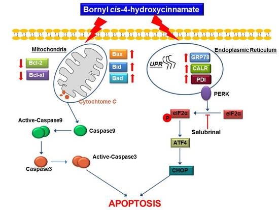
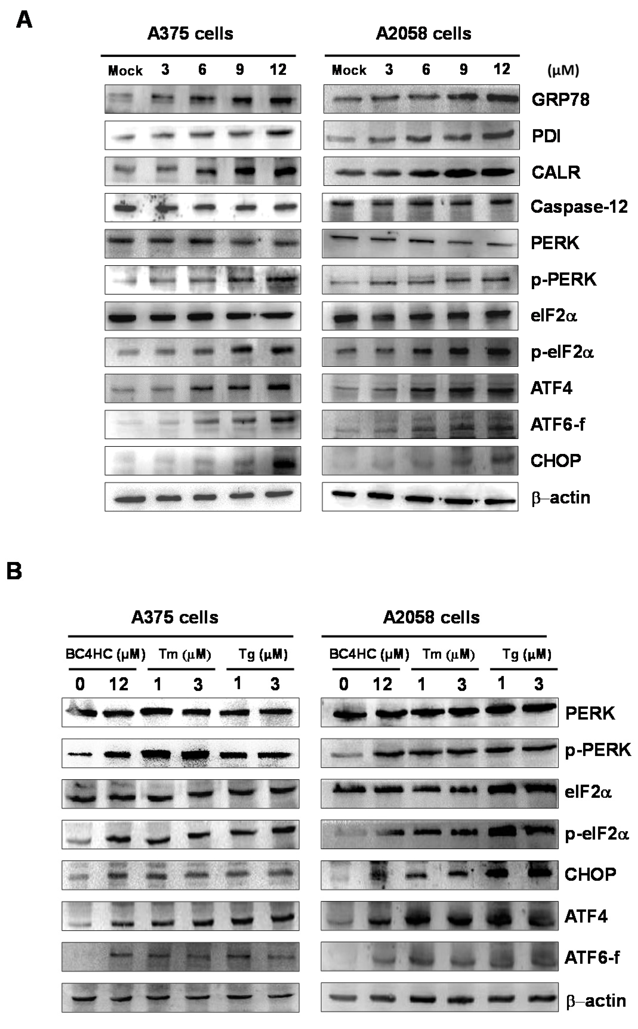
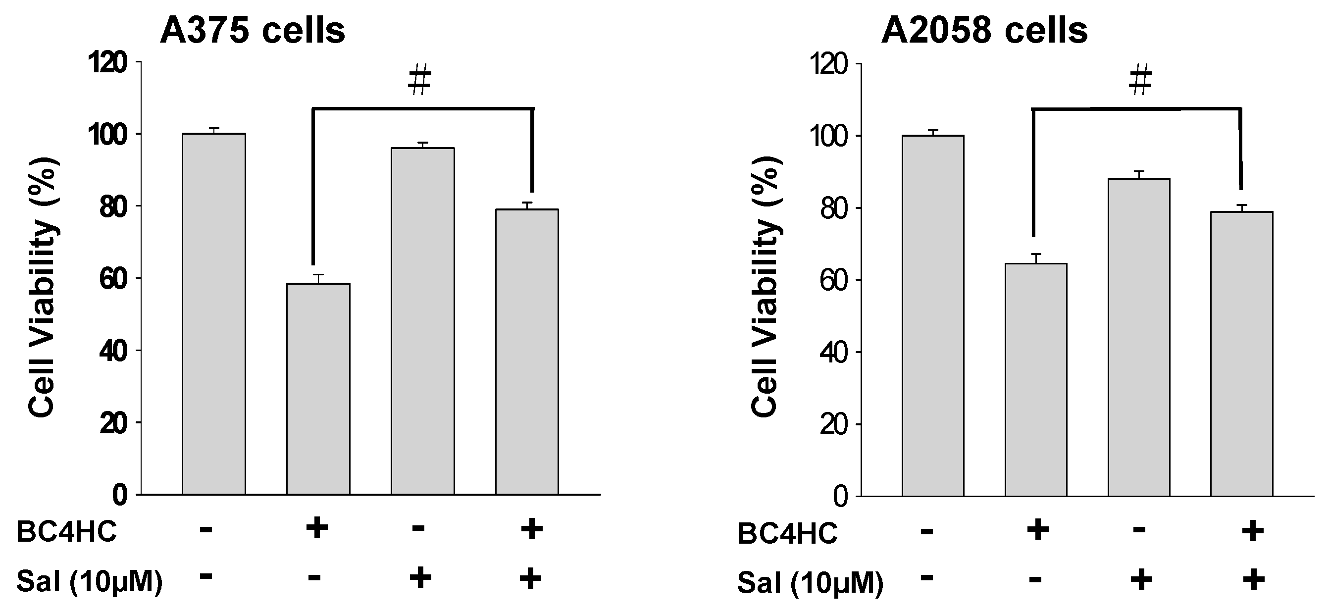
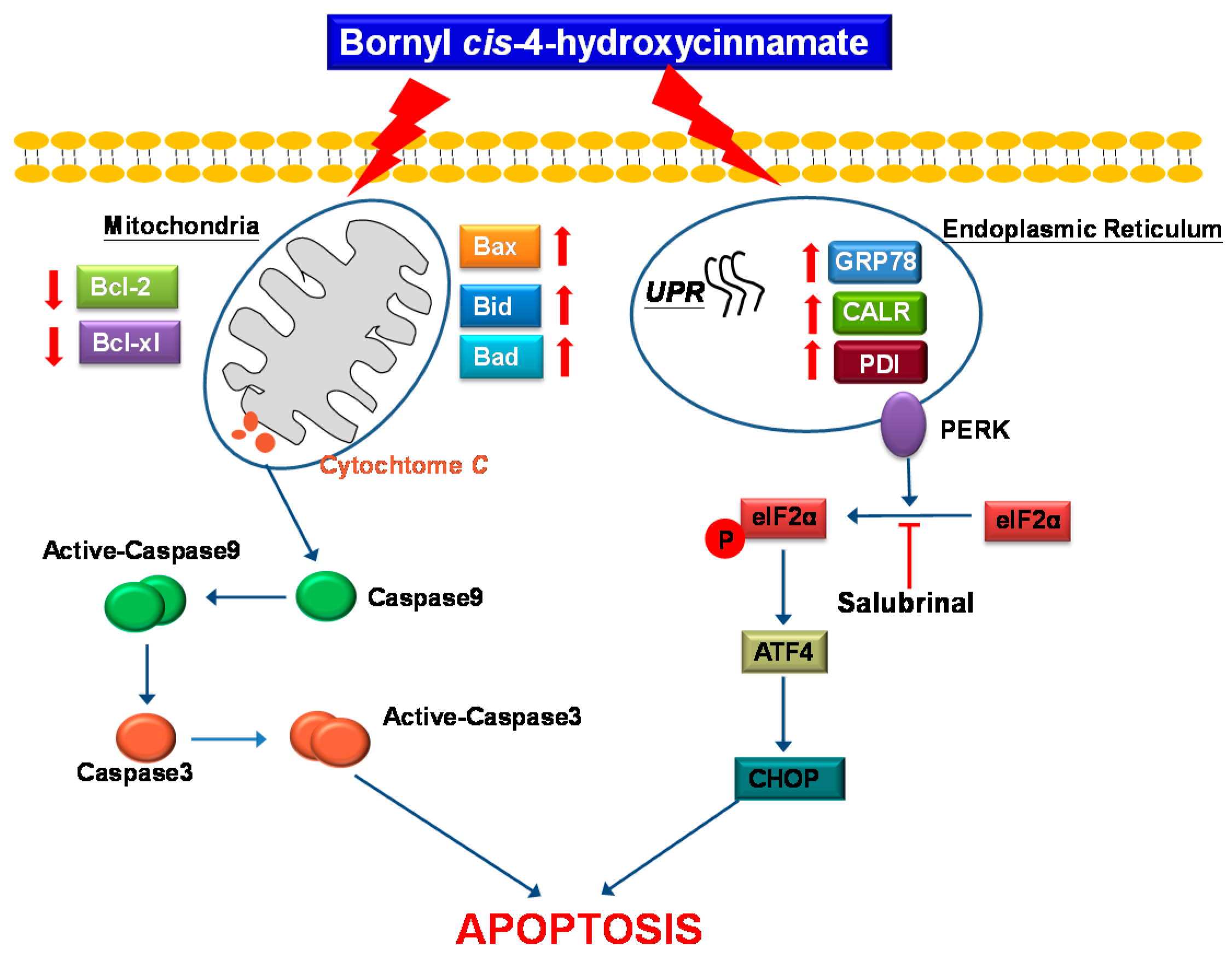
2.7. Bornyl cis-4-Hydroxycinnamate Treatment Induces the Endoplasmic Reticulum (ER) Stress Pathway

Next, we investigated whether the ER stress pathway was involved in bornyl cis-4-hydroxycinnamate-induced apoptosis in melanoma cells. Using western blot analysis, the expression levels of ER-related proteins GRP78 and TER ATPase were found to increase in the melanoma cells after bornyl cis-4-hydroxycinnamate treatment in a dose-dependent manner (Figure 4B). We then further verified the expression of three ER-resident transmembrane sensor proteins, i.e., endoribonuclease inositol-requiring enzyme 1α (IRE1α), protein kinase RNA-like endoplasmic reticulum kinase (PERK), and activating transcription factor 6 (ATF6), as well as of caspase-12 by western blot. The results showed that the expression of IRE1α and caspase-12 was unchanged after bornyl cis-4-hydroxycinnamate treatment. Under ER stress, endogenous ATF6 (p90 ATF6) is cleaved into a 50 KDa fragment transcription factor (p50 ATF6) and enters the nucleus to activate the GRP78 genes [16]. Our results showed that p-PERK and p-eIF2α expression increased after bornyl cis-4-hydroxycinnamate treatment. The level of transcription factor ATF4, a downstream target of PERK in the PERK–eIF2α pathway, was also increased after treatment in A2058 and A375 melanoma cells. Additionally, the expression and nuclear translocation of ER stress-induced CCAAT/enhancer-binding protein (C/EBP)-homologous protein (CHOP) was also increased by bornyl cis-4-hydroxycinnamate treatment (Figure 7A).
Bornyl cis-4-hydroxycinnamate increased the ER stress-related protein expression levels similar to the two ER stress inducer agents tunicamycin (Tm) and thapsigargin (Tg), used to monitor the ER stress response. These results imply that bornyl cis-4-hydroxycinnamate-triggered cell apoptosis is mediated by the ER stress pathway in A2058 and A375 melanoma cells (Figure 7B).
To further demonstrate that bornyl cis-4-hydroxycinnamate-induced apoptosis occurs through ER stress-related pathways as described above, salubrinal (an inhibitor) was tested to examine PERK-activated cell apoptosis upon bornyl cis-4-hydroxycinnamate treatment. An increase in cell viability from 64% to 78% was observed in bornyl cis-4-hydroxycinnamate-treated A2058 and A375 cells following treatment with salubrinal at a concentration of 10 μM (Figure 8).

3. Discussion

In the current study, we investigated the cytotoxic effect of bornyl cis-4-hydroxycinnamate, isolated from P. betel stems, and studied the signaling pathways involved in the apoptosis induced by bornyl cis-4-hydroxycinnamate in melanoma cells. Our results demonstrate that bornyl cis-4-hydroxycinnamate inhibited cell proliferation (Figure 2) and increased early apoptosis rates (Figure 3) in A2058 and A375 melanoma cells, in a concentration-dependent manner. These findings support the conclusion that bornyl cis-4-hydroxycinnamate possesses activity against cell proliferation and may induce apoptosis in melanoma cells.
The molecular mechanism involved in bornyl cis-4-hydroxycinnamate-induced apoptosis in A375 melanoma cells was elucidated from proteomic data, which showed significant changes in the expression of several crucial proteins, including sodium/potassium-transporting ATPase subunit α-1, ATP synthase subunit β, prohibitin, stress 70 protein, dihydrolipoyl dehydrogenase, and 3-hydroxyacyl-CoA dehydrogenase type-2, which are associated with mitochondrial function or apoptosis in melanoma cells. Prohibitin is located in the mitochondria and is known to regulate apoptosis, cellular signaling, cell migration, and cell proliferation and to stabilize mitochondria proteins [17,18,19,20]. The translocation of prohibitin to mitochondria together with p53 has been shown to be highly correlated with the suppression of cancer growth [21,22]. Therefore, the antiproliferative effects of bornyl cis-4-hydroxycinnamate are likely mediated by the mitochondria in A2058 and A375 cells and associated with the enhancement of prohibitin.
The apoptosis induced by bornyl cis-4-hydroxycinnamate is also a good indicator of the potential success of this molecule in the treatment of melanoma, as the development of anticancer drugs has often been based on the apoptosis effect as a potential mechanism of chemotherapy [23]. The mechanisms of apoptosis can be subdivided into two major pathways, i.e., the extrinsic and the intrinsic pathways. The former is also called the death receptor pathway, and the latter is also known as the mitochondrial pathway [24]. A study has suggested that the two pathways are related and influence each other [25], and molecules in the intrinsic pathway are located in either the ER or the mitochondria [26]. In addition, increased expression levels of the pro-apoptotic Bax and Bak proteins and decreased expression levels of the anti-apoptotic Bcl-2, Bcl-xl, and Mcl-1 proteins are known to play important roles in the intrinsic pathway and are associated with changes in the mitochondrial membrane potential and with the release of mitochondrial apoptotic factors [27]. The collapse of Δψm subsequently leads to cytochrome C release into the cytosol, resulting in caspase-9 activation, and further activates the downstream effector caspase-3. This process then causes poly(ADP-ribose) polymerase cleavage [28]. Existing information and our findings showing decreased Δψm and cytochrome C release from the mitochondria (Figure 5B) suggest that bornyl cis-4-hydroxycinnamate-induced apoptosis in A2058 and A375 cells occurs via the mitochondrial pathway. Additionally, the suppression of the anti-apoptotic Bcl-2, Bcl-xl, and Mcl-1 proteins and the augmented expression of the pro-apoptotic Bad and Bax proteins (Figure 5B), as well as the activation of caspase-3, caspase-9, and PARP (Figure 6), indicate that the caspase activation cascades are crucial in bornyl cis-4-hydroxycinnamate-induced apoptosis in A2058 and A375 cells.
The ER plays many important roles in regulating essential cellular functions, including proper protein folding, protein synthesis, and calcium homeostasis [29]. When too many unfolded proteins accumulate in the ER, the homeostasis of the ER is destroyed, causing stress and leading to cells activating a self-rescue program or triggering apoptosis. In the presence of stress, the ER may react in various ways, including the induction of ER-associated degradation (ERAD), of the unfolded protein response (UPR), and of apoptosis [30,31]. Continued and severe ER stress increases the UPR, which will trigger apoptosis [32]. The UPR process is regulated by three ER signaling sensors, named protein kinase RNA-like endoplasmic reticulum kinase (PERK), inositol requiring enzyme 1-α (IRE1-α), and activating transcription factor 6 (ATF6). When unfolded proteins accumulate in the ER, chaperones, such as GRP78/GRP94, will release the transmembrane proteins IRE1-α, PERK, and ATF6, resulting in ER dysfunction. The PERK pathway either leads to cell survival controlled by autophagy [33] or to apoptosis by increasing ATF4/CHOP [34]. In the presence of ER stress, the cells activate a self-rescue program to trigger the UPR, which prevents cell death. Persistent ER stress initiates caspase-dependent apoptosis, leading to cell death [35]. When cells are under oxidative stress, unfolded or misfolded proteins accumulate in the ER, causing ER stress, and this imposed ER stress subsequently causes the UPR to alleviate the stress and restore ER homeostasis. The process upregulates chaperone GRP78 and disulfide isomerase protein, which promote protein folding and alleviate protein aggregation, and modulates calreticulin activity for the storing of calcium in the ER [36,37]. Our immunostaining results demonstrated that the levels of GRP78 and calreticulin were increased in the presence of a high concentration of bornyl cis-4-hydroxycinnamate, indicating that these proteins were activated to restore normal protein folding and reduce ER stress. If the UPR is insufficient to relieve cells from ER stress, the PERK, IRE1-α, and ATF6 pathways may activate downstream proteins to induce apoptosis in order to destroy the ER stress-damaged cells [38]. It was observed that PERK activates eIF2α, ATF4, and CHOP, causing apoptosis. The immunostaining results of this study also showed that the levels of p-PERK, p-eIF2α, and downstream ATF4 were increased, as was the expression of ATF6. These responses led to the activation of CHOP and increased its expression to induce apoptosis. The level of caspase-12 expression did not change, suggesting that the apoptotic mechanism in our study was mediated by the PERK–eIF2α–ATF4–CHOP signaling pathways in the ER (Figure 7A). In order to verify our hypothesis that bornyl cis-4-hydroxycinnamate-induced apoptosis in melanoma cells is associated with ER stress, we used ER stress agonists, namely, Tm and Tg (1 and 3 μM, respectively), to treat the cells. The results showed that the treatment with increasing concentrations of Tm and Tg resulted in trends of the expression of PERK, p-PERK, eIF2α, p-eIF2α, CHOP, ATF4, and ATF6 consistent with those of cells treated with bornyl cis-4-hydroxycinnamate. This finding suggests that bornyl cis-4-hydroxycinnamate-induced apoptosis occurs through a mechanism that involves ER stress (Figure 7B). We also used salubrinal, an eIF2α inhibitor, to further confirm our hypothesis. An MTT assay showed that the survival rate of cells treated with bornyl cis-4-hydroxycinnamate and salubrinal together was significantly higher than that of cells treated with bornyl cis-4-hydroxycinnamate alone (Figure 8). In conclusion, the results indicate that bornyl cis-4-hydroxycinnamate initiated ER stress and subsequently induced apoptosis in melanoma cells.
Overall, our results demonstrate that bornyl cis-4-hydroxycinnamate-induced apoptosis in A2058 and A375 cells is mediated by a dysfunction of the mitochondrial pathway, the activation of caspase cascades, and ER stress.

4. Materials and Methods

4.1. Reagents

Bornyl cis-4-hydroxycinnamate was isolated from P. betel stem and dissolved in DMSO at a concentration of 100 mM as a stock solution and stored at −20 °C. The compounds 3-(4,5-dimethylthiazol-2-yl)-2,5-diphenyltetrazolium bromide (MTT), Z-DEVD-FMK (caspase-3 inhibitor), Z-VAD-FMK (caspase-9 inhibitor), tunicamycin (Tm), thapsigargin (Tg), salubrinal, and dimethyl sulfoxide (DMSO), the protease inhibitor cocktail, and rabbit anti-human β-actin antibodies were purchased from Sigma (St Louis, MO, USA). The chemiluminescent horseradish peroxidase (HRP) substrate was obtained from Pierce (Rockford, IL, USA). Isoelectric focusing (IEF) strips and immobilized pH gradient (IPG) buffer were obtained from GE Healthcare (Buckinghamshire, UK). Dulbecco’s modified Eagle’s medium was obtained from Biowest (Nuaillé, France). An annexin V/FITC Apoptosis Detection kit was purchased from Pharmingen (San Diego, CA, USA). The cationic dye JC-1 was purchased from Biotium (Hayward, CA, USA). Rabbit anti-human cytochrome C, PARP-1, pro-casapse-3, cleaved caspase-3, pro-caspase-8, pro-caspase-9, cleaved caspase-9, caspase-12, Bax, Bad, p-Bad, Bcl-2, Bcl-xl, Mcl-1, PDI, calreticulin, ATP synthase, stress 70, GRP78, prohibitin, protein DJ-1, transitional endoplasmic reticulum ATPase, IRE1α, PERK, p-PERK, eIF2α, p-eIF2α, ATF4, ATF6-f, and CHOP antibodies were obtained from Cell Signaling Technology (Danvers, MA, USA). Goat anti-rabbit and horseradish peroxidase-conjugated IgG was purchased from Millipore (Bellerica, MA, USA).

4.2. General Instrumental Operation for the Isolation and Identification of Compounds

NMR spectra were analyzed in CDCl3 at room temperature using a Varian Mercury plus 400 NMR spectrometer, and the solvent resonance was used as the internal shift reference (tetramethyl silane [TMS] as standard). EI–MS were recorded on a SX-102A mass spectrometer (JEOL USA, Inc., Peabody, MA, USA). Thin-layer chromatography was used on silica gel 60 F254 plates (Merck KGaA, Darmstadt, Germany), and the spots were visualized by spraying with 10% H2SO4 solution. Silica gels (230–400 mesh ASTM, Merck KGaA) were used for column chromatography. Semi-preparative HPLC was performed using LiChrosorb Si 60 column, (7 μm, 250 × 10 mm; (Merck KGaA) on a LDC Analytical-III system.

4.3. Source, Extraction, Fractionation, and Purification of P. betle Stem Compounds

Stems of P. betle were collected in Pingtung County, Taiwan. Air-dried stems of P. betle (3 kg) were ground using an electric high-speed grinder until a fine powder that passed through a 10-mesh sieve was obtained, and the powder was extracted with methanol (solid/solvent ratio = 1:5) at room temperature three times (7 d each). The filtrates were pooled and dried under reduced pressure to allow the solvent to evaporate. The dry crude residue was then suspended in water before being successively partitioned with ethyl acetate and n-butanol.
The ethyl acetate fraction (65 g) was separated into 24 fractions on a silica gel column (5 × 60 cm), using step-gradient elution with solvent mixtures of n-hexane and ethyl acetate of increasing polarity as eluents. Fraction 10 (4.2 g) was further chromatographed on a silica gel column (3 × 45 cm) using step-gradient elution with CH2Cl2–EtOAc (99:1 to 1:1) to give six fractions (each about 400 mL), 10 A–10 F. Semipreparative HPLC was used to separate Fr. 10 C by using n-hexane–CH2Cl2–EtOAc (16:6:1) to elute the sample (32.1 mg; Figure 1).

4.4. Cell Lines and Cell Culture Conditions

A2058 and A375 melanoma cells were obtained from the Food Industry Research and Development Institute (Hsinchu, Taiwan). A2058 and A375 melanoma cells were grown in Dulbecco’s modified Eagle’s medium (DMEM) supplemented with 10% (v/v) fetal bovine serum, 1 mM sodium pyruvate, 4 mM l-glutamine, 100 μg/mL streptomycin and 100 U/mL penicillin in a humidified incubator at 37 °C with 5% CO2.

4.5. Cytotoxicity Assessment

A2058 and A375 cells were seeded in 96-well plates at a density of 1 × 10⁵/well with 200 μL of Dulbecco’s modified Eagle medium (DMEM) and 10% fetal bovine serum in a humidified, 5% CO2 atmosphere at 37 °C. The cells in each well were treated with bornyl cis-4-hydroxycinnamate (at concentrations of 0, 3, 6, 9, 12, 15, and 18 μM) for 24 h. Cells treated with DMSO without bornyl cis-4-hydroxycinnamate were used as a control. After a 24 h incubation period, 50 μL of MTT (0.5 mg/mL stock) was added to the cells, followed by incubation for 2 h. The medium was then removed, and DMSO (200 μL) was added to dissolve the formazan. The optical density (OD) was determined at 595 nm on an ELISA reader (Bio-Rad, Hercules, CA, USA) to provide a relative estimate of cell viability. All experiments were performed in at least triplicate to confirm their reproducibility.

4.6. Flow Cytometric Analysis

To analyze apoptosis in A2058 and A375 melanoma cells after bornyl cis-4-hydroxycinnamate treatment, the cells were stained with an annexin V-FITC apoptosis staining kit (Pharmingen, San Diego, CA, USA) and analyzed as described in previous reports [39,40]. Briefly, 1 × 10⁶ cells were seeded into 5 cm Petri dishes and treated with different concentrations of bornyl cis-4-hydroxycinnamate (3, 6, and 12 μM) for 24 h. Then, the cells were harvested and stained with annexin V-FITC/propidium iodide (PI) in a cell culture incubator for 30 min. Samples were then assessed using a FACScan flow cytometer (Becton-Dickinson, Mansfield, MA, USA), and the data were analyzed using FlowJo software v10.4.2 (TreeStar, Inc., Ashland, OR, USA).

4.7. Protein Lysate Preparation

After bornyl cis-4-hydroxycinnamate treatment (0, 3, 6, 9 and 12 μM) for 24 h, A2058 and A375 cells were lysed with a cell extraction buffer (BioSource International, Camarillo, CA, USA). The supernatants were quantified using a Bio-Rad protein assay for sodium dodecyl sulfate polyacrylamide gel electrophoresis (SDS-PAGE). In addition, some of each supernatant was precipitated using a 10% cold trichloroacetic acid (TCA)/acetone solution containing 20 mM dithiothreitol (DTT) overnight at −20 °C. The protein pellet was dissolved in a rehydration buffer (2 M thiourea, 6 M urea, 20 mM DTT, 0.5% IPG buffer, 0.5% 3-[(3-cholamidopropyl)dimethylammonio]-1-propanesulfonate (CHAPS), and 0.002% bromophenol blue) at 4 °C, and the protein concentrations were determined using a 2-D Quant Kit (GE Healthcare) for 2-DE analysis.

4.8. Two-Dimensional Gel Electrophoresis and Differential Proteomic Analyses

For two-dimensional gel electrophoresis (2-DE), protein samples (200 μg) from the control cells and the cells treated with 12 μM bornyl cis-4-hydroxycinnamate were analyzed using a GE Healthcare Ettan IPGphor 3. For second-dimension electrophoresis, the equilibrated strip was placed onto the top of a 12.5% SDS-PAGE gel and run on an SE 600 Ruby Vertical electrophoresis system (Hoefer, Holliston, MA, USA). Each sample was run in triplicate, and the spots on the 2-DE gels were visualized by silver staining. The gels were scanned and then analyzed using PDQuest image analysis software v8.0.1 (Bio-Rad). Each triplicate sample was normalized prior to statistical analysis. The protein spots with an intensity difference between the control and bornyl cis-4-hydroxycinnamate-treated cells greater than 1.5-fold were identified, and statistically significant differences in 2-DE between 11-epi-sinulariolide acetate-treated HA22T cells and the control [41] were identified. The protein spots of interest were excised from the 2-DE gels, and in-gel digestion was performed with trypsin. The digested samples were analyzed by LC/MS/MS using a QTRAP 5500Q mass spectrometer (AB Sciex, Framingham, MA, USA). MS scanning ranged from m/z 100 to1000. The raw MS data were converted into the text file format WIFF using Analyst 1.5.1 (AB Sciex, Framingham, MA, USA).

4.9. Assessment of the Mitochondrial Membrane Potential (Δψm)

The mitochondrial membrane potential (Δψm) was determined using cells stained with the cationic dye JC-1. Briefly, A2058 and A375 cells at 1 × 10⁵ cells/well in a 12-well plate were treated with different concentrations of bornyl cis-4-hydroxycinnamate (0, 3, 6, and 12 μM). The treated cells were collected and washed twice with PBS, incubated with 70 μL of JC-1 staining solution, and placed in a cell culture incubator for 30 min. Following washing with a buffer, the cells were then directly observed under a fluorescence microscope [42].

4.10. Antibody and Western Blot Analysis

Western blot analysis was performed according to previous reports. The primary antibodies used were: anti-human stress 70, GRP78, ATP synthase, prohibitin, protein DJ-1, Transitional endoplasmic reticulum ATPase, Bax, Bad, p-Bad, Mcl-1, Bcl-2, Bcl-xl, PARP-1, pro-caspase-3, cleaved-caspase-3, caspase-8, pro-caspase-9, cleaved-caspase-9, cytochrome C, PDI, calreticulin, IRE1α, caspase-12, PERK, p-PERK, elf2α, p-eIF2α, ATF4, ATF6-f, CHOP, and β-actin antibodies. Secondary antibodies (horseradish peroxidase-conjugated goat anti-rabbit, 1:5000 in blocking solution) were added, followed by an incubation for 2 h. The signals were visualized using a chemiluminesence detection kit (Pierce Biotechnology, Rockford, IL, USA).

4.11. Statistical Analysis

All experiments were repeated at least in triplicate. Data analysis was performed using Student’s t test (Sigma-Stat 2.0, San Rafael, CA, USA); p values < 0.05 were considered significant.

5. Conclusions

Our results established that bornyl cis-4-hydroxycinnamate extracted from P. betel stems possesses the ability to induce apoptosis in A2058 and A375 melanoma cells. A differential proteomic analysis identified several important mitochondrial and endoplasmic reticulum proteins and proved that mitochondrial dysfunction and the activation of endoplasmic reticulum stress pathways are key molecular mechanisms in the apoptosis induced by bornyl cis-4-hydroxycinnamate (Figure 9). Our findings provide evidence that bornyl cis-4-hydroxycinnamate has the potential to be developed as new therapeutic agent for melanoma treatment.

Author Contributions

M.-L.W. and Y.-J.W. conceived and designed the experiments. C.-I.C. performed the sample collections, extraction, isolation, and structures determination; the pharmacological experiments were carried out by T.-Y.Y. and Y.-J.W.; C.-C.C. contributed reagents and analysis tools; T.-Y.Y., M.-L.W., and Y.-J.W. participated in data interpretation, wrote the manuscript, and revised the paper.

Acknowledgments

This study was supported in part by a grant from the National Science Council Research (MOST 105-2313-B-276-001-MY3) by Y.-J.W.

Conflicts of Interest

The authors declare no conflict of interest.

References

  1. Parkin, D.M.; Bray, F.; Ferlay, J.; Pisani, P. Global cancer statistics, 2002. CA A Cancer J. Clin. 2005, 55, 74–108. [Google Scholar] [CrossRef]
  2. American Cancer Society. Melanoma Skin Cancer. Available online: https://www.cancer.org/cancer/melanoma-skin-cancer/about.html (accessed on 19 November 2017).
  3. Fuglede, N.; Brinck-Claussen, U.; Deltour, I.; Boesen, E.; Dalton, S.; Johansen, C. Incidence of cutaneous malignant melanoma in denmark, 1978–2007. Br. J. Dermatol. 2011, 165, 349–353. [Google Scholar] [CrossRef] [PubMed]
  4. Sasse, A.D.; Sasse, E.C.; Clark, L.; Ulloa, L.; Clark, O. Chemoimmunotherapy versus chemotherapy for metastatic malignant melanoma. Cochrane Database Syst. Rev. 2007, 24, CD005413. [Google Scholar] [CrossRef] [PubMed]
  5. Tawbi, H.A.; Buch, S.C. Chemotherapy resistance abrogation in metastatic melanoma. Clin. Adv. Hematol. Oncol. 2010, 8, 259–266. [Google Scholar] [PubMed]
  6. Chang, J. Cutaneous melanoma: Taiwan experience and literature review. Chang Gung Med. J. 2010, 33, 602–612. [Google Scholar] [PubMed]
  7. Essner, R. Surgical treatment of malignant melanoma. Surg. Clin. 2003, 83, 109–156. [Google Scholar] [CrossRef]
  8. Bleehen, N.; Newlands, E.; Lee, S.M.; Thatcher, N.; Selby, P.; Calvert, A.; Rustin, G.; Brampton, M.; Stevens, M. Cancer research campaign phase II trial of temozolomide in metastatic melanoma. J. Clin. Oncol. 1995, 13, 910–913. [Google Scholar] [CrossRef] [PubMed]
  9. Treisman, J.; Garlie, N. Systemic therapy for cutaneous melanoma. Clin. Plast. Surg. 2010, 37, 127–146. [Google Scholar] [CrossRef] [PubMed]
  10. Acquavella, N.; Kluger, H.; Rhee, J.; Farber, L.; Tara, H.; Ariyan, S.; Narayan, D.; Kelly, W.; Sznol, M. Toxicity and activity of a twice daily high-dose bolus interleukin 2 regimen in patients with metastatic melanoma and metastatic renal cell cancer. J. Immunother. 2008, 31, 569–576. [Google Scholar] [CrossRef] [PubMed]
  11. Ibrahim, N.; Haluska, F.G. Molecular pathogenesis of cutaneous melanocytic neoplasms. Annu. Rev. Pathol. Mech. Dis. 2009, 4, 551–579. [Google Scholar] [CrossRef] [PubMed]
  12. Ng, P.L.; Rajab, N.F.; Then, S.M.; Yusof, Y.A.M.; Ngah, W.Z.W.; Pin, K.Y.; Looi, M.L. Piper betle leaf extract enhances the cytotoxicity effect of 5-fluorouracil in inhibiting the growth of HT29 and HCT116 colon cancer cells. J. Zhejiang Univ. Sci. B 2014, 15, 692–700. [Google Scholar] [CrossRef] [PubMed]
  13. Rai, M.P.; Thilakchand, K.R.; Palatty, P.L.; Rao, P.; Rao, S.; Bhat, H.P.; Baliga, M.S. Piper betel Linn (betel vine), the maligned southeast asian medicinal plant possesses cancer preventive effects: Time to reconsider the wronged opinion. Asian Pac. J. Cancer Prev. 2011, 12, 2149–2156. [Google Scholar] [PubMed]
  14. Venkadeswaran, K.; Muralidharan, A.R.; Annadurai, T.; Ruban, V.V.; Sundararajan, M.; Anandhi, R.; Thomas, P.A.; Geraldine, P. Antihypercholesterolemic and antioxidative potential of an extract of the plant, Piper betle, and its active constituent, eugenol, in triton WR-1339-induced hypercholesterolemia in experimental rats. Evid.-Based Complement. Altern. Med. 2014, 2014, 478973. [Google Scholar] [CrossRef] [PubMed]
  15. Al-Adhroey, A.H.; Nor, Z.M.; Al-Mekhlafi, H.M.; Amran, A.A.; Mahmud, R. Antimalarial activity of methanolic leaf extract of Piper betle L. Molecules 2011, 16, 107–118. [Google Scholar] [CrossRef] [PubMed]
  16. Toyota, M.; Saito, T.; Matsunami, J.; Asakawa, Y. A comparative study on three chmo-type of the liverwort conocephalum conicum using volatile constituents. Phytochemistry 1997, 44, 1265–1270. [Google Scholar] [CrossRef]
  17. Haze, K.; Yoshida, H.; Yanagi, H.; Yura, T.; Mori, K. Mammalian transcription factor ATF6 is synthesized as a transmembrane protein and activated by proteolysis in response to endoplasmic reticulum stress. Mol. Biol. Cell 1999, 10, 3787–3799. [Google Scholar] [CrossRef] [PubMed]
  18. Fusaro, G.; Dasgupta, P.; Rastogi, S.; Joshi, B.; Chellappan, S. Prohibitin induces the transcriptional activity of p53 and is exported from the nucleus upon apoptotic signaling. J. Biol. Chem. 2003, 278, 47853–47861. [Google Scholar] [CrossRef] [PubMed]
  19. Nijtmans, L.G.; de Jong, L.; Sanz, M.A.; Coates, P.J.; Berden, J.A.; Back, J.W.; Muijsers, A.O.; van der Spek, H.; Grivell, L.A. Prohibitins act as a membrane-bound chaperone for the stabilization of mitochondrial proteins. EMBO J. 2000, 19, 2444–2451. [Google Scholar] [CrossRef] [PubMed]
  20. Rajalingam, K.; Wunder, C.; Brinkmann, V.; Churin, Y.; Hekman, M.; Sievers, C.; Rapp, U.R.; Rudel, T. Prohibitin is required for ras-induced Raf–MEK–ERK activation and epithelial cell migration. Nat. Cell Biol. 2005, 7, 837–843. [Google Scholar] [CrossRef] [PubMed]
  21. Winter, A.; Kämäräinen, O.; Hofmann, A. Molecular modeling of prohibitin domains. Proteins Struct. Funct. Bioinform. 2007, 68, 353–362. [Google Scholar] [CrossRef] [PubMed]
  22. Wong, P.-F.; Cheong, W.-F.; Shu, M.-H.; Teh, C.-H.; Chan, K.-L.; AbuBakar, S. Eurycomanone suppresses expression of lung cancer cell tumor markers, prohibitin, annexin 1 and endoplasmic reticulum protein 28. Phytomedicine 2012, 19, 138–144. [Google Scholar] [CrossRef] [PubMed]
  23. Yang, H.-B.; Song, W.; Chen, L.-Y.; Li, Q.-F.; Shi, S.-L.; Kong, H.-Y.; Chen, P. Differential expression and regulation of prohibitin during curcumin-induced apoptosis of immortalized human epidermal hacat cells. Int. J. Mol. Med. 2014, 33, 507–514. [Google Scholar] [CrossRef] [PubMed]
  24. Liu, C.-I.; Wang, R.Y.-L.; Lin, J.-J.; Su, J.-H.; Chiu, C.-C.; Chen, J.-C.; Chen, J.Y.-F.; Wu, Y.-J. Proteomic profiling of the 11-dehydrosinulariolide-treated oral carcinoma cells Ca9–22: Effects on the cell apoptosis through mitochondrial-related and ER stress pathway. J. Proteom. 2012, 75, 5578–5589. [Google Scholar] [CrossRef] [PubMed]
  25. Denicourt, C.; Dowdy, S.F. Targeting apoptotic pathways in cancer cells. Science 2004, 305, 1411–1413. [Google Scholar] [CrossRef] [PubMed]
  26. Igney, F.H.; Krammer, P.H. Death and anti-death: Tumour resistance to apoptosis. Nat. Rev. Cancer 2002, 2, 277. [Google Scholar] [CrossRef] [PubMed]
  27. Liao, C.-T.; Chang, J.T.-C.; Wang, H.-M.; Ng, S.-H.; Hsueh, C.; Lee, L.-Y.; Lin, C.-H.; Chen, I.-H.; Huang, S.-F.; Cheng, A.-J. Analysis of risk factors of predictive local tumor control in oral cavity cancer. Ann. Surg. Oncol. 2008, 15, 915–922. [Google Scholar] [CrossRef] [PubMed]
  28. Nicholson, D.W.; Thornberry, N.A. Life and death decisions. Science 2003, 299, 214–215. [Google Scholar] [CrossRef] [PubMed]
  29. Amarante-Mendes, G.P.; Kim, C.N.; Liu, L.; Huang, Y.; Perkins, C.L.; Green, D.R.; Bhalla, K. Bcr-abl exerts its antiapoptotic effect against diverse apoptotic stimuli through blockage of mitochondrial release of cytochrome c and activation of caspase-3. Blood 1998, 91, 1700–1705. [Google Scholar] [PubMed]
  30. Rao, R.V.; Poksay, K.S.; Castro-Obregon, S.; Schilling, B.; Row, R.H.; del Rio, G.; Gibson, B.W.; Ellerby, H.M.; Bredesen, D.E. Molecular components of a cell death pathway activated by endoplasmic reticulum stress. J. Biol. Chem. 2004, 279, 177–187. [Google Scholar] [CrossRef] [PubMed]
  31. Kim, I.; Xu, W.; Reed, J.C. Cell death and endoplasmic reticulum stress: Disease relevance and therapeutic opportunities. Nat. Rev. Drug Discov. 2008, 7, 1013–1030. [Google Scholar] [CrossRef] [PubMed]
  32. Xu, C.; Bailly-Maitre, B.; Reed, J.C. Endoplasmic reticulum stress: Cell life and death decisions. J. Clin. Investig. 2005, 115, 2656–2664. [Google Scholar] [CrossRef] [PubMed]
  33. Boyce, M.; Yuan, J. Cellular response to endoplasmic reticulum stress: A matter of life or death. Cell Death Differ. 2006, 13, 363–373. [Google Scholar] [CrossRef] [PubMed]
  34. Kouroku, Y.; Fujita, E.; Tanida, I.; Ueno, T.; Isoai, A.; Kumagai, H.; Ogawa, S.; Kaufman, R.; Kominami, E.; Momoi, T. ER stress (PERK/eIF2α phosphorylation) mediates the polyglutamine-induced LC3 conversion, an essential step for autophagy formation. Cell Death Differ. 2007, 14, 230–239. [Google Scholar] [CrossRef] [PubMed]
  35. Oyadomari, S.; Mori, M. Roles of CHOP/GADD153 in endoplasmic reticulum stress. Cell Death Differ. 2004, 11, 381–389. [Google Scholar] [CrossRef] [PubMed]
  36. Ron, D.; Walter, P. Signal integration in the endoplasmic reticulum unfolded protein response. Nat. Rev. Mol. Cell Biol. 2007, 8, 519–529. [Google Scholar] [CrossRef] [PubMed]
  37. Bollo, M.; Paredes, R.M.; Holstein, D.; Zheleznova, N.; Camacho, P.; Lechleiter, J.D. Calcineurin interacts with perk and dephosphorylates calnexin to relieve ER stress in mammals and frogs. PLoS ONE 2010, 5, e11925. [Google Scholar] [CrossRef] [PubMed]
  38. Muller, C.; Bandemer, J.; Vindis, C.; Camaré, C.; Mucher, E.; Guéraud, F.; Larroque-Cardoso, P.; Bernis, C.; Auge, N.; Salvayre, R. Protein disulfide isomerase modification and inhibition contribute to ER stress and apoptosis induced by oxidized low density lipoproteins. Antioxid. Redox Signal. 2013, 18, 731–742. [Google Scholar] [CrossRef] [PubMed]
  39. Teske, B.F.; Wek, S.A.; Bunpo, P.; Cundiff, J.K.; McClintick, J.N.; Anthony, T.G.; Wek, R.C. The eIF2 kinase PERK and the integrated stress response facilitate activation of ATF6 during endoplasmic reticulum stress. Mol. Biol. Cell 2011, 22, 4390–4405. [Google Scholar] [CrossRef] [PubMed]
  40. Lin, Y.-H.; Chiu, J.-H. Use of chinese medicine among patients with liver cancer in taiwan. J. Altern. Complement. Med. 2010, 16, 527–528. [Google Scholar] [CrossRef] [PubMed]
  41. Lin, J.-J.; Wang, R.Y.; Chen, J.-C.; Chiu, C.-C.; Liao, M.-H.; Wu, Y.-J. Cytotoxicity of 11-epi-sinulariolide acetate isolated from cultured soft corals on HA22T cells through the endoplasmic reticulum stress pathway and mitochondrial dysfunction. Int. J. Mol. Sci. 2016, 17, 1787. [Google Scholar] [CrossRef] [PubMed]
  42. Wu, Y.-J.; Wong, B.-S.; Yea, S.-H.; Lu, C.-I.; Weng, S.-H. Sinularin induces apoptosis through mitochondria dysfunction and inactivation of the pi3k/akt/mtor pathway in gastric carcinoma cells. Mar. Drugs 2016, 14, 142. [Google Scholar] [CrossRef] [PubMed]
Figure 1. Chemical structure of bornyl cis-4-hydroxycinnamate isolated from the stem of Piper betle.
Ijms 19 01370 g001
Figure 2. Evaluation of the cytotoxic and antiproliferative effects of bornyl cis-4-hydroxycinnamate on melanoma cells. (A) The cell viability of melanoma cells (A2058 and A375 cells) was inhibited in a concentration-dependent manner, as observed by MTT assay. Data are presented as mean ± SD. of at least three experiments independently. The results were analyzed with the statistical approach Student’s t-test (* p < 0.001, compared with the control). (B) Morphological changes and reduced cell populations of A2058 and A375 melanoma cells treated with different concentrations of bornyl cis-4-hydroxycinnamate (3, 6, and 12 μM). Scale bar: 50 μm.
Ijms 19 01370 g002
Figure 3. Bornyl cis-4-hydroxycinnamate-induced apoptosis in A2058 and A375 melanoma cells. Detection of apoptotic A2058 and A375 cells after bornyl cis-4-hydroxycinnamate treatment by flow cytometry based-annexin V-fluorescein isothiocyanate (FITC)/propidium iodide (PI) analysis.
Ijms 19 01370 g003
Figure 4. (A) The protein spots marked on 2-DE maps considered differentially expressed were identified by LC-MS/MS. The analysis was repeated three times. Isoelectric focusing (IEF) studies using 11-cm IPG strips (pI 3–10, Immobiline DryStrip). (B) Western blotting assay to validate the identified selected proteins from 2-DE, including stress 70, prohibitin, protein DJ-1, glucose-regulated protein 78 (GRP78), ATP synthase, and TER ATPase. Mock: control, dimethyl sulfoxide (DMSO)-treated cells. β-actin was used as the internal control. The solid arrows indicate upregulated proteins; the dashed arrows indicate downregulated proteins.
Ijms 19 01370 g004
Figure 5. Bornyl cis-4-hydroxycinnamate induced apoptosis through mitochondria potential (Δψm) change and the mitochondrial-mediated pathway in A2058 and A375 melanoma cells. (A) A2058 and A375 melanoma cells were treated with or without bornyl cis-4-hydroxycinnamate; Δψm in melanoma cells was detected by JC-1 staining and analyzed using fluorescence microscopy. Scale bar: 50 μm. (B) Changes in Bcl-2, Bcl-xl, Mcl-1, Bad, p-Bad, Bax, and cytosolic cytochrome C expression in two melanoma cells treated with different concentrations of bornyl cis-4-hydroxycinnamate visualized by western blotting analysis. β-actin was used as the internal control.
Ijms 19 01370 g005
Figure 6. Bornyl cis-4-hydroxycinnamate induced apoptosis through caspase-dependent pathways. (A) The western blots show changes in apoptosis-associated protein expression levels in A2058 and A375 melanoma cells after treatment with bornyl cis-4-hydroxycinnamate. (B) Caspase-3 and caspase-9 inhibitors affected the viability of A2058 and A375 melanoma cells treated with bornyl cis-4-hydroxycinnamate. The cells were seeded onto a 24-well plate and pretreated with or without Z-DEVD-FMK (caspase-3 inhibitor) and Z-VAD-FMK (caspase-9 inhibitor), then treated with 12 µM bornyl cis-4-hydroxycinnamate. An MTT assay was performed for the evaluation of cell viability. Data are presented as mean ± SD. of at least three experiments independently. The results were analyzed with the statistical approach Student’s t-test (# p < 0.05, compared with bornyl cis-4-hydroxycinnamate treatment groups). (BC4HC: bornyl cis-4-hydroxycinnamate).
Ijms 19 01370 g006
Figure 7. Expressions of endoplasmic reticulum (ER) stress-induced proteins in A375 and A2058 melanoma cells after bornyl cis-4-hydroxycinnamate treatment. (A) Expressions levels of ER stress response-related proteins in A2058 and A375 melanoma cells after bornyl cis-4-hydroxycinnamate treatment. (BC4HC: bornyl cis-4-hydroxycinnamate) (B) The increases in the expression levels of bornyl cis-4-hydroxycinnamate-induced ER stress-related protein were similar to those produced by the two ER stress-inducers agents, tunicamycin (Tm) and thapsigargin (Tg).
Ijms 19 01370 g007
Figure 8. Cell survival after bornyl cis-4-hydroxycinnamate treatment was verified using the inhibitor salubrinal (Sal). The cells incubated with bornyl cis-4-hydroxycinnamate and salubrinal displayed increased survival compared to those treated with bornyl cis-4-hydroxycinnamate alone. Salubrinal treatment confirmed bornyl cis-4-hydroxycinnamate-induced apoptosis and inhibition of cell proliferation in melanoma cells. Data are presented as mean ± SD. of at least three experiments independently. The results were analyzed with the statistical approach Student’s t-test (# p < 0.05, compared with BC4HC treatment groups).
Ijms 19 01370 g008
Figure 9. Bornyl cis-4-hydroxycinnamate-induced apoptotic pathway in A2058 and A375 melanoma cells. The anti-ancer effect of bornyl cis-4-hydroxycinnamate is mediated by the induction of mitochondria dysfunction and ER stress signaling pathways.
Ijms 19 01370 g009
Table 1. The regulation (fold changes) of the differentially expressed proteins refers to a 24 h treatment with bornyl cis-4-hydroxycinnamate.
Spot No.Protein NameAccession. No.Mw/pIPeptide MatchedSequence Covered (%)MASCOT ScoreRegulation (Fold-Change)Cellular ComponentProtein Function
1NucleophosminP0674832.72/4.6440301737+1.53NucleusRegulation of ARF–p53 tumor suppressor pathway
2Sodium/potassium-transporting ATPase subunit α-1P05023114.15/5.3347282125+2.76MitochondrionSodium/potassium-exchanging ATPase activity
3Hsp90P0823883.56/4.97113493086+2.74CytoplasmProtein folding
4Hypoxia upregulated protein 1 (heat shock protein 70 family)Q9Y4L1111.49/5.166652+2.21Endoplasmic reticulumStress response
5ATP synthase subunit βP0657656.52/5.2651571865+2.89MitochondrionMitochondrial membrane ATP synthase
6VimentinP0867053.67/5.065958+2.72CytoplasmCytoskeletal protein
7Transitional endoplasmic reticulum ATPaseP5507289.96/5.142920432+3.12Endoplasmic reticulumInvolved in the formation of the transitional endoplasmic reticulum (tER)
8Voltage-dependent anion-selective channel proteinP2179630.87/8.6242681261−1.55CytoplasmInvolved in cell volume regulation and apoptosis
9Proliferating cell nuclear antigenP1200429.09/4.5761731246+1.36NucleusDNA repair and damage
10Tubulin β-4BP6837150.26/4.792740601+3.4CytoplasmStructural constituent of cytoskeleton
11Prohibitin P3523229.84/5.5773782345+1.51MitochondrionInhibits DNA synthesis
12Clathrin heavy chain 1Q00610193.29/5.4841291104−1.51CytoplasmIntracellular trafficking
13C-1-tetrahydrofolate synthaseP11586102.19/6.896654−1.56CytoplasmHydrolase
14Glycogen phosphorylaseP1121697.33/6.4043441331+1.59MitochondrionGlycosyltransferase
15Serine hydroxymethyltransferaseP3489756.42/8.762218198−2.11MitochondrionAssociates with mitochondrial DNA
16Poly(rC)-binding protein 2P4055537.99/6.663028491−1.52CytoplasmChaperone
17Protein DJ-1Q9949720.05/6.3356682434−3.21Endoplasmic ReticulumProtects cells against oxidative stress and cell death
18Prelamin-A/CP0254574.38/6.5767482452−3.13NucleusStructural molecule activity
19Bifunctional UDP-N-acetylglucosamine 2-epimerase/N-acetylmannosamine kinaseQ9Y22380.21/6.3232102−1.52CytoplasmRegulates and initiates biosynthesis of N-acetylneuraminic acid
20Fructose-bisphosphate aldolase AP0407539.85/8.30610121+3.25CytoplasmPlays a key role in glycolysis and gluconeogenesis
21α-enolaseP0673347.48/7.01355758369−1.81CytoplasmMultifunctional enzyme
(Transcription regulation)
22Elongation factor Tu,P4941149.85/7.26201695544−2.36MitochondrionElongation factor
23α-enolaseP0673347.48/7.01143763720−2.94CytoplasmLyase
24Stress-70 proteinP3864673.92/5.874327211,267+3.15MitochondrionChaperone
25Dihydrolipoyl dehydrogenase, mitochondrialP0962254.72/7.952827630−2.73MitochondrionOxidoreductase
26Heat-shock protein 105 kDaQ9259897.73/5.28120602474−1.52CytoplasmResponse to unfolded protein
27Tryptophan-tRNA ligaseP2338153.48/5.8357521802−1.53CytoplasmATP binding
28GRP78P1102172.40/2.0749531322+3.23Endoplasmic reticulumStress response
29Glyceraldehyde-3-phosphate dehydrogenase (GAPDH)P0440636.20/8.5788841998+1.54CytoplasmAssembly of the cytoskeleton
30Triosephosphate isomeraseP6017431.06/5.6554701494−2.78CytoplasmTriose-phosphate isomerase activity
31ATP synthase subunit βP0657656.52/5.262337524+2.29MitochondrionMitochondrial membrane ATP synthase
32RNA-binding protein 8AQ9Y5S919.93/5.501533348+1.57NucleusmRNA binding
33Ran-specific GTPase-activating proteinP4348723.47/5.192454484+1.52CytoplasmGTPase activation
34Elongation factor 2P1363996.26/6.41175582493−1.51CytoplasmGTPase activity
353-hydroxyacyl-CoA dehydrogenase type-2Q9971427.13/7.6659861770−1.55MitochondrionMitochondrial tRNA maturation

