Article

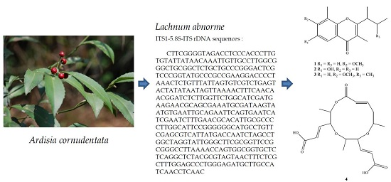
Secondary Metabolites of the Endophytic Fungus Lachnum abnorme from Ardisia cornudentata

1
Graduate Institute of Natural Products, College of Pharmacy, Kaohsiung Medical University, Kaohsiung 807, Taiwan
2
School of Pharmacy, College of Pharmacy, Kaohsiung Medical University, Kaohsiung 807, Taiwan
3
Bioresource Collection and Research Center, Food Industry Research and Development Institute, Hsinchu 300, Taiwan
*
Authors to whom correspondence should be addressed.
Int. J. Mol. Sci. 2016, 17(9), 1512; https://doi.org/10.3390/ijms17091512
Submission received: 8 July 2016 / Revised: 16 August 2016 / Accepted: 31 August 2016 / Published: 8 September 2016
(This article belongs to the Section Bioactives and Nutraceuticals)

Abstract

:
Fractionation of an EtOAc-soluble fraction of the solid fermentate of an endophytic fungus, Lachnum abnorme Mont. BCRC 09F0006, derived from the endemic plant, Ardisia cornudentata Mez. (Myrsinaceae), resulted in the isolation of three new chromones, lachnochromonins D–F (13), one novel compound, lachabnormic acid (4), along with nine known compounds (513). Their structures were elucidated by spectroscopic analyses. Alternariol-3,9-dimethyl ether (6) was given the correct data as well as 2D spectral analyses for the first time. This is the first report of the isolation of one unprecedented compound 4 from Lachnum genus, while all known compounds were also found for the first time from Lachnum. The effects of some isolates (3, 4, 79, 10, and 13) on the inhibition of nitric oxide (NO) production in lipopolysaccharide (LPS)-activated RAW 264.7 murine macrophages were also evaluated. Several compounds exhibited weak inhibitory activity on lipopolysaccharide (LPS)-stimulated NO production in RAW 264.7 macrophages.

Graphical Abstract

1. Introduction

Plant endophytes are regarded as sorts of particular-biotope microorganisms by residing in the tissues of living plants without causing any immediate obvious negative effects in their host plants [1,2]. Recently, some endophytic fungi have been applied in medicine and agriculture. For example, taxol, an antineoplastic agent, was isolated from Taxomyces andreanae derived from Taxus brevifolia; crytocandin and cyptocin, antimycotic agents, were separated from Cryptosporiopsis cf. quercina residing in Tripterygium wilfordii [3,4,5]. A series of investigation programs on Formosan endemic plants’ endophytes, some secondary metabolites from fungal strains, showed bioactive potential for exploration. One of these fungal strains, Lachnum abnorme Mont. (Hyaloscyphaceae) BCRC 09F0006, was isolated from the stem of an endemic plant, Ardisia cornudentata Mez. (Myrsinaceae). In previous chemical investigations of the genus Lachnum, three species and one unidentified species were studied, and 35 secondary metabolites were reported, including benzenoids, coumarins, isocoumarins, chromones, phthalides, etc. [6,7,8,9,10,11]. Investigation of the EtOAc-soluble fraction of the solid fermentate from L. abnorme led to the isolation of four new compounds, lachnochromonins D (1), E (2), F (3), and lachabnormic acid (4), along with nine known compounds (513) (Figure 1). The structure identification of the new compounds and the NO inhibitory in macrophage RAW 264.7 cells of the main isolates will be described below.

2. Results

2.1. Purification and Characterization

The solid state fermentate of L. abnorme was extracted with methanol and the resulted extracts were partitioned into the EtOAc and H2O soluble layers. The EtOAc layer was purified by conventional chromatographic techniques to obtain thirteen compounds (113), and the structures were elucidated by 1D and 2D NMR spectra and compared with literature data.

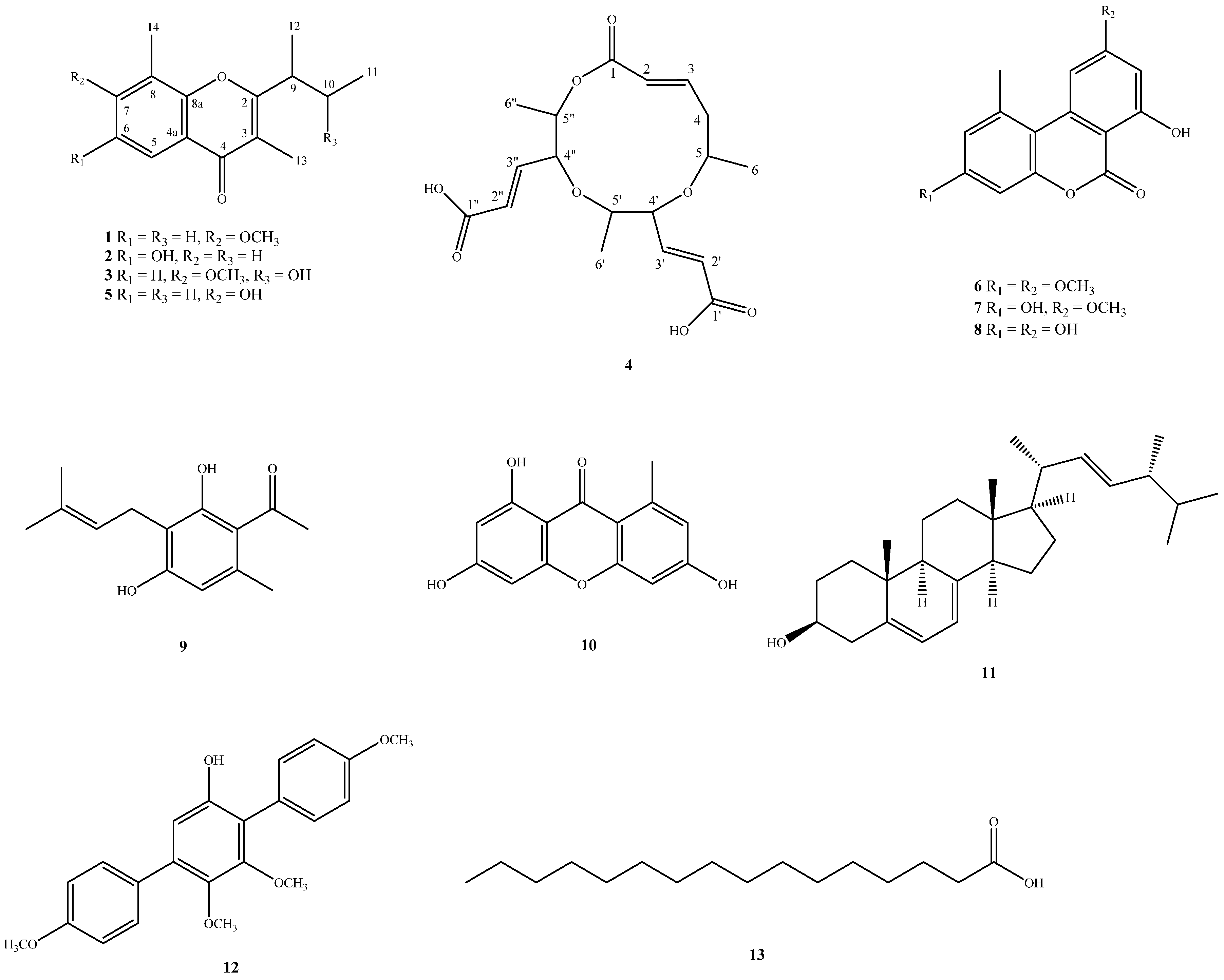
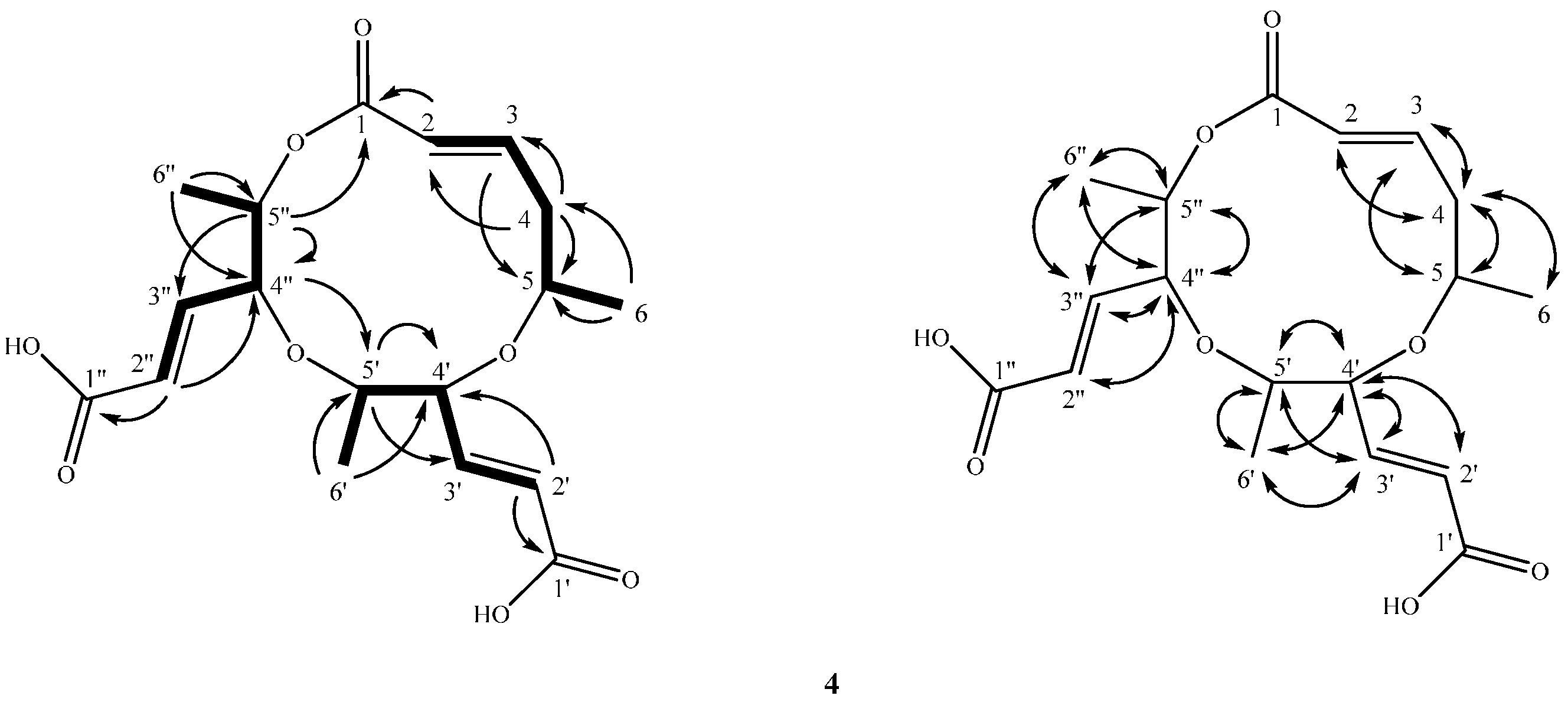
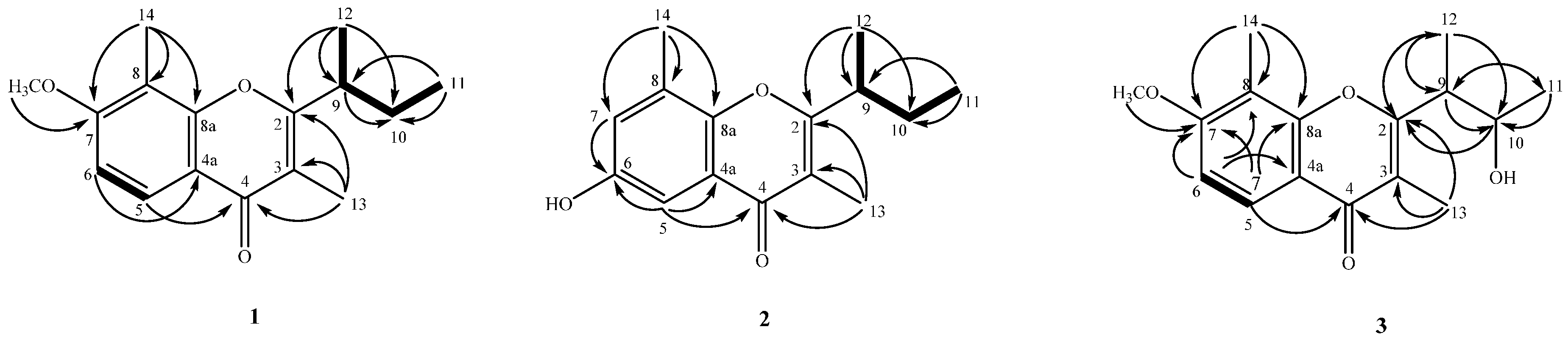
2.2. Structure Elucidation of Compounds 14

Compound 1 was obtained as an optically active colorless oil with [ α ] D 25 −33.6 (c 0.47, CHCl3), and its molecular formula was established as C16H20O3 by ESI and HRESIMS with seven degrees of unsaturation. The UV absorptions at 231, 246, 254 sh, and 298 nm and IR absorptions at 1632 cm−1 (a conjugated carbonyl group), 1604 and 1579 cm−1 (an aromatic ring) accompanying the 13C NMR resonnance at δ 178.4 (C-4), suggested a chromone skeleton [12]. The 1H NMR spectrum showed two aromatic protons with ortho-coupling at δ 6.92 (1H, d, J = 8.8 Hz, H-6) and 8.03 (1H, d, J = 8.8 Hz, H-5), which was assignable due to being next to the carbonyl group of chromone moiety. The fragments of C-12-C-9-C-10-C-11 were observed by COSY analysis. The presence of a sec-butyl group was further confirmed by HMBC spectrum showing the correlation between H-12 to C-2, C-9, C-10, and H-11 to C-9, and C-10. The HMBC (Figure 2) spectrum revealed the correlation between H-5 and C-4, C-6 (δC 108.0), C-7 (δC 160.6), and C-8a (δC 156.0); H-6 and C-7, C-8 (δC 113.3), C-4a (δC 116.5); OCH3H 3.91) and C-7; H-14 and C-7, C-8, and C-8a; H-9 and C-2, C-3; H-13 and C-2, C-3 and C-4, thus the location of methyl group (δH 2.27) at C-8, methoxyl group at C-7, another methyl group (δH 2.05) at C-3 and the connection of a sec-butyl group to C-2 can be confirmed. The 1H NMR spectrum (Table 1) of 1 was similar to that of lachnochromonin C (5) [13], also obtained in this study, except a methoxy group at C-7 in (1) was in place of a hydroxy group (δH 8.91, br s) in 5. According to the above data, the structure of 1 was unequivocally determined as 2-sec-butyl-7-methoxy-3,8-dimethyl-4H-chromen-4-one, which was further confirmed by COSY (Figure 2), 13C NMR (Table 1), DEPT, HSQC, HMBC (Figure 2), and NOESY techniques, and designated as lachnochromonin D. Hence, compound 1 had the proposed structure, leaving the C-9 configuration undetermined.
Compound 2 was obtained as colorless needles with [ α ] D 25 −57.2 (c 0.17, CHCl3). The ESIMS and HRESIMS were used to establish the molecular formula of 2 as C15H18O3 with seven degrees of unsaturation. The UV absorptions at 224, 245, 251, and 299 nm and after addition of KOH aqueous. solution gave a bathochromic shift. The IR absorption at 3194 cm−1 (a hydroxy group), 1626 cm−1 (a conjugated carbonyl group), 1584 and 1482 cm−1 (an aromatic ring) suggested the presence of a phenolic chromone skeleton [12], which was also supported by a 13C NMR resonance of C-4 (δC 178.3) and δH 7.35 (1H, br s, OH-6) in 1H NMR spectrum. The 1H NMR data was similar to that of 5 except two meta-coupled protons of H-5 (δ 6.90) and H-7 (δ 7.96), one hydroxy group in C-6 of 2 replaced two ortho-coupled protons of H-5 and H-6, and one hydroxy group (δ 8.91, br s) in 5. Thus, the structure of 2 was proposed to be 2-sec-butyl-6-hydroxy-3,8-dimethyl-4H-chromen-4-one, which was further confirmed by COSY (Figure 2), 13C NMR (Table 1), DEPT, HSQC, HMBC (Figure 2), and NOESY experiments, and namely lachnochromonin E. At present, the absolute configuration at C-9 of 2 remains uncertain.
Compound 3 was obtained as an optically active yellowish powder with [ α ] D 25 –68.5 (c 0.22, CHCl3). The molecular formula was established as C16H20O4 by the ESIMS and HRESIMS with seven degrees of unsaturation. Its UV spectra showed absorption bands at 228, 244, 252, and 297 nm, and the IR spectrum revealed the presence of a hydroxy group (3397 cm−1), and a conjugated carbonyl group (1627 cm−1). Analyses of the 1H NMR spectrum indicated the presence of two aromatic protons with ortho-coupling at δ 6.94 (1H, d, J = 8.9 Hz, H-6) and 8.03 (1H, d, J = 8.9 Hz, H-5), one methoxy group at δ 3.94 (3H, s, OCH3-7), four methyl groups at δ 1.32 (3H, d, J = 7.2 Hz, H-12), 1.34 (3H, d, J = 6.0 Hz, H-11), 2.09 (3H, s, H-13), and 2.29 (3H, s, H-14), one methine and one oxymethine at δ 3.11 (1H, qd, J = 7.2, 6.6 Hz, H-9) and 4.15 (1H, dq, J = 6.6, 6.0 Hz, H-10), respectively. Meanwhile, the 13C NMR spectrum exhibited 16 signals that were clearly classified into four methines, five methyls, and seven quaternary carbons based on the HSQC, suggesting 3 might be a chromone, similar to 1, which was corroborated by its 2D NMR spectra involving COSY, HSQC, HMBC and NOESY. The only difference was that one CHOH group (δ 4.15 (1H, dq, J = 6.6, 6.0 Hz, H-10)) attached on C-10 in 3, replacing a CH2 group (δ 1.68/1.82 (each 1H, m, CH2-10)) at C-10 in 1, which was further confirmed in this proposed structure by the 2D NMR data (COSY and HMBC) (Figure 2), allowed determining the structure of 3. The configurations of C-9 and C-10 in 3 remain uncertain. Thus, the structure of 3 was fully established and named lachnochromonin F.
Compound 4 was isolated as a whitish powder with [ α ] D 25 ±0° (c 0.38, MeOH). The ESIMS and HRESIMS data provided the molecular formula C18H24O8 with seven degrees of unsaturation. The UV spectrum exhibited the absorption at 260 nm. The IR spectrum showed absorptions at 3455, 1720 cm−1 for the OH group and the carbonyl group, respectively. The 1H NMR, 13C NMR, DEPT, and HSQC spectra displayed signals for three methyl doublets at δ 1.28 (3H, d, J = 6.0 Hz, H-6)/21.2 (C-6), 1.31 (3H, d, J = 6.0 Hz, H-6′)/18.4 (C-6′), and 1.34 (3H, d, J = 6.0 Hz, H-6′′)/18.5 (C-6′′), three trans-form olefinic groups at δ 5.83 (1H, ddd, J = 15.6, 1.8, 0.9 Hz, H-2)/125.3 (C-2), 6.86 (1H, ddd, J = 15.6, 9.6, 5.9 Hz, H-3)/146.3 (C-3), 5.93 (1H, dd, J = 15.9, 0.9 Hz, H-2′)/123.95 (C-2′), 6.81 (1H, dd, J = 15.9, 7.6 Hz, H-3′)/149.1 (C-3′), 5.97 (1H, dd, J = 15.7, 1.2 Hz, H-2′′)/123.98 (C-2′′), and 6.83 (1H, dd, J = 15.7, 7.2 Hz, H-3′′)/148.5 (C-3′′), one non-equivalent CH2 at δ 2.43 (1H, dddd, J = 14.4, 9.6, 9.6, 0.9 Hz, H-4b), 2.59 (1H, dddd, J = 14.4, 5.9, 3.0, 1.8 Hz, H-4a)/40.0 (C-4), five oxymethines at δ 5.10 (1H, dqd, J = 9.6, 6.0, 3.0 Hz, H-5)/70.3 (C-5), 4.12 (1H, m, H-4′)/75.2 (C-4′), 4.84 (1H, m, H-5′)/73.1 (C-5′), 4.17 (1H, m, H-4′′)/74.7 (C-4′′), 4.82 (1H, m, H-5′′)/73.9 (C-5′′), and three carbonyl groups at δ 165.8 (C-1), 165.9 (C-1′), and 166.1 (C-1′′). The above observation, followed by the COSY (Figure 3) and HSQC spectrum of 4, established the presence of the partial three substitutes: (A) C-1 to C-6, (B) C-1′ to C-6′, and (C) C-1′′ to C-6′′, for skeleton of 4. The entire skeleton of 4 was constructed by the aid of the HMBC spectrum (Figure 3). In the HMBC spectrum of 4 (Figure 3), there were 2J, 3J-correlations from H-2 to C-1, C-3, and C-4; from H-3 to C-1, C-2, C-4, and C-5; from H-4 to C-2, C-3, and C-5; from H-6 to C-4 and C-5; from H-2′ to C-1′ and C-4′; from H-3′ to C-4′ and C-5′; from H-4′ to C-2′, C-3′, C-5′, and C-6′; from H-6′ to C-4′ and C-5′; from H-2′′ to C-1′′, C-3′′, and C-4′′; from H-3′′ to C-4′′ and C-5′′; from H-4′′ to C-2′′, C-3′′, C-5′′, and C-6′′, H-6’ to C-4’ and C-5’. Moreover, the key HMBC correlations of H-4′′/ C-5′; H-5′′/C-1; H-4′/C-5 verified the junction of the each two substitutes A to B unit at C-5, B to C unit at C-5′, and C to A at C-5′′ through the oxygen atom between C-5/C-4′, C-5′/C-4′′, and C-5′′/C-1, respectively. This structure was composed of three (E)-5-hydroxy-2-hexenoic acid groups. Unfortunately, the NOESY (Figure 3) spectrum only showed the correlations within the fragments, and therefore the relative configurations of 4 could not be determined. Because of the optical inactivity, 4 was also proposed to be racemic. 1H and 13C NMR, COSY (Figure 3), HMBC (Figure 3), and NOESY (Figure 3) experiments confirmed the planar structure as 4 and was named as a lachabnormic acid.
Alternariol-3,9-dimethyl ether (6) was a known compound that was isolated in this study. The 1H NMR data of compound 6 was compared with the two reference data [14,15] very closely, but only the chemical shift of H-11 in compound 6 is different. In Yang’s report [15], δ 2.03 (Me-11) was too high field to be assigned as a methyl group in ring A of 6H-benzo[c]chromen-6-one skeleton in comparison with the reference compounds alternariol-9-methyl ether (7) [16,17] and alternariol (8) [18]. The chemical shift of H-11 of compound 6 should appear at δ 2.81, and the chemical shift of H-11 in the reference [15] displayed at δ 2.03 is erroneous. We had asked the corresponding author [15] to provide us with their original 1H and 13C NMR spectrum of compound 6 for further confirmation. It was caused by the author’s typo. According to the donated spectrum, the chemical shift of H-11 of 6 indeed appeared at δ 2.81 (in CDCl3). We also provided the correct data and 2D NMR spectrum to further confirm the structure.
The known compounds, lachnochromonin C (5) [13], alternariol-9-methyl ether (7) [16,17], alternariol (8) [18], pestalorionol (9) [19], 1,3,6-trihydroxy-8-methylxanthone (10) [20], ergosterol (11) [21], 6′-hydroxy-4,2′,3′,4′′-tetramethoxy-p-terphenyl (12) [22], and palmitic acid (13) [23] were identified by comparison of their physical and spectroscopic data with values reported in the literature.
A literature review revealed that lachnochromonin C (5) has been reported in the patent literature concerning its strong anti-inflammatory activity [13]. In our study, we tested the anti-inflammatory activity by measuring the nitric oxide (NO) production in lipopolysaccharide (LPS)-activated RAW 264.7 macrophages. The iNOS inhibitory activity of compounds 3, 4, 7, 8, 9, 10, and 13 at a concentration of 100 μM was presented in Table 2, showing most compounds had nitrite production values of 77.75%–101.83% under LPS induction. The result indicated that all compounds that we tested exhibited a weak anti-inflammatory activity. The increase in cell viability of compound 8 suggests that this compound might have some activities in cell proliferation, which is worthy to be explored by other evaluation models.

3. Discussion

Natural products from medicinal plants and microorganisms are the most concordant and productive source for the discovery of new pharmaceuticals and lead compounds. Recently, a great deal of interest has been generated by discovery of remarkable pharmacological agents from endophytic fungi. In our ongoing pharmacological screening program on biodiversity of endophytic fungi from endemic Formosana plants, we found out that the extracts of an endophytic fungus of the ascomycetes family growing on an endemic plant, A. cornudentata Mez. (Myrsinaceae), named L. abnorme, showed approximately 95% inhibition at a concentration of 10 μg/mL. It is remarkable to inhibit lipopolysaccharide (LPS)-induced nitric oxide (NO) release in RAW 264.7 murine macrophages. Careful investigation the EtOAc-soluble fraction of the solid fermentate from L. abnorme led to the isolation of three new chromones, lachnochromonins D–F (13), and one novel compound, lachabnormic acid (4), together with nine known isolates. Alternariol-3,9-dimethyl ether (6) was given the correct data as well as 2D spectral analyses for the first time. Interestingly, the unique side chain 3-hydroxybutan-2-yl in lachnochromonin F (3) so far was only isolated from this plant. Various substitutents of the bulky or small side chain [24] at the C-2 position of chromone isolated from different species indicated the distinct and diverse metabolic pathway of Lachnum. To our knowledge, the chromone skeleton has seldom been previously reported in the genus Lachnum. This type of compound could stimulate future phytochemical studies.

4. Materials and Methods

4.1. General

General. TLC: silica gel 60 F254 precoated plates (Merck, Kenilworth, NJ, USA). Column chromatography (CC): silica gel 60 (70–230 or 230–400 mesh, Merck) and Spherical C18 100A Reversed Phase Silica Gel (RP-18) (particle size: 20–40 μm) (Silicycle). UV Spectra: Jasco V-530 UV/VIS spectrophotometer (Tokyo, Japan); λmax (log ε) in nm. Optical rotation: Jasco P-2000 polarimeter; in CHCl3. IR Spectra: Jasco FTIR-4200 spectrophotometer; ν in cm−1. 1H, 13C and 2D NMR spectra: Varian-VNMRS-600 (Palo Alto, CA, USA), and Varian-Mercury-400 spectrometers; δ in ppm rel. to Me4Si, J in Hz. EI–MS: VG-Biotech Quatro-5022 mass spectrometer (Palo Alto, CA, USA); m/z (rel. %). ESI and HRESIMS: Bruker APEX-II mass spectrometer (Billerica, MA, USA); in m/z.

4.2. Fungus Material

The fungal strain Lachnum abnorme was isolated from the stem of Ardisia cornudentata, which was collected in Wutai Township, Pingtung County, Taiwan, during August of 2008, and identified by Ih-Sheng Chen, one of the authors. The fungal strain was identified as L. abnorme (family Hyaloscyphaceae) by Sung-Yuan Hsieh, one of the authors, basing on cultural and anamorphic data.
Fungus cultivation and fermentation were carried out by a previously reported method [25], and the sequence data derived from the fungal strain has been submitted and deposited at GenBank with accession no. AB267638. BLAST (nucleotide sequence comparison program) search result displayed that the sequence was the most resembling (100%) to the sequence of L. abnorme. The strain is preserved at the Bioresource Collection and Research Center (BCRC) of the Food Industry Research and Development Institute (FIRDI), under the ID No. 09F0006.

4.3. Extraction and Isolation

Solid fermentate of L. abnorme (5 kg) was extracted with cold MeOH to yield a MeOH extract (150 g) which was partitioned in EtOAc–H2O (1:1) to produce an EtOAc layer (20 g) and an H2O layer. The H2O layer was further partitioned with BuOH, and the BuOH layer (50 g) was separated. The EtOAc layer was subjected to C.C. (silica gel; hexane/acetone gradient) to get eight fractions (Frs. 1–8). Fr. 3 was subjected to MPLC (silica gel; hexane/acetone, 5:1) to produce 18 fractions, Frs. 3.1–3.18. Fr. 3.7 was subjected to MPLC (silica gel; hexane/CH2Cl2/acetone, 30:1:1) to produce 10 fractions, Frs. 3.7.1–3.7.10. Fr. 3.7.7 furnished 13 (170 mg). Fr. 4 was subjected to MPLC (silica gel; CH2Cl2/acetone, 30:1) to produce 11 fractions, Frs. 4.1–4.11. Fr. 4.3 was subjected to MPLC (silica gel; CH2Cl2/EtOAc, 100:1) to produce six fractions, Frs. 4.3.1–4.3.6. Fr. 4.3.3 was subjected to MPLC (silica gel; hexane/EtOAc, 10:1) to produce 12 fractions, Frs. 4.3.3.1–4.3.3.12. Fr. 4.3.3.7 was further purified with prep. TLC (hexane/acetone, 15:1) to obtain 9 (Rf 0.45; 2.2 mg). Fr. 4.3.3.10 was further purified with prep. TLC (hexane/acetone, 10:1) to obtain 6 (Rf 0.48; 0.9 mg). Fr. 4.4 was subjected to MPLC (silica gel; hexane/EtOAc, 4:1) to produce 11 fractions, Frs. 4.4.1–4.4.11. Fr. 4.4.2 was furnished 1 (45 mg). Fr. 5 was recrystallized from acetone and CH3OH to gain 11 (140 mg). Fr. 6 was subjected to MPLC (silica gel; hexane/acetone, 4:1) to produce 10 fractions, Frs. 6.1–6.10. Fr. 6.5 was subjected to MPLC (RP–18; MeOH/H2O 1:1) to produce six fractions, Fr. 6.5.1–6.5.6. Fr. 6.5.4 was further purified to MPLC (silica gel; CH2Cl2/MeOH, 50:1) to yield 2 (4.8 mg). Fr. 6.5.6 was purified to MPLC (silica gel; CH2Cl2/MeOH, 50:1) to afford 12 (3.3 mg). Fr. 7 was subjected to MPLC (silica gel; hexane/EtOAc, 1:1) to produce 18 fractions, Frs. 7.1–7.18. Fr. 7.5 was subjected to MPLC (silica gel; hexane/acetone, 2:1) to produce 11 fractions, Frs. 7.5.1–7.5.11. Fr. 7.5.2 was subjected to MPLC (silica gel; CHCl3/acetone, 30:1) to produce 22 fractions, Frs. 7.5.2.1–7.5.2.22. Fr. 7.5.2.7 afforded 7 (6.2 mg). Fr. 7.5.2.15 was subjected to MPLC (silica gel; CHCl3/acetone, 40:1) to produce six fractions, Frs. 7.5.2.15.1–7.5.2.15.6. Fr. 7.5.2.15.4 afforded 5 (152 mg). Fr. 7.5.5 was subjected to MPLC (silica gel; hexane/acetone, 4:1) to produce five fractions, Frs. 7.5.5.1–7.5.5.5. Fr. 7.5.5.2 was subjected to MPLC (silica gel; CHCl3/CH3OH, 60:1) to produce five fractions, Frs. 7.5.5.2.1–7.5.5.2.5. Fr. 7.5.5.2.5 was subjected to MPLC (silica gel; hexane/acetone, 5:2) to produce four fractions, Frs. 7.5.5.2.5.1–7.5.5.2.5.4. Fr. 7.5.5.2.5.2 was subjected to MPLC (silica gel; CH2Cl2/CH3OH, 40:1) to produce seven fractions, Frs. 7.5.5.2.5.2.1–7.5.5.2.5.2.7. Fr. 7.5.5.2.5.2.6 yielded 10 (3.7 mg). Fr. 7.6 was purified with TLC (RP–18; acetone/H2O 2:1) to obtain 8 (Rf 0.58; 2.1 mg) Fr. 7.9 was subjected to MPLC (silica gel; CHCl3/CH3OH, 40:1) to produce 22 fractions, Frs. 7.9.1–7.9.22. Fr. 7.9.7 was purified with TLC (RP–18; acetone/H2O 1:1) to obtain 3 (Rf 0.38; 2.2 mg) Fr. 7.9.14 was subjected to MPLC (silica gel; CH2Cl2/CH3OH, 30:1) to produce 10 fractions, Frs. 7.9.14.1–7.9.14.10. Fr. 7.9.14.4 was purified with TLC (RP–18; acetone/H2O 2:1) to obtain 4 (Rf 0.68; 3.8 mg).

4.3.1. Lachnochromonin D (1)

Colorless oil; [ α ] D 25 −33.6 (c 0.47, CHCl3); UV λmax (MeOH) (log ε) 231 (4.34), 246 (4.11), 254 sh (4.06), 298 (4.09) nm; IR νmax (ATR) 1632 (C=O), 1604, 1579 (aromatic ring) cm−1; 1H and 13C NMR: see Table 1; ESIMS: m/z 261 ([M + H]+); HRESIMS: m/z 261.14841 ([M + H]+, calcd for C16H21O3, 261.14852).

4.3.2. Lachnochromonin E (2)

Colorless needles (MeOH-CH2Cl2); M.p.: 231–233 °C; [ α ] D 25 −57.2 (c 0.17, CHCl3); UV λmax (MeOH) (log ε) 224 (4.30), 245 (4.05), 251 (4.05), 299 (4.07) nm; UV λmax (MeOH + KOH) (log ε) 215 (4.39), 254 (4.25), 338 (4.13) nm; IR νmax (ATR) 3194 (OH), 1626 (C=O), 1584, 1482 (aromatic ring) cm−1; 1H and 13C NMR: see Table 1; ESIMS: m/z 247 ([M + H]+); HRESIMS: m/z 247.13266 ([M + H]+, calcd for C15H19O3, 247.13287).

4.3.3. Lachnochromonin F (3)

Yellowish powder; [ α ] D 25 −68.5° (c 0.22, CHCl3); UV λmax (MeOH) (log ε): 228 (3.90), 244 (3.62), 252 sh (3.57), 297 (3.61) nm; IR νmax (KBr): 3397 (OH), 1627 (C=O) cm−1; 1H and 13C NMR: see Table 1; ESIMS: m/z 277 [M + H]+; HRESIMS: m/z 277.1432 ([M + H]+, calcd for C16H21O4, 277.1434).

4.3.4. Lachabnormic Acid (4)

Whitish powder; [ α ] D 25 ±0° (c 0.38, MeOH); UV λmax (MeOH) (log ε): 260 (2.88) nm; IR νmax (KBr): 3455 (OH), 1720 (C=O) cm−1; 1H NMR (CDCl3, 600 MHz) δ 1.28 (3H, d, J = 6.0 Hz, H-6), 1.31 (3H, d, J = 6.0 Hz, H-6′), 1.34 (3H, d, J = 6.0 Hz, H-6′′), 2.43 (1H, dddd, J = 14.4, 9.6, 9.6, 0.9 Hz, H-4b), 2.59 (1H, dddd, J = 14.4, 5.9, 3.0, 1.8 Hz, H-4a), 4.12 (1H, m, H-4′), 4.17 (1H, m, H-4′′), 4.82 (1H, m, H-5′′), 4.84 (1H, m, H-5′), 5.10 (1H, dqd, J = 9.6, 6.0, 3.0 Hz, H-5), 5.83 (1H, ddd, J = 15.6, 1.8, 0.9 Hz, H-2), 5.93 (1H, dd, J = 15.9, 0.9 Hz, H-2′), 5.97 (1H, dd, J = 15.7, 1.2 Hz, H-2′′), 6.81 (1H, dd, J = 15.9, 7.6 Hz, H-3′), 6.83 (1H, dd, J = 15.7, 7.2 Hz, H-3′′), 6.86 (1H, ddd, J = 15.6, 9.6, 5.9 Hz, H-3); 13C NMR (CDCl3, 150 MHz) δ: 18.4 (C-6′), 18.5 (C-6′′), 21.2 (C-6), 40.0 (C-4), 70.3 (C-5), 73.1 (C-5′), 73.9 (C-5′′), 74.7 (C-4′′), 75.2 (C-4′), 123.95 (C-2′), 123.98 (C-2′’), 125.3 (C-2), 146.3 (C-3), 148.5 (C-3′′), 149.1 (C-3′), 165.8 (C-1), 165.9 (C-1′), 166.1 (C-1′′); ESIMS: m/z 369 [M + H]+; HRESIMS: m/z 391.1367 ([M + Na]+ , calcd for C18H24O8Na, 391.1369).

4.3.5. Lachnochromonin C (5)

Colorless needles; M.p.: 160–162 °C; [ α ] D 25 +5.66° (c 0.06 MeOH); UV λmax (MeOH) (log ε): 229 (4.61), 246 (4.49), 253 sh (4.45), 298 (4.39) nm; UV λmax (MeOH + KOH) (log ε): 221 sh (4.55), 260 (4.62), 337 (4.30) nm; 1H NMR (CDCl3, 400 MHz) δ 0.93 (3H, dd, J = 7.6, 7.2 Hz, H-11), 1.33 (3H, d, J = 6.8 Hz, H-12), 1.68 (1H, m, H-10b), 1.85 (1H, m, H-10a), 2.11 (3H, s, H-13), 2.35 (3H, s, H-14), 3.05 (1H, m, H-9), 7.03 (1H, d, J = 8.8 Hz, H-6), 7.90 (1H, d, J = 8.8 Hz, H-5), 8.91 (1H, br s, OH-7, D2O exchangeable); 13C NMR (CDCl3, 100 MHz) δ: 7.9 (C-14), 9.6 (C-13), 12.0 (C-11), 18.0 (C-12), 27.8 (C-10), 37.8 (C-9), 114.2 (C-6), 111.4 (C-8), 115.1 (C-3), 115.6 (C-4a), 123.8 (C-5), 155.9 (C-8a), 159.5 (C-7), 168.2 (C-2), 179.2 (C-4); ESIMS: m/z 247 [M + H]+.

4.3.6. Alternariol-3,9-dimethyl Ether (6)

Colorless needles; M.p.: 179–181 °C; UV λmax (MeOH) (log ε): 254 (4.35), 286 (3.63), 297 (3.61), 331 (3.66) nm; UV λmax (MeOH + KOH) (log ε): 250 (417), 304 (3.46), 354 (3.60) nm; IR νmax (KBr): 3400 (OH), 1669 (C=O) cm1; 1H NMR (CDCl3, 600 MHz) δ 2.81 (3H, s, H-11), 3.86 (3H, s, OCH3-3), 3.92 (3H, s, OCH3-9), 6.55 (1H, d, J = 1.5 Hz, H-8), 6.75 (1H, d, J = 2.4 Hz, H-2), 6.76 (1H, d, J = 2.4 Hz, H-4), 7.27 (1H, d, J = 1.5 Hz, H-10), 12.0 (1H, s, OH-7); 13C NMR (CDCl3, 150 MHz) δ 25.7 (C-11), 55.5 (OCH3-3), 55.7 (OCH3-9), 98.9 (C-8), 99.4 (C-6a), 100.0 (C-4), 104.5 (C-10), 110.9 (C-10b), 116.9 (C-2), 138.0 (C-10a), 138.2 (C-1), 153.2 (C-4a), 160.0 (C-3), 165.2 (C-6), 165.4 (C-7), 166.4 (C-9); ESIMS m/z 287 [M + H]+.

4.3.7. Alternariol-9-methyl Ether (7)

Colorless needles; M.p.: 245–248 °C; UV λmax (MeOH) (log ε): 219 sh (3.74), 256 (3.99), 288 (3.37), 299 (3.35), 340 (3.44) nm; UV λmax (MeOH + KOH) (log ε): 265 (3.84), 277 sh (3.73), 311 (3.41), 365 (3.41) nm; IR νmax (ATR): 3314 (OH), 1711 (C=O) cm1; 1H NMR (Acetone-d6, 400 MHz) δ 2.80 (3H, s, H-11), 3.97 (3H, s, OCH3-9), 6.57 (1H, d, J = 2.2 Hz, H-8), 6.70 (1H, d, J = 2.8 Hz, H-4), 6.80 (1H, d, J = 2.8 Hz, H-2), 7.30 (1H, d, J = 2.2 Hz, H-10), 9.22 (1H, br s, OH-3), 11.8 (1H, s, OH-7); 13C NMR (Acetone-d6, 100 MHz) δ 26.3 (C-11), 56.9 (OCH3-9), 100.5 (C-6a), 100.6 (C-8), 103.4 (C-4), 105.2 (C-10), 111.3 (C-10b), 119.1 (C-2), 139.8 (C-10a), 140.3 (C-1), 154.8 (C-4a), 160.0 (C-3), 166.7 (C-6, C-7), 168.2 (C-9); ESIMS m/z 273 [M + H]+.

4.3.8. Alternariol (8)

Yellowish powder; UV λmax (MeOH) (log ε): 254 (3.93), 286 (3.45), 331 sh (3.34) nm; UV λmax (MeOH + KOH) (log ε): 276 (3.92), 317 sh (3.49), 346 sh (3.44) nm; IR νmax (ATR): 3350 (OH), 1657 (C=O) cm1; 1H NMR (CDCl3, 600 MHz) δ 2.77 (3H, s, H-11), 6.37 (1H, d, J = 1.8 Hz, H-8), 6.62 (1H, d, J = 2.5 Hz, H-4), 6.70 (1H, d, J = 2.5 Hz, H-2), 7.27 (1H, d, J = 1.8 Hz, H-10); 13C NMR (CDCl3, 150 MHz) δ 25.8 (C-11), 99.1 (C-6a), 102.0 (C-8), 102.8 (C-4), 105.5 (C-10), 111.0 (C-10b), 118.6 (C-2), 139.8 (C-1), 140.1 (C-10a), 154.5 (C-4a), 159.9 (C-3), 166.2 (C-7), 166.9 (C-9), 167.0 (C-6); ESIMS m/z 259 [M + H]+.

4.3.9. Pestalotionol (9)

Yellowish oil; UV λmax (MeOH) (log ε): 219 sh (3.81), 295 (3.61) nm; UV λmax (MeOH + KOH) (log ε): 252 (3.51), 338 (3.90) nm; IR νmax (ATR): 3401 (OH), 1648 (C=O) cm1; 1H NMR (CDCl3, 600 MHz) δ 1.75 (3H, br s, H-10), 1.81 (3H, br s, H-11), 2.46 (3H, s, H-13), 2.64 (3H, s, H-14), 3.41 (2H, d, J = 7.5 Hz, H-7), 5.26 (1H, br t, J = 7.5 Hz, H-8), 5.83 (1H, br s, OH-4, D2O exchangeable), 6.23 (1H, s, H-5), 12.17 (1H, s, OH-2, D2O exchangeable); 13C NMR (CDCl3, 150 MHz) δ 13.1 (C-13), 17.9 (C-11), 22.1 (C-7), 24.6 (C-14), 25.8 (C-10), 111.70 (C-5), 111.74 (C-3), 115.4 (C-1), 121.4 (C-8), 135.4 (C-9), 138.8 (C-6), 158.9 (C-4), 160.5 (C-2), 198.2 (C-12); ESIMS m/z 273 [M + K]+.

4.3.10. 1,3,6-Trihydroxy-8-methylxanthone (10)

Yellowish needles (MeOH); M.p.: 228–230 °C; UV λmax (MeOH) (log ε): 239 (4.24), 254 sh (3.96), 268 sh (3.68), 3.09 (4.00) nm; UV λmax (MeOH + KOH) (log ε): 235 (4.23), 263 (4.03), 295 sh (3.59), 357 (4.40) nm; IR νmax (ATR): 3511 (OH), 1655 (C=O) cm1; 1H NMR (CD3OD, 400 MHz) δ 2.77 (3H, s, H-10), 6.11 (1H, d, J = 2.4 Hz, H-4), 6.22 (1H, d, J = 2.4 Hz, H-2), 6.59 (1H, d, J = 2.4 Hz, H-5), 6.60 (1H, d, J = 2.4 Hz, H-7); 13C NMR (CD3OD, 100 MHz) δ 23.6 (C-10), 94.2 (C-2), 98.8 (C-4), 101.5 (C-5), 104.0 (C-9a), 112.9 (C-8a), 117.0 (C-7), 144.7 (C-8), 158.6 (C-1), 160.8 (C-5a), 164.2 (C-6), 164.9 (C-4a), 166.1 (C-3), 183.5 (C-9); ESIMS m/z 259 [M + H]+.

4.3.11. Ergosterol (11)

Colorless needles (CH2Cl2-MeOH); M.p.: 159–161 °C; IR νmax (ATR): 3408 (OH) cm1; 1H NMR (Acetone-d6, 600 MHz) δ 0.67 (3H, s, H-18), 0.85 (3H, d, J = 7.2 Hz, H-26), 0.86 (3H, d, J = 7.2 Hz, H-27), 0.94 (3H, d, J = 6.6 Hz, H-28), 0.95 (3H, s, H-19), 1.07 (3H, d, J = 6.6 Hz, H-21), 1.29 (1H, m, H-1b), 1.30 (1H, m, H-12b), 1.32 (1H, m, H-17), 1.38 (1H, m, H-16b), 1.39 (1H, m, H-15b), 1.46 (1H, m, H-2b), 1.49 (1H, m, H-25), 1.64 (1H, m, H-11b), 1.67 (1H, m, H-15a), 1.76 (1H, m, H-11a), 1.81 (1H, m, H-16a), 1.83 (1H, m, H-2a), 1.88 (2H, m, H-1a, H-24), 1.92 (1H, m, H-14), 1.96 (1H, m, H-9), 2.09 (2H, m, H-12a, H-20), 2.23 (1H, m, H-4b), 2.40 (1H, ddd, J = 14.4, 4.8, 2.4 Hz, H-4a), 3.50 (1H, m, H-3), 3.64 (1H, d, J = 4.8 Hz, OH-3), 5.24 (2H, dd, J = 15.6, 7.5 Hz, H-22), 5.28 (2H, dd, J = 15.6, 7.2 Hz, H-23), 5.37 (1H, m, H-7), 5.53 (1H, dd, J = 6.0, 3.0 Hz, H-6); 13C NMR (Acetone-d6, 150 MHz) δ 13.1 (C-18), 17.4 (C-19), 18.8 (C-28), 20.7 (C-26), 21.0 (C-27), 22.3 (C-21), 22.5 (C-11), 24.4 (C-15), 29.7 (C-16), 33.6 (C-2), 34.6 (C-25), 38.6 (C-10), 40.0 (C-1), 40.6 (C-12), 42.0 (C-20), 42.5 (C-4), 44.3 (C-13), 44.5 (C-24), 47.9 (C-9), 56.0 (C-14), 57.3 (C-17), 71.0 (C-3), 118.1 (C-7), 120.7 (C-6), 133.5 (C-23), 137.3 (C-22), 142.1 (C-5), 142.3 (C-8); ESIMS m/z 419 [M + Na]+.

4.3.12. 6′-Hydroxy-4,2′,3′,4′′-tetramethoxy-p-terphenyl (12)

Colorless needles (MeOH); M.p.: 228–230 °C; UV λmax (MeOH) (log ε): 209 (4.24), 227 sh (3.96), 274 (3.68) nm; UV λmax (MeOH + KOH) (log ε): 212 (4.23), 233 sh (3.59), 276 (4.40) nm; IR νmax (ATR): 3446 (OH) cm1; 1H NMR (CDCl3, 400 MHz) δ 3.45 (3H, s, H-3′), 3.75 (3H, s, H-2′), 3.85 (3H, s, H-4′′), 3.88 (3H, s, H-4), 5.93 (1H, br s, OH-3′, D2O exchangeable), 6.47 (1H, s, H-5′), 7.00 (2H, d, J = 8.4 Hz, H-3′′, 5′′), 7.41 (2H, d, J = 8.8 Hz, H-2, 6), 7.59 (2H, d, J = 8.8 Hz, H-2′′, 6′′); ESIMS m/z 367 [M + H]+.

4.3.13. Palmitic Acid (13)

Whitish powder; IR νmax (ATR): 3300–2700 (OH), 1706 (C=O) cm1; 1H NMR (CDCl3, 400 MHz) δ 0.88 (3H, t, J = 6.8 Hz, H-16), 1.25 (24H, s, H-4–15), 1.63 (2H, quint, J = 7.4 Hz, H-3), 2.34 (2H, t, J = 7.4 Hz, H-2); EIMS m/z 256 [M]+ (44), 227 (12), 213 (35), 199 (21), 185 (35), 171 (36),157 (42), 143 (28), 129 (67), 115 (31), 87 (100), 73 (80), 57 (52).

4.4. Determination of NO Cell Viability Assay

4.4.1. NO Activity Assay

The murine macrophage cell line RAW 264.7 cells obtained from the Bioresource Collection and Research Center (BCRC; Hsinchu, Taiwan), were cultured in Dulbecco’s modified Eagle medium (DMEM) supplemented with 10% fetal calf serum (FCS) and penicillin–streptomycin, and incubated at 37 °C in a humidified atmosphere with 5% CO2.
The nitrite measurement was based on our published technique [26]. A cell density of 2 × 10⁵ cells/100 μL were seeded into 96-well plates for 24 h. After pre-treating the cells with the compounds, a positive control, aminoguanidine (a selective iNOS inhibitor; 100 μM), or the DMSO (0.1%) vehicle before 200 ng/mL of lipopolysaccharide (LPS) induction. Twenty-four hours after LPS induction, the culture supernatant was collected and assayed for nitrite concentration by the Griess reagent. The absorbance at 550 nm was measured with a microplate spectrophotometer (Bio-Tek Instrument, Inc., Winooski, VT, USA). Fresh medium was used as the blank. The results were expressed as a percentage of the LPS induction without compound treatment group.

4.4.2. Cell Viability Assay

After the culture supernatant had been removed for nitrite determination, we used the MTT (3-(4,5-dimethylthiazol-2-yl)-2,5-diphenyltetrazolium bromide) solution to assess the cell viability as previously described [27]. In brief, 0.5 mg/mL of MTT in DMEM was added to each well of cells and incubated in cell culture condition for 3 h. Then, the supernatant was removed from each well by suction, and 100 μL of DMSO was added to dissolve the formazan crystals. The absorbance at 570 nm was measured with a microplate spectrophotometer. The results were expressed as a percentage of the LPS induction without compound treatment group.

Supplementary Materials

Supplementary materials can be found at www.mdpi.com/1422-0067/17/9/1512/s1.

Acknowledgments

This work is kindly supported by the Ministry of Economic Affairs, Taiwan, to the Food Industry Research & Development Institute (FIRDI).

Author Contributions

Hsun-Shuo Chang, Yi-Shuan Chen, Ming-Jen Cheng, Shan-Yu Lin, Yue-Jin Lin performed the research and recorded the spectra. Hing-Yuen Chan and Sung-Yuan Hsieh performed the isolation and identification of the fungal strain Hui-Chun Wang did the bioactivity experiments. Ih-Sheng Chen and Gwo-Fang Yuan designed the research. Ming-Jen Cheng, Chu-Hung Lin, and Ho-Cheng Wu completed the manuscript draft.

Conflicts of Interest

The authors declare no conflict of interest.

References

  1. Gunatilaka, A.A. Natural products from plant-associated microorganisms: Distribution, structural diversity, bioactivity, and implications of their occurrence. J. Nat. Prod. 2006, 69, 509–526. [Google Scholar] [CrossRef] [PubMed]
  2. Tan, R.X.; Zou, W.X. Endophytes: A rich source of functional metabolites. Nat. Prod. Rep. 2001, 18, 448–459. [Google Scholar] [CrossRef] [PubMed]
  3. Stierle, A.; Strobel, G.; Stierle, D. Taxol and taxane production by Taxomyces andreanae, an endophytic fungus of Pacific yew. Science 1993, 260, 214–216. [Google Scholar] [CrossRef] [PubMed]
  4. Strobel, G.A.; Miller, R.V.; Martinez-Miller, C.; Condron, M.M.; Teplow, D.B.; Hess, W.M. Cryptocandin, a potent antimycotic from the endophytic fungus Cryptosporiopsis cf. quercina. Microbiology 1999, 145, 1919–1926. [Google Scholar] [CrossRef] [PubMed]
  5. Li, J.Y.; Strobel, G.; Harper, J.; Lobkovsky, E.; Clardy, J. Cryptocin, a potent tetramic acid antimycotic from the endophytic fungus Cryptosporiopsis cf. quercina. Org. Lett. 2000, 2, 767–770. [Google Scholar] [CrossRef] [PubMed]
  6. Matumoto, T.; Hosoya, T.; Shigemori, H. Palmariols A and B, two new chlorinated dibenzo-α-pyrones from discomycete Lachnum palmae. Heterocycles 2010, 81, 1231–1237. [Google Scholar] [CrossRef]
  7. Shan, R.; Stabler, M.; Anke, H.; Sterner, O. Naphthalenone and phthalide metabolites from Lachnum papyraceum. J. Nat. Prod. 1997, 60, 804–805. [Google Scholar] [CrossRef]
  8. Shan, R.; Stadler, M.; Sterner, O.; Anke, H. New metabolites with nematicidal and antimicrobial activities from the ascomycete Lachnum papyraceum (Karst.) Karst. VIII. Isolation, structure determination and biological activities of minor metabolites structurally related to mycorrhizin A. J. Antibiot. 1996, 49, 447–452. [Google Scholar] [CrossRef] [PubMed]
  9. Stadler, M.; Anke, H.; Sterner, O. Metabolites with nematicidal and antimicrobial activities from the ascomycete Lachnum papyraceum (Karst.) Karst. V. Production, isolation and biological activities of bromine-containing mycorrhizin and lachnumon derivatives and four additional new bioactive metabolites. J. Antibiot. 1995, 48, 149–153. [Google Scholar] [PubMed]
  10. Stadler, M.; Anke, H.; Sterner, O. Metabolites with nematicidal and antimicrobial activities from the ascomycete Lachnum papyraceum (Karst.) Karst. III. Production of novel isocoumarin derivatives, isolation, and biological activities. J. Antibiot. 1995, 48, 261–266. [Google Scholar] [CrossRef] [PubMed]
  11. Ondeyka, J.; Harris, G.; Zink, D.; Basilio, A.; Vicente, F.; Bills, G.; Platas, G.; Collado, J.; Gonzalez, A.; de la Cruz, M.; et al. Isolation, structure elucidation, and biological activity of virgineone from Lachnum virgineum using the genome-wide Candida albicans fitness test. J. Nat. Prod. 2009, 72, 136–141. [Google Scholar] [CrossRef] [PubMed]
  12. Ganguly, B.K.; Bagchi, P. Studies on the ultraviolet absorption spectra of coumarins and chromones. Part I. J. Org. Chem. 1956, 21, 1415–1419. [Google Scholar] [CrossRef]
  13. Yano, K.; Tanaka, I.; Miyauchi, T. Novel Compound, Lachnochromonin Compound. WO 2009101959 A1, 20 August 2009. [Google Scholar]
  14. Tan, N.; Tao, Y.W.; Pan, J.H.; Wang, S.Y.; Xu, F.; She, Z.G.; Lin, Y.C.; Jones, E.B.G. Isolation, structure elucidation, and mutagenicity of four alternariol derivatives produced by the mangrove endophytic fungus No. 2240. Chem. Nat. Compd. 2008, 44, 296–300. [Google Scholar] [CrossRef]
  15. Yang, Z.J.; Yang, T.; Luo, M.Y.; Xia, X.; Chen, D.J.; Qian, X.P. A new sesquiterpenoid from fungus Colletotrichum sp. and its cytotoxicity. Acta Pharm. Sin. 2013, 48, 891–895. [Google Scholar]
  16. Liang, D.; Luo, H.; Liu, Y.F.; Hao, Z.Y.; Wang, Y.; Zhang, C.L.; Zhang, Q.J.; Chen, R.Y.; Yu, D.Q. Lysilactones A-C, three 6H-dibenzo[b,d]pyran-6-one glycosides from Lysimachia clethroides, total synthesis of lysilactone A. Tetrahedron 2013, 69, 2093–2097. [Google Scholar] [CrossRef]
  17. Freeman, G.G. Isolation of alternariol and alternariol monomethyl ether from Alternaria dauci (Kühn) Groves et Skolko. Phytochemistry 1965, 5, 719–725. [Google Scholar] [CrossRef]
  18. Aly, A.H.; Edrada-Ebel, R.; Indriani, I.D.; Wray, V.; Müller, W.E.G.; Totzke, F.; Zirrgibel, U.; Schächtele, C.; Kubbutat, M.H.G.; Lin, W.H.; et al. Cytotoxic metabolites from the fungal endophyte Alternaria sp. and their subsequent detection in its host plant Polygonum senegalense. J. Nat. Prod. 2008, 71, 972–980. [Google Scholar] [CrossRef] [PubMed]
  19. Arunpanichlert, J.; Rukachaisirikul, V.; Phongpaichit, S.; Supaphon, O.; Sakayaroj, J. Meroterpenoid, isocoumarin, and phenol derivatives from the seagrass-derived fungus Pestalotiopsis sp. PSU-ES194. Tetrahedron 2015, 71, 882–888. [Google Scholar] [CrossRef]
  20. Mutanyatta, J.; Matapa, B.G.; Shushu, D.D.; Abegaz, B.M. Homoisoflavonoids and xanthones from the tubers of wild and in vitro regenerated Ledebouria graminifolia and cytotoxic activities of some of the homoisoflavonoids. Phytochemistry 2003, 62, 797–804. [Google Scholar] [CrossRef]
  21. Kanokmedhakul, S.; Kanokmedhakul, K.; Phonkerd, N.; Soytong, K.; Kongsaeree, P.; Suksamrarn, A. Antimycobacterial anthraquinone-chromanone compound and diketopiperazine alkaloid from the fungus Chaetomium globosum KMITL-N0802. Planta Med. 2002, 68, 834–836. [Google Scholar] [CrossRef] [PubMed]
  22. Tian, S.Z.; Pu, X.; Luo, G.; Zhao, L.X.; Xu, L.H.; Li, W.J.; Luo, Y. Isolation and characterization of new p-terphenyls with antifungal, antibacterial, and antioxidant activities from halophilic actinomycete Nocardiopsis gilva YIM 90087. J. Agric. Food Chem. 2013, 61, 3006–3012. [Google Scholar] [CrossRef] [PubMed]
  23. Walter, E.D. Isolation of α-spinasteryl-d-glucoside and α-spinasterol from Alfalfa. J. Pharm. Sci. 1964, 24, 1917–1919. [Google Scholar] [CrossRef]
  24. Rukachaisirikul, V.; Chantaruk, S.; Pongcharoen, W.; Isaka, M.; Lapanun, S. Chromone derivatives from the filamentous fungus Lachnum sp. BCC 2424. J. Nat. Prod. 2006, 69, 980–982. [Google Scholar] [CrossRef] [PubMed]
  25. Hsiao, Y.; Chang, H.S.; Liu, T.W.; Hsieh, S.Y.; Yuan, G.F.; Cheng, M.J.; Chen, I.S. Secondary metabolites and bioactivity of the endophytic fungus Phomopsis theicola from Taiwanese endemic plant. Rec. Nat. Prod. 2016, 10, 189–194. [Google Scholar]
  26. Ting, Y.C.; Ko, H.H.; Wang, H.C.; Peng, C.F.; Chang, H.S.; Hsieh, P.C.; Chen, I.S. Biological evaluation of secondary metabolites from the roots of Myrica adenophora. Phytochemistry 2014, 103, 89–98. [Google Scholar] [CrossRef] [PubMed]
  27. Wang, H.C.; Wu, C.C.; Cheng, T.S.; Kuo, C.Y.; Tsai, Y.C.; Chiang, S.Y.; Wong, T.S.; Wu, Y.C.; Chang, F.R. Active constituents from Liriope platyphylla root against cancer growth in vitro. J. Evid. Based Complement. Altern. Med. 2013, 2013. [Google Scholar] [CrossRef]
Figure 1. Chemical structures of compounds 113.
Ijms 17 01512 g001
Figure 2. Key HMBC (H→C) and COSY (bold line) correlations for compounds 13.
Ijms 17 01512 g002
Figure 3. Key HMBC (H→C), COSY (bold line), and NOESY (H↔H) correlations for compound 4.
Ijms 17 01512 g003
Table 1. 1H (400 MHz) and 13C (100 MHz) NMR data of 1, 2, and 5 (CDCl3); 1H (600 MHz) and 13C (150 MHz) NMR data of 3 (CDCl3); δ in ppm, J in Hz.
Position1235
δ (J in Hz)δCδ (J in Hz)δCδ (J in Hz)δCδ (J in Hz)δC
2 167.2 167.7 165.0 168.2
3 115.1 115.6 116.8 115.1
4 178.4 178.3 178.2 179.2
4a 116.5 115.9 116.5 115.6
58.03 d (8.8)124.16.90 s101.88.03 d (8.9)124.47.90 d (8.8)123.8
66.92 d (8.8)108.0 156.26.94 d (8.9)108.2 114.2
7 160.67.96 s127.1 160.87.03 d (8.8)159.5
8 113.3 123.4 113.2 111.4
8a 156.0 159.4 154.8 155.9
93.00 m37.63.00 m37.53.11 dq (7.2, 6.6)44.93.05 m37.8
101.82 m27.71.77 m27.54.15 dq (6.6, 6.0)70.21.85 m27.8
1.68 m1.64 m1.68 m
110.89 t (7.4)11.90.89 t (7.6)12.01.34 d (6.0)21.10.93 dd (7.6, 7.2)12.0
121.29 d (6.8)18.01.27 d (6.8)17.81.32 d (7.2)14.61.33 d (6.8)18.0
132.05 s9.42.09 s9.72.09 s9.72.11 s9.6
142.27 s7.82.33 s15.62.29 s8.02.35 s7.9
OH-6 7.35 br s
OH-7 8.91 br s
OCH3-73.91 s55.9 3.94 s56.0
Table 2. Inhibitory effects of the seven isolates from L. abnorme on LPS-activated NO production in RAW 264.7 macrophages a.
CompoundsNitrite Production (%)Emax (%) bCell Viability (%)
lachnochromonin F (3)88.48 ± 6.4511.52 ± 6.4598.17 ± 13.65
lachabnormic acid (4)82.46 ± 1.6717.54 ± 1.6773.62 ± 7.68
alternariol-9-methyl ether (7)101.83 ± 0.69−1.83 ± 0.6983.00 ± 4.67
alternariol (8)86.65 ± 2.2213.35 ± 2.22106.41 ± 8.19
pestalorionol (9)91.62 ± 2.248.38 ± 2.24107.33 ± 5.87
1,3,6-trihydroxy-8-methyl-xanthone (10)83.77 ± 0.5316.23 ± 0.5396.40 ± 11.07
palmitic acid (13)77.75 ± 2.8922.25 ± 2.8992.83 ± 0.13
aminoguanidine35.59 ± 0.278.74 ± 0.6488.87 ± 2.98
a Results of these are shown as the mean ± standard error of means (SE) from three independent experiments; b Emax indicates mean maximum inhibitory effect of nitrite production at a concentration of 100 μM. iNOS inhibitor aminoguanidine is a positive control.

